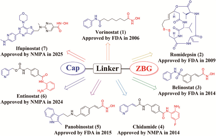
Figure 1.
The chemical structures of currently approved HDACis and its representative pharmacophore composition: Cap, linker, and ZBG.

Hydrazide-based HDAC inhibitors and PROTAC degraders in drug discovery: Recent developments and therapeutic applications
Hao Li , Yu-Xuan Zhao , Jin-Bo Niu , Jian Song , Sai-Yang Zhang


Epigenetic regulation, a key mechanism governing heritable gene expression without altering the DNA sequence itself, operates through reversible modifications of chromatin structure, such as histone post-translational modifications and DNA methylation, and plays a central role in maintaining cellular homeostasis [1]. However, widespread dysregulation of the epigenome is a well-established driver of aberrant transcriptional programs, directly contributing to the initiation, malignant progression, and therapeutic resistance observed in various human diseases, including cancer [2,3]. Among the diverse epigenetic marks, the reversible acetylation and deacetylation of histone lysine residues are particularly critical, representing dynamic and highly regulated post-translational processes that profoundly influence chromatin structure, transcription factor recruitment, and essential cellular functions [4,5]. Histone deacetylases (HDACs), a class of zinc-dependent metalloenzymes, catalyze the removal of acetyl groups from lysine residues on histone tails, thereby mediating chromatin condensation and effectively repressing the transcription of specific genes [6]. Under pathological conditions, such as cancer, aberrant HDAC activity or expression drives malignant phenotypes, including uncontrolled proliferation [7]/metastasis [8], chemoresistance [9-11], and evasion of apoptosis [12], primarily through the repression of tumor suppressor genes and the activation of pro-survival signaling pathways. Given this central pathogenic role, HDACs have emerged as pivotal targets for developing novel epigenetic-targeted therapeutics [13].
Eighteen human HDACs isoforms have been identified and classified based on cofactor dependence and sequence homology into Zn2+-dependent classes (Ⅰ, Ⅱ, Ⅳ) and NAD+-dependent class Ⅲ enzymes (sirtuins, SIRT1-7) [14,15]. Class Ⅰ HDACs (HDAC1/2/3/8) play crucial roles in proliferation control, cell differentiation, and transcriptional repression. Class Ⅱ HDACs are subdivided into class IIa (HDAC4/5/7/9), which regulate nucleocytoplasmic shuttling and specific DNA binding, and class IIb (HDAC6/10), with HDAC6 modulating cell motility, adhesion, and chaperone function, and HDAC10 implicated in angiogenesis. Class Ⅲ sirtuins (SIRT1-7) are involved in processes such as DNA repair, cell survival, cell invasion, and energy metabolism. Class Ⅳ solely consists of HDAC11, which participates in DNA replication and immune regulation by modulating interleukin-1 (IL-1) expression [16,17]. Substantial evidence indicates that HDACs are aberrantly overexpressed in diverse pathologies, including colorectal cancer [18], breast cancer [19], inflammation [20], neurodegenerative diseases [21,22], and diabetes [23], establishing them as significant therapeutic targets for drug development [24,25].
Histone deacetylase inhibitors (HDACis) exert their therapeutic effects across malignancies (e.g., leukemia [26], lymphoma [27], and solid tumors [28]), neurodegenerative diseases [21], and inflammatory/autoimmune conditions [20,29] by potently and selectively inhibiting HDAC enzymatic activity through their binding to the catalytic Zn2+ within HDAC enzymes, thereby restoring histone acetylation. Structurally, most HDACis comprise three pharmacophoric elements, including a zinc-binding group (ZBG), a cap group (Cap), and a connecting linker [30-32]. The ZBG, positioned at one terminus of the molecule, is classified primarily into bidentate and other types based on its coordination mode with the Zn2+. Bidentate ZBGs, such as hydroxamates, o-aminobenzamides, and hydrazides, typically utilize a carbonyl oxygen atom and an adjacent nitrogen atom to form a bidentate ligand, recognizing the Zn2+ ion at the bottom of the HDAC catalytic pocket and coordinating with Zn2+, often forming a five-membered chelate ring [33]. In contrast, other ZBG types coordinate Zn2+ through different atomic arrangements. For instance, carboxylic acids coordinate through two oxygen atoms, while thiols coordinate via a single sulfur atom [34]. Binding of the ZBG to Zn2+ prevents the Zn2+ from activating a water molecule for nucleophilic attack on the acetyl carbonyl carbon of the lysine substrate, thereby blocking HDAC-catalyzed histone deacetylation. Furthermore, the chemical structure and properties of the ZBG critically influence key pharmacological and physicochemical characteristics of HDACis, including inhibitory potency and isoform selectivity, solubility, cell permeability, and pharmacokinetic (PK) properties. The distal Cap group, typically a diverse aromatic or heterocyclic structure [32,35], binds via hydrophobic, van der Waals, or hydrogen-bonding interactions to amino acid residues at the entrance of the surface channel of HDAC enzymes, blocking substrate access to the active site, and also influencing isoform selectivity, affinity, physicochemical properties, and drug-likeness of HDACis. Meanwhile, the linker unit, connecting the Cap to the ZBG, positions the Cap over the channel entrance, precisely directs the ZBG for effective chelation with the catalytic Zn2+, and mimics the aliphatic chain of the natural lysine substrate to fill the narrow hydrophobic channel, with length and conformation of the linker critically impacting the inhibitory ability of HDACis to fit the channel and thereby influencing isoform selectivity, potency, and physicochemical properties [36,37]. Furthermore, HDACis are structurally classified by their ZBGs into four major categories, hydroxamates (e.g., vorinostat/SAHA, panobinostat), short-chain fatty acids (e.g., sodium butyrate, valproic acid) [38], o-aminobenzamides (e.g., entinostat, mocetinostat) and cyclic peptides (e.g., romidepsin, apicidin) [31]. Different HDACis typically possess distinct classes of ZBGs, along with unique combinations of ZBG, Cap, and linker, which collectively confer diverse HDAC isoform inhibitory activity, pharmacological properties, and clinical applications.
Currently, there are seven HDACis have been approved by U.S. Food and Drug Administration (FDA) and National Medical Products Administration (NMPA) (Fig. 1). Among them, these encompass FDA-approved hydroxamates (vorinostat, belinostat, panobinostat) and cyclic peptides (romidepsin), along with NMPA-approved agents chidamide and entinostat. In addition, vorinostat [39] and romidepsin have been approved for the treatment of cutaneous T-cell lymphoma (CTCL) [27], romidepsin [40,41], belinostat [42,43], and chidamide [44,45] have been approved for the treatment of peripheral T-cell lymphoma (PTCL), and panobinostat is approved for multiple myeloma (MM) [46]. Significantly, chidamide [47] and entinostat [48] have been approved for breast cancer and HR+/HER2− advanced breast cancer, suggesting the expansion of HDACis clinical applications from hematological malignancies to solid tumors. Notably, ifupinostat (BEBT-908), the worldwide first-in-class dual-target HDACi simultaneously regulating the phosphatidylinositol 3-kinase (PI3K) pathway and HDAC epigenetics, received NMPA approval on July 2, 2025, for the treatment of relapsed/refractory diffuse large B-cell lymphoma (r/r DLBCL) in patients who failed at least two prior lines of therapy [49], demonstrating that the therapeutic rationale and broad potential of HDAC-based dual-target inhibitors.
Despite significant therapeutic promise as epigenetic targets, HDACis in clinical use or development face considerable challenges, including dose-limiting toxicities, inadequate systemic exposure due to poor metabolic stability, and limited efficacy against solid tumors [50-52]. Inhibitors employing hydroxamate as the ZBG, exemplified by vorinostat and panobinostat, demonstrate potent enzymatic inhibitory activity but are constrained in clinical application by potential genotoxicity, short plasma half-lives, and a consequently narrow therapeutic window [53-56]. Similarly, o-aminobenzamide-based HDACis are frequently compromised by chemical stability issues or off-target effects [57]. Thiols (-SH) exhibit potent activity but suffer from oxidative instability [38] and short-chain carboxylic acids (-COOH) provide favorable safety profiles yet possess weaker inhibitory potency [58]. Consequently, developing novel, potent, and safer ZBG alternatives to circumvent the PK liabilities and toxicity profiles of current HDACis represents a critical research priority for advancing HDAC-targeted epigenetic therapies [59]. In the design of HDACis, the hydrazide group (-C(O)-NH-NH2) serves as a ZBG and functions as a bidentate ligand, chelating the catalytic Zn2+ via the carbonyl oxygen and hydrazide nitrogen to establish inhibitory activity [60]. The carbonyl carbon connects to the linker domain, while the terminal amino group engages conserved hydrogen-bonding interactions with residues (e.g., histidine, aspartic/glutamic acid) or bound water molecules [59]. Additionally, the solvent-exposed nature of ZBG contributes favorably to molecular solubility and PK [61].
Compared with classic ZBGs such as hydroxamates, hydrazides could offer superior zinc-chelating capability, enhanced oral bioavailability, and favorable PK properties [33]. Notably, their Zn2+ affinity typically exceeds that of monodentate oxygen donors (e.g., carboxylic acids) or sulfur donors (e.g., thiols), rivaling the binding strength of hydroxamates [62]. Significantly, the hydrazide bond (-NH-NH-) confers enhanced conformational flexibility, enabling superior adaptation to geometric variations across HDAC isoform active sites, thereby improving steric fit within the enzyme cavity for refined isoform selectivity or broader inhibitory activity profiles [60]. Furthermore, hydrazides possess lower molecular weight (MW) and structural simplicity relative to hydroxamates or o-aminobenzamides, facilitating control over overall MW and lipophilicity, which aid compliance with Rule of Five criteria [60], mitigate druglikeness risks associated with excessive size or hydrophobicity, and enhance membrane permeability and oral bioavailability [34]. Notably, the terminal -NH2 group serves as a key modification site via N-alkylation or introduction of substituents such as acyl, sulfonyl, or heterocyclic moieties [63], which enables the tuning of physicochemical properties (solubility, logP), metabolic stability (cytochrome P450 (CYP450)-mediated), membrane permeability, and selectivity for specific HDAC isoforms (particularly class Ⅰ HDACs) (Fig. 2).
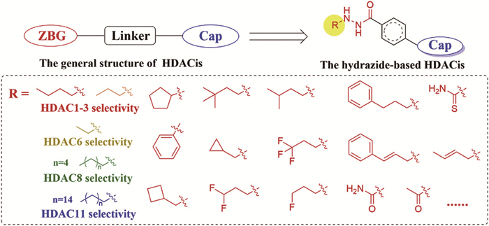
Currently, hydrazide-based HDACis have demonstrated considerable potential in the treatment of many human diseases (e.g., cancer, inflammation, and immune dysregulation). Therefore, this review summarizes recent advances in hydrazide-based HDACis, including single-target inhibitors, dual-target inhibitors and proteolysis-targeting chimera (PROTAC) degraders, and focuses on rational design strategies, structure-activity relationships (SAR), and pharmacological profiles of hydrazide-based HDACis, aiming to provide new references for the design and development of HDAC-targeting compounds with superior selectivity, potency, and better safety characteristics.
To clearly present the development trajectory and evolving strategies in the development of hydrazide-based HDACis, the subsequent three sections are organized chronologically based on the publication year of the studies. This arrangement allows readers to seamlessly follow the rational design processes, SAR studies, inhibitory and/or degradative potency as well as the progressive improvement of pharmacological profiles (e.g., potency, selectivity, metabolic stability, PK properties) over time.
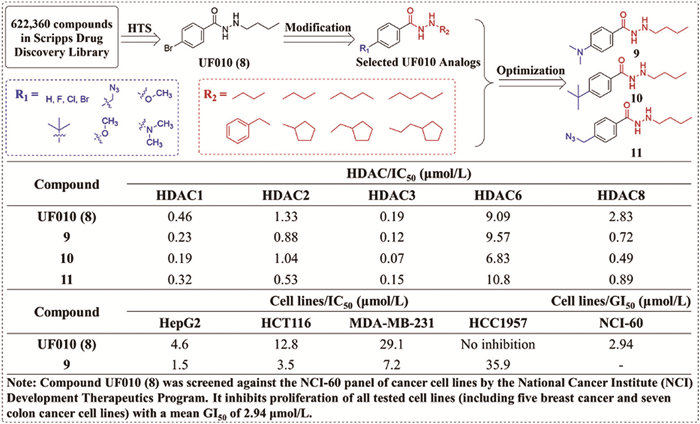
To overcome the limitations of HDACis, such as poor isoform selectivity, unfavorable PK properties, and clinical toxicity [50], Wang et al. [64] identified the benzoylhydrazide-based lead compound UF010 (8) via high-throughput screening of 620,000 compounds in 2014 (Fig. 3). Compound UF010 (8) demonstrated broad-spectrum antiproliferative activity, showing a mean growth inhibition of 50% (GI50) of 2.94 μmol/L (Fig. 3) across the NCI-60 cell line panel and significantly inhibited MDA-MB-231 cell migration. Mechanistic studies revealed compound UF010 (8) arrests MDA-MB-231 cells at the G1/S phase, markedly increases histone H3/H4 acetylation without affecting HDAC6 substrate α-tubulin and induces p53 acetylation to activate the tumor-suppressive p53/Rb pathway while suppressing MYC/KRAS oncogenic signaling. Building upon compound UF010 (8), a series of HDAC1-3 selective inhibitors were designed by employing a benzene ring linker bearing diverse Cap groups (e.g., halogen, methoxy, tert-butyl) at the para-position and a hydrazide ZBG with alkylated terminal nitrogen. SAR analysis established that n-butyl substitution on the terminal ZBG nitrogen conferred optimal HDAC inhibitory potency, and elongating the alkyl chain (e.g., n-pentyl) or introducing branched chains significantly reduced potency against HDAC1/2/3. Furthermore, the bulky Cap groups (e.g., dimethylamino, tert-butyl, azidomethyl) para to the hydrazide carbonyl markedly enhanced HDAC inhibitory potency. Among these derivatives, compound 9 with Cap as dimethylamino, compound 10 with Cap as tert-butyl, and compound 11 with Cap as azidomethyl, exhibited potent class Ⅰ HDAC inhibitory effect (half-inhibitory concentrations (IC50) = 0.07–1.04 μmol/L), and compound 9 showed 2–5-fold greater potency than compound UF010 (8) against HDAC1/2/3 (IC50 = 0.23, 0.88 and 0.12 μmol/L, respectively) (Fig. 3).
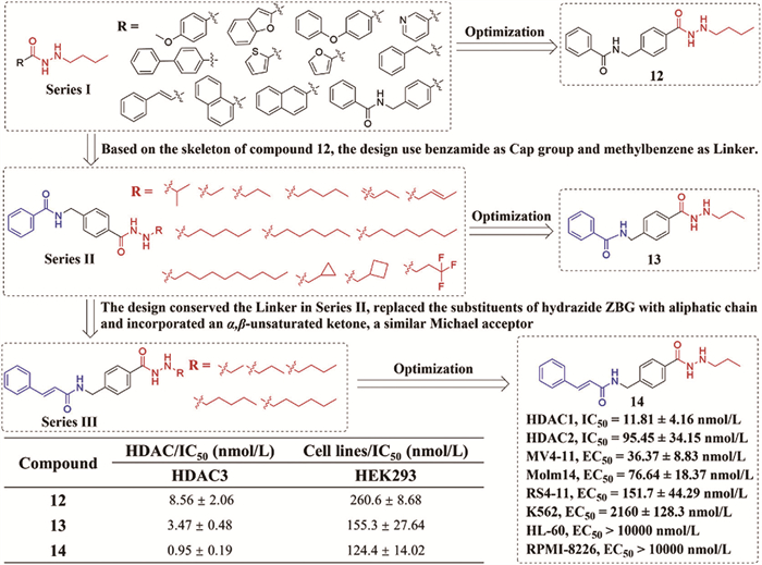
To address the clinical limitations of hydroxamate-based HDACis, which undergo rapid metabolic inactivation via UGT1A1 glucuronidation leading to high doses and dose-limiting toxicities [53-56], McClure et al. [26] reported novel allosteric class Ⅰ selective HDACis bearing a hydrazide ZBG for the treatment of acute myeloid leukemia (AML) (Fig. 4). This strategy replaced the hydroxamate ZBG, yielding three inhibitor series. Series 1, featuring diverse cyclic Cap/linkers (e.g., benzene, thiophene, naphthalene) with a butyl hydrazide ZBG, identified the benzamide-benzyl analogue 12 as a potent HDAC3 inhibitor (IC50 = 8.56 nmol/L) (Fig. 4). Optimization of the hydrazide alkyl chain length in Series 2, retaining the N-(4-(hydrazide) benzyl) benzamide Cap and benzene linker from compound 12, yielded compound 13 (propyl hydrazide as ZBG) with enhanced HDAC3 potency (IC50 = 3.47 nmol/L) (Fig. 4). Further modification in Series 3 introduced a cinnamamide Cap containing an α,β-unsaturated ketone (Michael acceptor) and aliphatic hydrazide chains (C2-C6), affording compound 14 (propyl hydrazide as ZBG) as the most potent HDAC3 inhibitor (IC50 = 0.95 nmol/L) (Fig. 4). Molecular docking (HDAC3/PDB ID: 4A69/NCoR2) [65] revealed compounds 13 and 14 bind an allosteric pocket distinct from the catalytic zinc site, with their propyl chains extending into a hydrophobic tunnel, exhibiting significantly lower binding energies than vorinostat. Compound 14 demonstrated potent anti-proliferative effects against leukemia and MM cell lines, showing highest selectivity for MV4-11 cells (half maximal effective concentration (EC50) = 36.37 nmol/L) (Fig. 4) and more than 100-fold selectivity over solid tumor cells HEK293 and HeLa, alongside reduced toxicity towards normal PBMCs compared to panobinostat. Mechanistically, compounds 12, 13, and 14 dose-dependently upregulated histone H3/H4 acetylation, outperforming vorinostat and entinostat, without affecting acetylated α-tubulin. Crucially, PK studies confirmed resistance to glucuronidation in human liver microsomes for hydrazide compounds 12, 13, and 14, with no metabolites detected, contrasting starkly with the significant glucuronidation and inactivation observed for vorinostat.
To address the limitations of suboptimal potency and glucuronidation-mediated inactivation of lead compound 15 [50,55], Li et al. [66] rationally replaced the ZBG with a hydrazide moiety, constructing novel hydrazide-based selective class Ⅰ HDACis based on the o-aminobenzamide-based class Ⅰ HDACi 15 (Fig. S1 in Supporting information). Optimization of the ZBG yielded a series of linear alkyl hydrazide derivatives, wherein the propyl hydrazide analog compound 16 exhibited the strongest inhibitory activity against HDAC1/2/3 (IC50 = 5.63–287.1 nmol/L) with enhanced selectivity over HDAC8 (IC50 = 557.3 nmol/L) (Fig. S1). Subsequent linker truncation of compound 16 afforded compound 17, which achieved low-nanomolar potency against HDAC1/2/3 (IC50 = 1.41–28.04 nmol/L) and a 26-fold increase in anti-proliferative activity against MV4-11 leukemia cells (EC50 = 34.7 nmol/L) compared to compound 15 (EC50 = 911.8 nmol/L) (Fig. S1). Mechanistically, compound 17 could induce G1/S arrest and caspase-3-dependent apoptosis in p53-wild-type MV4-11 cells but only cause G2/M arrest without apoptosis in p53-null PC-3 cells, highlighting the p53-dependent mechanistic divergence of compound 17.
To circumvent potential off-target protein modifications associated with the Michael acceptor properties of the cinnamamide moiety in the previously reported HDAC3 inhibitor 14 [26,67,68], Jiang et al. [69] implemented a ring-closure strategy on HDACis 14 and 18, through replacing the α,β-unsaturated carbonyl system with benzofused heterocycles (e.g., naphthamide, indole-2-carboxamide), to yield novel HDACis with modifications to the Cap group (Fig. S2 in Supporting information). Most compounds retained HDAC1-3 inhibitory potency comparable to compounds 14 and 18, and representative indole-2-carboxamide compound 19 conserved the binding pose in HDAC1/3 (PDB: 4BKX/4A69) [65,70] catalytic pockets, exhibiting potent and selective inhibitory activity of HDAC1-3 (IC50 = 0.43–3.01 nmol/L, 2–5-fold enhancement vs. compound 14) with minimal HDAC6 activity (IC50 > 10,000 nmol/L) (Fig. S2). In MV4-11 AML cells, compound 19 could suppress proliferation (IC50 = 19.23 nmol/L) (Fig. S2), induce p53-dependent apoptosis, and specifically elevate histone H3/H4 acetylation. Crucially, compound 19 could demonstrate exceptional PK improvements, including an oral bioavailability of 112% (compared to 19.8% of compound 18 and 4.62% of panobinostat), a 332-fold higher Cmax, and a 1146-fold greater AUC0-inf vs. panobinostat, alongside favorable t1/2. Oral administration of compound 19 (4 mg/kg) for 24 days significantly inhibited tumor growth in AML xenograft models (total growth inhibition (TGI) = 78.9%), whereas panobinostat failed due to insufficient tissue exposure.
To address the issues of non-selective toxicity and metabolic instability due to the glucuronidation associated with hydroxamate-based HDACis [54,55], Sravani et al. [33] reported novel HDACis based on a benzohydrazide ZBG in 2022 (Fig. S3 in Supporting information). Building upon the compound UF010 (8) [64] and retaining its phenyl ring as the linker, they replaced the traditionally employed, metabolically labile ZBGs like hydroxamate or o-aminobenzamide with a hydrazide moiety. The hydrazide terminus was functionalized with n-butyl or n-propyl groups, while diverse amino substituents (e.g., aryl and aroyl groups) were introduced at the para-position of the hydrazide ZBG, yielding a series of new HDACis designed for improved selectivity and metabolic stability. SAR analysis revealed that incorporating a propyl group at the hydrazide ZBG terminus (e.g., compound 20) significantly enhanced selectivity and inhibitory potency towards HDAC3 compared to a butyl group. Among the synthesized compounds, compound 20 emerged as a highly selective HDAC3 inhibitor (IC50 = 15.41 nmol/L) (Fig. S3), exhibiting selectivity indices of 18.72-to > 1000-fold over HDAC1/2/6/8, which was superior to compound UF010 (8) (IC50 = 256.7 nmol/L) and the known HDAC3-selective inhibitors BG-45 (30) (IC50 = 566 nmol/L) (Fig. S3). Molecular docking studies (HDAC3 PDB: 4A69) [65] revealed that the hydrazide group of compound 20 forms a bidentate chelation with the catalytic Zn2+, while the phenyl ring of compound 20 engages in π-π stacking with His172 and Phe200, and the amide group forms a hydrogen bond with Asp93. In vitro, compound 20 demonstrated potent antiproliferative activity against multiple cancer cell lines with low toxicity towards normal cells (Fig. S3). Notably, compound 20 could induce G2/M phase arrest and significantly promote apoptosis in B16F10 and 4T1 cells and was more effectively than UF010. Rat PK studies indicated that compound 20 had a plasma half-life (t1/2) of 2.4 h (i.v.) and 4 h (i.p.), respectively.
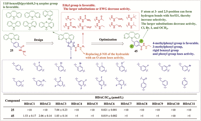
In 2022, capitalizing on the structural distinction of the shallower catalytic pocket of HDAC6 vs. class Ⅰ HDACs [71,72], Li et al. [20] designed hydrazide-based HDACis by shortening the ZBG tail chain (from 3–4 carbons in compound 13 to 1–2 carbons in compounds 22/23), enhancing the HDAC6 potency and selectivity while conferring anti-inflammatory activity (Fig. S4 in Supporting information). This research was based on a previous work by McClure et al. (2016) [26], which was mentioned in the introduction of compounds 12–14 in this chapter. Subsequent introduction of bulky rigid Cap groups (e.g., phenothiazine, dibenzazepine) and benzyl linkers yielded compound 25 featuring an ethyl hydrazide ZBG and a tricyclic Cap, which achieved exceptional HDAC6 selectivity (IC50 = 19 nmol/L, HDAC1/6 selectivity index >80-fold) (Fig. S4). Molecular docking revealed that HDAC6 (PDB: 5EDU) [73] selectivity of compound 25 originates from hydrophobic interactions between the Cap and Leu749/Pro501, π-π stacking of the benzyl linker with Phe620/Phe680, bidentate zinc chelation by the hydrazide, and hydrophobic contacts of the ethyl tail with Cys621/Pro608 (Fig. S4). In J774A.1 cells, compound 25 could selectively inhibit HDAC6 to induce α-tubulin acetylation without affecting histone H3/H4, suppress NLRP3 inflammasome activation by disrupting HDAC6-dynein interaction, and reduce lipopolysaccharide (LPS)/ATP-induced IL-1β release (IC50 = 2.61 μmol/L). Crucially, compound 25 exhibited excellent PK properties, with the 93.4% oral bioavailability (vs. 5.9% for tubastatin A), 76.8-fold higher AUC0-inf (10,292 ng h mL−1), t1/2 = 6.8 h (oral at 20 mg/kg) and significant blood-brain barrier penetration (brain/plasma ratio = 1.26–1.51). And compound 25 also exhibited slow binding/dissociation kinetics, low cytotoxicity and acute toxicity (median lethal dose (LD50) > 500 mg/kg), which support the potential of compound 25 as an oral candidate for NLRP3-mediated disorders.
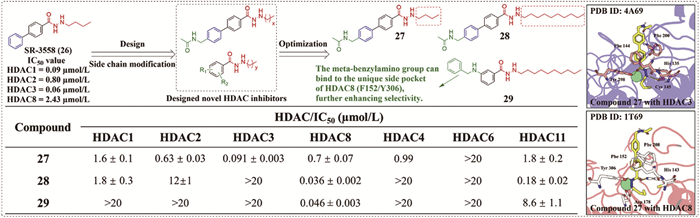
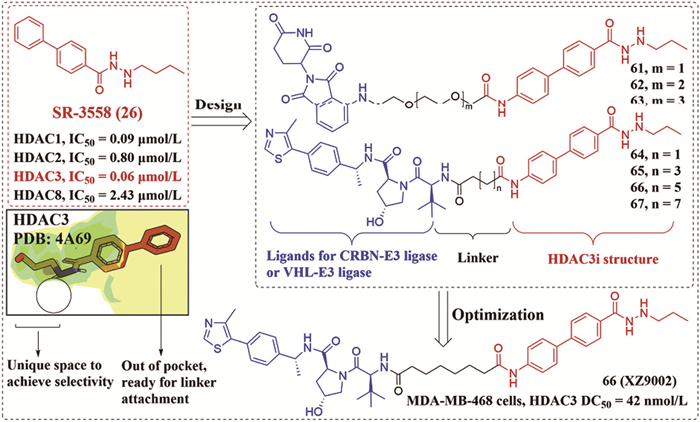
In 2022, leveraging distinct foot pocket lengths between HDAC3 (PDB: 4A69) [65] and HDAC8 (PDB: 1T69) [74], Wolfgang Sippl et al. [75] optimized the canonical pharmacophore of hydrazide-based inhibitor SR-3558 (26) (ZBG/linker/Cap) to develop alkylhydrazide-based selective class Ⅰ HDACis (Fig. 5). By systematically varying the N-alkyl chain length (C2-C6) to target the hydrophobic foot pocket, and incorporating diverse linkers (biphenyl, aminopyrimidine, piperazine-pyrimidine) and Cap groups (acetamidomethyl, benzylamine, indole derivatives), they identified compound 28 (hexyl chain) as a potent HDAC8 inhibitor (IC50 = 36 nmol/L) (Fig. 5) with > 550-fold selectivity over HDAC3 (IC50 > 20 μmol/L) (Fig. 5). In contrast, the propyl analog compound 27 selectively inhibited HDAC3 (IC50 = 91 nmol/L) (Fig. 5), confirming alkyl chain length governs subtype specificity. Molecular docking revealed compound 27 chelates the catalytic Zn2+ in the HDAC3 (PDB: 4A69) [65] active site, forms hydrogen bonds with His135 and Tyr298, and engages in π-π stacking with Phe144 and Phe200. The n-propyl side chain optimally occupies the foot pocket. In HDAC8 (PDB: 1T69) [74], while compound 27 also coordinates the Zn2+ and positions its biphenyl linker between Phe152 and Phe208, the shorter alkyl chain is less optimally accommodated, rationalizing its reduced inhibitory activity against this isoform (Fig. 5). In Jurkat T cells, compound 28 could selectively increase acetylation of HDAC8 substrates (H3K27/SMC3). Additionally, compound 28 demonstrated exceptional stability and low cytotoxicity.
Targeting the unmet therapeutic need in triple-negative breast cancer (TNBC) and leveraging the established overexpression of HDAC3 in this malignancy, Pulya et al. (2023) [31] rationally designed and optimized a series of novel pyrazine-hydrazide-based selective HDAC3 inhibitors building upon previously reported lead compounds UF010 (8) [64] and BG-45 (30) (Fig. S5 in Supporting information) [76]. Employing a hybrid structure strategy, the design retained the pyrazine ring from BG-45 (30) as the linker, replaced the o-aminobenzamide ZBG of BG-45 (30) with the hydrazide moiety from UF010 (8), and introduced diverse Cap (e.g., benzylamine, p-methoxybenzylamine, or indole moiety) onto the pyrazine ring. SAR analysis revealed that incorporating a propyl or butyl chain at the ZBG terminus enhanced HDAC3 selectivity, inhibitory potency, and metabolic stability, with the propyl hydrazide proving significantly superior to the butyl chain. Compound 31, featuring a p-methoxybenzylamine Cap and a propyl hydrazide ZBG, emerged as the most potent HDAC3 inhibitor (IC50 = 14.00 nmol/L) (Fig. S5), exhibiting more than 121-fold selectivity over HDAC1/2 and negligible activity against HDAC6/8. Molecular docking (HDAC3 PDB: 4A69) [65] elucidated that the hydrazide carbonyl of compound 31 formed a salt bridge with the catalytic Zn2+ of HDAC3, the amide-NH formed a hydrogen bonded with Asp93, and the pyrazine ring engaged in π-π stacking with Phe144, Phe200 and His172, which accounts for the high selectivity of compound 31 towards HDAC3. Compound 31 demonstrated potent antiproliferative activity against TNBC cell lines 4T1 and MDA-MB-231 (IC50 = 0.55 and 0.74 μmol/L, respectively) (Fig. S5), with low toxicity toward normal cells. Furthermore, compound 31 could induce G2/M phase arrest, promote ROS generation, and trigger apoptosis in 4T1 and MDA-MB-231 cells. Notably, compound 31 could reverse oxaliplatin resistance in oxaliplatin-resistant MDA-MB-231 cells, demonstrating significant chemosensitizing activity. PK evaluation revealed improved metabolic stability for compound 31 (rat liver microsome, t1/2 = 38.3 min) and favorable systemic exposure following intraperitoneal administration (5 and 15 mg/kg, t1/2 was 7.19 and 7.20 h, respectively). In the 4T1-Luc tumor-bearing mouse model, compound 31 (15 mg/kg, biw) achieved a remarkable 97.92% TGI and effectively suppressed lung metastasis.
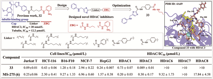
In 2024, Sun et al. [77] structurally optimized dual-targeting inhibitor 32 to overcome the non-selectivity, toxicity, and PK limitations of clinical HDACis [50] by replacing the o-phenylenediamine ZBG with a hydrazide moiety, removing the 3,4,5-trimethoxyphenyl (TMP) moiety (a tubulin-binding group) and refining the linker, yielding novel hydrazide-based HDACis featuring a 5-((phenylamino)methyl) thiazol-2-yl Cap (Fig. 6). Although o-phenylenediamine analogs showed superior Jurkat T-cell anti-proliferative activity, the hydrazide-based compound 33 achieved potent HDAC3-selective inhibitory activity (IC50 = 89 nmol/L, HDAC3/HDAC1 selectivity ratio ≈ 10.3) (Fig. 6) with broad cytotoxicity (IC50 = 0.09 μmol/L in Jurkat cells, 0.24 μmol/L in HepG2 cells) (Fig. 6). Molecular docking revealed that compound 33 chelates Zn2+ in the HDAC3 (PDB: 4A69) [65] catalytic pocket, forms a key hydrogen bonding with His172, and engages in π-π stacking with Phe144/Phe199 within a hydrophobic environment constituted by Phe200 and Leu266 (Fig. 6). In B16-F10 melanoma cells, compound 33 could induce G2/M arrest and mitochondrial apoptosis. Significantly, compound HQ-30 also could downregulate programmed cell death ligand 1 (PD-L1) via a cathepsin B (CTSB)-dependent lysosomal pathway (degradation concentration 50% (DC50) = 5.7 μmol/L). Oral administration (25 mg/kg) for 9 days in B16-F10 models achieved 67% TGI, surpassing MS-275 (37% TGI) and PD-L1 inhibitor NP19 (37% TGI). And compound 33 exhibited favorable PK properties, with the 57% oral bioavailability, 214 min of t1/2 (oral at 20 mg/kg), aqueous solubility (15.6 μg/mL) and safety (LD50 > 1000 mg/kg).
In 2024, Kraft et al. [10] developed novel hydrazide-based HDACis targeting cisplatin-resistant malignancies (Fig. S6 in Supporting information) to address cisplatin resistance in solid tumors and leverage the potential of epigenetic agents to synergize with or reverse cisplatin resistance [78]. Utilizing the o-aminobenzamide-based HDACi VK1 (35) as a starting point, they retained its peptidomimetic Cap and benzyl linker while replacing the o-aminobenzamide ZBG with a hydrazide moiety. Systematic optimization of the hydrazide R-group with diverse alkyl (e.g., linear, branched, cyclic) or phenyl substituents yielded a series of novel hydrazide-based HDACis. Compounds 36 and 37 (featuring propyl and butyl hydrazide ZBGs, respectively) exhibited potent HDAC1 and HDAC3 inhibitory activity, with HDAC3 IC50 values of 0.004 for compound 36 and 0.008 μmol/L for compound 37 (Fig. S6), surpassing the activity of VK1, entinostat, and vorinostat. Notably, compound 36 displayed significantly improved solubility (16.8 μg/mL) vs. VK1 (0.98 μg/mL). Molecular docking indicated that the propyl chain of compound 36 optimally occupies the hydrophobic foot pocket of HDAC3 (PDB: 4A69) [65], explaining the pronounced HDAC3 potency of compound 36. Compounds 36 and 37 also demonstrated low micromolar cytotoxicity (IC50 = 0.151–0.295 μmol/L) against both cisplatin-sensitive (Cal27 and A2780) and cisplatin-resistant (Cal27 CisR and A2780 CisR) cell lines (Fig. S6). Mechanistic studies revealed that compounds 36 and 37 could synergistically enhance cisplatin-induced DNA damage in Cal27 and Cal27 CisR cells, upregulate p21 and pro-apoptotic BIM expression, and induce caspase-dependent apoptosis. Significantly, compounds 36 and 37 completely reversed cisplatin resistance in Cal27 CisR and A2780 CisR cells, restoring chemosensitivity.
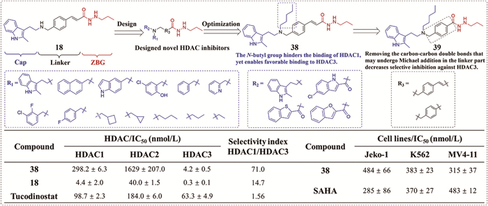
To enhance HDAC3 isoform selectivity, Li et al. [79] leveraged structural insights from HDAC1/3 (PDB: 5ICN [80]/4A69 [65], HDAC3 has a wider pocket) allosteric pocket differences to rationally modified pan-HDAC1-3 inhibitor 18 by introducing sterically bulky R1 groups (n-butyl, cyclobutylmethyl, naphthylmethyl, etc.) at the secondary amine nitrogen, and optimizing the Cap group (e.g., replacing indole with benzothiophene/benzofuran) (Fig. 7). Crucially, removing the R3 linker double bond to avoid Michael addition side reactions abolished HDAC3 selectivity, suggesting the role of double bond in maintaining the bioactive conformation. Compound 38 (R1 = n-butyl) exhibited exceptional HDAC3 selectivity (IC50 = 4.2 nmol/L) with more than 70-fold over HDAC1 (IC50 = 298.2 nmol/L), and low activity against class Ⅱ/Ⅳ HDACs (IC50 > 10 μmol/L) (Fig. 7). Docking results demonstrated the butyl group of compound 38 engages the allosteric pocket via hydrophobic interactions and hydrogen bonds with Lys449/Phe451 in HDAC3 (PDB: 4A69) [65], while steric hindrance in HDAC1 (PDB: 5ICN) [80] weakens binding. In MV4-11 AML cells, compound 38 could suppress proliferation (IC50 = 315 nmol/L) (Fig. 7), selectively elevate histone H3/H4 acetylation (Ac-H3/Ac-H4), and synergize with venetoclax (combination index (CI) < 1) by downregulating Mcl-1 and upregulating cleaved caspase-3 and cleaved-poly ADP-ribose polymerase (cleaved-PARP). Compound 38 also showed stability in simulated gastric/intestinal fluids and rat plasma (>48 h).
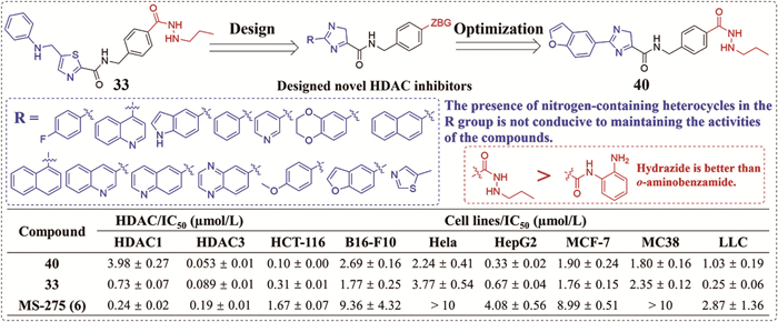
Building on the solvent-exposed Cap group of selective inhibitor 33 [77], Chen et al. [81] designed novel hydrazide-based HDAC3 inhibitors by incorporating hydrophobic aromatics (e.g., benzofuran) as Cap replacements while retaining hydrazide or o-aminobenzamide ZBGs (Fig. 8). SAR studies suggested compound 40 (with a benzofuran Cap and propyl hydrazide) exhibited the most potent HDAC3 inhibitory effect (IC50 = 53 nmol/L), showing an approximately 10-fold higher selectivity index (HDAC3/HDAC1 = 75) than the lead compound 33 (Fig. 8) and minimal inhibitory activity against HDAC6/7/8 (IC50 > 10 μmol/L) as well. Notably, nitrogen-containing heterocyclic Caps reduced HDAC inhibitory potency, and hydrazide ZBG outperformed o-aminobenzamide ZBG in HDAC inhibitory activity and selectivity. Molecular docking showed that compound 40 could effectively fit into the HDAC3 binding pocket (PDB: 4A69) [65], the hydrazine group could be inserted into the pocket and chelate with the catalytic Zn2+, and the benzofuran and imidazole ring were exposed to the solvent region. Compound 40 demonstrated broad antiproliferative activity across 7 solid tumor cell lines (IC50 = 0.10–2.69 μmol/L) (Fig. 8), and inhibited MC38 cell via ROS generation, mitochondrial dysfunction, and G2/M arrest. Crucially, compound 40 could dose-dependently upregulate PD-L1 expression in MC38 cells and xenografts, which effectively block by BRD4 inhibitor JQ1, indicating epigenetic regulation. In MC38 models, compound 40 (50 mg/kg, p.o.) achieved 63% TGI as monotherapy, and sensitized tumors to PD-L1 blockade with NP19, which could achieve 80% TGI and increase CD8+ T-cell infiltration.
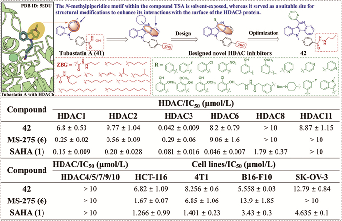
Capitalizing on the longer substrate channel of HDAC3 (PDB: 4A69) [65] vs. HDAC6 (PDB: 5EDU) [73], Xiao et al. [82] rationally designed HDAC3 inhibitors by replacing the hydroxamate ZBG of tubastatin A (41) with a hydrazide moiety and modifying the Cap region with aromatic amines (e.g., pyridine, indole) to enhance selectivity (Fig. 9). In addition, substituting the aliphatic amine (N-methylpiperidine) of trichostatin A (TSA) with aromatic Cap groups further reduced HDAC10 inhibitory activity by disrupting key hydrogen bonds. SAR studies of 49 derivatives identified compound 42 with N’-propyl acetylhydrazone group as ZBG and the pyridin-2-yl group as Cap as the most optimal compound, exhibiting potent HDAC3 inhibitory activity (IC50 = 42 nmol/L) with >161-fold selectivity over HDAC1/2/6 (Fig. 9). Compound 42 demonstrated broad in vitro antitumor activity (IC50 = 5.558–12.79 μmol/L in HCT-116/4T1/B16-F10/SK-OV-3 cells) (Fig. 9), suppressed migration/invasion, and induced G1 arrest/apoptosis in B16-F10 cells. Significantly, compound 42 could degrade PD-L1 via CTSB-dependent lysosomal pathway (DC50 = 4.027 μmol/L) and synergize with olaparib to enhance DNA damage. Compound 42 also exhibited exceptional oral PK in rats, with a F of 95.34%, t1/2 of 0.63 h (i.v., 2 mg/kg, n = 6) and 2.026 h (p.o., 20 mg/kg, n = 6). In vivo, monotherapy of compound 42 (50 mg/kg) inhibited melanoma growth (TGI = 67%), while combination with PD-L1 inhibitor NP-19 (compound 42 + NP-19, 50 + 20 mg/kg) increased CD3+CD4+/CD8+ T-cell infiltration (TGI = 80%), and combination with olaparib enhanced DNA damage response (TGI = 91% in breast cancer), without significant toxicity.
In 2025, Stopper et al. [11] developed a series of potent ethyl hydrazide-based HDACis designed as chemosensitizers (Fig. S7 in Supporting information) to address the need to overcome chemotherapy resistance [78] and circumvent the genotoxicity risks and PK limitations associated with approved hydroxamate-based HDACis (e.g., vorinostat, belinostat) [55]. Based on the classical HDACi pharmacophore, the design retained peptide-like Cap groups while optimizing the linker and employed an ethyl hydrazide moiety as the ZBG. Among these compounds, compound 44 demonstrated potent and selective inhibitory potency of HDAC1/2/3/6 (IC50 = 0.029, 0.123, 0.0226, and 0.367 μmol/L, respectively) with weak activity against HDAC4/8 (Fig. S7). Compound 44 also exhibited low micromolar cytotoxicity against both cisplatin-sensitive and cisplatin-resistant tumor cell lines A2780, A2780 CisR, Cal27 and Cal27 CisR. Crucially, compound 44 could effectively reverse cisplatin resistance, showing high sensitization effects (shift factor = 6.15 and 18.7 in Cal27 CisR and A2780 CisR cells, respectively) and strong synergic effects (CI < 0.2). Mechanistically, compound 44 could enhance DNA damage, induce p21 expression, and trigger caspase-dependent apoptosis. Pharmacokinetically, compound 44 displayed favorable metabolic stability in human liver microsomes (t1/2 = 33.93 min), superior to VK1 (35) (t1/2 = 17.45 min), and possessed suitable lipophilicity (logD7.4 = 2.69).
To address dose-dependent off-target effects on HDAC1-3 from the hydrazide-based HDAC6 inhibitor 25 (HDAC6: IC50 = 19 nmol/L) (Fig. 10) [20], Li et al. [83] optimized selectivity through strategic fluorination of the benzene ring in the linker while retaining the critical 11H-benzo[b]pyrido[4,3-f]azepine Cap and ethylhydrazide ZBG (Fig. 10). SAR analysis revealed the fluorination uniquely enhanced HDAC6 selectivity vs. other halogen substitutions, with the 2,5-difluoro analog 45 maintaining potent HDAC6 inhibitory activity (IC50 = 21 nmol/L) while achieving more than 335-fold selectivity over HDAC3 (IC50 = 7.04 μmol/L), and minimal HDAC1/2 activity (IC50 > 10 μmol/L) (Fig. 10). Mechanistically, compound 45 could suppress NLRP3 inflammasome activation by disrupting HDAC6-dynein interaction, reducing IL-1β release in LPS/ATP-stimulated macrophages. Orally administered compound 45 (20–40 mg/kg) attenuated serum IL-1β in LPS-induced peritonitis, improved colon pathology/IL-1β expression in dextran sulfate sodium (DSS) colitis, and alleviated skin inflammation in IMQ-induced psoriasis. Additionally, compound 45 also exhibited exceptional PK properties (rat, 20 mg/kg, 97% oral bioavailability, Cmax = 9813 ng/mL, AUC0-inf = 13,945 ng h mL−1 and t1/2 = 0.49 h), establishing compound 45 as a fluorination-engineered HDAC6 inhibitor with optimized selectivity and PK profiles for NLRP3-driven inflammatory diseases.
To develop HDACis with low toxicity and favorable PK properties for cancer therapy through immunomodulation and DNA damage, Hu et al. (2025) [61] designed a series of novel hydrazide-based selective HDAC3 inhibitors, leveraging the HDAC6 inhibitor 46 as a lead compound to create enhancers for immunotherapy and DNA damage response (Fig. S8 in Supporting information). The design replaced the hydroxamate ZBG of 46 with a hydrazide moiety to improve PK and incorporated diverse Cap (e.g., 4-NH-Boc-phenyl) as well as extended alkyl linkers to enhance potency. Compound 47, featuring a 4-NH-Boc-phenyl Cap and a heptyl linker, exhibited potent HDAC3 inhibitory activity (IC50 = 0.31 μmol/L, SI > 32 vs. HDAC1/2/6/8), superior to MS-275 (IC50 = 0.45 μmol/L) (Fig. S8). Compound 47 demonstrated broad-spectrum antiproliferative activity against five HDAC3-overexpressing cancer cell lines (e.g., SK-OV-3, 4T1 cells) with low cytotoxicity toward normal HK-2 cells (Fig. S8). SAR indicated that long-chain aliphatic linkers usually enhanced the antiproliferative activity, while bulky groups and/or heterocycles (e.g., indole, naphthalene, pyrene) in the Cap group lowered the antiproliferative activity. Molecular docking (HDAC3 PDB: 4A69) [65] revealed the hydrazide ZBG formed a hydrogen bond with Glu156, the Boc group interacted with Lys474, and the thiophene engaged in π-π stacking with Lys475 (Fig. S8). More importantly, as a selective HDAC3 inhibitor, compound 47 also functioned as an immunomodulator and DNA damage enhancer. In B16-F10 melanoma cells, compound 47 outperformed SAHA in inhibiting migration/invasion, inducing apoptosis, and causing G2/M arrest. Mechanistically, compound 47 could target HDAC3 in B16-F10 cells, concentration-dependently downregulating PD-L1 expression (DC50 = 4.174 μmol/L) via a CTSB-mediated lysosomal pathway, dose-dependently upregulating IFN-γ to promote T-cell infiltration, and downregulating NCOR2. Furthermore, compound 47 could synergistically enhance DNA damage when combined with the PARP inhibitor olaparib, significantly upregulating γ-H2AX and downregulating PARP1. In vivo, compound 47 monotherapy (50 mg/kg) inhibited melanoma and breast cancer growth (TGI = 59% and 64%, respectively) (Fig. S8). The combination effect of compound 47 was more significant, with a TGI of 65% in combination with the PD-L1 inhibitor NP-19 (compound 47 + NP-19, 25 + 10 mg/kg) in a B16-F10 mice model and 88% in combination with olaparib (compound 47 + olaparib, 25 + 10 mg/kg) in a 4T1 mice model.
To capitalize on the unique dual catalytic domains and shallow hydrophobic active site of HDAC6 [17], Khan Hashim Ali et al. (2025) [56] synthesized a series of 3-substituted indazole-based HDAC6 inhibitors, for the treatment of cancer, particularly colorectal cancer. Employing rational drug design, the strategy utilized the indazole ZBG to mimic the γ-carboline Cap of tubastatin A (41), aiming to enhance interactions with key L1 pocket residues (Pro464/Leu712). An aryl linker was incorporated, and the hydroxamate group was prioritized as ZBG to optimize binding within the catalytic center. Among the synthesized compounds, the 2-furyl substituted compound 48 emerged as a highly selective HDAC6 inhibitor (IC50 = 1.2 nmol/L) (Fig. S9 in Supporting information). Compound 48 demonstrated significant growth inhibition in HCT116 cells (GI50 = 3.1 μmol/L). However, compound 48 suffered from poor PK properties, including low oral bioavailability in mice (1.2%), a short plasma half-life, and significant inhibitory effect of CYP450 isoforms (1A2, 2C9, 3A4). To address these limitations, the hydroxamate ZBG of compound 48 was replaced with an ethyl hydrazide moiety, yielding compound 49, which substantially improved oral bioavailability in rats (53%) and maintained a reasonable plasma half-life after i.v. dosing (5 mg/kg, t1/2 = 1.2 ± 0.2 h). However, compound 49 exhibited reduced HDAC6 inhibitory potency (IC50 = 170 nmol/L) and selectivity, along with attenuated antiproliferative activity against HCT-116 cells (GI50 = 6.3 μmol/L). Compound 49 could induce histone H3 acetylation, indicating a shift in inhibitory potency profile towards Class Ⅰ HDACs. Despite the decreased activity relative to the hydroxamate-based HDACi 48, compound 49 successfully addressed critical PK liabilities.
Although significant progress has been made in the development of inhibitors targeting individual HDAC isoforms, their clinical applications still face challenges such as limited efficacy. Leveraging the pleiotropic effects of HDACis and their highly modifiable Cap group, the development of HDAC-based dual-target inhibitors by combining them with other pathologically relevant targets may yield more potent therapeutic outcomes [84].
Dual-target drug design represents a promising approach to enhance therapeutic efficacy against significant diseases, including cancer [85], inflammation [86], and immune disorders [87]. While combination therapy could demonstrate promising therapeutic efficacy, it suffers from inherent challenges such as polypharmacy burden, complex dosing regimens, and potential toxicity overlap [88-91]. In contrast, rationally designed single-agent therapies based on the philosophy of poly-pharmacology drugs could achieve superior efficacy and safety through synergistic modulation of interacting pathways [92-94]. The highly modifiable Cap of HDACis provides an ideal modifiable point for constructing bifunctional agents [95]. By fusing a hydrazide-based ZBG with diverse pharmacophores (e.g., kinase inhibitors, epigenetic modulators), bifunctional molecules simultaneously targeting HDAC and key pathways, such as vascular endothelial growth factor receptor (VEGFR) [96], nicotinamide phosphoribosyltransferase (NAMPT) [97], PD-L1 [98] and PI3K [99] have been developed, which achieved enhanced synergistic therapeutic effects. Collectively, this HDAC-based synergistic approach might offer an innovative therapeutic strategy for overcoming challenges in treating complex diseases [92,100].
Inspired by the VEGFR inhibitor 50 [101], Xue et al. (2021) [96] employed a pharmacophore hybridization strategy to design novel hydrazide-based HDAC/VEGFR dual inhibitors derived from pazopanib (an approved VEGFR inhibitor), aiming at overcoming the off-target effects associated with o-aminobenzamides (Fig. S10 in Supporting information). Key modifications included replacing the o-diaminophenyl ZBG with a hydrazide ZBG to balance dual-target activity and metabolic stability. Compound 51 demonstrated HDAC1 inhibitory selectivity like MS-275 (IC50 = 420 nmol/L) with more than 21-fold over HDAC6 (IC50 > 8.96 μmol/L) (Fig. S10), potent VEGFR1-3 inhibitory activity (IC50 = 25–37 nmol/L), and potent antiproliferative activity against six solid tumor cells (IC50 = 2.59–4.82 μmol/L) (Fig. S10). Mechanistically, compound 51 could selectively increase histone H4 acetylation while suppressing VEGFR2 phosphorylation and effectively inhibit angiogenesis in HUVEC tube formation and rat aortic ring assays. However, rat liver microsomal stability remained suboptimal (t1/2 = 4.1–9.2 min vs. 15.2 min for pazopanib), necessitating further optimization for drug-like properties.
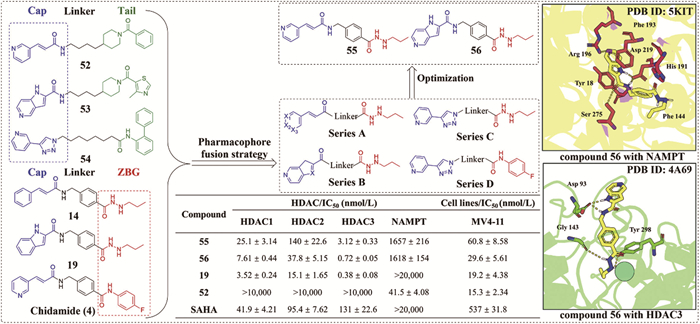
In 2024, capitalizing on structural homology between HDAC and NAMPT inhibitors and the known synergy of 53 in enhancing HDACis cytotoxicity against hematological malignancies, Chou et al. [97] designed nitrogenous heterocyclic Cap-based novel HDAC/NAMPT dual inhibitors via pharmacophore fusion strategy (Fig. 11). Representative compounds 55 and 56 with the hydrazide as ZBG and the aminomethylbenzyl as linker potently and selectively inhibited HDAC1-3 (IC50 = 0.72–25.1 nmol/L) with minimal activity against HDAC4-11 (IC50 > 10 μmol/L), while moderately inhibiting NAMPT (IC50 ≈ 1600 nmol/L) (Fig. 11). Molecular docking revealed that compound 56 chelates the catalytic Zn2+ in the HDAC3 (PDB: 4A69) [65] active site and forms three hydrogen bonds with Gly143, Tyr298, and Asp93. For NAMPT (PDB: 5KIT) [102], compound 56 occupies the substrate pocket, forming hydrogen bonds with Ser275 and Asp219, and the core of compound 56 is stabilized by hydrophobic contacts with Phe144 and Phe193, and a cation-π interaction with Tyr18 (Fig. 11), which collectively partly rationalize the dual inhibitory activity of compound 56. Compounds 55 and 56 could exhibit nanomolar antiproliferative activity (IC50 < 100 nmol/L) (Fig. 11) in MV4-11 cells and crucially overcame intrinsic resistance in p53-null AML cells HL60 and PL21 to single-target agents (e.g., compounds 19 or 52). Unlike cytostatic monotherapies, compounds 55 and 56 could induce caspase-3-dependent apoptosis (LD50 ≈ 600 nmol/L) by concurrently disrupting epigenetic regulation and NAD+ biosynthesis [103,104]. PK studies in rats (5 mg/kg i.v.) showed compound 56 exhibited favorable exposure (AUC0-inf = 3356 ng h mL−1), prolonged half-life (t1/2 = 9.19 h), and moderate systemic clearance (Clp = 25.1 mL min–1 kg–1).
Despite the breakthrough of monoclonal antibodies targeting the programmed cell death protein 1 (PD-1)/PD-L1 pathway in cancer immunotherapy, their clinical utility is hampered by high resistance/relapse rates, low objective response rates, and safety concerns with combination therapies [105-108]. Inspired by the structural features of the previously reported HDAC3 inhibitor 14 [26] and the PD-L1 inhibitor 57 [109], Wang et al. employ a pharmacophore fusion strategy to rationally designed novel PD-L1/HDAC3 dual-target inhibitors (Fig. S11 in Supporting information) [98] and overcome the limitations mentioned above. Integrating the biphenylpyridine Cap from PD-L1 inhibitor 57 and the N-propyl hydroxamate ZBG from HDAC3 inhibitor 14, they synthesized hydrazide-based compounds with diverse linkers (e.g., flexible alkyl chains, piperidine, aromatic rings and nitrogen-containing heterocycles). Compound 58, featuring a tetramethylene linker, potently inhibited PD-L1 and HDAC3 (IC50 = 89.4 and 107 nmol/L, respectively) with more than 21-fold selectivity for HDAC3 over HDAC1/6/8 (Fig. S11). Molecular docking with HDAC3 (PDB: 4A69) [65] indicated that the N-propyl hydroxamate ZBG of compound 58 chelates Zn2+ and forms H-bonds with Gly143/His172 in the foot pocket of HDAC3, explaining the HDAC3 selectivity of compound 58. Within the hydrophobic pocket of PD-L1 (PDB: 5J89) [110], the biphenylpyridine Cap engages in π-π stacking with Tyr56, the tail NH group bonds with Asp122 through a hydrogen bond, and the linker occupies the adjacent hydrophobic channel. Compound 58 inhibited proliferation in multiple cancer cell lines (e.g., B16-F10, A375, IC50 = 2.33–4.66 µmol/L) (Fig. S11), inducing histone H3 acetylation, apoptosis, and G0/G1 cell cycle arrest. In the A375/Jurkat T-cell co-culture model, compound 58 dose-dependently enhanced T-cell-mediated killing of A375 cells. In the B16-F10 melanoma model, compound 58 dose-dependently suppressed tumor growth (20 mg/kg, TGI = 48.9% and 40 mg/kg, TGI = 66.5%), surpassing single agent NP-19 (20 mg/kg, TGI = 30.8%) or the NP19 + MS-275 (6) combination (10 + 10 mg/kg, TGI = 37.0%) without observable significant toxicity.
To circumvent the burdens of multi-drug therapy and complex dosing regimens inherent in combination therapy [90] and leverage the synergistic antitumor effects of dual PI3K/HDAC inhibitory potency on proliferation suppression and apoptosis induction [111], Hou et al. (2025) employed a pharmacophore fusion strategy to design novel morpholine-triazine-based dual inhibitors (Fig. S12 in Supporting information) [99]. The design fused the morpholine-triazine pharmacophore of the oral pan-class Ⅰ PI3K inhibitor compound 59 with the propyl hydrazide ZBG from the HDAC-selective inhibitor 19, optimized the linker (e.g., benzamide, piperazine, 1-methyl-4-ethylbenzene), and yielded a series of hydrazide-based dual inhibitors exhibiting enhanced pro-apoptotic activity in mantle cell lymphoma (MCL). Compound 60 emerged as a potent and selective lead compound, demonstrating significant activity against HDAC1 (IC50 = 75.5 nmol/L), HDAC2 (IC50 = 70.9 nmol/L), and HDAC3 (IC50 = 1.9 nmol/L), with IC50 > 10 μmol/L against HDAC4/5/6/7/9/11 (Fig. S12). Concurrently, compound 60 acted as a potent pan-PI3K inhibitor (IC50 = 2.5, 80.5, 10.0 and 57.2 nmol/L against PIK3α/β/δ/γ, respectively) (Fig. S12). Compound 60 displayed potent antiproliferative activity against multiple cancer cell lines (e.g., MV4-11, Jeko-1, IC50 = 0.2–1.5 μmol/L) without significant toxicity toward normal hepatocytes (HL7702). In Jeko-1 MCL cells, compound 60 significantly induced apoptosis, outperforming the combination. This superior efficacy stemmed from synergistic inhibitory potency of the PI3K/protein kinase B (AKT) pathway and HDAC function.
While dual-target inhibitors demonstrate synergistic therapeutic benefits, their action remains confined to the transient suppression of target protein functions. In contrast, PROTAC technology serves as a more thorough and sustained therapeutic approach, offering a powerful tool for both epigenetic research and therapeutic development [112].
Targeted protein degradation by proteolysis targeting chimeras (PROTACs) has emerged as a promising strategy to address undruggable targets and overcome resistance to traditional inhibitors [113-116]. Currently, several PROTAC degraders targeting different HDAC isoforms have been developed successfully [117,118], demonstrating relatively high degradation efficiency in hematologic malignancies [37,119] and neurodegenerative diseases [120]. Compared with traditional HDACis, which reversibly inhibit HDAC activity by competitively occupying the catalytic pocket, HDAC-based PROTAC degraders simultaneously engage the target HDAC protein and an E3 ubiquitin ligase, forming a ternary complex that triggers HDAC ubiquitination and degradation [121]. In addition, HDAC-based PROTAC degraders also exhibit significant advantages such as sustained pharmacological effects enabling long-lasting target degradation following single-dose administration, enhanced subtype selectivity achieved through rational linker design and the capability to overcome acquired resistance due to target mutations or overexpression [122]. However, there are also challenges in HDAC-based PROTAC degraders, including poor membrane permeability (due to high MW/polarity) [123,124], potential off-target toxicity (from tissue-specific E3 ligase expression) [125], and insufficient degradation selectivity for specific HDAC isoforms [126].
Notably, hydrazide-based HDAC PROTAC degraders demonstrate high potency, superior selectivity, nanomolar-range DC50 (a metric indicating show degradation potency) [127,128] and promising therapeutic efficacy against hematologic malignancies [37], thereby representing a promising advancement in the field of HDAC-based PROTAC degraders.
Employing the benzoylhydrazide-based HDAC3-selective inhibitor SR-3558 (26) as a warhead, Xiao et al. [129] developed a first-in-class HDAC3-selective PROTAC degrader XZ9002 (66) by conjugating a Von Hippel-Lindau (VHL) E3 ligase to the warhead via a linker attached to the solvent-exposed terminal phenyl group of the warhead (Fig. 12). Since the lowest binding energy pose of SR-3558 (26) revealed the solvent-exposed terminal phenyl ring provides a feasible linking site for E3 ligase ligand tethering, CRBN-based PROTAC degraders 61–63 and VHL-based PROTAC degraders 64–67 were subsequently obtained. Notably, CRBN-based PROTAC degraders 61–63 with PEG linkers preserved HDAC inhibitory potency, whereas VHL-based PROTAC degraders 64–67 bearing alkyl linkers exhibited a reduction in HDAC inhibitory activity, with further slight decreases observed as linker length increased. In MDA-MB-468 and T47D cells, all VHL-based PROTAC degraders 64–67 effectively mediated HDAC3 degradation at 100 nmol/L, while among CRBN-based analogs, only 63 showed marginal effects at 1.0 μmol/L. Remarkably, the VHL-recruiting PROTAC degrader 66 (XZ9002) exhibited potent HDAC3 degradation activity in the low nanomolar range (Fig. 12) without significantly affecting HDAC1/2/6 isoforms, suggesting that converting inhibitors into PROTAC degraders may confer additional selectivity beyond inherent binding preferences through ternary complex cooperativity and isoform-specific ubiquitination mechanisms. Mechanistic investigations demonstrated that compound 66 could mediate targeted protein degradation via the ubiquitin-proteasome pathway, relying on the recruitment of the VHL E3 ligase. In contrast to the non-degradable negative control compound 66-NC, compound 66 exhibited superior efficacy in suppressing clonogenic growth of breast cancer cells, underscoring the therapeutic advantage of degradation over conventional enzymatic inhibition. Notably, compound 66 induced dose-dependent histone acetylation with reduced potency compared to CRBN-based PROTAC degraders, which was a phenomenon consistent with its selective degradation of HDAC3 rather than broad inhibitory potency of HDAC1–3.
The study by Xiao et al. [128] reported a novel HDAC3/8 dual degrader, YX968 (80), which achieves highly selective targeted degradation through the PROTAC technology (Fig. S13 in Supporting information). Firstly, based on compound 72, this design obtained compounds 70 and 71 containing hydrophilic piperazine groups. Compound 70, with an N-methylpiperazine group, exhibited the highest potency, and its IC50 values on HDAC1/3/8 were almost the same as those of compound 68. Subsequently, VHL-based PROTAC degraders were synthesized based on compounds 69 and 72. Among them, compound 80 unexpectedly displayed significant degradation ability for HDAC3/8 in MDA-MB-231 cells (DC50 = 1.7 and 6.1 nmol/L, 8 h treatment) without degrading HDAC1 or HDAC2 in MCF-7 and MDA-MB-231 cells, while compound 66 did not show a similar effect in MDA-MB-231 cells (Fig. S13). Mechanistic studies showed that compound 80-mediated degradation was through the ubiquitin-proteasome system (UPS), and the degradation process was rapid and reversible. Notably, global histone hyperacetylation (such as H3K27ac, H4K8ac) was not induced at effective degradation concentrations and only increased at high concentrations. In vitro, compound 80 exhibited potent anti-proliferative activity in breast cancer (MCF-7, MDA-MB-231, etc.) and lung cancer cell lines (A549), with IC50 reaching the sub-nanomolar level, and induced apoptosis of MDA-MB-231 cells by activating CASP3. In addition, compared with the HDAC3-selective inhibitor RGFP-966 and the HDAC8 inhibitor PCI 34051 used alone or in combination, compound 80 had significantly enhanced potency in the colony-formation inhibition assay, highlighting the synergistic advantage of the dual-degradation strategy.
HDAC8 is a promising therapeutic target implicated in various cancers, including diffuse large B-cell lymphoma (DLBCL), breast cancer, and melanoma [75]. In 2024, Xiao et al. [37] rationally designed novel hydrazide-based HDAC8 PROTAC degraders (Fig. S14 in Supporting information) to overcome limitations of current inhibitors, including insufficient selectivity, ineffective disruption of non-enzymatic scaffolding functions, and limited efficacy in DLBCL models. Building on lead compound 80 and leveraging PROTAC technology for selective degradation, their design exploited structural differences in the HDAC8 acetate release channel. Modifications involved altering the propyl hydrazide ZBG of compound 80 and incorporating diverse linkers. SAR studies revealed that longer alkyl substituents on the hydrazide ZBG enhanced HDAC8 selectivity over HDAC3, while shorter alkyl or cyclopropyl groups increased HDAC3 selectivity. Linker modifications (e.g., elongation, shortening, or cyclic structures like piperazine/piperidine) markedly reduced degradation potency. Compound 83, featuring a hexyl hydrazide ZBG, emerged as the most potent HDAC8 degrader, efficiently recruiting the VHL E3 ligase to selectively degrade HDAC8 without affecting other HDAC isoforms (Fig. S14). Mechanistic studies confirmed compound 83-mediated degradation is UPS dependent and blocked by VHL ligands or proteasome inhibitors. Crucially, compound 83 demonstrated superior antiproliferative activity in DLBCL cells (SU-DHL-2 cells, IC50 = 0.72 μmol/L), superior to PCI-34051 (SU-DHL-2 cells, IC50 > 5 μmol/L). Degradation was reversible (recovery within 24 h post-washout) with no observed hook effect (≤8 μmol/L), and quantitative proteomics confirmed target specificity.
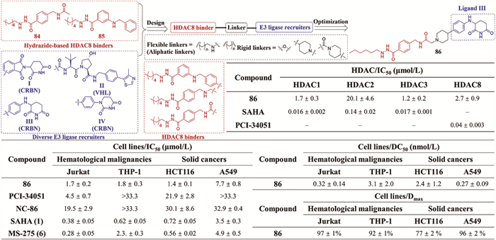
Given the dual enzymatic and scaffolding roles of HDAC8 in malignancies (e.g., T-cell leukemia [130], AML [131], solid tumors [132,133]) and the limitations or suboptimal efficacy of existing inhibitors, PROTAC technology provides a strategic solution to overcome these challenges by degrading the entire HDAC8 protein via the UPS. In 2024, Zhao et al. [134] designed hydrazide-based HDAC8-selective PROTAC degraders (Fig. 13) to address potency and selectivity deficits in reported degraders. Molecular docking revealed the acetyl group of compound 84 engaged the active site of HDAC (PDB: 1T69) [74], while steric clashes and suboptimal hydrogen bonding precluded binding to HDAC6 (PDB: 5EDU) [73] and HDAC2 (PDB: 4LXZ) [135], rationalizing selectivity for HDAC8. A series of compounds were synthesized employing a dual CRBN/VHL ligand strategy, which involved linking compound 84 to either CRBN ligand Ⅰ or VHL ligand Ⅱ via flexible C2-C8 aliphatic chains, with the hydrazide moiety serving as the ZBG. A subsequent series incorporated rigid linkers (e.g., piperidincsse) to optimize physicochemical properties, degradation efficiency, and hydrolytic stability while minimizing off-target effects. From these, compound 86 achieved high HDAC8 degradation (DC50 = 0.32–3.1 nmol/L, Dmax = 77%–97%) in Jurkat, A549, HCT116, and THP-1 cells at 100 nmol/L with negligible off-target degradation (HDAC1/2/3/4/6/7/11) (Fig. 13). Mechanistic studies confirmed ternary complex-driven degradation, with compound 86 inducing HDAC8-specific SMC3 hyperacetylation at low concentrations and off-target histone H3 hyperacetylation at higher doses. Compound 86 exhibited potent antiproliferative activity across hematologic and solid tumor models through cell type-dependent mechanisms, including ferroptosis (via lipid peroxidation) and G0/G1 arrest in Jurkat cells, apoptosis in THP-1 cells, combined lipid peroxidation, G2/M arrest, and apoptosis in HCT116 cells, as well as partial apoptosis in A549 cells.
To circumvent potential genotoxicity associated with hydroxamate ZBGs in HDACis, Hansen et al. [136] pioneered ethyl hydrazide-based PROTAC degraders by redesigning the HDAC6 ligand of lead compound 87 (Fig. S15 in Supporting information). They replaced the hydroxamate ZBG with an ethyl hydrazide moiety, incorporated a benzyl linker for enhanced HDAC6 selectivity, and conjugated CRBN (pomalidomide-derived) or VHL (VH032-derived) E3 ligase ligands via alkyl (C6-C8) or PEG linkers with amide/triazole connectors, yielding eight hydrazide-based HDAC PROTAC degraders. SAR analysis revealed CRBN-targeting PROTAC degraders with alkyl chains (88) outperformed PEG variants, and triazole linkers (88) surpassed amide analogs, while VHL-based constructs failed to degrade HDAC6. Among them, compound 88 achieved the most potent HDAC6 degradation in MM.1S cells (DC50 = 14 nmol/L, at 0.5 μmol/L, 24 h). Molecular docking results (HDAC6: 5EDU [73], CRBN: 4CI1 [137]) showed that ethyl hydrazide of compound 88 coordinates Zn2+, forms a hydrogen bond with Tyr782, and positions the pomalidomide moiety in the Phe pocket of CRBN, stabilizing the ternary complex (Fig. S15). Degradation of compound 88 was CRBN-dependent, which was abolished by the methylated control NC-88, and blocked by proteasome inhibitor MLN4924, CRBN competitor pomalidomide, or HDAC6 inhibitor tubastatin A, confirming ubiquitin-proteasome pathway mediation. Crucially, compound 88 could selectively degrade HDAC6 (>1.5-fold) and CRBN neo-substrates (IKZF3, MIER1) without affecting other HDAC isoforms.
Hydrazide-based HDACis have emerged as a research hotspot, representing an attractive alternative that effectively addresses key limitations associated with hydroxamate-based inhibitors and o-aminobenzamide-based inhibitors. Compared to the hydroxamate ZBG, the hydrazide moiety exhibits superior physicochemical properties and effectively circumvents Phase Ⅱ metabolism (e.g., glucuronidation) [60], thereby significantly improving PK profiles and demonstrating enhanced inhibitory activity both in vitro and in vivo. Furthermore, hydrazide-based HDACis address the limitations associated with o-aminobenzamide-based inhibitors, which are prone to N-hydroxylation at the free amino group and challenging for developing subtype-selective analogs. Capitalizing on their superior membrane permeability, PK advantages associated with bypassing glucuronidation, and potent HDAC inhibitory activity, the development of hydrazide HDACis constitutes an active area of research. Current efforts focus on the design and optimization of novel hydrazide scaffolds and the evaluation of their biological activity across diverse disease models, including cancer, driving the continued pursuit of potent and selective HDACis.
Like classical HDACis, hydrazide-based HDACis feature the canonical three-domain architecture (Cap/linker/ZBG). The Cap group, positioned over the entrance of the HDAC catalytic pocket, serves as the primary region for enzyme surface interactions, including hydrophobic contacts, π-π stacking, hydrogen bonding, electrostatic forces, and van der Waals interactions, critical for achieving subtype selectivity. The linker precisely positions the ZBG at the catalytic Zn2+ in the pocket floor while anchoring the Cap group at the entrance surface. The length of linker must precisely match the depth of the HDAC catalytic channel, whereas the rigidity and chemical composition (predominantly hydrophobic or hydrogen-bonding motifs) of linker govern conformational entropy penalties, channel binding efficiency, and subtype selectivity. Notably, ZBG tail length is a key determinant of subtype selectivity in hydrazide HDACis. Short tails (2 carbons) favor HDAC6 binding because the shallow active site of HDAC6 lacks the deep hydrophobic foot pocket of class Ⅰ HDACs [75]. Medium tails (3–4 carbons, e.g., propyl/butyl chains) engage the deep hydrophobic pocket unique to class Ⅰ HDACs (particularly HDAC3), with propyl chains exhibiting superior HDAC3 selectivity (attributed to the more constricted pocket of HDAC3 vs. HDAC1/2) [75]. Long tails (>5 carbons, e.g., hexyl chains) accommodate the expanded hydrophobic pocket of HDAC8 (formed by Trp141/Ile34) but incur steric clashes with Met24/Leu133 in HDAC3 [82]. Therefore, varying the modification of the terminal hydrazide amino group and combining it with different Cap groups enables the design of HDACis with diverse physicochemical properties, metabolic stability, and selectivity towards specific HDAC isoforms.
Despite significant advances in hydrazide-based HDACis demonstrating potent inhibitory activity, improved bioavailability, and potential subtype selectivity, their clinical translation remains challenging. Currently, no hydrazide HDACi has advanced to clinical studies, primarily due to neurotoxicity risks associated with specific structural motifs, which is a critical safety concern requiring further elucidation [60]. Future studies should focus on modulating the tail chain length of hydrazide-based inhibitors to accommodate the internal pockets of various HDAC classes, while incorporating conformationally restricted cap groups and optimizing the length and flexibility of the linker chain. These modifications are expected to enhance HDAC subtype selectivity and reduce off-target effects. Regarding neurotoxicity, strategies such as dual-targeting approaches, PROTAC technology, and the development of hydrazide-based prodrugs should be employed to improve drug efficacy, reduce dosage, and mitigate adverse effects.
Hao Li: Writing – original draft, Validation, Investigation, Formal analysis. Yu-Xuan Zhao: Writing – original draft, Investigation, Formal analysis. Jin-Bo Niu: Writing – original draft, Validation, Investigation, Formal analysis. Jian Song: Writing – review & editing, Validation, Supervision, Project administration. Sai-Yang Zhang: Writing – review & editing, Supervision, Project administration, Funding acquisition, Conceptualization.
The authors declare that they have no known competing financial interests or personal relationships that could have appeared to influence the work reported in this paper.
This work was supported by the National Natural Science Foundation of China (Nos. 82273782, 82504589), the Natural Science Foundation of Henan (No. 242300421472), and the Training Program for Young Key Teachers of Colleges and Universities in Henan Province (No. 2023GGJS008).
Supplementary material associated with this article can be found, in the online version, at doi:
A. Bird, Nature 447 (2007) 396–398. doi: 10.1038/nature05913
S.J. Hogg, P.A. Beavis, M.A. Dawson, R.W. Johnstone, Nat. Rev. Drug Discov. 19 (2020) 776–800. doi: 10.1038/s41573-020-0077-5
K. Nepali, J.P. Liou, J. Biomed. Sci. 28 (2021) 27. doi: 10.1186/s12929-021-00721-x
M. Shvedunova, A. Akhtar, Nat. Rev. Mol. Cell Biol. 23 (2022) 329–349. doi: 10.1038/s41580-021-00441-y
S. Zhao, W. Xu, W. Jiang, et al., Science 327 (2010) 1000–1004. doi: 10.1126/science.1179689
P. Gallinari, S. Di Marco, P. Jones, M. Pallaoro, C. Steinkühler, Cell Res. 17 (2007) 195–211. doi: 10.1038/sj.cr.7310149
Y. He, L. Xu, J. Feng, K. Wu, Y. Zhao, H. Huang, Mol. Ther. Oncolytics 18 (2020) 623–630. doi: 10.1016/j.omto.2020.08.003
E. Cambria, M.F. Coughlin, M.A. Floryan, et al., Nat. Rev. Cancer 24 (2024) 216–228. doi: 10.1038/s41568-023-00656-5
Y. Sun, X. Bao, Y. Ren, et al., Cell Death Dis. 10 (2019) 400. doi: 10.1038/s41419-019-1597-y
F.B. Kraft, L. Biermann, L. Schäker-Hübner, et al., J. Med. Chem. 67 (2024) 17796–17819. doi: 10.1021/acs.jmedchem.4c01817
D. Stopper, L. Biermann, P.R. Watson, et al., J. Med. Chem. 68 (2025) 4426–4452. doi: 10.1021/acs.jmedchem.4c02373
F. Condorelli, I. Gnemmi, A. Vallario, A.A. Genazzani, P.L. Canonico, Br. J. Pharmacol. 153 (2008) 657–668. doi: 10.1038/sj.bjp.0707608
T. Liang, F. Wang, R.M. Elhassan, et al., Acta Pharm. Sin. B 13 (2023) 2425–2463. doi: 10.1016/j.apsb.2023.02.007
T.C.S. Ho, A.H.Y. Chan, A. Ganesan, J. Med. Chem. 63 (2020) 12460–12484. doi: 10.1021/acs.jmedchem.0c00830
W.W. Hancock, T. Akimova, U.H. Beier, Y. Liu, L. Wang, Ann. Rheum. Dis. 71 (2012) i46–i54. doi: 10.1136/annrheumdis-2011-200593
C. Brancolini, T. Gagliano, M. Minisini, Pharmacol. Ther. 238 (2022) 108190. doi: 10.1016/j.pharmthera.2022.108190
S. Pulya, S.A. Amin, N. Adhikari, et al., Pharmacol. Res. 163 (2021) 105274. doi: 10.1016/j.phrs.2020.105274
F. Wang, Y. Jin, M. Wang, et al., Nat. Med. 30 (2024) 1035–1043. doi: 10.1038/s41591-024-02813-1
X. He, Y. Li, J. Li, et al., Adv. Sci. 12 (2025) e2413121. doi: 10.1002/advs.202413121
K. Yue, S. Sun, G. Jia, et al., J. Med. Chem. 65 (2022) 12140–12162. doi: 10.1021/acs.jmedchem.2c00853
D.M. Wilson 3rd, M.R. Cookson, L. Van Den Bosch, et al., Cell 186 (2023) 693–714. doi: 10.1016/j.cell.2022.12.032
A. Chakrabarti, I. Oehme, O. Witt, et al., Trends Pharmacol. Sci. 36 (2015) 481–492. doi: 10.1016/j.tips.2015.04.013
L. Ning, X. Rui, W. Bo, G. Qing, Cell Death Dis. 12 (2021) 734. doi: 10.1038/s41419-021-04019-6
Y. Liao, J. Cheng, X. Kong, et al., Theranostics 10 (2020) 9644–9662. doi: 10.7150/thno.47651
Y. Tang, Y.H. Lin, H.Y. Ni, et al., J. Am. Heart Assoc. 6 (2017) e007236. doi: 10.1161/JAHA.117.007236
J.J. McClure, C. Zhang, E.S. Inks, et al., J. Med. Chem. 59 (2016) 9942–9959. doi: 10.1021/acs.jmedchem.6b01385
S. Jain, J. Zain, O. O’Connor, J. Hematol. Oncol. 5 (2012) 24. doi: 10.1186/1756-8722-5-24
A. Zhuang, X. Gu, T. Ge, et al., Cancer Commun. 43 (2023) 1185–1206. doi: 10.1002/cac2.12471
R.M. Tordera, M. Cortés-Erice, Role of histone deacetylases in monocyte function in health and chronic inflammatory diseases, in: S.H.F. Pedersen (Ed.), Reviews of Physiology, Biochemistry and Pharmacology, Reviews of Physiology, Biochemistry and Pharmacology, 180, Springer, Cham, 2021, pp. 1–47.
C. Micelli, G. Rastelli, Drug Discov. Today 20 (2015) 718–735. doi: 10.1016/j.drudis.2015.01.007
S. Pulya, A. Himaja, M. Paul, et al., J. Med. Chem. 66 (2023) 12033–12058. doi: 10.1021/acs.jmedchem.3c00614
M. Zhang, J.B. Ying, S.S. Wang, et al., J. Cell Biochem. 121 (2020) 3162–3172. doi: 10.1002/jcb.29583
S. Pulya, T. Patel, M. Paul, et al., Eur. J. Med. Chem. 238 (2022) 114470. doi: 10.1016/j.ejmech.2022.114470
K. Yue, M. Qin, C. Huang, et al., Bioorg. Med. Chem. Lett. 70 (2022) 128797. doi: 10.1016/j.bmcl.2022.128797
P.C. Chen, V. Patil, W. Guerrant, P. Green, A.K. Oyelere, Bioorg. Med. Chem. 16 (2008) 4839–4853. doi: 10.1016/j.bmc.2008.03.050
S. Chung, J.B. Parker, M. Bianchet, L.M. Amzel, J.T. Stivers, Nat. Chem. Biol. 5 (2009) 407–413. doi: 10.1038/nchembio.163
Y. Xiao, N. Awasthee, Y. Liu, et al., J. Med. Chem. 67 (2024) 12784–12806. doi: 10.1021/acs.jmedchem.4c00761
T.S. Ibrahim, T.A. Sheha, N.E. Abo-Dya, et al., Bioorg. Chem. 99 (2020) 103797. doi: 10.1016/j.bioorg.2020.103797
P.A. Marks, R. Breslow, Nat. Biotechnol. 25 (2007) 84–90. doi: 10.1038/nbt1272
E. Bachy, V. Camus, C. Thieblemont, et al., J. Clin. Oncol. 40 (2022) 242–251. doi: 10.1200/jco.21.01815
S.J. Whittaker, M.F. Demierre, E.J. Kim, et al., J. Clin. Oncol. 28 (2010) 4485–4491. doi: 10.1200/JCO.2010.28.9066
Y. Zhang, W. Xu, H. Liu, J. Li, J. Hematol. Oncol. 9 (2016) 37. doi: 10.1186/s13045-016-0267-0
P. Reimer, S. Chawla, J. Hematol. Oncol. 6 (2013) 69. doi: 10.1186/1756-8722-6-69
J. Wu, D. Hu, H. Yu, et al., Cell Death Dis. 16 (2025) 522. doi: 10.1038/s41419-025-07775-x
J. Lin, Z. Huang, Z. Cai, et al., J. Hematol. Oncol. 17 (2024) 101. doi: 10.1186/s13045-024-01624-8
L. Naymagon, M. Abdul-Hay, J. Hematol. Oncol. 9 (2016) 52. doi: 10.1186/s13045-016-0282-1
Y. Li, H. Zhang, Y. Merkher, et al., J. Hematol. Oncol. 15 (2022) 121. doi: 10.1186/s13045-022-01341-0
A. Sonnenblick, S.A. Im, K.S. Lee, et al., Ann. Oncol. 32 (2021) S479.
P. Liu, Y. Li, Y. Li, et al., J. Hematol. Oncol. 43 (2025) 7050. doi: 10.1200/jco.2025.43.16_suppl.7050
M.Q. Shi, Y. Xu, X. Fu, et al., J. Hematol. Oncol. 17 (2024) 37. doi: 10.1186/s13045-024-01551-8
M. Zhou, M. Yuan, M. Zhang, et al., Eur. J. Med. Chem. 226 (2021) 113825. doi: 10.1016/j.ejmech.2021.113825
H. Yang, B. Sun, K. Xu, et al., EBioMedicine 69 (2021) 103457. doi: 10.1016/j.ebiom.2021.103457
S.P. Kang, J. Ramirez, L. House, et al., Pharmacogenet. Genom. 20 (2010) 638–641. doi: 10.1097/FPC.0b013e32833e1b37
N.S. Wong, E.Z. Seah, L.Z. Wang, Pharmacogenet. Genom. 21 (2011) 760–768. doi: 10.1097/FPC.0b013e32834a8639
L.Z. Wang, J. Ramírez, W. Yeo, et al., PLoS One 8 (2013) e54522. doi: 10.1371/journal.pone.0054522
K.H. Ali, S.K. Gupta, Y.J. Oh, et al., Eur. J. Med. Chem. 295 (2025) 117785. doi: 10.1016/j.ejmech.2025.117785
W.X. Zhang, J. Huang, X.Y. Tian, et al., Eur. J. Med. Chem. 259 (2023) 115673. doi: 10.1016/j.ejmech.2023.115673
T.S. Ibrahim, A.M. Malebari, M.F.A. Mohamed, Pharmaceuticals 14 (2021) 1177. doi: 10.3390/ph14111177
S. Banerjee, S.K. Baidya, N. Adhikari, T. Jha, B. Ghosh, Curr. Top. Med. Chem. 23 (2023) 2343–2372. doi: 10.2174/1568026623666230405124207
A. Raucci, C. Zwergel, S. Valente, A. Mai, J. Med. Chem. 68 (2025) 14171–14194. doi: 10.1021/acs.jmedchem.5c01677
Z. Hu, S. Li, W. Pan, H. Wu, X. Peng, Eur. J. Med. Chem. 291 (2025) 117601. doi: 10.1016/j.ejmech.2025.117601
L. Zhang, J. Zhang, Q. Jiang, L. Zhang, W. Song, J. Enzyme Inhib. Med. Chem. 33 (2018) 714–721. doi: 10.1080/14756366.2017.1417274
S. Sun, Q. Jia, Z. Zhang, Bioorg. Med. Chem. Lett. 29 (2019) 2535–2550. doi: 10.1016/j.bmcl.2019.07.033
Y. Wang, R.L. Stowe, C.E. Pinello, et al., Chem. Biol. 22 (2015) 273–284. doi: 10.1016/j.chembiol.2014.12.015
P.J. Watson, L. Fairall, G.M. Santos, J.W.R. Schwabe, Nature 481 (2012) 335–340. doi: 10.1038/nature10728
X. Li, Y.K. Peterson, E.S. Inks, et al., J. Med. Chem. 61 (2018) 2589–2603. doi: 10.1021/acs.jmedchem.8b00136
R.P. Paulino, R.B. Alves, H. Silva, R.P. de Freitas, Med. Chem. Res. 33 (2024) 2110–2121. doi: 10.1007/s00044-024-03307-y
M. Piesche, J. Roos, B. Kühn, et al., Front. Pharmacol. 11 (2020) 1297. doi: 10.3389/fphar.2020.01297
Y. Jiang, J. Xu, K. Yue, et al., J. Med. Chem. 65 (2022) 285–302. doi: 10.1021/acs.jmedchem.1c01472
C.J. Millard, P.J. Watson, I. Celardo, et al., Mol. Cell 51 (2013) 57–67. doi: 10.1016/j.molcel.2013.05.020
J.H. Kalin, J.A. Bergman, J. Med. Chem. 56 (2013) 6297–6313. doi: 10.1021/jm4001659
V.G. Magupalli, R. Negro, Y. Tian, et al., Science 369 (2020) eaas8995. doi: 10.1126/science.aas8995
Y. Hai, D.W. Christianson, Nat. Chem. Biol. 12 (2016) 741–747. doi: 10.1038/nchembio.2134
J.R. Somoza, R.J. Skene, B.A. Katz, et al., Structure 12 (2004) 1325–1334. doi: 10.1016/j.str.2004.04.012
P. Sun, J. Wang, K.S. Khan, J. Med. Chem. 65 (2022) 16313–16337. doi: 10.1021/acs.jmedchem.2c01132
J. Minami, R. Suzuki, R. Mazitschek, Leukemia 28 (2014) 680–689. doi: 10.1038/leu.2013.231
Z. Sun, C. Xu, J. Cheng, J. Med. Chem. 67 (2024) 13067–13088. doi: 10.1021/acs.jmedchem.4c01062
F. Li, Z. Zheng, W. Chen, et al., Drug Resist. Update. 68 (2023) 100938. doi: 10.1016/j.drup.2023.100938
E. Liu, Y. Chen, M. Qin, et al., Eur. J. Med. Chem. 276 (2024) 116663. doi: 10.1016/j.ejmech.2024.116663
P.J. Watson, C.J. Millard, A.M. Riley, et al., Nat. Commun. 7 (2016) 11262. doi: 10.1038/ncomms11262
Z. Sun, J. Cheng, C. Xu, et al., J. Med. Chem. 68 (2025) 3048–3064. doi: 10.1021/acs.jmedchem.4c02296
S. Li, Z. Hu, W. Pan, et al., J. Med. Chem. 68 (2025) 3212–3237. doi: 10.1021/acs.jmedchem.4c02445
K. Yue, S. Sun, Z. Yin, et al., J. Med. Chem. 68 (2025) 9279–9302. doi: 10.1021/acs.jmedchem.4c02883
R. Liu, W. Duan, W. Yan, J. Zhang, J. Cheng, Chin. Chem. Lett. 35 (2024) 108136. doi: 10.1016/j.cclet.2023.108136
B. Biersack, S. Polat, M. Höpfner, Semin. Cancer Biol. 83 (2022) 472–486. doi: 10.1016/j.semcancer.2020.11.005
Z. Feng, G. Kang, J. Wang, et al., Biomed. Pharmacother. 158 (2023) 114174. doi: 10.1016/j.biopha.2022.114174
G. Monteleone, A. Moscardelli, A. Colella, I. Marafini, S. Salvatori, Autoimmun. Rev. 22 (2023) 103410. doi: 10.1016/j.autrev.2023.103410
R.L. Siegel, K.D. Miller, N.S. Wagle, A. Jemal, CA Cancer J. Clin. 73 (2023) 17–48. doi: 10.3322/caac.21763
R.L. Siegel, A.N. Giaquinto, A. Jemal, CA Cancer J. Clin. 74 (2024) 12–49. doi: 10.3322/caac.21820
S. Sharmin, M.M. Rahaman, M. Martorell, et al., Cancer Cell Int. 21 (2021) 612. doi: 10.1186/s12935-021-02309-9
X. Li, X. Li, F. Liu, S. Li, D. Shi, J. Med. Chem. 64 (2021) 10581–10605. doi: 10.1021/acs.jmedchem.1c00683
J. Song, X. Liu, Y.F. Zhang, et al., Bioorg. Chem. 131 (2023) 106328. doi: 10.1016/j.bioorg.2022.106328
A. Anighoro, J. Bajorath, G. Rastelli, J. Med. Chem. 57 (2014) 7874–7887. doi: 10.1021/jm5006463
X.H. Makhoba, C. Viegas Jr., R.A. Mosa, F.P.D. Viegas, O.J. Pooe, Drug Des. Devel. Ther. 14 (2020) 3235–3249. doi: 10.2147/dddt.s257494
Q. Sun, Q. Dai, C. Zhang, et al., Chin. Chem. Lett. 32 (2021) 2479–2483. doi: 10.1016/j.cclet.2021.02.004
X. Xue, Y. Zhang, Y. Liao, et al., Invest. New Drugs 40 (2022) 10–20. doi: 10.1007/s10637-021-01169-4
K. Yue, S. Sun, E. Liu, et al., Eur. J. Med. Chem. 266 (2024) 116127. doi: 10.1016/j.ejmech.2024.116127
Z. Wang, H. He, X. Liao, et al., J. Med. Chem. 68 (2025) 8046–8064. doi: 10.1021/acs.jmedchem.4c02529
B. Hou, G. Jia, Z. Li, et al., Eur. J. Med. Chem. 292 (2025) 117658. doi: 10.1016/j.ejmech.2025.117658
W.B. Liu, J. Song, S.Y. Zhang, Future Med. Chem. 17 (2025) 5–7. doi: 10.1080/17568919.2024.2437975
J. Zang, X. Liang, Y. Huang, et al., J. Med. Chem. 61 (2018) 5304–5322. doi: 10.1021/acs.jmedchem.8b00384
M. Zak, P.W. Yuen, X. Liu, et al., J. Med. Chem. 59 (2016) 8345–8368. doi: 10.1021/acs.jmedchem.6b00697
N. Böhnke, M. Berger, N. Griebenow, et al., Bioconjug. Chem. 33 (2022) 1210–1221. doi: 10.1021/acs.bioconjchem.2c00178
J. Yoshino, J.A. Baur, S.I. Imai, Cell Metab. 27 (2018) 513–528. doi: 10.1016/j.cmet.2017.11.002
C. Sun, Y. Cheng, X. Liu, et al., Acta. Pharm. Sin. B 12 (2022) 4446–4457. doi: 10.1016/j.apsb.2022.04.007
M. Jiang, M. Liu, G. Liu, et al., MAbs 15 (2023) 2236740. doi: 10.1080/19420862.2023.2236740
A. Hollebecque, H.C. Chung, M.J. de Miguel, et al., Clin. Cancer Res. 27 (2021) 6393–6404. doi: 10.1158/1078-0432.ccr-21-0261
K. Pang, Z.D. Shi, L.Y. Wei, et al., Drug Resist. Updat. 66 (2023) 100907. doi: 10.1016/j.drup.2022.100907
T. Wang, S. Cai, M. Wang, et al., J. Med. Chem. 64 (2021) 7390–7403. doi: 10.1021/acs.jmedchem.1c00010
K.M. Zak, P. Grudnik, K. Guzik, et al., Oncotarget 7 (2016) 30323–30335. doi: 10.18632/oncotarget.8730
K.E. de Visser, J.A. Joyce, Cancer Cell 41 (2023) 374–403. doi: 10.1016/j.ccell.2023.02.016
Q. Gong, J. Song, Y. Song, et al., Chin. Chem. Lett. 36 (2025) 110456. doi: 10.1016/j.cclet.2024.110456
M. Békés, D.R. Langley, C.M. Crews, Nat. Rev. Drug Discov. 21 (2022) 181–200. doi: 10.1038/s41573-021-00371-6
Y. Xiong, K.A. Donovan, N.A. Eleuteri, et al., Cell Chem. Biol. 28 (2021) 1514–1527 e1514. doi: 10.1016/j.chembiol.2021.07.002
H. Zhu, J. Wang, Q. Zhang, X. Pan, J. Zhang, Pharmacol. Ther. 244 (2023) 108371. doi: 10.1016/j.pharmthera.2023.108371
C. Zhang, Y. Liu, G. Li, et al., Sci. Bull. 69 (2024) 1776–1797. doi: 10.1016/j.scib.2024.03.056
X. Wang, Z.L. Qin, N. Li, et al., Eur. J. Med. Chem. 267 (2024) 116166. doi: 10.1016/j.ejmech.2024.116166
S. Chen, Y. Zheng, B. Liang, et al., Eur. J. Med. Chem. 260 (2023) 115746. doi: 10.1016/j.ejmech.2023.115746
Y. Sun, X. Luo, Z. Yang, et al., Chin. Chem. Lett. 34 (2023) 107924. doi: 10.1016/j.cclet.2022.107924
S. Darwish, E. Ghazy, T. Heimburg, et al., Int. J. Mol. Sci. 23 (2022) 7535. doi: 10.3390/ijms23147535
X. Wang, N. Li, Y.H. Liu, et al., Eur. J. Med. Chem. 276 (2024) 116678. doi: 10.1016/j.ejmech.2024.116678
Y. Liu, J. Liang, M. Zheng, et al., Chin. Chem. Lett. 36 (2025) 110317. doi: 10.1016/j.cclet.2024.110317
X. Teng, X. Zhao, Y. Dai, et al., J. Am. Chem. Soc. 146 (2024) 27382–27391. doi: 10.1021/jacs.4c06402
C. Wang, Y. Zhang, W. Yu, J. Xu, D. Xing, Int. J. Biol. Macromol. 261 (2024) 129864. doi: 10.1016/j.ijbiomac.2024.129864
M. Cornu, T. Lemaitre, C. Kieffer, A.S. Voisin-Chiret, Drug Discov. Today 30 (2025) 104376. doi: 10.1016/j.drudis.2025.104376
H.S. Ibrahim, M. Guo, S. Hilscher, et al., Bioorg. Chem. 153 (2024) 107887. doi: 10.1016/j.bioorg.2024.107887
H.K. Garcha, O.O. Olaoye, A. Sedighi, et al., J. Med. Chem. 68 (2025) 6165–6177. doi: 10.1021/acs.jmedchem.4c02021
Y. Xiao, S. Hale, N. Awasthee, et al., Cell Chem. Biol. 30 (2023) 1421–1435 e1412. doi: 10.1016/j.chembiol.2023.07.010
Y. Xiao, J. Wang, L.Y. Zhao, et al., Chem. Commun. 56 (2020) 9866–9869. doi: 10.1039/d0cc03243c
S. Balasubramanian, J. Ramos, W. Luo, et al., Leukemia 22 (2008) 1026–1034. doi: 10.1038/leu.2008.9
Q. Zhao, H. Liu, J. Peng, et al., Eur. J. Med. Chem. 280 (2024) 116972. doi: 10.1016/j.ejmech.2024.116972
M.F. Emmons, R.L. Bennett, A. Riva, Nat. Commun. 14 (2023) 7759. doi: 10.1038/s41467-023-43519-1
W. Yang, Y. Feng, J. Zhou, et al., Sci. Transl. Med. 13 (2021) eaaz6804. doi: 10.1126/scitranslmed.aaz6804
C. Zhao, J. Zhang, H. Zhou, J. Med. Chem. 67 (2024) 14016–14039. doi: 10.1021/acs.jmedchem.4c00836
B.E.L. Lauffer, R. Mintzer, R. Fong, et al., J. Biol. Chem. 288 (2013) 26926–26943. doi: 10.1074/jbc.M113.490706
D. Stopper, I. Honin, F. Feller, F.K. Hansen, ACS Med. Chem. Lett. 16 (2025) 487–495. doi: 10.1021/acsmedchemlett.5c00033
E.S. Fischer, K. Böhm, J.R. Lydeard, et al., Nature 512 (2014) 49–53. doi: 10.1038/nature13527

Figure 1 The chemical structures of currently approved HDACis and its representative pharmacophore composition: Cap, linker, and ZBG.

Figure 2 General structure of hydrazide-based HDACis and some representative ZBG modifications, as well as their influences on the selectivity of compounds for different HDAC isoforms.

Figure 3 Discovery of a lead compound UF010 (8) with benzoylhydrazide ZBG through HTS and design of UF010 analogs 8–11 as hydrazide-based HDAC1/2/3 selective inhibitors.

Figure 4 Design and optimization of three series of hydrazide-based compounds and different modifications on Cap, linker, and ZBG as novel HDAC class Ⅰ selective hydrazide-based HDACis.

Figure 5 Design strategy of alkylhydrazide-based selective class Ⅰ HDACis and the molecular docking models of compound 27 with HDAC3/8.

Figure 6 Design strategy of novel hydrazide-based HDAC3 selective inhibitor 33 featuring a 5-((phenylamino)methyl) thiazol-2-yl Cap and based on the previous compound 32 with TMP Cap.

Figure 7 Design strategy of hydrazide-based HDAC3 selective inhibitor 39 based on lead compound 18 and the molecular docking models of lead compound 18 in HDAC1 and HDAC3.

Figure 8 Design strategy of a hydrazide-based HDAC3 selective inhibitor 40 based on HDAC3 inhibitor 33 with thiazole moiety.

Figure 9 Design strategy of a novel HDAC3 selective hydrazide-based HDAC3 inhibitor 42 based on tubastatin A (41) and the inhibitory potency of compound 42.

Figure 10 Design and SAR of ethyl hydrazide-based HDAC6 selective inhibitor 45 with 2,5-difluoro substitution and the inhibitory potency of compound 45.

Figure 11 Design of HDAC/NAMPT dual inhibitors 55 and 56 based on nitrogenous heterocyclic Cap and hydrazide ZBG through a pharmacophore fusion strategy, and docking pose of compound 56 in HDAC3 and NAMPT.

Figure 12 Design of a novel hydrazide-based HDAC PROTAC degrader 66 based on benzoylhydrazide-based class Ⅰ selective HDACi SR-3558 (26).

 扫一扫看文章


扫一扫关注我们
 DownLoad:
 下载:

 下载: