School of Pharmaceutical Sciences, Key laboratory of smart drug delivery (Ministry of Education), MOE Innovative Center for New Drug Development of Immune Inflammatory Diseases, State Key Laboratory of Advanced Drug Formulations for Overcoming Delivery Barriers, Fudan University, Shanghai 201203, China
b.
Department of Pathology, Shanghai Fifth People’s Hospital, Fudan University, Shanghai 200240, China
Received Date:
09 July 2025 Accepted Date:
08 December 2025 Revised Date:
06 December 2025 Available Online:
15 November 2026
Abstract:
Because of the high aggressiveness and particularity of glioblastoma (GBM), the post-surgical minimal residual lesion is usually unavoidable and represents a crucial factor to the postoperative recurrence. Upon their combination with chemotherapy, the targeted delivery of therapeutic agents to the residual lesion is greatly challenged, ultimately causing the unfavorable prognosis. Inspired by the histopathological features of GBM tumors, a bioinspired fibrin hydrogel (FHL) system loading natural albumin nanoparticles was optimized from 4 different formulations by characterizing the targeting and penetrating ability in the postoperative residual lesion of GL261-induced GBM model. After surgical resection of GBM tumors, the intracranial injection of FHL loading albumin-paclitaxel nanoparticles exhibited specific targeting and notable intratumor penetration in the residual lesion, thereby significantly retarding the tumor relapse and producing a 2.16-fold prolongation of survival time. Therefore, the bioinspired FHL loading albumin nanoparticles provides an encouraging targeted delivery platform for postoperative GBM therapy.
Glioblastoma (GBM) is the most common and aggressive malignant brain tumor in adults with a 5-year survival of 7.2% [1–4]. Currently, maximum surgical resection followed by adjuvant chemoradiotherapy is the standard treatment strategy [5,6]. However, the highly invasive nature and the particularity location of GBM leads to the intermingling of tumor cells with the normal brain parenchyma, making complete resection of GBM tumor extremely difficult [7,8]. The post-surgical minimal residual lesion is usually unavoidable and represents a crucial contributor to the postoperative recurrence, with the median time to recurrence of ~9.5 months [9,10]. Rationally, delivering therapeutic agent to GBM cancer cells in the residual lesion would be a feasible method to inhibit the postoperative recurrence.
Due to the dense blood-brain barrier and intricate intratumor delivery barrier in GBM, systemic administration of therapeutic agents is difficult to reach the residual lesion, even when they are loaded into nano-sized formulations [11–13]. Alternatively, local administration of therapeutic agents with hydrogel system in the resection cavity can directly access the residual lesion and eliminate residual GBM cells [14]. However, the loaded drug molecules in the hydrogel system are usually ineffective to circumvent the intratumor delivery barrier and penetrate the residual lesion, and also nonspecifically damage the normal brain tissues to cause unexpected side effects [15,16]. By integrating the merits of hydrogel and nanoparticles, designing nanoparticle-loaded hydrogel system would be reasonable to facilitate their targeting to the residual lesion for postoperative GBM therapy.
Of note, within the multifunctional hydrogel system, the loaded nanoparticles are expected to diffuse across the hydrogel matrix and penetrate the residual lesion to achieve the therapeutic outcomes. The hydrogel system and the loaded nanoparticles should be sophisticatedly considered. Fibrin hydrogel (FHL) system is a natural protein network composed of fibrinogen and thrombin, which have been explored as biocompatible matrix loading antibodies or chimeric antigen receptor (CAR)-T cells for postoperative cancer therapy [17–19]. In our preliminary experiments in a U251-induced postoperative GBM model, the loading of a peptide-modified lipid nanoparticles in the FHL system exhibited better distribution profiles in the residual lesion vs. that in the commercially available Surgiflo gelatin sponge (Fig. S1 in Supporting information), providing FHL as potential hydrogel matrix to load nanoparticles for postoperative GBM therapy.
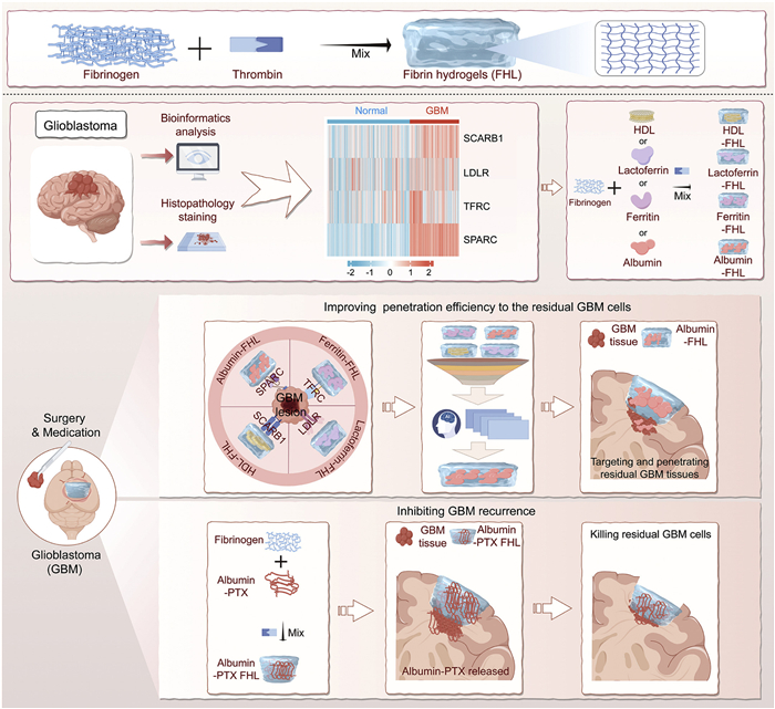
Herein, we designed a multifunctional FHL system loading tumor-penetrating nanoparticles to facilitate their targeting to the residual lesion for postoperative GBM therapy (Fig. 1). The physicochemical properties of FHL and nanoparticle-loaded FHL system were characterized. The loaded nanoparticles were chosen based on the histopathological features of GBM tumors and the bioinformatics analysis. FHL loading albumin nanoparticles were selected to target the residual lesion and inhibit GBM tumor recurrence in a GL261-induced postoperative GBM tumor model.
Figure 1
Figure 1.
Schematic illustration of histopathological features-inspired FHL loading nanoparticles to target and penetrate postoperative residual lesion for GBM therapy. The FHL is fabricated with fibrinogen and thrombin. Inspired by the histopathology of GBM tumors from GBM patients, a FHL loaded albumin (Albumin-FHL) system was optimized from 4 formulations with profound penetrating ability in postoperative lesion of GBM. Then, an Albumin-PTX-FHL was designed for postoperative GBM therapy.
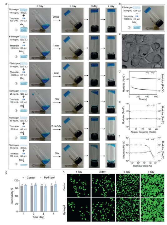
Initially, the formation of FHL system and their physicochemical properties were characterized. To optimize the applicability of FHL system for local administration, 6 FHL formulations were fabricated with fibrinogen ranging from 10 mg/mL to 100 mg/mL and thrombin at 50 or 100 U/mL, respectively (Fig. 2a). The properties and gelation time of these fibrin-based hydrogels were evaluated to determine the optimal concentration of fibrinogen and thrombin (Table S1 in Supporting information). By characterizing the shape fidelity and high elasticity, the optimized FHL formulations was composed of 40 µL of 100 U/mL thrombin, 200 µL fibrinogen (50 mg/mL) and 160 µL phosphate buffered saline (PBS, pH 7.4) (Fig. 2b). The three-dimensional and crosslinked network structure morphology of FHL were confirmed by scanning electron microscopy (SEM) measurements (Fig. 2c and Fig. S2 in Supporting information). As viscoelastic materials, hydrogels show energy storage and energy dissipation characteristics [20]. Rheology test has been employed to evaluate the viscoelastic properties of hydrogel, shear moduli (Pa) of resulting hydrogel were recorded in storage modulus (G′) as indicative of the elastic component and in loss modulus (G″) as indicative of the viscous component of the material [21]. The G′ in the optimal FHL was approximately 10-fold higher than the G″ during the time sweep within 10 min, indicating the formation of the hydrogel network (Fig. 2d). The stability of hydrogel was assessed based on the ratio G′/G″ (~10 in our case), and both G′ and G″ were independent of frequency, where a higher fold difference indicates elastic behavior of stable hydrogels (Fig. 2e). The G′ was dominant over the G″ over a broad strain range, suggesting that FHL had a good linear viscoelastic region (Fig. 2f). Hydrogel swelling was calculated via weight changes initially and up to 72 h after incubating in PBS or artificial cerebral spinal fluid (ACSF) at 37 ℃ (Fig. S3 in Supporting information). Hydrogels demonstrated a low degree of swelling (< 3%). Fig. S4 (Supporting information) showed that the degradation profiles of FHLs in PBS or ACSF containing different concentrations of plasmin. The degradation profile of the FHLs demonstrates that degradation occurs more rapidly in PBS or ACSF containing 10 U plasmin than in PBS or ACSF containing 1 U plasmin. In addition, upon the incubation of FHL with GL261 cells, the cell viability was approximately 100% and negligible cytotoxicity was detected, confirming the good biocompatibility of FHL and their feasibility for biomedical applications (Figs. 2g and h). Moreover, upon the incubation of FHL with BV2 or bEnd3 cells, the cell viability was also approximately 100% (Fig. S5 in Supporting information). As a result, the FHL system exhibited good performance to load nanoparticles for postoperative GBM therapy.
Figure 2
Figure 2.
Preparation and characterization of FHL. (a) Inverted vials of the pristine and hybrid FHL with different fibrinogen/thrombin ratios. (b) Gelation of the optimal FHL. (c) SEM images of the optimal FHL. Scale bar: 100 µm. (d) Dynamic storage modulus (G′) and loss modulus (G″) of the optimal FHL during time sweep analysis. (e) Frequency dependence of dynamic G′ and G″ of the optimal FHL. (f) Representative oscillatory rheological analysis results of the optimal FHL. (g) Viability of cells cultured in FHL extract. (h) Live/dead staining of cells cultured in the control media and FHL media. The living cells were denoted as green signals. Data are presented as mean ± standard deviation (SD) (n = 4). Scale bar: 200 µm.
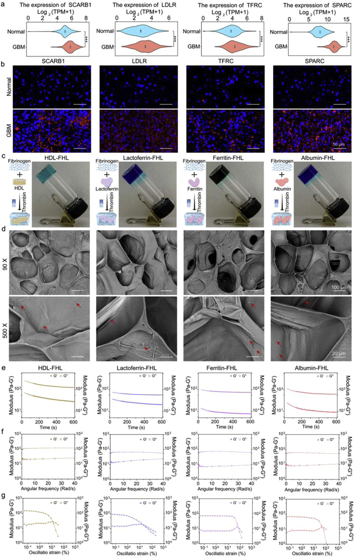
Compelling evidence has verified the advantages of bioinspired nanosized particles as drug delivery carriers to ameliorate their penetration in versatile malignant tumors [22–24]. To develop nanoparticles-loaded FHL system as natural ligand-protein macromers with dual functions of both protein assembly and molecular recognition to specifically target the residual lesion of postoperative GBM, the loading of natural nanosized particles in FHL system was designed in a histopathology-inspired strategy. This targeting strategy that imparts precisely matched, ligand-receptor interactions to efficiently enhance the retention of nanoparticles at the residual lesion of postoperative GBM. The design and selection of targeting ligands mostly depend on the overexpressed receptors in GBM tissues. Characterization of the overexpressed receptors in GBM tissues is a key prerequisite to designing receptor-mediated targeting strategies. The bioinformatics analysis from the database of The Cancer Genome Atlas (TCGA) indicated that some typical receptors, such as scavenger receptor class B type I (SCARB1), low-density lipoprotein receptor (LDLR), transferrin receptor (TFRC), and secreted protein acidic and rich in cysteine (SPARC), were abnormally expressed in the GBM tumors vs. that in normal brain tissues (Fig. 3a). Meanwhile, the aberrant upregulation of these receptors in tumor vs. peritumor tissues from GBM patients was confirmed by immunofluorescence assays (Fig. 3b). The signals of SCARB1, LDLR, TFRC, and SPARC in GBM tumors were obviously detected with stronger fluorescence intensity over those in the peritumor tissues (Figs. 3a and b, Figs. S6–S9 in Supporting information). These highly upregulated receptors provide substantial opportunities to choose natural targeting nano-ligands of high-density lipoprotein (HDL) for SCARB1, lactoferrin for LDLR, ferritin for TFRC, and albumin for SPARC, respectively, which could be loaded in FHL system to target the postoperative residual lesion of GBM [25].
Figure 3
Figure 3.
Design and characterizations of multifunctional FHL system. (a) The violin plots of SCARB1, LDLR, TFRC and SPARC expression in GBM patients (n = 1157) and healthy people (n = 689) isolated from TCGA and Genotype-Tissue Expression determined by Gene Expression Profiling Interactive Analysis. The data were transformed as log2(TPM + 1). (b) SCARB1, LDLR, TFRC and SPARC expression (red signals) in normal and tumor tissues from GBM patients. Scale bars: 50 µm. (c) Schematic illustration and photographs of nanoparticle-loaded multifunctional FHL systems. (d) SEM images of the microstructure of FHL loading different nanoparticles. Arrows indicated nanocarrier particles. Scale bars: 100 µm (90× image), 20 µm (500× image). (e) Dynamic time sweep of all 4 FHL, at the strain of 1.00% and at the frequency of 10 rad/s. (f) Dynamic frequency sweep of all 4 FHL, measured at 1% strain. (g) Strain-dependent oscillatory shear rheology of all 4 FHL with a fixed frequency of 10 rad/s.
On this basis, multifunctional bioinspired FHL system loading these natural nanosized particles (including re-constructed HDL, lactoferrin, ferritin, and albumin) were respectively designed (named as HDL-FHL, Lactoferrin-FHL, Ferritin-FHL, and Albumin-FHL) to evaluate their targeting to the postoperative residual lesion of GBM (Fig. 3c). The solid-like state of these multifunctional hydrogel systems remained unchanged even after 7 days and the results showed that these multifunctional hydrogel systems could still exist stably after 7 days (Fig. S10 in Supporting information). The SEM images showed that these multifunctional FHL systems exhibited porous structure with nanosized particles distributed in the skeleton of the hydrogel, indicating the effective loading of nanoparticles in the FHL system (Fig. 3d, Figs. S11 and S12 in Supporting information). As shown in Fig. 3e, the G′ value for these 4 FHL system were much higher than the G″ value, indicating the formation of hydrogel. In a frequency sweep test at a constant shear strain of 1%, the modulus of each FHL system was independent of the frequency, suggesting that the FHL system could maintain at a high elasticity after intracranial injection (Fig. 3f). Moreover, the G′ values were greater than the G″ values across the swept frequency, and both the G′ and G″ values of these 4 FHL system maintained at a constant value in a certain range of strain, indicating that each FHL exhibited elastic properties within this range. By comparing the G′ and G″ values of these 4 FHL system to the blank-FHL, these nanosized particles would be extensively interpenetrated in the hydrogel network (Fig. S13 in Supporting information). Rheological studies showed that the G′ value of the nanoparticle-loaded FHL system decreased quickly when the strain increased to a certain value, and the overlap of the G′ and G″ curve indicated the complete destruction of the hydrogel system (Fig. 3g). The oscillatory shear strain-to-break values for the HDL-, Lactoferrin-, Ferritin- and Albumin-FHL were 13.65%, 20.46%, 84.25% and 51.83%, respectively (Figs. S14 and S15 in Supporting information), suggesting that the stability of the FHL system would be affected by the loaded nanoparticles in the following order: HDL < lactoferrin < albumin < ferritin.
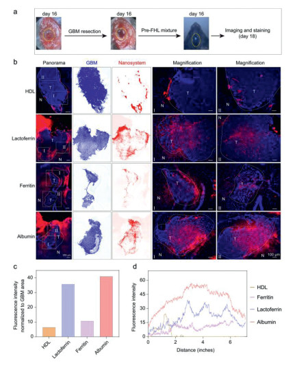
Most importantly, the preferential targeting and penetration in the residual lesion were essential prerequisites to postoperative GBM therapy [26]. The nanosized particles of re-constructed HDL, lactoferrin, ferritin, and albumin were respectively labeled with fluorescence probes, such as DiIC18(7) (DiR) for re-constructed HDL and cyanine5 (Cy5) for other 3 nanoparticles, for the measurements. Their distribution profiles of these nanoparticle-loaded FHL system in the postoperative residual lesion were evaluated in a GL261 with stable expression of luciferase (GL261-luc)-induced orthotopic GBM tumor model. All animal procedures were performed in accordance with the Guidelines for Care and Use of Laboratory Animals of Fudan University and experiments were approved by the Animal Ethics Committee of the School of Pharmacy, Fudan University (approval No. 2025-DWYY-082). At 48 h of intracranial injection, the brain tissue from each group was collected to monitor the fluorescence signals using confocal laser scanning microscopy (CLSM) system (Fig. 4a). As shown in Fig. 4b, the red fluorescence signals of Albumin-FHL and Lactoferrin-FHL system were extensively distributed in the residual tumor lesion with strong fluorescence intensity. In contrast, the signals of HDL-FHL and Ferritin-FHL were only distributed in the margins of the residual GBM tumor tissues (Fig. 4b and Fig. S16 in Supporting information). The image analysis indicated the normalized fluorescence intensity in the tumor area and the fluorescence intensity from tumor edge to interior regions of tumor lesion was at the highest level in Albumin-FHL system, followed by Lactoferrin-FHL, and remained at lower level in Ferritin-FHL and HDL-FHL groups (Figs. 4c and d). These data confirmed the intriguing preference of Albumin-FHL system for targeting and penetrating the postoperative residual lesion, which could be used for postoperative GBM therapy.
Figure 4
Figure 4.
Specific targeting and intratumor distribution of 4 nanoparticle-loaded FHL in postoperative residual GBM tumors. (a) Surgical tumor debulking of GL261-luc induced orthotopic GBM model at 16 after inoculation. Yellow circles show the cranial window for surgery. (b) Specific targeting of DiR or Cy5-labeled multifunctional FHL (red signals) to the postoperative residual GBM lesions (inner-circle). Scale bars: 250, 100 µm. The segmentation images displayed nanosystem distribution (red) within the tumor tissue (dark blue areas). N = normal, T = tumor. (c) The normalized fluorescence intensity of nanoparticle-loaded FHL in residual tumor. (d) Image analysis of nanoparticle-loaded FHL in residual tumor using ImageJ software.
Encouraged by the tumor-targeting and penetrating ability of Albumin-FHL in postoperative residual lesion GBM tumor, the Cy5-labeled albumin-paclitaxel nanoparticle loaded FHL system (Albumin-PTX-Cy5-FHL) was prepared to evaluate their physicochemical properties. The in vitro release results indicate that Albumin-Cy5-PTX-FHL could promote the sustained release of the loaded cargoes in ACSF. The former rapid-release phase was likely due to the unencapsulated Albumin-PTX-Cy5 during gel formation, and the later sustained-release phase was probably attributed to the degradation of the scaffold (Fig. S17 in Supporting information). The cellular uptake of Cy5-labeled Albumin-PTX-FHL system in GL261, BV2 or bEnd3 was confirmed by CLSM imaging (Fig. S18 in Supporting information). Cy5-labeled Albumin-PTX was mainly internalized by GL261 cells and less uptake by bEnd3 cells. In addition, the internalization of Cy5-labeled Albumin-PTX by BV2 cells was observed due to the phagocytic activity of microglia. Based on the above results, the Albumin-Paclitaxel nanoparticle loaded FHL system (Albumin-PTX-FHL) was prepared to evaluate their therapeutic efficacy in the GL261-luc induced orthotopic GBM tumor model (Fig. 5a). The SEM images showed that the Albumin-PTX loaded FHLs exhibited porous structure with nanosized particles distributed in the skeleton of the hydrogel, indicating the effective loading of Albumin-PTX nanoparticles in the FHL system (Fig. S19 in Supporting information). The specific targeting and penetrating of Cy5-labeled Albumin-PTX-FHL system to the postoperative residual lesion was confirmed by CLSM imaging (Fig. 5b), which was comparable to that of aforementioned albumin-FHL system. Then, the therapeutic effect of Albumin-PTX-FHL was evaluated in postoperative GBM model by measuring tumor recurrence and survival prolongation. The procedure of surgical tumor resection from the GL261-luc induced orthotopic GBM tumor model and hydrogel implantation was shown in Fig. 5c. On day 16 of tumor inoculation, the orthotopic GBM tumors were performed surgical resections, and then intracranially administered with blank FHL or Albumin-PTX-FHL at 5 mg/kg of PTX after surgery. The tumor recurrence was monitored by measuring the bioluminescence signals from each group. As presented in Fig. 5d, the bioluminescent signal of the mice brain in the blank FHL group was significantly increased within 25 days, suggesting rapid tumor recurrence. As illustrated by the bioluminescence imaging, the residual lesions in blank FHL treated group regrew rapidly after surgery and caused 100% fatality at day 14 after surgery (Figs. 5d and e). In contrast, in Albumin-PTX-FHL treated group, the increase of the bioluminescent signals was largely reduced and maintained at lower level within 25 days, indicating the effective retardation of tumor recurrence (Figs. 5d and e). Moreover, Albumin-PTX-FHL treatment was effective in prolonging survival time and rate with a median survival time of 43 days (27 days after surgery), which was much longer that in the blank-FHL group (28.5 and 12.5 days after surgery) (Fig. 5f). As a result, the Albumin-PTX-FHL treatment could produce a 2.16-fold prolongation of survival time vs. the FHL group, confirming the effectiveness of this multifunctional nanoparticle-loaded FHL system in postoperative GBM therapy. In addition, the major organs of mice were stained for histopathological examinations. The microstructures of the major organs from the FHL and Albumin-PTX-FHL group were clear and cellular morphogenesis were in regular arrangement. Moreover, no hemorrhage, necrosis, or inflammatory cell infiltration were observed (Fig. S20 in Supporting information). These data indicated the good biocompatibility of the FHL treatment.
Figure 5
Figure 5.
Therapeutic efficacy of intracranial Albumin-PTX-FHL in postoperative GBM model. (a) Schematic illustration of designing Albumin-PTX-FHL. (b) Specific targeting of Cy5-labeled Albumin-PTX-FHL (red signals) to postoperative GBM tumor lesion. N = normal, T = tumor. Scale bar: 100 µm. (c) Schematic illustration of evaluation. (d) Representative images of GBM recurrence from each treatment. (e) Quantified results of bioluminescent signals from each group. (f) Survival of intracranial Albumin-PTX-FHL treatment in postoperative GBM model. Data are mean ± SD (n = 6). **P < 0.01.
In summary, inspired by the histopathological features of GBM, an albumin nanoparticle loaded FHL system (Albumin-FHL) was optimized from 4 formulations with notable targeting and penetrating ability in postoperative residual lesion of GBM. Compared to the control group, the Albumin-PTX-FHL treatment dramatically retarded the GBM tumor recurrence after surgery and produced a 2.16-fold prolongation of survival in GL261-induced orthotopic postoperative GBM model. Therefore, the histopathological features-inspired FHL loading albumin nanoparticles provides an encouraging delivery platform to target and penetrate postoperative residual lesion for GBM therapy.
The authors declare that they have no known competing financial interests or personal relationships that could have appeared to influence the work reported in this paper.
Acknowledgments
Financial supports by the National Key Research and Development Program of China (Nos. 2023YFE0104300, 2022YFC2304303), National Natural Science Foundation of China (Nos. 82574334, 32271461), Scientific Research Project of Shanghai Fifth People’s Hospital, Fudan University (No. 2023WYRH08) and the Postdoctoral Fellowship Program of CPSF (No. GZC20230547), were acknowledged.
Supplementary materials
Supplementary material associated with this article can be found, in the online version, at doi:10.1016/j.cclet.2025.112232.
[1]
L.B. Wang, A. Karpova, M.A. Gritsenko, et al., Cancer Cell 39 (2021) 509–528 e20. doi: 10.22331/q-2021-07-26-509
Figure 1
Schematic illustration of histopathological features-inspired FHL loading nanoparticles to target and penetrate postoperative residual lesion for GBM therapy. The FHL is fabricated with fibrinogen and thrombin. Inspired by the histopathology of GBM tumors from GBM patients, a FHL loaded albumin (Albumin-FHL) system was optimized from 4 formulations with profound penetrating ability in postoperative lesion of GBM. Then, an Albumin-PTX-FHL was designed for postoperative GBM therapy.
Figure 2
Preparation and characterization of FHL. (a) Inverted vials of the pristine and hybrid FHL with different fibrinogen/thrombin ratios. (b) Gelation of the optimal FHL. (c) SEM images of the optimal FHL. Scale bar: 100 µm. (d) Dynamic storage modulus (G′) and loss modulus (G″) of the optimal FHL during time sweep analysis. (e) Frequency dependence of dynamic G′ and G″ of the optimal FHL. (f) Representative oscillatory rheological analysis results of the optimal FHL. (g) Viability of cells cultured in FHL extract. (h) Live/dead staining of cells cultured in the control media and FHL media. The living cells were denoted as green signals. Data are presented as mean ± standard deviation (SD) (n = 4). Scale bar: 200 µm.
Figure 3
Design and characterizations of multifunctional FHL system. (a) The violin plots of SCARB1, LDLR, TFRC and SPARC expression in GBM patients (n = 1157) and healthy people (n = 689) isolated from TCGA and Genotype-Tissue Expression determined by Gene Expression Profiling Interactive Analysis. The data were transformed as log2(TPM + 1). (b) SCARB1, LDLR, TFRC and SPARC expression (red signals) in normal and tumor tissues from GBM patients. Scale bars: 50 µm. (c) Schematic illustration and photographs of nanoparticle-loaded multifunctional FHL systems. (d) SEM images of the microstructure of FHL loading different nanoparticles. Arrows indicated nanocarrier particles. Scale bars: 100 µm (90× image), 20 µm (500× image). (e) Dynamic time sweep of all 4 FHL, at the strain of 1.00% and at the frequency of 10 rad/s. (f) Dynamic frequency sweep of all 4 FHL, measured at 1% strain. (g) Strain-dependent oscillatory shear rheology of all 4 FHL with a fixed frequency of 10 rad/s.
Figure 4
Specific targeting and intratumor distribution of 4 nanoparticle-loaded FHL in postoperative residual GBM tumors. (a) Surgical tumor debulking of GL261-luc induced orthotopic GBM model at 16 after inoculation. Yellow circles show the cranial window for surgery. (b) Specific targeting of DiR or Cy5-labeled multifunctional FHL (red signals) to the postoperative residual GBM lesions (inner-circle). Scale bars: 250, 100 µm. The segmentation images displayed nanosystem distribution (red) within the tumor tissue (dark blue areas). N = normal, T = tumor. (c) The normalized fluorescence intensity of nanoparticle-loaded FHL in residual tumor. (d) Image analysis of nanoparticle-loaded FHL in residual tumor using ImageJ software.
Figure 5
Therapeutic efficacy of intracranial Albumin-PTX-FHL in postoperative GBM model. (a) Schematic illustration of designing Albumin-PTX-FHL. (b) Specific targeting of Cy5-labeled Albumin-PTX-FHL (red signals) to postoperative GBM tumor lesion. N = normal, T = tumor. Scale bar: 100 µm. (c) Schematic illustration of evaluation. (d) Representative images of GBM recurrence from each treatment. (e) Quantified results of bioluminescent signals from each group. (f) Survival of intracranial Albumin-PTX-FHL treatment in postoperative GBM model. Data are mean ± SD (n = 6). **P < 0.01.


 DownLoad:


 下载:


 下载:

