wnwang@ahmu.edu.cn (W. Wang). 1 These authors contributed equally to this work.
Received Date:
06 July 2025 Accepted Date:
04 December 2025 Revised Date:
03 December 2025 Available Online:
15 November 2026
Abstract:
The pathogenesis of inflammatory bowel disease (IBD) is complex and multifactorial, involving intestinal microbiota imbalance, immune system abnormalities, genetic predispositions, and other factors. Therefore, the attainment of a complete cure for IBD through a single therapeutic modality remains a formidable challenge. In recent years, probiotics have received considerable attention in the treatment of IBD due to their properties of regulating intestinal microecology, immune activation, anti-infection and metabolic regulation. In order to further improve the safety, efficacy and clinical translation prospects of probiotics in the treatment of IBD, the engineering modification of probiotics has become the current research focus. In this review, we systematically summarize the probiotic species with promising clinical applications. Subsequently, the primary methods of engineering modifications (physical, chemical, genetic, etc.) are described and the role of these methods and their mechanisms in the treatment of IBD using engineered probiotics are discussed. Finally, we explore in depth the current challenges in the field and proposes potential future solutions. This review aims to present a comprehensive perspective on the development of a new generation of engineered probiotics and their application in IBD therapy.
Inflammatory bowel disease (IBD) is a chronic immune-mediated disease of the gastrointestinal tract, classified as Crohn’s disease (CD) and ulcerative colitis (UC). The aetiology of IBD is thought to be multifactorial, with genetic susceptibility, immune system dysfunction and environmental influences being recognised as potential risk factors [1,2]. At present, the clinical treatment of IBD is primarily focused on approaches such as immune modulation, anti-inflammatory therapies, and microbiome regulation [3]. In addition, some studies have attempted to treat IBD by promoting mucosal healing without immunosuppression [4]. The drugs commonly used to treat IBD include 5-aminosalicylate, antibiotics, etc., which can relieve the symptoms of IBD but cannot achieve a complete cure. Furthermore, the clinical utilization of these medications is constrained by the severity and location of the disease, and their adverse effects cannot be disregarded [5-7]. For instance, although the utilization of corticosteroids (CSs) has been demonstrated to alleviate mild CD, it has also been observed to cause adverse effects, including hypertension and opportunistic infections [8,9]. Similarly, 11%–40% of patients treated with Sulfasalazine experience side effects such as fatigue, nausea, abdominal pain, and diarrhea [10]. However, when probiotics are used as the primary therapeutic modality, the situation changes.
A plethora of microorganisms have been identified within the intestinal tract, with the composition of these microorganisms exerting a substantial influence on the health of the gastrointestinal system [11-14]. This phenomenon has prompted us to explore the potential application of probiotics in the treatment of IBD. In fact, probiotics possess a number of properties that make them promising candidates for treating IBD. First, probiotics (e.g., Bifidobacterium bifidum) can compete with harmful bacteria in the gut for intestinal surface receptors and reduce their binding in the gut, thus preventing the deterioration of IBD [15]. Secondly, some of the active substances secreted by probiotics (e.g., short-chain fatty acids) have also been shown to slow down inflammation [16,17]. Concurrently, intestinal probiotics are the components of the body’s immune system, and the bioactive substances secreted by probiotics, such as lactobacilli, can regulate the intestinal immune function [18]. Despite the non-negligible potential of probiotics in clinical therapy, there are still some challenges in the clinical management of IBD. First, probiotics often struggle to survive within the intestinal environment and exhibit limited targeting capabilities, which diminishes their therapeutic efficacy. Additionally, the excessive activity of probiotics may become a latent risk in IBD treatment. For example, in patients with compromised intestinal mucosa, probiotics may translocate into the bloodstream through lesions, leading to bacteremia [19]. Furthermore, the toxic substances produced by the metabolism of probiotics have the potential to adversely impact patient health. Lactobacillus spp. has been associated with the development of D-lactic acidosis in patients with short bowel syndrome [20]. These shortcomings limit the utilization of probiotics in the management of IBD. Consequently, there is a pressing need for engineering modification of probiotics to mitigate these adverse effects.
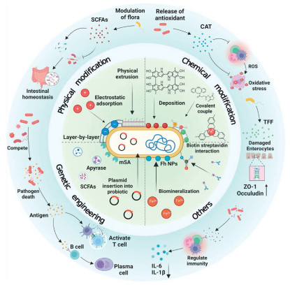
In recent years, probiotic engineering technology and novel material science have been rapidly developed, and they have attracted extensive attention from scientists seeking to modify the functional properties of probiotics. Through physicochemical modification or encapsulation with nanomaterials, probiotics can be microencapsulated, thereby resisting the interference of gastric acid and enhancing their survivability in the intestinal tract [21,22]. Furthermore, the clearance of probiotics by the immune system of the host represents a limiting factor for this approach. However, certain novel nanomaterials and biological membranes can modulate the host immune response, preventing the clearance of probiotics and thereby enhancing the bioactivity and bioavailability of oral formulations [23-26]. The encapsulation of probiotics with adhesive materials has been demonstrated to enhance the ability of probiotics to colonize the intestines, thereby facilitating prolonged treatment within the intestinal tract [22]. Furthermore, genetically engineered probiotics can act as “cell factories” for therapeutic molecules (expressing therapeutic substances directly on the cell surface) [27]. In comparison to wild-type probiotics, engineered probiotics offer distinct advantages, including enhanced aggregation at sites of inflammation, facilitation of targeted drug delivery, and sustained release. In summary, physical and chemical engineering modifications of probiotics can improve their survival in the intestine while enhancing their colonization ability. Genetic engineering further endows probiotics with novel functionalities, thereby enhancing their potential for the treatment of IBD (Fig. 1).
Figure 1
Figure 1.
Schematic synthesis of engineered modified probiotics. Mechanism of engineered modified probiotics for the treatment of IBD can be divided into chemical modification, physical modification and genetic engineering and other methods. By exploiting the structure of probiotics, the surface composition and structure of the bacteria can be altered by surface modification, or the biological properties of the bacteria can be altered and given new functions by the importation of target genes.
In this review, we firstly highlight several probiotics with clinical therapeutic potential and explore the reasons for their suitability in the treatment of IBD, including the secretion of anti-inflammatory substances and the antagonism of harmful microbial populations. Next, we further elucidated the impact of physical, chemical and genetic engineering modifications on probiotic therapies for IBD, such as enabling targeted colonization and transforming probiotics into cellular factories for the secretion of therapeutic agents. Finally, we discuss the prospects and potential challenges associated with the use of engineered probiotics in the treatment of IBD (Table S1 in Supporting information).
2.
Probiotics with potential therapeutic value in the treatment of IBD
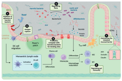
With the ongoing advancements in science and technology, it has been acknowledged that certain microorganisms may offer therapeutic potential. These microorganisms are referred to as probiotics, defined as “non-pathogenic, live microorganisms that, when administered in sufficient quantities, provide health benefits to the host [28]”. Probiotics have demonstrated a range of beneficial properties, including immunomodulatory, anti-inflammatory, beneficial substances secretion and regulation of intestinal barrier permeability. These properties have contributed to the widespread application in the medical field, particularly in the prevention and treatment of diseases [29-33]. Subsequently, some probiotics with the potential to be used in clinical therapy will be presented. By engineering these probiotics and developing corresponding probiotic supplements, it is expected that the therapeutic efficacy in the treatment of intestinal inflammation can be further enhanced (Fig. 2). For details of potential probiotics for IBD treatment, please refer to Sections S2.1-S2.3 (Supporting information) [34-74].
Figure 2
Figure 2.
Intestinal probiotics play beneficial roles in the gut. (A) Probiotics inhibit harmful bacteria from adhering to the intestine. (B) Dendritic (DC) cells capture antigens to achieve the differentiation of immune cells. (C) Probiotics regulate intestinal flora. (D) The intestinal immune environment regulated by probiotics. (E) Bifidobacteria secrete SCFAs to regulate the release of various cytokines. (F) Probiotics repair the intestinal mucosa by upregulating the expression of various intestinal proteins.
3.
Methods of engineering modified probiotics and their therapeutic effects
In order to enhance the therapeutic efficacy of probiotics in patients with IBD, it is imperative to ascertain that a sufficient number of probiotics are capable of successfully colonizing the gut and sustaining stable proliferation. Currently, the engineering of probiotics is undergoing rapid development, with the emergence of novel materials providing a valuable opportunity to address the aforementioned issues [26,75,76]. Through the application of engineering techniques, the survival and colonization capabilities of probiotics within the intestinal environment can be enhanced. Furthermore, the implementation of particular nano-coating technologies enables the incorporation of exogenous functions, such as immunomodulation, antioxidant and anti-inflammatory properties in probiotics. This process serves to augment their efficacy in the treatment of colitis, while concurrently ensuring the safety of the treatment process [77,78]. Therefore, by employing synthetic biology or physicochemical modification techniques, we can engineer probiotics to enhance their clinical therapeutic effects. This can be achieved by introducing new functional groups on the surface of probiotics or by enabling the expression of specific membrane ligands and receptors, thereby altering the interactions between probiotics and host extracellular matrix proteins (e.g., fibrinogen, collagen) [79].
3.1
Physical modification
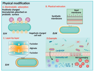
Probiotics are rich in proteins, and their isoelectric point is usually below 5.0, which is usually lower than the pH of the intestine. In addition, the cell walls of probiotics are rich in peptidoglycans and teichoic acids, which together result in the negative charge characteristics of the probiotic surface. This provides the ability to adsorb positively charged materials, thus providing a feasible way for probiotic surface modification. At this stage, physical methods such as electrostatic adsorption and mechanical extrusion have been applied in the field of probiotic engineering modification (Fig. 3).
Figure 3
Figure 3.
Schematic diagram of physical modification technology for probiotics in IBD treatment. (A) Technological mechanism of modification of probiotics by electrostatic interactions. (B) Technological mechanism of modification of probiotics by physical extrusion. (C) Technological mechanism of modification of probiotics by layer-by-layer assembly. (D) Physical modification of probiotics achieves alleviation of IBD by scavenging intestinal ROS, regulating intestinal flora and inhibiting anti-inflammatory responses in the intestine.
Bacterial cell walls carry abundant peptidoglycans and teichoic acids, resulting in a predominantly negative charge on the surface of most bacterial cells. Consequently, cationic polymers or biomaterials with a positive charge, can be readily adsorbed onto bacterial surfaces through electrostatic interactions, which offers a promising approach for bacterial modification [80]. The microbial-small molecule drug synergistic therapeutic system holds significant potential for disease intervention, yet its clinical application is hindered by challenges such as limited bioavailability and premature release of non-specific drugs. To address these challenges, Kuang et al. developed a reactive oxygen species (ROS)-responsive delivery system, employing a multistage mechanism. First, cationized drug conjugates are electrostatically adsorbed onto the Escherichia coli Nissle 1917 (EcN) surface, forming a protective nanocoating. This coating enhances EcN survival in acidic gastric environments and under antibiotic exposure, while synchronizing therapeutic drug release with cinnamaldehyde. Thereafter, charge reversal, achieved by negatively charged thiolated chitosan, induces dissociation of the nanocoating, leading to synchronous release of living bacteria, enabling targeted EcN release in the intestinal compartment [81].
3.1.2
Physical extrusion
Physical extrusion is a novel technique for preparing cell membrane-encapsulated bacteria. This method is based on the principle that extrusion disrupts the cell membrane structure and allows it to reintegrate around the bacteria. This technique not only facilitates the effective circumvention of numerous challenges encountered by probiotic therapies in vivo applications, such as rapid clearance and low therapeutic efficiencies, but also contributes to the mitigation of adverse effects associated with immunogenicity. Lin et al. successfully encapsulated EcN within a yeast membrane (YM) that is rich in β-glucan by employing a physical co-extrusion method. The encapsulated EcN was able to activate Dectin-1 receptor-mediated phagocytosis of M cells in the intestinal epithelium, promoting a strong mucosal immune response, which effectively inhibited the growth of pathogens such as Salmonella and Escherichia-Shigella. At the same time, it ensured the stable colonization of commensal bacteria in the infected intestine. Notably, the structural design can markedly reduce intestinal barrier damage and limit the translocation of probiotics and systemic inflammatory response, thereby effectively preventing the occurrence of bacteremia during the treatment process [82].
3.1.3
Layer by layer (LbL) assembly
LbL assembly is a very effective technique for encapsulating probiotics by sequentially depositing small amounts of oppositely charged polymers on the probiotic surface. Chitosan is widely used in microbial encapsulation as classic LbL self-assembly materials. Han et al. encapsulated EcN in liposomes containing curcumin, which were subsequently encapsulated with chitosan-containing polymers using a layer-by-layer technique. Under simulated gastrointestinal conditions, the survival rate, adhesion to colonic mucus and ROS scavenging ability of the encapsulated EcN were significantly improved. Furthermore, it was determined that the coating could act as a protective barrier, thereby ensuring the effective scavenging of reactive oxygen radicals and the survival of probiotics within an inflammatory environment [83].
For details on the advantages of physically engineered modifications of probiotics for IBD, please refer to Sections S3.1 (1)-(3) (Supporting information) [21,84-87].
3.2
Chemical modification
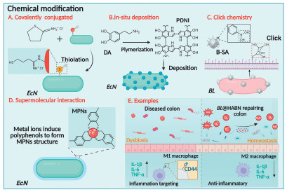
Proteins, phospholipids, and other functional groups present on the surface of probiotics, which are capable of reacting with various chemicals, form the foundation for their chemical modification. Consequently, when microorganisms interact with functional materials, the process of coating probiotics with these materials through chemical reactions involving functional groups is classified as chemical modification. This chemical coating enables synergistic treatment, leading to more effective attenuation of inflammatory infiltrates and the associated pathological processes (Fig. 4).
Figure 4
Figure 4.
Schematic diagram of chemical modification technology for probiotics in IBD treatment. (A) Technological mechanisms of probiotic modification by covalent binding. (B) Technological mechanisms of probiotic modification by in situ deposition. (C) Technological mechanisms of probiotic modification by click chemistry. (D) Technological mechanisms of probiotic modification by supramolecular interactions. (E) Chemically engineered modification of probiotic bacteria for treatment of IBD by scavenging intestinal ROS, regulating intestinal flora, and inhibiting the intestinal anti-inflammatory response.
Covalent binding is a prevalent method of chemical modification, which principally involves the formation of covalent bonds between covalent binding sites on the surface of the probiotic and a chemical substance, thereby achieving particular probiotic surface modifications. The introduction of functional groups on the surface of the probiotic by covalent binding serves to alter the interaction with the gut, which can be employed to enhance the colonization of the probiotic in the intestine. Liu et al. utilized Traut’s reagent to introduce free thiol groups onto the surface of EcN, thereby enabling it to undergo a catalyst-free thiol-disulfide exchange with mucins while exhibiting thiolation level-dependent attachment. Bacteria containing 9.3 × 10⁷ thiols exhibited a 170-fold increase in attachment capacity in mucin-rich jejunum, thus demonstrating a strategy that significantly improves the adhesion of orally administered probiotics in the gut and solves the problem of probiotic colonization in the intestine [88].
3.2.2
In-situ deposition
In-situ deposition technology is a strategy developed from nanocomposites that involves the deposition of colloidal particles or inorganic minerals on or within the surface of bacteria. This process is induced through the co-incubation of the probiotic with reactive monomers and associated catalysts and driven by covalent bonding and non-covalent interactions, resulting in the formation of multilayered protective encapsulated structures [89]. Polydopamine (PDA), a synthetic material inspired by an adhesive protein found in mussels, has been widely used as a coating for various surfaces due to the strong adhesion of its catechol groups [90]. The study by Pan et al. has demonstrated that the PDA coating significantly enhances the survival rate and intestinal colonization capacity of EcN under simulated gastrointestinal stress conditions. Additionally, the electrostatic interactions between the positively charged chitosan and the PDA coating further functionalize the modification, improving EcN’s mucosal adhesion and drug-carrying capacity [91].
3.2.3
Click chemistry
The mild reaction conditions (insensitivity to water, oxygen, etc.) and bio-orthogonality (low levels of azides and alkynes in vivo) of click chemistry have greatly facilitated its biomedical applications. Recently, Cao et al. has developed an artificial enzyme-armed Bifidobacterium longum (BL) using click chemistry. In this platform, a single-atom catalyst (B-SA) was attached to the surface of BL by click chemistry to form BL@B-SA50, and the artificial enzyme on the surface could mimic the antioxidant enzymes of superoxide dismutase (SOD) and catalase (CAT) to scavenge various ROS. Treatment of mice with this bacterium resulted in a reduction in the levels of interleukin-6 (IL-6) and myeloperoxidase (MPO), while the abundance of Lachnospiraceae was increased and the intestinal barrier was restored, thereby demonstrating the potential of this bacterium in the treatment of IBD [92].
3.2.4
Supramolecular interaction
Metal-ligand supramolecular self-assembly represents a significant component of supramolecular interactions, with metal ions capable of inducing the self-assembly of certain chemicals into specific structures. The adsorption of metal ions onto the surface of a biofilm has been shown to guide the self-assembly of chemicals onto probiotic bacteria, thereby achieving multifunctionality of the bacterial surface. Liu et al. encapsulated EcN by vortexing with dioleoylphosphatydic acid (DOPA) and cholesterol in calcium phosphate buffer and formed lipid membrane coated bacteria, which was more tolerant to acidic environments, and its survival and bioavailability in the mouse stomach was significantly increased compared with that of the unencapsulated bacterium. Furthermore, the treated mice with enteritis exhibited a substantial reduction in inflammation and exhibited significantly lower serum IL-6 and IFN-γ levels, thereby substantiating the augmented preventive and therapeutic efficacy of the engineered bacteria in mouse models of Salmonella typhimurium and dextran sodium sulfate (DSS)-induced colitis [93].
For details on the advantages of chemically engineered modifications of probiotics for IBD, please refer to Sections S3.2 (1)-(5) (Supporting information) [94-109].
3.3
Genetic engineering
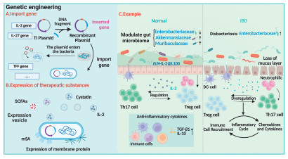
The genetic engineering of probiotics has been demonstrated to endow these strains with novel functionalities that were not present in their original state, thereby enhancing their efficacy in the treatment of IBD. These new features mitigate therapeutic side effects by either secreting therapeutic substances not expressed by the probiotic (e.g., IL-2) or by reducing the toxicity of the probiotic itself. Furthermore, this method of modifying strains can be used for a number of other applications, such as vaccine or drug delivery, mimicking surface receptors, targeting specific toxins or pathogens, and modulating host immune responses [110]. The subsequent discussion will address the alterations imposed by various prevalent genetic engineering techniques on probiotics, along with their implications in the management of IBD (Fig. 5).
Figure 5
Figure 5.
Schematic diagram of genetic engineering technology for probiotics in IBD treatment. (A) Introduction of target genes into probiotic bacteria. (B) Genetically modified probiotics have been found to express vesicles that secrete SCFAs, interleukins, or membrane proteins on their surface. (C) EcN regulates intestinal flora and the intestinal immune system by secreting IL-2 in the gut after genetic modification and physicochemical modifications.
3.3.1
Introduce target genes to enhance probiotic colonization and targeted therapeutic efficacy
The precise colonization of probiotics in intestinal delivery is a prerequisite for the use of probiotics in the treatment of IBD. To date, many systems have been explored to achieve controlled probiotic therapy, and optogenetics stands out among the many regulatory systems for precise intestinal delivery due to its precision and controllability within a narrow spatiotemporal window. Cui et al. used this principle to genetically modify EcN to secrete “functional cellular glues” induced by transdermal blue light in vitro to achieve precise spatiotemporal colonization in the intestine, while further secretion of transforming growth factor beta 1 (TGF-β1) at the site of inflammation also restored the levels of IL-6, tumor necrosis factor alpha (TNF-α), interferon gamma (IFN-γ) and MPO in the intestine to normal levels to achieve control of the intestinal inflammatory response [111]. In addition, an important issue in this therapeutic area is the retention time of microorganisms in the gastrointestinal tract. To address the challenge, Heavey et al. drawing inspiration from a range of gut-resident microbes, probiotics and several pathogens that express cell surface proteins known as adhesins, has developed a platform that expresses monomeric streptavidin on the surface of the Saccharomyces boulardii (S.b.) cell. This platform utilizes tunable antibody achieve binding of S.b. to extracellular matrix proteins, thereby enabling targeted therapy. The results demonstrated that this strategy significantly prolonged the residence time of S.b. in the gastrointestinal tract, while concomitantly increasing the concentration of S.b. by 100-fold. Furthermore, the study revealed that this genetic modification did not affect the probiotics’ original gene expression ability [112].
In order to enhance the precision and manageability of probiotic therapy, apart from the aforementioned strategies, it is feasible to establish inducible systems in which their expression can be activated in response to inducers, thereby modulating the synthesis of exogenous proteins. IBD, akin to numerous other diseases, possesses numerous biomarkers, including nitric oxide (NO), calprotectin, and others [113]. Genetically engineered E. coli by McKay et al. was utilized to establish a smart “engineered probiotic” platform, wherein the native promoter of EcN strain promoted NO sensing at the site of inflammation, which activates gene expression and regulates macrophage-colony stimulating factor synthesis and release. Concurrently, it accumulates at the site of inflammation via pseudotaxis, thereby realizing targeted aggregation at the inflammation site. This approach has been shown to effectively reduce systemic side effects and improve therapeutic efficacy, thus enhancing the effectiveness of IBD therapy [114].
3.3.2
Introduce targeted genes to turn probiotics into “cell factories” for therapeutics
In comparison with oral and parenteral drug delivery, probiotics, as endogenous “drug factories”, are more suitable for the delivery of small molecule proteins and peptides. This is due to the fact that they can effectively avoid drug degradation and immune effects [115]. Concurrently, gene modification has the capacity to compensate for defects in the encoded product, thereby enhancing its efficacy in actual treatment scenarios.
For details on the advantages of genetically engineered modifications of probiotics for IBD, please refer to Sections S3.3 (1)-(4) (Supporting information) [116-151].
3.4
Other engineering methods to modify probiotics
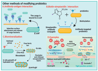
In addition to the modifications of probiotics’ surfaces or genes through physical, chemical, and genetic engineering, probiotics themselves are capable of being metabolized and biosynthesized on their surfaces. Furthermore, the biological activity of substances such as surface proteins can be modified by coupling specific therapeutic substances to the surface through receptor-ligand interactions or biotin-streptavidin interactions (Fig. 6).
Figure 6
Figure 6.
Schematic diagram of other modification technology for probiotics in IBD treatment. (A) Diagram of the mechanism of antigen-antibody interaction modification of probiotics. (B) Diagram of the mechanism of biotin-streptavidin interaction modification of probiotics. (C) Diagram of the mechanism of in situ mineralization modification of probiotics. (D) Engineered probiotics alleviate IBD by mimicking artificial enzyme activity, modulating the intestinal microenvironment, decreasing neutrophil infiltration, and repairing the intestinal mucosa.
Receptor-ligand interaction is the basis for the modification of various cargoes, including small-molecule drugs and biomolecules (e.g., proteins, DNA, and carbohydrates) on the surface of probiotic bacteria. This modification process can be used for sensitive identification of early-stage disease biomarkers, and the development of innovative materials for biomedical applications. Fernandes et al. developed a two-antibody-based method for attaching and releasing motile, cargo-carrying bacteria from patterned surfaces. The employment of antibodies in this method ensures a high degree of selectivity, allowing for the targeted attachment of bacteria (e.g., E. coli) of a specific species and the placement of these biomaterials at designated receptors on the bacterium. The methodology for surface attachment utilized in this study enables the coating of surfaces with bacteria-cargo conjugates, which can be released in a time-dependent manner and in response to specific chemical triggers [152].
3.4.2
Biotin-streptavidin coupling
Biotin-streptavidin coupling is a bio-coupling method based on an extremely strong non-covalent interaction between biotin (a small molecule of vitamin B7) and streptavidin (a protein from probiotic bacteria), which is considered to be the strongest protein-ligand interaction known in nature [153]. Due to its high affinity, it is widely utilized in fields such as immunohistochemistry and protein purification, and becomes a common strategy for probiotic modification. Vargason et al. utilized N-hydroxysuccinimide chemistry to couple biotin to the surface of EcN, subsequently binding monoclonal antibodies modified with streptavidin to the biotinylated EcN. This modification significantly enhanced the ability of the probiotic to adhere to Caco-2 cells. Furthermore, the synthesized adhesins resulted in a stronger adhesion of the pairs of probiotics to the gut, thereby reducing the attachment of pathogenic bacteria to the gut to a certain extent [154].
3.4.3
Biomineralization
A significant number of unicellular organisms possess outer membranes that serve as templates for biomineralization, facilitating the formation of functional coatings in the form of thin mineral layers. Inspired by natural mineralization, modifying cell surfaces by introducing chemical functionality into cells through biomimetic mineralization strategies may become a universal strategy for improving organisms [155,156]. Chen et al. developed in situ growth of Fh NPs (hydrated iron nanoparticles) on Lactobacillus casei, which exhibited robust catalase-like activity over a wide pH range to effectively degrade ROS, while the encapsulation of the surface coating ensured better viability of the probiotics in the gut. The fucoidan-derived protective shield further encapsulated the probiotic and endowed it with the inflammatory targeting function, and inflammatory factors such as IL-6, IL-1β were downregulated in treated enteritis mice, while the intestinal flora is also remodelled, therefore decelerating the progression of intestinal inflammation [157].
Interactions such as receptor-ligand interactions and biotin-streptavidin have been demonstrated to facilitate the targeting of probiotics in intestinal therapy and their adherence in the gut. Furthermore, the introduction of therapeutic substances such as Fh NPs into the surface of probiotics through biomineralization has been shown to maintain the balance of the intestinal flora and achieve the alleviation of IBD through the regulation of intestinal oxidative stress.
4.
Conclusion and perspectives
This review outlines the limitations of current clinical therapies for the treatment of IBD, introduces several probiotics with promising clinical applications, and provides a brief overview of the basic methods for engineering the modification of these probiotics and their use in the treatment of IBD. The multifunctional properties of probiotics, including regulation of the intestinal microenvironment, immune system stimulation, anti-infection properties, and metabolic regulatory functions, render them promising candidates for drug delivery platforms in the treatment of IBD. Probiotics are engineered to exhibit significantly higher resistance to gastric acids and enzymes in the intestinal tract, thereby ensuring their enhanced ability to survive in this environment. The construction of response systems has also been demonstrated to promote the targeting of probiotics in the intestinal tract, thereby improving the course of treatment. In addition, the antioxidant capacity conferred by surface-coated biomaterials has been shown to significantly inhibit oxidative stress in the gut and promote mucosal healing by altering the composition of the flora. In the context of IBD treatment with probiotics, the potential for probiotics to infect wounds and cause bacteraemia due to excessive motility has been a subject of concern. This issue can be addressed by encapsulating a more adhesive substance on the surface of the probiotic, which not only prevents the probiotic from being rapidly cleared in the gut, but also enables prolonged treatment. Genetically engineered probiotics can also be used as “cell factories” to achieve immune regulation and anti-inflammatory therapy by secreting therapeutic substances in situ at the inflammation site, so as to further improve the therapeutic effect and make up for the low efficacy of probiotic therapy. The engineering of probiotics to modify their function is indicative of a promising future for their utilization in the treatment of IBD (Table S1).
The present strategy for engineering modified probiotics for the treatment of IBD still faces numerous challenges: (1) In terms of therapeutic intervention for IBD, many studies have explored the use of immunosuppression as a means to curtail the mucosal inflammatory response, thereby facilitating intestinal mucosal healing. However, it is imperative to acknowledge the potential ramifications of such prolonged utilization of this approach, which may engender an augmented risk of infection and cancer [158-160]. Furthermore, while the modification of probiotic genes to express various substances appears to be an ideal approach for treating IBD, the risks associated with gene transfer cannot be overlooked [161]. The integration of specific target genes into the human cellular genome has the potential to trigger unforeseeable consequences, with the most fundamental impact being the potential for alteration of cellular metabolism. From a clinical perspective, the paucity of practical application cases has resulted in the safety assessment remaining challenging. This has led to the conclusion among scientists that further evaluation of its development and application is warranted. (2) The safety of probiotics, including Saccharomyces, EcN, Bifidobacteria, and similar organisms, is also a prominent issue. Despite their widespread acceptance as beneficial and relatively innocuous to the human organism, their capacity for autonomous proliferation as living entities constitutes a substantial component of their ability to proliferate uncontrollably within the human organism. This can lead to adverse effects, including metastatic infections, bacteraemia, toxaemia, or severe immune reactions [162]. (3) Secondly, a plethora of biomaterials, including nanoparticles, synthetic polymers and hydrogels, find application in the design of probiotic delivery systems. Although the antioxidant and immunomodulatory properties of these biomaterials have facilitated the use of probiotics in the treatment of IBD, their biocompatibility and degradability should be considered when used. It is noteworthy that certain biomaterials have been observed to elicit an immune response within the intestinal tract, which can result in the onset of further inflammation. This phenomenon not only fails to achieve the therapeutic objective, but also serves to exacerbate the severity of the underlying disease [163]. Concurrently, certain biomaterials are characterized by a lack of biodegradability, which can lead to result in deleterious side effects caused by their long-term retention in the intestinal tract. A further significant challenge for practical application pertains to the capacity of probiotics to preserve their cellular viability and structural integrity, metabolic activity and probiotic therapeutic properties when subjected to encapsulation by biomaterials (Fig. 7).
Figure 7
Figure 7.
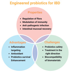
Overview chart of engineered-modified probiotics for IBD treatment. Advantages of probiotics in the treatment of IBD, changes in the treatment of IBD brought about by engineered modifications to probiotics and challenges encountered in engineering modified probiotics for the treatment of IBD.
In view of the aforementioned problems, the search for rational therapeutic directions and the discovery of new therapeutic targets remains significant issues in the treatment of IBD. In comparison with immunosuppression, the utilization of Teduglutide-like intestinal regeneration factors, or the development of probiotics to promote intestinal mucosal healing by obstructing the myosin light chain kinase (MLCK) signaling pathway [164]. The application of metabolomics has the potential to facilitate a more profound comprehension of the pathogenesis of IBD, thereby enabling the development of novel therapeutic targets [165,166]. Furthermore, existing biomaterials and modification technologies may still have limitations in terms of their biological affinity to the body and ensuring the vitality and function of probiotics. In order to ensure that biomaterials do not cause side effects such as inflammation in the treatment of the body, and that probiotics do not lose their activity under the encapsulation of biomaterials, it is necessary to continue to develop new biodegradable biomaterials and to design new probiotic modification methods and delivery systems. These are directions that need to be addressed in the engineering modification of probiotics for the treatment of IBD. More significantly, the side effects engendered by the proliferation of probiotics in the body, including infection and bacteraemia, are of significance and must be given due consideration. The establishment of an appropriate tracer system to observe and control the proliferation of these organisms in the intestine is also a key point to note in the research, which will minimize the potential pathogenicity risk and improve biosafety. Apart from the discovery of the anti-inflammatory and immunomodulatory properties of probiotic outer membrane vesicles provide novel insights into the utilization of probiotics in the development of novel therapeutic platforms for IBD [166-168]. Moreover, it has been demonstrated that genetically modified probiotics can enhance in vivo diagnostic capabilities. Daeffler et al. designed a transgenic EcN for monitoring IBD through detecting thiosulphate in intestinal [169]. Consequently, this has led to the exploration of probiotics as a diagnostic instrument for IBD. As our understanding of the structure and genome of probiotics continues to evolve, alongside the emergence of novel biomaterials, gene sequences and modification methods, the future application of probiotic-based engineered modifications in the field of IBD therapy is poised to advance towards a more precise, efficient and safe modality [170-173]. Through the combination of fundamental research and clinical application, as well as more rational and targeted design and implementation, it is anticipated that the utilization of probiotics will become a significant adjunct or primary therapeutic instrument in the management of IBD in the future.
The authors declare that they have no known competing financial interests or personal relationships that could have appeared to influence the work reported in this paper.
Acknowledgments
This work was partially supported by the National Natural Science Foundation of China (Nos. 82200610, 32301169), the Natural Science Foundation of Anhui Province (No. 2308085QH281), Key Project for the Cultivation of Outstanding Young Teachers of Anhui Province (Nos. YQZD2025016, YQZD2025019), Youth fund of Zhongshan Hospital Affiliated to Fudan University (No. 2024ZSQN12).
Supplementary materials
Supplementary material associated with this article can be found, in the online version, at doi:10.1016/j.cclet.2025.112219.
[1]
J. Wehkamp, M. Götz, K. Herrlinger, et al., Dtsch. Arztebl. Int. 113 (2016) 72–82.
[2]
M.J. Buie, J. Quan, J.W. Windsor, et al., Clin. Gastroenterol. Hepatol. 21 (2023) 2211–2221. doi: 10.1016/j.cgh.2022.06.030
[3]
P.T. Santana, S.L.B. Rosas, B.E. Ribeiro, et al., Int. J. Mol. Sci. 23 (2022) 3464. doi: 10.3390/ijms23073464
S. Hu, R. Zhao, Y. Xu, et al., J. Mat. Chem. B 12 (2024) 13–38. doi: 10.1039/D3TB02302H
Figure 1
Schematic synthesis of engineered modified probiotics. Mechanism of engineered modified probiotics for the treatment of IBD can be divided into chemical modification, physical modification and genetic engineering and other methods. By exploiting the structure of probiotics, the surface composition and structure of the bacteria can be altered by surface modification, or the biological properties of the bacteria can be altered and given new functions by the importation of target genes.
Figure 2
Intestinal probiotics play beneficial roles in the gut. (A) Probiotics inhibit harmful bacteria from adhering to the intestine. (B) Dendritic (DC) cells capture antigens to achieve the differentiation of immune cells. (C) Probiotics regulate intestinal flora. (D) The intestinal immune environment regulated by probiotics. (E) Bifidobacteria secrete SCFAs to regulate the release of various cytokines. (F) Probiotics repair the intestinal mucosa by upregulating the expression of various intestinal proteins.
Figure 3
Schematic diagram of physical modification technology for probiotics in IBD treatment. (A) Technological mechanism of modification of probiotics by electrostatic interactions. (B) Technological mechanism of modification of probiotics by physical extrusion. (C) Technological mechanism of modification of probiotics by layer-by-layer assembly. (D) Physical modification of probiotics achieves alleviation of IBD by scavenging intestinal ROS, regulating intestinal flora and inhibiting anti-inflammatory responses in the intestine.
Figure 4
Schematic diagram of chemical modification technology for probiotics in IBD treatment. (A) Technological mechanisms of probiotic modification by covalent binding. (B) Technological mechanisms of probiotic modification by in situ deposition. (C) Technological mechanisms of probiotic modification by click chemistry. (D) Technological mechanisms of probiotic modification by supramolecular interactions. (E) Chemically engineered modification of probiotic bacteria for treatment of IBD by scavenging intestinal ROS, regulating intestinal flora, and inhibiting the intestinal anti-inflammatory response.
Figure 5
Schematic diagram of genetic engineering technology for probiotics in IBD treatment. (A) Introduction of target genes into probiotic bacteria. (B) Genetically modified probiotics have been found to express vesicles that secrete SCFAs, interleukins, or membrane proteins on their surface. (C) EcN regulates intestinal flora and the intestinal immune system by secreting IL-2 in the gut after genetic modification and physicochemical modifications.
Figure 6
Schematic diagram of other modification technology for probiotics in IBD treatment. (A) Diagram of the mechanism of antigen-antibody interaction modification of probiotics. (B) Diagram of the mechanism of biotin-streptavidin interaction modification of probiotics. (C) Diagram of the mechanism of in situ mineralization modification of probiotics. (D) Engineered probiotics alleviate IBD by mimicking artificial enzyme activity, modulating the intestinal microenvironment, decreasing neutrophil infiltration, and repairing the intestinal mucosa.
Figure 7
Overview chart of engineered-modified probiotics for IBD treatment. Advantages of probiotics in the treatment of IBD, changes in the treatment of IBD brought about by engineered modifications to probiotics and challenges encountered in engineering modified probiotics for the treatment of IBD.


 DownLoad:

 下载:


 下载:

