Citation:
Hufeng Fang, Xueyun Sun, Dan Su, Lidan Tang, Jinhong Gong, Hongliang Xin. Molybdenum-based nanomaterials, the charming candidates for biomedical applications: A systematic review[J]. Chinese Chemical Letters,
2026, 37(11): 112212.
doi:
10.1016/j.cclet.2025.112212
Molybdenum-based nanomaterials, the charming candidates for biomedical applications: A systematic review
English
Molybdenum-based nanomaterials, the charming candidates for biomedical applications: A systematic review
Department of Pharmacy, The Second People’s Hospital of Changzhou, the Third Affiliated Hospital of Nanjing Medical University, Changzhou 213000, China
b.
College of Pharmacy, Dalian Medical University, Dalian 116044, China
c.
Department of Pharmaceutics, Pharmacy of School, Nanjing Medical University, Nanjing 211166, China
xhl@njmu.edu.cn (H. Xin). 1 These authors contributed equally to this work.
Received Date:
22 July 2025 Accepted Date:
03 December 2025 Revised Date:
24 November 2025 Available Online:
15 November 2026
Abstract:
Molybdenum (Mo) was initially discovered in 1778, which was recognized as an essential element. Over the past few decades, Mo-based materials emerged as popular member in the field of industrial manufacture such as electrodes and catalysts due to their appealing strength and corrosion resistance. In recent years, with the vigorous development of nanotechnology, Mo-based nanomaterials have gradually drawn extensive attention in biomedical field due to their physicochemical versatility including excellent biocompatibility, high catalytic activity, redox chemistry and strong photothermal conversion efficiency (PCE), which greatly promoted diversified applications of Mo-based nanomaterials in biomedical area. Up to date, scientific workers have exhibited thousands of bio-applications of Mo-based nanomaterials, but a systematical overview in this filed is still rare. In this review, the specific applications outline of Mo-contained nanomaterials in biomedical field are summarized via six aspects including reactive oxygen species (ROS) scavenging, photodynamic therapy (PDT) and photothermal therapy (PTT), antibacterial performance, drug delivery, bioimaging, and biosensors. And the unique properties of Mo-based nanomaterial momentous to its biomedical applications are accordingly expounded. Finally, the relevant existing challenges and future objectives of Mo-contained nanomaterials applied in biomedicine are thoroughly discussed.
Molybdenum (Mo) is a crucial transition metal element that is widely distributed in nature [1]. As an essential trace element, Mo plays an indispensable role in biological systems [2]. Mo serves as catalytic center of enzyme, such as xanthine oxidoreductases [3], participating in various metabolic reactions. Besides, it is well-known that the deficiency of Mo is a risky factor for human health that can elevate the occurrence of neurological lesion and esophageal cancer [4]. Undoubtedly, Mo-based nanomaterial is regarded as one of the most promising biocompatible materials [5]. Despite the fact that conventional metal nanomaterials (such as gold, silver, platinum) have achieved significant milestones in biomedical applications, which widely behaved as drug carriers, bioimaging agent, antimicrobial dressings, etc. [6]. These metal nanomaterials still face some limitations. For instance, the nonnegligible toxicity of gold and silver nanomaterials remains a concern in clinical uses [7]. Additionally, the high cost of gold and platinum presents a barrier to large-scale use [8]. Therefore, current research should focus on enhancing the safety, efficacy, and clinical translatability of metal nanomaterials. Timely, Mo-based nanomaterials have emerged as a promising alternative, not only because of a more economic source, but the improving biosafety and biodegradability [9]. Rivetingly, the special physicochemical properties of Mo, such as multiple valence states, unique photoelectric absorption effect, abundant surface groups and considerable vacancy sites [10], make Mo-based nanomaterials multifunctional in biomedical research, especially in disease diagnosis and treatment, drug delivery and so on [11].
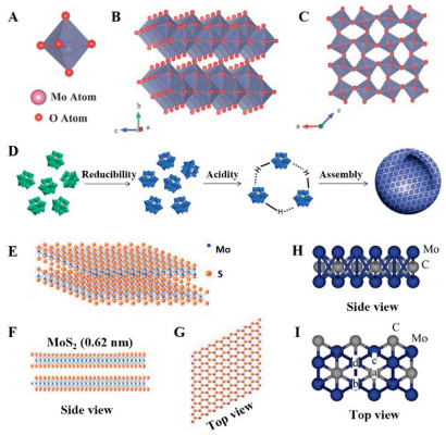
Mo-based nanomaterials are mainly composed by Mo oxides (e.g., MoO3-x), Mo based polyoxometalates (e.g., POMs), Mo dichalcogenides (e.g., MoS2), Mo-based MXenes (e.g., Mo2C), etc. [12]. The chemical structures of Mo-based nanomaterials are displayed in Fig. 1. Mo oxide is formed by octahedral MoO6 basic structural units, in which Mo atoms locate at the center and oxygen atoms distribute in the endpoints (Fig. 1A) [13]. Stoichiometric MoO3 mainly includes the thermodynamically stable orthogonal α-phase (Fig. 1B) and the metastable monocline β-phase (Fig. 1C) [14]. Mo-POM is one kind of polyanion metal-oxide cluster with precise chemical composition and architecture, which is composed by oxygen and high oxidation state Mo (Fig. 1D) [15]. MoS2 is the most commonly used nanostructure among Mo-based nanomaterials [11]. As shown in Fig. 1E, single-layer MoS2 consists of three layers of atoms (S-Mo-S), Mo atoms are sited between two layers of S atoms, shaping a sandwich structure in the side view (Fig. 1F), and forming a hexagonal honeycomb structure by weak van der Waals interactions in the top view (Fig. 1G) [16,17]. Resembling to MoS2, Mo2C presents a two-dimensional (2D) sheet-like morphology (Figs. 1H and I) [18]. Additionally, A comprehensive discussion about the differences of various Mo-based materials can be found in Table S1 and Section S1 (Supporting information).
Figure 1
Figure 1.
Schematic overview of the chemical structure of Mo-based nanomaterials. (A) Mono octahedral MoO6 structure. (B) Layered thermodynamically stable orthorhombic α-MoO3. (C) Metastable monoclinic β-MoO3. Copied with permission [13]. Copyright 2017, Wiley. (D) The architecture of Mo-POM and the aggregated construction of reducibility/acidity induced self-assembly composite. Copied with permission [15], Copyright 2020, The Royal Society of Chemistry. (E) MoS2 crystal structure: (F) side view and (G) top view. Copied with permission [17]. Copyright 2012, Wiley. The side view (H) and top view (I) of Mo2C structure. Copied with permission [18]. Copyright 2005, American Chemical Society.
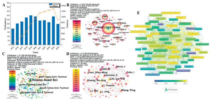
Nowadays, bibliometrics analysis has become a systematic method to quantitatively study the characteristics of scientific literatures [19]. In this review, we captured the data about the biomedical researches of Mo-based nanomaterials by the Web of Science (WOS) Core Collection and analyzed by VOSviewer and Citespace (Fig. 2). Obviously, Mo-engineered materials hold great potential for biomedical applications. The detailed descriptions are provided in Section S2 (Supporting information).
Figure 2
Figure 2.
Bibliometric quantitative analysis. (A) The annual publication volume and citations, (B) the network of national/regions cooperation, (C) the institutional cooperation network, (D) the network diagram of authors, and (E) keywords co-occurrence of visualization map of Mo-based engineered nanomaterials in biomedical field.
However, there is still a lack of a review that comprehensively analyze the research progress in biomedical field from a relative holistic perspective. Therefore, in this review, recent research trends in the biomedical applications of Mo-based nanomaterials were presented by the following aspects including scavenging reactive oxygen species (ROS), photothermal therapy (PDT)/photothermal therapy (PTT), antibacterial therapy, drug delivery, bioimaging and biosensors. Moreover, the key challenges and future outlook of Mo-based nanomaterials in biomedical applications will be discussed and proposed.
2.
ROS scavenging
ROS, such as hydrogen peroxide (H2O2), superoxide anion (O2•−), and hydroxyl radical (•OH), are naturally generated during cellular metabolism and operate as crucial signaling mediators in various physiological processes, including immune response and cellular homeostasis [20]. Nonetheless, excessive ROS accumulation induces oxidative stress, which is widely implicated in the pathogenesis of inflammatory, neurodegenerative, cardiovascular disorders, as well as cancer [21]. Multifunctional Mo-based nanomaterials have appeared as antioxidant-like activity platforms in recent years due to their charming Mo6+/Mo5+ redox electron couples and the promise of acting as nanozymes [22], nanomaterials with enzyme-like activity can mimic the activity of natural enzymes [23], like peroxidase (POD), superoxide dismutase (SOD) and catalase (CAT) (Fig. S1A and Table S2 in Supporting information).
2.1
Mo-based nanozymes for ROS removal
MoS2 is considered as the most studied member of the 2D transition metal dichalcogenides (TMDs) family [24]. Individual sandwiched S-Mo-S layers in MoS2 are assembled together [25]. The unique spatial structure effectively increased the exposure of the active site [26]. In recent years, a large number of reports have certified that MoS2-based nanomaterials removed harmful ROS by exerted as nanozymes. For example, Chen et al. reported that fullerene-like MoS2 (F-MoS2) nanoparticles (NPs) were effective lubricants and antioxidants for osteoarthritis with great SOD and CAT activity [27]. Besides, enhancing the dispersity of MoS2 to prevent disordered aggregation was an intelligent strategy to promote the removal of ROS [28], such as MoS2 modified TiO2 nanoribbons (MoS2@TiO2) [29]. The proposed nanoribbons exhibited excellent CAT and SOD activities, and demonstrating the better performance in ROS scavenging and reducing inflammatory factors in the mice neck oxidative stress model (Fig. S1B in Supporting information).
Mo2C, MoSe2, MoO3-x, etc. also showed great nano-enzymic effect on ROS removing. The active site was fully exposed on the surface of 2D Mo2C, which improved the efficiency of ROS removal [30]. MoSe2 was similar to MoS2 both in structure and function, and exhibited enzyme-like activity to clear ROS. The intracellular ROS level in acute pancreatitis 266.6 cells was significantly reduced to 10% of the original level via MoSe2@PVP NSs treating [31]. Moreover, the ultra-small MoO3-x nanodots (NDs) played a positive role in the treatment of Alzheimer’s disease (AD), which drastically alleviated amyloamyloid-β-mediated oxidative stress due to the excellent CAT and SOD mimic activities of MoO3-x NDs [32].
2.2
Neutralizing ROS based on redox reactions
Mo-POMs is considered as “electron sponge”, which present a unique property to accept or release electrons [33]. Rich in Mo6+ and Mo5+, Mo-POMs can be used to fast reverse the multi-electron redox reactions to clean ROS [34]. It is well known that Mo-POMs tended to aggregate under acidic conditions, and resulted in a long retention time in the digestive tract rather than absorption [35]. Hence, Mo-POMs showed a great advantage in the diagnosis and treatment of ROS-induced gastrointestinal diseases. For example, Yang et al. found that Mo-POMs showed strong ROS clearance performance in vitro and ameliorated the abnormal production of intestinal metabolites in ulcerative colitis (Fig. S1C in Supporting information) [36]. Furthermore, the hydrodynamic diameter of Mo-POMs (~1–2 nm in pH 7.4) [37] was lower than renal filtration threshold (5.5 nm) [38], it was easy to be excreted within a certain time scale [39], which promoted the application in kidney diseases. A significant recovery of renal functions, and obviously reduction in kidney injury molecule-1 and heme oxygenase-1 indexes were observed in the acute kidney injury model mice after Mo-POMs treatment [40]. Besides, Mo-POMs showed great potential in the treatment of other inflammatory diseases. Shi et al.’s report pointed that Mo-POMs exhibited a super-strong antioxidant capacity and the number of synovial inflammatory cells were significantly reduced by Mo-POMs [41]. Another scientific result showed that ultra-small multivalent MoO3-x NDs also exhibited a key function in diabetic ulcers treating by ROS consummation attributed to Mo5+-to-Mo6+ transformation, and the cell survival rate was increased from 46.7% to 79.6% [42].
In summary, benefit to the structure and composition, Mo-based nanomaterials have made great progress in the removing of ROS. However, some worries should be considered and worked out. The catalytic activity regulating mechanisms and catalytic kinetics of Mo-based nanozymes are still remain unclear [43]. Another concern is that the ROS-scavenging abilities of Mo-based nanomaterials showed a universality to various diseases. Highly selective Mo-based ROS removal nanomaterials with specific therapeutic effects should be further designed [44]. Furthermore, Systematic assessment of the biocompatibility, in vivo metabolism and mechanisms of Mo-based antioxidants is crucial. Finally, owing to the inherent complexity of the disease [23], it is necessary to continuously explore the combination of Mo-based nanomaterials with drugs or physical therapy to enhance the effect of disease treatment.
3.
PDT-PTT
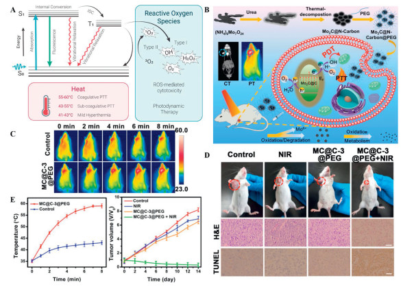
Benefiting from the merits of minimal invasiveness, spatiotemporal selectivity and negligible toxicity on normal tissue, PDT-PTT become an attractive tool for disease intervention [45]. Exactly, PDT refers to a treatment that uses specific laser irradiation to activate photosensitizers (PSs) to produce ROS, which induces cell apoptosis or necrosis [46]. Additionally, PTT is a new therapeutic strategy that mostly utilizes the near-infrared (NIR) photothermal agents to ablate abnormal cells by generating hyperthermia [47]. The simplified mechanism of PDT-PTT process was exhibited in Fig. 3A. The specific mechanism was elaborated in Section S3.1 (Supporting information). Currently, it was fortunate to find that not only Mo-based nanomaterials can be used as carriers to load PSs, such as chlorin E6 (Ce 6), to realize PDT [48], but Mo-based nanomaterials exhibited a high photothermal conversion efficiency (PCE) to promote widely application in PTT [49].
Figure 3
Figure 3.
The principle of PDT-PTT and the therapeutic effects of Mo-engineered PDT-PTT nanoplatforms. (A) Simplified mechanism illustration of Mo-based engineered nanomaterials in PDT and PTT process. (S0: ground state, S1: excited singlet state, T1: excited triplet state, ISC: intersystem crossing). Copied with permission [59]. Copyright 2023, American Chemical Society. (B) Mo2C@N-Carbon@PEG nanomaterials fabrication and their application for tumor PTT/PDT and CT imaging. (C) The infrared thermal images of tumor-bearing mice with MC@C-3@PEG + NIR treatment and control group. (D) Digital images of tumors size in each group mice and the corresponding hematoxylin and eosin (H&E) (scale bar: 50 µm) and terminal deoxynucleotidyl transferase deoxyuridine triphosphate nick end labeling (TUNEL) (scale bar: 25 µm) staining tumor slices. (E) Temperature curves of MC@C-3@PEG + NIR and control group (n = 3) and tumor volume of each group tumor mice treated for 14 days. Copied with permission [58]. Copyright 2022, Elsevier.
It has been found that synergetic PTT and PDT provided more efficacy compared to photothermal therapy alone or standalone photodynamic treatment [50], which were discussed in Section S3 (Supporting information). It could be concluded in three ways to achieve synergetic PDT-PTT tactics for Mo-based nanomaterials. (1) Loading PSs into Mo-based nanomaterials was a general method to achieve a synergetic PTT-PDT therapy pattern. Mo-based nanomaterials were commonly modified with kinds of PSs by encapsulation [48], electrostatic adsorption [51] or chemical functionalization [52]. Muhammad anchored indocyanine green (ICG) onto AuNRs/MoS2 hybrid (PCE = 68.8%) via electrostatic interaction to achieve simultaneous PTT-PDT. A complete tumor eradication was noticed at day 14 in the Au/MoS2-ICG treated group, while individual PTT groups Au NRs or MoS2 NSs slightly delayed the tumor growth rather than growth inhibition, which suggested synergistic PDT-PTT increased the chance to completely eradicate the tumor [53]. (2) Doping with multivalent metal elements to stimulate the generation of ROS with laser irradiation [54]. An acid-responsive MoS2/CoFe2O4 nanostructure (MSCO-PEG) (PCE = 41.16%) was synthesized for the treatment of breast cancer. MSCO-PEG can amplify the edge potential by forming Z-scheme heterojunction. Under NIR radiation, the Z-scheme heterojunction produced electrons (e−) and holes (h+) with higher reducibility and oxidation ability at the band edge, which promoted the cascaded conversion of H2O to ROS for attacking tumor tissues to achieve PDT. The results showed that MSCO-PEG-laser treated tumor began to shrink at the fourth day and almost disappeared after two weeks’ phototherapy, which exhibited a better antitumor effect than the group of MoS2-laser (only PTT) with tumor growth reaching around 8 times of the initial size after 14 days treatment [55]. (3) MoS2 [56], Mo2C [57] NPs were confirmed as “one-for-all” theranostic agents, which exhibited great PCE for photothermal therapy and synchronously promoted the generation of ROS via NIR-photocatalysis, triggering a PDT-PTT combination effect without intricate assemblies. Mo2C@N-carbon@PEG NPs (PCE = 52.7%) were constructed to reveal the synergic PTT-PDT effect (Fig. 3B). Firstly, in order to explore the individual contribution of PDT and PTT, glutathione (GSH) was used to shield the generated ROS, HepG-2 cell viability increased to 39.1% ± 1.2%. It was indicated that the cytotoxicity from PTT was 60.9% ± 3.6% and the contribution for PDT was about 31.5% ± 2.4%. Whereas, without GSH masking, the Mo2C@N-carbon@PEG treated HepG-2 cell viability reduced to 7.6% ± 1.2%. To further investigated the synergic PTT-PDT effect, the photothermal imaging photos revealed that after treated with Mo2C@N-carbon@PEG under NIR irradiation for 8 min, the temperature of tumor increased to nearly 59.0 ± 0.9 ℃, whereas the control group only exhibited 43.1 ± 0.8 ℃ (Fig. 3C). Besides, the therapeutic group also showed prominent tumor inhibition performance as exhibited in Figs. 3D and E [58].
Although Mo-based nanomaterials have become potential PDT/PTT materials due to their outstanding performance, there are still plenty of challenges in their clinical translation. Firstly, most Mo-based nanomaterials need to be surface modified to increase the efficiency of PTT, this process usually changes the structure and makes it difficult to enter the target tissue. Secondly, PDT/PTT need to be carried out under NIR or visible light irradiation, which may cause damage to normal tissues because it was difficult to control the optimal irradiation time and intensity. Thirdly, further research is essential to develop more efficient and safer therapeutic strategies.
4.
Antibacterial properties and mechanisms
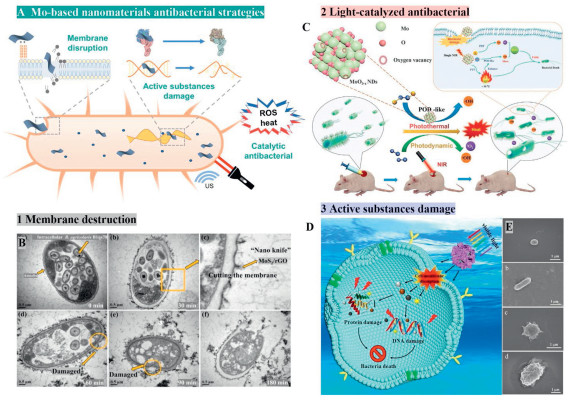
Infectious diseases caused by bacteria have always been the main threat to human health [60]. Although a large number of antibiotics have been invented, the abuse of antibiotics is still widespread, even antibiotics resistant strains have been surged [61]. It is urgent to develop safe and reliable antibacterial strategy. Currently, Mo-based antimicrobial agents have aroused global interest [62]. Up to now, the antibacterial mechanism of Mo-based nanomaterials has been mainly deduced in three pathways (Fig. 4A).
Figure 4
Figure 4.
Antibacterial strategies of Mo-based nanomaterials. (A) Schematic illustrations of three modes of antibacterial mechanisms. (B) Transmission electron microscopy (TEM) photos of amoeba morphology with “nano knife” MoS2/rGO treatment from 0 min to 180 min (scale bar: 0.5 µm). Copied with permission [71]. Copyright 2024, Elsevier. (C) Scheme of the treatment of wound infection based on the combination of nanozymatic/photothermal/photodynamic triple-therapy synergetic effects launched by MoO3-x NDs. Copied with permission [84]. Copyright 2021, Wiley. (D) CuFe2O4/MoS2@Ag composites triggered damage to intracellular proteins, lipids, and DNA, resulting in bacteria death. Copied with permission [97]. Copyright 2025, Elsevier. (E) Morphological changes of S. aureus and E. coli before and after incubated with PEG-MoS2/rGO-SS (scale bar: 1 µm). Copied with permission [98]. Copyright 2020, Elsevier.
The cell membrane is an important substance to maintain the structure of bacteria, while any membrane damage, such as antibiotic destruction or disinfectant action, could result in cytoplasm leakage, biofunctions disorder and further induce the death of bacteria [63]. Currently, there were some attempts about increasing the interaction with bacterial cell membrane by introducing Mo-based nanomaterials to enhance antimicrobial performance. For instance, it was discovered that MoOx QDs showed great damage on Escherichia coli and S. aureus membrane with fatality rates about 58.1% and 53.3%, respectively [64]. The toxicity was attributed to molybdic acid/MoOx hydroxonium ions [65], which diffused into cell membranes and disturbed the protein synthesis processes and frameworks [66].
Moreover, it is convenient for positively charged Mo-based nanomaterials to adhere to bacteria by electrostatic adsorption, resulting in a physical damage or integrity destruction of the bacterial membrane [67]. Cao’s group designed a polyethyleneimine (PEI)-modified MoS2 nanocomposite. Cationic polymer PEI changed the zeta potential of MoS2 from negative to positive, thus promoted the combination of MoS2-PEI with E. coli and S. aureus cell membrane by electrostatic interaction. The antibacterial activity increased in a concentration-dependent manner (20–80 µg/mL). Typically, 20 µg/mL PEI-MoS2 showed good killing rates to E. coli and S. aureus (68.1% and 66.2%, respectively), higher than that of MoS2 (46.0% and 45.6%) [68]. The same antimicrobial effect and mechanism were also found in positively charged compounds decorated MoS2, such as chitosan (CS) [69] or polydopamine [70]. What is more, Mo-based nanomaterials acted as “nano knife”, directly adhering the cell wall and membrane of bacteria for physical mechanical cutting, due to their sharp edges. As the pictures shown in Fig. 4B, MoS2/rGO composites performed like blades inserted into amoeba’s cell wall and membrane [71].The physical action was insufficient to kill microorganism that the survival of B. agricolaris B1qs70 and amoeba decreased by 0.12 and 0.1-log in colony-forming units (CFUs) after 180 min of disinfection (2.0 mg/mL MoS2/rGO), but the disinfection synergistically increased to 8.15 and 4.18-log reduction in CFUs for B. agricolaris B1qs70 and amoeba under ultrasound assistance [71]. Finally, functionalizing was also a wise choice for Mo-based nanomaterials to disrupt microorganism integrity [72]. Multifunctional MoS2 nanotubes hydrogel (MTH) exhibited an inherent antimicrobial capacity. The imidazole moiety of MTH could effectively increase the permeability of bacterial membrane. The survival rates of E. coli and S. aureus were dropped to 60% and 30.4% after the treatment of 100 mg MTH for 15 min, and the antibacterial effect was further enhanced to 0.8% (E. coli) and 3.2% (S. aureus) survival rates under NIR irradiation [73].
4.2
Catalytic antibacterial
Under ultrasound or light stimulation, multifunctional Mo-based nanomaterials were induced to switch as catalysts to erupt numerous ROS/heat to disturb the normal physiological and metabolic functions of bacteria, and causing bacteria death finally [74]. MoOx@Mo2C nanonetworks were proposed as a nano-sonosensitizer to burst ROS for bacteria eradication. MoOx@Mo2C nanonetworks exhibited an inherent strong bacteria-capturing ability due to the 2D network with 310.7 nm surface area and intrinsic hydrophilicity. Specifically, after incubated with 25 µg/mL MoOx@Mo2C nanonetworks with US irradiation, the CFUs of E. coli, S. aureus, and methicillin-resistant S. aureus (MRSA) manifested remarkable reductions of 1.54, 1.53, and 1.96-log, respectively [75].
Photo-induced antibacterial behaviors of Mo-based materials can be classified into photoinduced-oxidative antibacterial [76] and photothermal antibacterial [23]. Mo-based nanomaterials usually triggered oxidative stress via exerting photocatalytic capacity, photo triggered enzyme-like activity or relying on PSs [77]. Typically, coupling another type of semiconductors with Mo-based nanomaterials to form a heterojunction structure at the interface was an effective method to achieve photo-induced antibacterial [78]. This heterojunction engendered a light induced electric field, which facilitated the separation of electrons and holes in local environment, thereby these escaped holes in the valence band of heterojunction reacted with H2O to generate •OH, boosting ROS production [79]. Just like MoO3-x/Zn2In2S5 [80], MoS2@Cu2O heterojunction [81] and so on. Rivetingly, defect-rich nanostructures also displayed strong possibility in antibacterial activity [82], especially, the photo-responsive peroxidase-like enzymatic activity. Generally, when bulk Mo-contained structures were cut into nanoscale dots, abundant surface oxygen deficiencies or distinctive edge sites have been exposed, which would play as the active sites for the nanozyme antimicrobial activity [83]. Like the reported MoO3-x NDs, worked as a peroxidase mimic, could effectively decompose H2O2 to generate •OH with significant enhancement under NIR radiation. The antibacterial results demonstrated that the CFUs of E. coli and S. aureus can totally be suppressed by MoO3-x NDs [84]. The peroxidase-like MoO3-x NDs could also accelerate the wound healing in vivo due to their efficient bactericidal activity against drug-resistant strains extended-spectrum β-lactamases (ESBL)-producing E. coli and MRSA (Fig. 4C). The antibacterial result indicated the bacterium were decreased by ~6-log CFUs in MoO3-x NDs treating group, which showed the best wound healing effect among all the tested groups. Besides, embedding with metal composites contained with Fe, Cu, Ag elements or utilizing multi-valence property could be another way to achieve the photo induced enzyme-like antibacterial activity for Mo-based materials [85], because these metal composites or Mo5+ exhibited Fenton-like catalytic efficiency to generate numerous ROS [86]. MoS2 NSs possessed a great potential on PDT when used it as photosensitizer [56]. According to Zhao’s group, MoS2 NSs were demonstrated to possess good antibacterial properties that the killing rate of MDR E. coli and MRSA even reached to 99.9999% in only 30 min [87]. Furthermore, benefit from the large surface area, Mo-based nanomaterials also can be used as the carrier of PSs to achieve photodynamic antibacterial effect by loading PSs [51]. Moreover, the prominent photothermal nature of Mo-based nanomaterials facilitate the wide application of Mo-based nanomaterials in antibacterial field. For example, MoSe2@CS (PCE = 45.7%) mediated PTT for H. pylori and exhibited a high bacteriostasis rate of 99.9% [88].
4.3
Disrupting bacterial active substances
Emerging evidences have shown that Mo-engineered nanomaterials exhibited charming inhibition on bacterial active substances by doping or loading with well-known antimicrobial agents and metal ions, or blocking gene transfer (Figs. 4D and E). More explanations were provided in Section S4 (Supporting information).
However, it was not sufficient to launch strong and long-term antibacterial efficiency because of the limited microbial damage caused by single antibacterial mode, especially for the mechanical destruction of cell membrane [89]. Combining multiple antibacterial pathways has become a new trend in synergistic antibacterial therapy [90]. It was primely evidenced by Xu’s team that they developed an effective antibacterial strategy by using Mo oxide nanowires (MoOxNWs) as multifunctional bacterial eradicating agents [91]. MoOxNWs not only played as photo-triggers to activate the performance of PDT and PTT, but also caused mechanical damage to bacteria due to their sharp ends structure. Delightfully, the synergistic antibacterial platform achieved significant antibacterial effect that the sterilization rates of ESBL-producing E. coli and MRSA were risen to 97.67% and 96.34% after the incubation with 100 µg/mL MoOxNWs. Another prominent triple-mode germicidal nanoplatform (Ag-Pd@MoS2) exhibited intrinsic catalytic, photothermal, and photodynamic properties, indicating a fantastic perspective in antibacterial protective clothing [92].
Although Mo-based nanomaterials have attracted widespread attention in antimicrobial field, there are some concerns that need to be addressed. First of all, some modification operations in fabricating process including grinding, spin coating or stirring may ignore the weakening of stability and bactericidal activity. Secondly, it is indeed necessary to expand the work to clarify the antibacterial spectrum of Mo-based nanomaterial, especially for the multidrug-resistant bacteria [93]. Though MoO3 NPs was reported to possess antibacterial activity against some multidrug-resistant pathogens based on the mechanism of hydroxonium ions release [94]. While there were pretty difficulties for MoO3 NPs to be introduced into clinic: (1) MoO3 NPs exhibited high antibacterial activity in a very acidic environment (2.52–2.77), but a completely abolished germicidal effect at neutral pH [66], which meant the antibacterial performance of MoO3 NPs need a strict condition. (2) Mo-based NPs showed a bad susceptibility to multidrug-resistant isolates that the minimum inhibitory concentrations were up to 0.7–0.8 g/L [66]. (3) Whether the metabolites of MoO3 NPs modulate the periplasmic substrate-binding protein, such like Mod A [95], to further promote the proliferation of multidrug-resistant pathogens still remain figure out. Finally, with incessant bacteria mutate [96] and the increasing use of new antibacterial nano-systems in clinic, whether bacteria will develop resistance to Mo-based nanomaterials, which is also a key issue that requires clarification.
5.
Drug delivery
Mo-based nanomaterials have been proved to be one of the most promising drug delivery systems due to their extremely attractive properties including high specific surface area, distinctive heteropoly acid structure, photothermal conversion effect, and so on [99].
5.1
Enhancing loading efficiency
It was evidenced that Mo-based nanomaterials presented as powerful tool to improve the drug loading capacity for drug delivery, due to their unique nanostructure and modification. Detailed descriptions are provided in Section S5 (Supporting information).
5.2
Controlled drug release
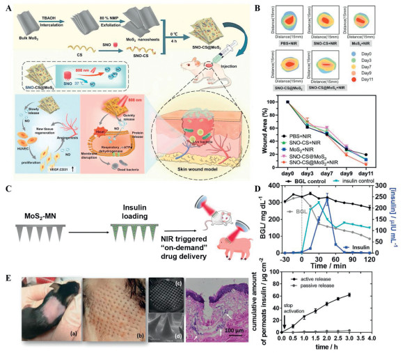
Drug release profile is of great importance of the drug delivery. The photothermal property of Mo-based nanomaterials play a crucial role in drug releasing, which can realize photo-heat-controlled drug release. For example, a NIR-triggered nitric oxide (NO) delivery nanostructure (SNO-CS@MoS2) was developed by Mu et al. [100]. In SNO-CS@MoS2, MoS2 played as photothermal agents, and S-nitrosothiol (SNO) was a NO gas donor that could substantially generate NO via photothermal stimulation. Releasing results indicated that after NIR activation SNO-CS@MoS2 exhibited a photo-heat-controlled NO releasing with a 6.88 µmol/L concentration and last for 18 h. Notably, SNO-CS@MoS2 + NIR group significantly accelerated wound healing with the wound area reduced to 2.95% on day 11, which was at least three times smaller than other groups (Figs. 5A and B). Additionally, Liu’s group prepared MoS2@DOX/MnO2-PEG (MDMP) nanomaterials. When the concentration of MDMP was only 200 µg/mL, the temperature of MDMP rose to 52 ℃ within 5 min and doxorubicin (DOX) releasing rate was reach to 65% [102].
Figure 5
Figure 5.
Illustration of the design and synthesis of Mo-based drug delivery system and the efficacy of drug loading, releasing and therapy. (A) SNO-CS@MoS2-nanosheets with NIR-triggered NO delivery and antibacterial treatment of tissue wound. (B) The charts about the wound healing efficiency of SNO-CS@MoS2. Reproduced with permission [100]. Copyright 2023, Springer. (C) The fabrication of insulin loaded MoS2-MN and the diagram of NIR induced on demand drug delivery. (D) The measurements of blood insulin and blood glucose level in mini-pigs carrying MoS2-MN patches and cumulative permeation profile of MoS2-MN with or without NIR activation. (E) Image of mouse skin transcutaneously treated with the MoS2-MN patch, scanning electron microscopy (SEM) images of the MoS2-MN array after skin penetration and H&E staining data of the mouse skin penetrated by one MoS2-MN array patch (white arrows show the infiltration of inflammatory cells) (scale bar: 100 µm). Reproduced with permission [101]. Copyright 2022, The Royal Society of Chemistry.
Microenvironment alteration was another key factor affecting drug release. For instance, Cai et al. synthesized a DOX loaded thiolated aptamer AS1411 and PEG dual modified MoS2 nanosheets (DOX@Apt-PEG-PDA-MoS2). Attributed to the acid pH-induced the protonation of DOX, the solubility of DOX was improved obviously, and DOX release rate was improved up to 58% under acidic conditions (pH 5.0), while only about 22% was released in pH 7.4, which indicated a good efficacy in cancer treatment [103].
5.3
Transdermal drug delivery
Beyond intravenous administration, Mo-based nanomaterials also can be used for transdermal drug delivery, which provided a new route of increasing medication compliance for chronic patients. For example, Zhang’s group developed cationic hydroxyethyl cellulose (JR400) surface-modified MoS2 NPs (JR400-MoS2 NPs) for the transdermal administration of atenolol. The loading efficiency of JR400-MoS2 NPs was increased to 90.4% ± 0.3%. And the skin penetration rate was increased by 2.3 times under NIR stimulation [104]. JR400-MoS2 NPs facilitated a better sustained-release profile than CS and alginate coated gold microcapsules [105]. Moreover, a novel MoS2 nanoplatform was designed for erythromycin transdermal delivery with encapsulation efficiency up to 62.8%. And there was still 201.83 µg/cm2 of drug after 24 h administration [106]. MoS2 nanosheets were also integrated into microneedles (MN) hydrogels to realize insulin on-demand releasing (Fig. 5C). Transdermal administration of insulin loaded MoS2-MN patch was more effectively to reduce the blood glucose levels compared to subcutaneous insulin injection according to Bilal’s assay (Figs. 5D and E) [101]. In contrast to graphene oxide (GO) and reduced graphene oxide (rGO) nanosheets transdermal insulin delivery systems, in which insulin was strongly adsorbed via H-bonding, π-π stacking. While MoS2 nanosheets showed a better insulin release profile due to the less firm interaction with aromatic residues of insulin [107].
Mo-based nanomaterials exhibited high drug loading and release rate due to their large specific surface area and good photothermal conversion performance. Such advancements have provided new strategies for diseases treatment. However, the application of Mo-based nanomaterials in drug delivery also exists certain limitations. For instance, the types of drugs delivered by Mo-based nanomaterials are very limited. Nevertheless, metal-organic framework (MOF) is an emerging class of hybrid porous materials and gradually become a promising candidate for drug delivery [108], preparing Mo-MOF nanomaterials may be a new method to improve the drug loading and releasing performance. Additionally, it has not been reported whether there are other serious side effects of Mo-based nanomaterials used for drug delivery, so it is necessary to keep going to explore and find more effective and comprehensive Mo-based nanomaterials.
6.
Bioimaging
Bioimaging, accurately identifying the location of lesions and monitoring disease progression, is a pivotal tool in modern medicine, and plays a vital role in disease diagnosis and treatment [109]. Mo-based nanomaterials have been demonstrated as promising imaging agents based on photoacoustic imaging, magnetic resonance imaging and fluorescence imaging (Fig. S2 in supporting information). The detailed discussion and comparison of different imaging technology are showed in Section S6 (Supporting information).
7.
Biosensing
Currently, Mo-based nanomaterials have been used as biosensors worldwide with satisfactory results (Fig. S3 in supporting information) [110], such as the detections of small biomolecules, biomarkers, nucleic acids. More recently, Mo-contained nanomaterial based wearable sensing devices has caught more and more focus in the medical field. A deep overview about Mo-contained nanomaterial used as biosensors was expounded in Section S7 (Supporting information).
8.
Opportunities and challenges of Mo-based nanomaterials in clinical translation
Considered as a potentially new biodegradable metal, Mo exhibited superior long-term corrosion resistance compared to the counterparts of Zn, Mg and Fe [111], which has facilitated the applications of Mo-based materials in medical field, such as drug delivery materials, implantable device and so on [112]. It was found that the degradation process of Mo was launched by a cascade oxidation reaction in the mimetic physiological DMEM/F-12 cell medium [113]. The corrosion products of Mo-based materials transitioned from low-valence oxides to high-valence oxides, and eventually changed into water-soluble molybdate (Mo6+-oxide species), a biocompatible cofactor in several proteins, within 14 days of immersion [113]. The fast biodegradability strongly enhanced the biosecurity of Mo-based materials. Furthermore, in vitro cell studies pointed to no apparent cytotoxicity of Mo-based nanostructures. Feng et al.’s report indicated that Mo2C-PVA (polyvinyl alcohol: PVA) nanoflakes exhibited both high biocompatibility to tumor cell lines (4T1 cells and MDA-MB-231 cells) and normal cell lines (L929 cells), even at high concentrations of up to 400 µg/mL for 48 h [114]. Similarly, MoS2 nanoflowers designed by Kanwar’s group showed high biocompatibility to human mesenchymal stem cells (hMSCs) that the half-maximal inhibitory concentration (IC50) of MoS2 nanoflowers was evidenced up to 400 µg/mL with 24 h of exposure to hMSCs [115]. The good in vitro biocompatibility of Mo -based nanomaterials promote their potential use in biomedicine.
Notably, the pharmacokinetics, biodistribution and clearance behaviors of nanomaterials hold great importance for their clinical translation. The pharmacokinetics of Mo-based nanomaterials were conformed well to the two-component model with a blood circulation half-time about 1–2 h [116]. Biodistribution experiments showed that Mo-based NPs were mainly accumulated in the reticuloendothelial system organs (liver, spleen and lung) [117]. Slow clearance and sufficient dosages of nanomaterials may increase the long-term retention in these organs which may be associated with a higher risk of toxicity [118]. Fortunately, numbers of literatures have pointed out that ultrasmall sized Mo-based nanomaterials, below the renal interception threshold, could be excreted effectively and completely via both renal-urinary pathway and the hepato-enteral excretory system within a month [117]. Moreover, further histological and blood analysis likewise showed no obvious long-term toxicity [119]. Owning to the charming biodegradability and relatively rapid excretion, Mo-based nanomaterials exhibit a promise for further clinical applications.
Up to now, many nanoscale materials evidenced their powerful value in clinical translation, such as Doxil, the first Food and Drug Administration (FDA)-approved doxorubicin HCl liposome, has demonstrated clear efficacy in the treatment of various cancers [120]. Meanwhile, ferumoxytol, iron oxide NP, was approved by FDA as magnetic resonance imaging (MRI) contrast agents, iron supplements for anemia and leukemia therapy [121]. Multifunctional Mo-based nanomaterials are garnering increasing attention for their significant role in biomedicine. However, none of these have been approved by the FDA for clinical use. There is still a long way to go before Mo-based nanomaterials can be applied in clinical practice. Firstly, some unwanted immune-responses were found in the early stage of degradation, which promoted the polarization of macrophages towards a pro-inflammatory phenotype leading to edema and tissue damage [122]. How to avoid or solve these problems still requires further research. Besides, most research conclusions were drawn based on the investigation of Mo6+ oxidation state, whether the other valence states of Mo are involved in immune regulation or aggravate serious adverse reactions like allergenicity and renal failure still remains unclear. Secondly, the cytotoxicity assessments were incomprehensive. For example, it was inevitable to introduce elements with high toxicity, such as selenide, for the purpose of better phototherapy performance [48]. And the contribution of by-products of the toxic elements is often overlooked in cytotoxicity studies. Thirdly, NP size is known to act a pivotal role in the retention and elimination of engineered NPs [123]. It was demonstrated that NPs sized below the renal interception threshold can be quickly cleared by urinary excretion within hours to days after administration [124], such as the ultrasmall Mo2C-PVA nanoflakes mentioned above. Nevertheless, larger-sized non-degradable NPs are more likely to trigger uptake by the mononuclear phagocyte system subsequent long-term retention in organs and tissues resulted in a higher risk of off-target and toxicity [125]. Last but not the least, current existing pharmacokinetics and toxicity profiles data were mainly obtained via animal experiments, which may lead to mismatches in actual clinical translation due to the cross-species differences. Therefore, it is extremely necessary for Mo-based nanomaterials to be considered for further investigation through clinical trials.
In addition, emphasis should also be given on the development of synthesis strategies that are safe, scalability, uniform, cost-effective and sustainable. And more detailed discussions of synthesis strategies were provided in Section S8 (Supporting information).
9.
Conclusion
The application prospects of Mo-based nanomaterials in biomedical field should not be underestimated. The characteristics of good biocompatibility and easy modification made Mo-based nanomaterials an emerging multi-functional strategy to overcome the difficulties faced by other nanomaterials, and exhibited unlimited potential in the biomedical field. As a class of functional metal nanomaterials, Mo-based nanomaterials not only exhibited fascinating performance in ROS scavenging, but presented optimistic foreground in drug delivery and nidus targeting that improved the drug concentration in diseased tissues. At the same time, several Mo-based nanostructures such like MoS2 were promising candidates for PDT and PTT, due to their strong absorption in the NIR region and high PCE (most examples were tested up to 40%), therefore Mo-based nanomaterials brought the possibility to overcome major diseases such as cancer and bacterial infection. Finally, based on the unique optical and electrical properties of Mo-based nanomaterials, they were constantly developed for bioimaging and biosensors. It provided a new technical support for the early detection of diseases.
Although Mo-based nanomaterials have made some progress in the field of biomedicine, the clinical translation of Mo-based nanomaterials still faced great challenges. The biological toxicity assessment, action processes and fabrication methods of Mo-based nanomaterials were still under further exploration. Therefore, it is necessary to increase the toxicity research of Mo-based nanomaterials in clinical toxicology experiments, clarifying the action mechanism of Mo-based nanomaterials, and developing green sustainable synthesis methods. Nonetheless, as an emerging force in biomedical science, Mo-based nanomaterials have made major breakthroughs in tumor diagnosis and treatment, antibacterial therapy, medical imaging, etc. With the continuous improvements of biocompatibility, preparation methods and clinical translation, we believe that Mo-based nanomaterials will become a fantastic tool for biomedicine.
The authors declare that they have no known competing financial interests or personal relationships that could have appeared to influence the work reported in this paper.
Acknowledgments
This work was supported by Changzhou Sci&Tech Program (No. CJ20245026), the Young Talent Development Plan of Changzhou Health Commission (No. CZQM2023015), Science and Technology Project of Changzhou Health Commission (No. QN202357)
Supplementary materials
Supplementary material associated with this article can be found, in the online version, at doi:10.1016/j.cclet.2025.112212.
J. Mills, F. Liu, T. Jarrett, N. Fletcher, K. Thurecht, Biomater. Sci. 10 (2022) 3029–3053. doi: 10.1039/d2bm00181k
Figure 1
Schematic overview of the chemical structure of Mo-based nanomaterials. (A) Mono octahedral MoO6 structure. (B) Layered thermodynamically stable orthorhombic α-MoO3. (C) Metastable monoclinic β-MoO3. Copied with permission [13]. Copyright 2017, Wiley. (D) The architecture of Mo-POM and the aggregated construction of reducibility/acidity induced self-assembly composite. Copied with permission [15], Copyright 2020, The Royal Society of Chemistry. (E) MoS2 crystal structure: (F) side view and (G) top view. Copied with permission [17]. Copyright 2012, Wiley. The side view (H) and top view (I) of Mo2C structure. Copied with permission [18]. Copyright 2005, American Chemical Society.
Figure 2
Bibliometric quantitative analysis. (A) The annual publication volume and citations, (B) the network of national/regions cooperation, (C) the institutional cooperation network, (D) the network diagram of authors, and (E) keywords co-occurrence of visualization map of Mo-based engineered nanomaterials in biomedical field.
Figure 3
The principle of PDT-PTT and the therapeutic effects of Mo-engineered PDT-PTT nanoplatforms. (A) Simplified mechanism illustration of Mo-based engineered nanomaterials in PDT and PTT process. (S0: ground state, S1: excited singlet state, T1: excited triplet state, ISC: intersystem crossing). Copied with permission [59]. Copyright 2023, American Chemical Society. (B) Mo2C@N-Carbon@PEG nanomaterials fabrication and their application for tumor PTT/PDT and CT imaging. (C) The infrared thermal images of tumor-bearing mice with MC@C-3@PEG + NIR treatment and control group. (D) Digital images of tumors size in each group mice and the corresponding hematoxylin and eosin (H&E) (scale bar: 50 µm) and terminal deoxynucleotidyl transferase deoxyuridine triphosphate nick end labeling (TUNEL) (scale bar: 25 µm) staining tumor slices. (E) Temperature curves of MC@C-3@PEG + NIR and control group (n = 3) and tumor volume of each group tumor mice treated for 14 days. Copied with permission [58]. Copyright 2022, Elsevier.
Figure 4
Antibacterial strategies of Mo-based nanomaterials. (A) Schematic illustrations of three modes of antibacterial mechanisms. (B) Transmission electron microscopy (TEM) photos of amoeba morphology with “nano knife” MoS2/rGO treatment from 0 min to 180 min (scale bar: 0.5 µm). Copied with permission [71]. Copyright 2024, Elsevier. (C) Scheme of the treatment of wound infection based on the combination of nanozymatic/photothermal/photodynamic triple-therapy synergetic effects launched by MoO3-x NDs. Copied with permission [84]. Copyright 2021, Wiley. (D) CuFe2O4/MoS2@Ag composites triggered damage to intracellular proteins, lipids, and DNA, resulting in bacteria death. Copied with permission [97]. Copyright 2025, Elsevier. (E) Morphological changes of S. aureus and E. coli before and after incubated with PEG-MoS2/rGO-SS (scale bar: 1 µm). Copied with permission [98]. Copyright 2020, Elsevier.
Figure 5
Illustration of the design and synthesis of Mo-based drug delivery system and the efficacy of drug loading, releasing and therapy. (A) SNO-CS@MoS2-nanosheets with NIR-triggered NO delivery and antibacterial treatment of tissue wound. (B) The charts about the wound healing efficiency of SNO-CS@MoS2. Reproduced with permission [100]. Copyright 2023, Springer. (C) The fabrication of insulin loaded MoS2-MN and the diagram of NIR induced on demand drug delivery. (D) The measurements of blood insulin and blood glucose level in mini-pigs carrying MoS2-MN patches and cumulative permeation profile of MoS2-MN with or without NIR activation. (E) Image of mouse skin transcutaneously treated with the MoS2-MN patch, scanning electron microscopy (SEM) images of the MoS2-MN array after skin penetration and H&E staining data of the mouse skin penetrated by one MoS2-MN array patch (white arrows show the infiltration of inflammatory cells) (scale bar: 100 µm). Reproduced with permission [101]. Copyright 2022, The Royal Society of Chemistry.


 DownLoad:

 下载:


 下载:

