School of Pharmaceutical Sciences and Minhang Hospital, Fudan University, Shanghai 201203, China
* Corresponding author. E-mail address: wfu@fudan.edu.cn (W. Fu). 1 These authors contributed equally to this work.
Received Date:
31 August 2025 Accepted Date:
27 November 2025 Revised Date:
26 November 2025 Available Online:
15 November 2026
Abstract:
Due to improvements in living standards and shifts in dietary patterns, the prevalence of hyperuricemia and gout has been steadily increasing, making it the second most common metabolic disorder after diabetes. Exogenous uricase has emerged as a promising therapeutic option for patients with severe and refractory gout, owing to its potent urate-lowering efficacy and ability to alleviate pain, thereby significantly improving patient quality of life. Pegloticase remains the only uricase-based drug approved by the U.S. Food and Drug Administration (FDA) for the treatment of chronic gout unresponsive to standard therapies. However, the current pipeline of exogenous uricase candidates is limited, and high immunogenicity continues to pose a major challenge, highlighting the urgent need for further innovation in this field. Here, we investigate the non-conserved amino acid residues located at the entrance of the uric acid channel and proposes a mutation-based strategy to enhance the catalytic activity of Arthrobacter globiformis uricase, achieving a maximum activity increase of 27%. Furthermore, the introduction of a K129C mutation enables the formation of a disulfide bond at the dimer–dimer interface within the tetramer, thereby significantly enhancing the thermal stability of the enzyme, with the tetrameric Tm increased by 5.88 ℃. The combination of activity-enhancing and thermostability-improving mutations resulted in uricase variants with both high catalytic efficiency and improved structural resilience. This study offers mechanistic insights into the structure–function relationship of the urate channel and proposes an innovative approach for engineering next-generation uricase therapeutics.
Gout is a prevalent and highly debilitating inflammatory arthritis caused by the deposition of monosodium urate (MSU) crystals within the joints and periarticular tissues [1]. Characterized by acute pain, joint erythema and edema, progressive joint damage resulting from recurrent attacks, as well as severe comorbidities, including hyperuricemia, chronic renal insufficiency, premature mortality, and an array of metabolic disorders [2]. Epidemiological studies indicate that the global prevalence of gout ranges from < 1% to 6.8%, with a discernible upward trend in recent years [3]. Sustained elevation of serum uric acid (SUA) levels (hyperuricemia) constitutes the central pathological driver of gout, rendering urate-lowering therapies (ULTs) critical for disease management [4–6].
Conventional treatments, xanthine oxidase inhibitors (e.g., allopurinol, febuxostat) and uricosuric agents (e.g., benzbromarone), lower SUA levels by either inhibiting uric acid production or facilitating its renal excretion [7,8]. However, their clinical utility is constrained by suboptimal efficacy, adverse side effects, and contraindications in patients with hepatic or renal impairment. Consequently, alternative therapies with improved efficacy and safety profiles are urgently needed.
In recent years, uricase-based enzyme replacement therapy (ERT) has emerged as a transformative paradigm in the field of gout treatment. Uricase catalyzes the oxidation of uric acid to allantoin, a soluble and readily excretable metabolite, has been garnered considerable attention as a potential therapeutic agent [9]. Although this enzyme is widespread among mammals, plants, and microorganisms, humans lost its function through evolutionary pseudogenization of the urate oxidase (UOX) gene, resulting in 3–4-fold higher SUA levels compared with uricase-expressing species [10]. Pegloticase, a PEGylated recombinant porcine uricase approved by the U.S. Food and Drug Administration (FDA) in 2010 particularly for chronic, refractory gout, demonstrates potent urate-lowering clinical efficacy but faces challenges including immunogenicity (anti-drug antibodies development in ~40% recipients), infusion reactions, and paradoxical gout flare-ups during initial treatment [11,12]. These challenges highlight the need for improved uricase variants with higher enzymatic activity, stability, and reduced immunogenicity [9,13,14].
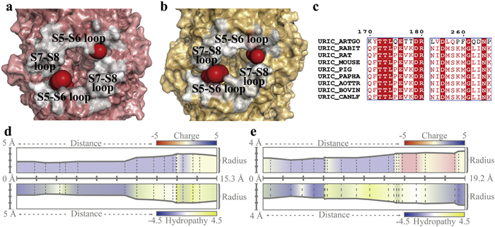
Building on our previous structural–functional studies of Arthrobacter globiformis uricase (AgUricase) [15], we investigated the role of non-conserved residues located at the exit of the uric acid channel and explored strategies to enhance enzymatic performance and thermal stability through rational mutagenesis and disulfide engineering. Uricase adopts a tetrameric barrel-like structure composed of two dimers, with an internal hydrophobic channel formed by β-sheets. Our previous analysis using the ChannelsDB 2.0 identified two positively charged channels located on the lateral sides of the tetramer, specifically at the dimer–dimer interfaces [15,16]. These channels were proposed to serve as entry pathways for uric acid into the enzyme's active pocket. Structural features indicated that the loops between β-strands S5–S6 and S7–S8 form the entry point into the pocket (Figs. 1a and b). Site-directed mutagenesis of two aromatic residues within the channel, H251 and F253, to alanine markedly reduced enzymatic activity, confirming the channel's functional relevance in guiding uric acid into the active site via π–π stacking interactions.
Figure 1
Figure 1.
The uric acid channel of uric acid from Arthrobacter globiformis differs significantly from that of mammalian uricases. (a) Surface view of uric acid entry in uricase from Arthrobacter globiformis(PDB ID: 2YZB). (b) Surface view of uric acid entry in uricase from mammalian(PDB ID: 4MB8). (c) Sequence alignment results of the uric acid entry site between uricases from Arthrobacter globiformis and mammalian. (d) Physicochemical properties of the uric acid channel in uricases form Arthrobacter globiformis. (e) Physicochemical properties of the uric acid channel in uricases form mammalian.
To further characterize channel properties, we employed the MOLE algorithm to predict the uric acid access channel in mammalian uricase (PDB ID: 4MB8) and compared it with that of AgUricase (PDB ID: 2YZB) (Figs. 1d and e) [17]. The mammalian uricase channel was narrower and longer (19.2 Å) and more negatively charged, whereas the AgUricase channel was neutral and hydrophilic, features favoring negatively charged uric acid entry. Given that uric acid carries a negative charge under physiological pH, these findings suggest that the features of the mammalian channel may be less conducive to uric acid entry.
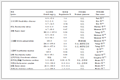
Due to the considerable conformational and chemical differences at the channel entrance, we further performed sequence alignments of the loop regions between S5–S6 (residues 170–180) and S7–S8 (residues 254–265) from AgUricase and various mammalian uricases (Fig. 1c). This analysis identified several non-conserved residues with distinct properties in the AgUricase, including K170, Q175, T177, T178, L254, P259, F260, Q262, D263 and P265. These residues were individually mutated to their corresponding counterparts in mammalian uricase to assess functional impact. Additionally, P259 and P265 were mutated to glutamine, an amino acid that is polar like lysine but electrically neutral, to further assess the influence of polarity at these positions. The purified proteins were subsequently analyzed by sodium dodecyl sulfate-polyacrylamide gel electrophoresis (SDS-PAGE) to confirm their molecular weight and assess their purity (Fig. S1 in Supporting information). The enzymatic activity and thermal stability of the mutants are summarized in Table 1 (Figs. S2 and S3 in Supporting information).
Table 1
Table 1.
Activity and thermostability of site-directed mutants.a
a The activity data are shown as mean ± SD and the thermostability data are shown as mean (n = 3 independent experiments). bTm1: melting temperature for tetramer dissociation to dimer. cTm2: melting temperature for dimer dissociation.
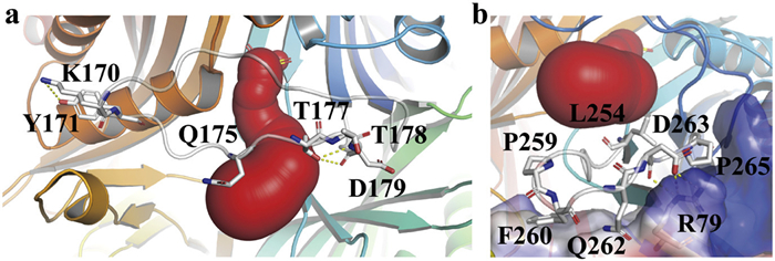
According to the crystal structure of AgUricase (PDB ID: 2YZB), residues K170, Q175, T177, and T178 are located within the loop between β-strands S5 and S6, which contributes to the formation of the uric acid entry site (Fig. 2a). These residues were individually mutated to the corresponding conserved amino acids found in mammalian uricases, resulting in the variants K170Q, Q175P, T177V, and T178K. Enzymatic activity and thermal stability assays revealed only minor reductions in catalytic activity relative to the wild-type enzyme, and no significant differences in thermal stability.
Figure 2
Figure 2.
The structure characteristics of S5-S6 loop and S7-S8 loop. (a) Positions of K170, Q175, T177 and T178 in the crystal structure of AgUricase, along with the interaction networks of K170 and T177 with surrounding amino acid residues. (b) Positions of L254, P259, F260, Q262, D263 and P265 in the crystal structure of AgUricase. Electrostatic surface potential analysis of a neighboring subunit. D263 interacts with R79 from the neighboring subunit, forming hydrogen bond and salt bridge interactions. The red region indicates the predicted uric acid access channel identified by the Channels DB 2.0 program. Hydrogen bonds are shown as yellow dashed lines.
Structural analysis suggests that K170 forms a hydrogen bond with the adjacent Y171 and may also engage in a cation–π interaction. Substitution of K170 with glutamine (K170Q) likely weakens these interactions. Q175 can form a hydrogen bond with the backbone of T173; mutation to proline (Q175P) disrupts this interaction and introduces a rigid pyrrolidine ring, potentially reducing the flexibility of the loop. Similarly, T177 is capable of forming a hydrogen bond with D179, which is abolished upon substitution with valine (T177V). In contrast, the side chain of T178 does not appear to participate in interactions with neighboring residues and extends toward the solvent; its replacement with lysine (T178K) retains hydrophilic character.
Residues L254, P259, F260, Q262, D263, and P265 are located within the loop between β-strands S7 and S8, which also contributes to the formation of the uric acid entry site (Fig. 2b). These residues were individually mutated to the conserved amino acids found in mammalian uricases, resulting in the variants L254N, P259K, F260M, Q262L, D263I, and P265K. In addition, P259 and P265 were also mutated to glutamine, an amino acid with similar polarity but distinct physicochemical properties compared to lysine, yielding the P259Q and P265Q mutants.
Enzymatic activity assays revealed that L254N, P259K, and P265Q exhibited significantly increased activity, while Q262L and P265K showed markedly reduced activity. Thermal stability analyses indicated that P259Q, F260M, and P265Q displayed reduced tetramer stability, whereas L254N resulted in noticeable destabilization of the dimeric form. Notably, the D263I mutation had minimal effects on both activity and stability, likely due to its relatively distant position from the channel entrance.
Residue 259 is a proline containing a pyrrolidine ring; however, substitution with lysine (P259K) increased catalytic activity, while replacement with glutamine (P259Q) led to no substantial change, likely due to favorable electrostatic attraction between the positively charged lysine and negatively charged uric acid, facilitating substrate capture. F260 is located at the interface between dimers and is relatively close to the solvent-exposed surface. Mutation to methionine (F260M) may increase susceptibility to oxidation, which could account for the observed reduction in thermal stability of the tetramer [18].
Electrostatic surface potential analysis revealed a negatively charged region surrounding the loop between strands S7 and S8, primarily contributed by residues Q262, D263, and the nearby R79 and D80 (Fig. 2b). This region differs significantly from its counterpart in mammalian uricases. Substitution of Q262 with a nonpolar isoleucine (Q262L) led to decreased activity, while mutation of P265 to the positively charged lysine (P265K) also resulted in activity loss, possibly due to disruption of the negative electrostatic patch. In contrast, replacement of P265 with glutamine (P265Q) which extended the negative surface, enhanced catalysis. Meanwhile, the mutation of D263 to isoleucine (D263I) did not affect activity or stability, suggesting that introduction of a nonpolar residue at this site has minimal impact on protein function (Table 1).
To further explore the structural basis of enhanced activity, comparative 100 ns molecular dynamics (MD) simulations were conducted for L254N, P259Q, P265Q and the wild type. All systems reached equilibrium within the 70–100 ns, as indicated by converged root-mean-square deviation (RMSD) fluctuations (Fig. S4 in Supporting information) [19,20]. Representative conformations were extracted from equilibrated trajectories using cluster analysis for subsequent structural evaluation.
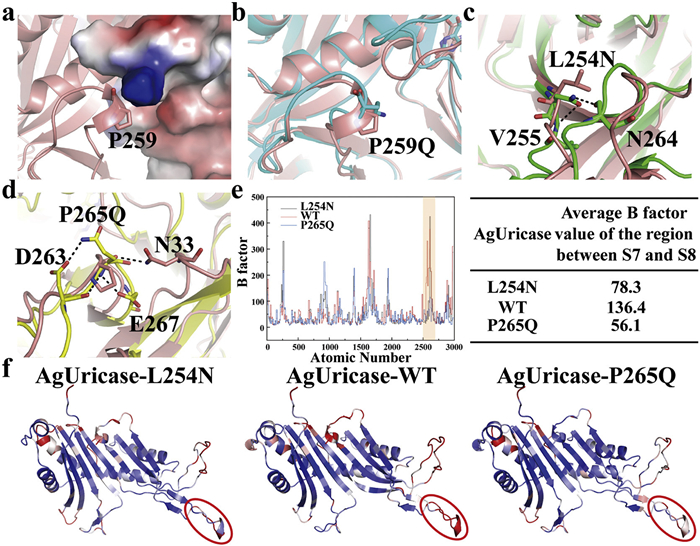
Residue P259 is located at the inter-subunit interface connecting S7 and S8, where its cyclic side chain fits precisely into a cleft on the adjacent subunit (Fig. 3a). This proline residue confers local rigidity essential for maintaining proper tetramer geometry. In contrast, substitution with glutamine (P259Q) increased interfacial flexibility and disrupted tetramer packing, accounting for the observed reduction in thermostability (Fig. 3b). These findings indicate that mutation at structurally pivotal residues contributing to the inter-subunit interface, such as P259, can compromise overall tetramer stability.
Figure 3
Figure 3.
Molecular dynamics simulations reveal the structural basis of mutation-mediated functional alterations. (a) Structural localization of P259 in WT AgUricase. (b) Conformational changes induced by the P259Q mutation. The P259Q mutant and WT AgUricase are represented in cyan and salmon, respectively. (c, d) Comparative analysis of hydrogen bonding patterns. The L254N mutant, P265Q mutant and WT AgUricase are represented in green, yellow and salmon, respectively. Hydrogen bonds are depicted as black dashed lines. (e, f) B factor analysis of monomers within the L254N (left), WT (middle) and P265Q (right) systems, with a color gradient ranging from blue (low) to white (intermediate) to red (high), indicating increasing residue flexibility and dynamic behavior. The regions between S7 and S8, which encompass residues 254 and 265, are circled or highlighted, and their average B factor values are quantified.
MD simulations further revealed the structural mechanisms responsible for enhanced activity in the L254N and P265Q variants. Both residues are situated within the S7-S8 loop adjacent to the substrate entry channel. Replacement of the hydrophobic leucine at position 254 with a polar asparagine introduced new hydrogen bonds with V255 and N264, reshaping the local loop and widening the channel entrance. Similarly, substitution of proline with glutamine at position 265 introduced additional hydrogen bonds with D263 and E267, in contrast to the single N33 interaction observed in the wild type, thereby stabilizing the loop conformation (Figs. 3c and d). B factor analysis revealed that both L254N and P265Q reduced conformational fluctuations in the S7-S8 loop (Figs. 3e and f). As this region constitutes a vital component of the substrate entry channel, enhanced local rigidity likely facilitates substrate access and contributes to the observed increase in enzymatic activity.
Thermal stability analysis of wild-type AgUricase revealed a biphasic denaturation profile, protein thermal shift assay showed two distinct transition peaks, with Tm values of 49.77 and 84.74 ℃ obtained through derivative fitting analysis. Given that uricase typically exists as a tetramer, the first transition peak (Tm1 = 49.77 ℃) corresponds to tetramer dissociation into dimers. The second transition (Tm2 = 84.74 ℃) represents subsequent dimer dissociation, during which the hydrophobic core becomes solvent-exposed. Notably, the thermal denaturation process does not involve complete dissociation into monomers; rather, the three-dimensional structure collapses directly from the dimeric state.
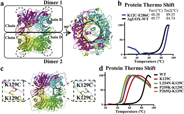
To enhance the thermostability of uricase, Eric Xu research team engineered disulfide bonds into AgUricase by introducing K12C and E286C mutations [21]. Structural rationale for these mutations stems from the proximity between K12 in one monomer and E286 in the adjacent monomer within the dimeric interface (Fig. 4a). Results of protein thermal shift assay demonstrated that the engineered disulfide bond significantly improved dimer stability, with the second transition temperature (Tm2) increasing from 84.74 ℃ to 89.55 ℃. However, the first transition temperature (Tm1) remained largely unchanged at 48.3 ℃, indicating minimal impact on tetramer stability (Fig. 4b). This observation confirms that the introduced disulfide bond specifically stabilizes the dimeric interface without substantially affecting the tetrameric assembly.
Figure 4
Figure 4.
Introduction of a disulfide bond between subunits to enhance protein thermal stability. (a) Schematic representation of the disulfide bond formed by the K12C–E286C mutation in the three-dimensional structure of uricase. The disulfide bond formed between K12C and E286C is indicated by the dashed circle. (b) Changes in the protein thermal shift curve of uricase following the introduction of the K12C–E286C disulfide bond. (c) AlphaFold-predicted formation of a disulfide bond between K129C residues across dimers. (d) The protein thermal shift curve of uricase following the introduction of the K129C disulfide bond.
To enhance the thermostability of uricase at the tetrameric level, this study focused on engineering inter-subunit interactions between dimeric units. Structural analysis revealed that due to the high symmetry of the uricase tetramer, residue K129 is strategically positioned at the dimer-dimer interface. Notably, the α-carbons of adjacent K129 residues from neighboring dimers are separated by only 5.2 Å, suggesting potential for disulfide bond formation. Computational modeling using AlphaFold predicted that mutation of K129 to cysteine (K129C) could facilitate the formation of an inter-subunit disulfide bridge between dimeric units (Fig. 4c and Fig. S5 in Supporting information) [22]. This engineered covalent linkage is expected to specifically stabilize the tetrameric assembly by reinforcing the critical dimer-dimer interface.
To investigate the effect of K129C mutation on AgUricase, we subsequently evaluated the activity and thermal stability of the mutants (Table 2, Figs. S6 and S7 in Supporting information). Upon introducing the K129C mutation, the thermal shift assay (TSA) curve exhibited only a single peak with a Tm value of 55.65 ℃. This indicates that substituting this residue with cysteine significantly enhances the thermostability of the tetramer. During heating, the protein denatures and dissociates; however, due to the presence of disulfide bonds between dimers, the overall protein structure collapses directly upon temperature increase, bypassing the dissociation process from tetramer to dimer, thus resulting in a single peak. Moreover, enzymatic activity assays revealed that introduction of this mutation led to an increase in uricase activity, reaching 27.61 U/mg. When this mutation was introduced into the previously enhanced variants L254N, P259K, and P265Q, the resulting mutants exhibited a single peak in their PTS profiles and showed improved thermal stability (Fig. 4d). Although a slight reduction in enzymatic activity was observed in these combinatorial mutants, their activity levels remained superior to that of the wild-type enzyme.
Table 2
Table 2.
Activity and thermostability of site-directed mutants.a
a The activity data are shown as mean ± SD and the thermostability data are shown as mean (n = 3 independent experiments). bTm1: melting temperature for tetramer dissociation to dimer. cTm2: melting temperature for dimer dissociation.
To further examine the formation of disulfide bonds between uricase monomers upon introduction of the K129C mutation, we compared SDS-PAGE results under reducing and non-reducing conditions (Fig. S8 in Supporting information). While TSA analysis demonstrated a pronounced enhancement in thermal stability, the gel images showed only a small fraction of oligomers. This observation suggests that the disulfide bonds formed between C129 residues are metastable, likely under conformational strain during protein folding. Consequently, although these bonds exert a significant stabilizing effect on the tetramer, they are relatively unstable and can only be detected as a minor population of oligomers under non-reducing conditions.
Compared with conventional oral ULTs, exogenous uricase has become a key therapeutic option for patients with severe or refractory gout, offering rapid urate reduction and tophi resolution when conventional ULTs fail [9]. Currently, pegloticase remains the only FDA-approved exogenous uricase for treating chronic gout unresponsive to standard therapies. However, further development of uricase therapeutics is hindered by high immunogenicity, underscoring the need for improved enzyme variants [13].
Building on our previous structure–activity analysis of AgUricase [15], the present study focuses on the loop regions between β-strands S5–S6 and S7–S8 that form the entrance of the uric acid channel. We aimed to modulate local conformation at this site through targeted mutations to enhance catalytic efficiency while introducing disulfide bonds to improve enzyme stability. Comparative analysis between the high-activity AgUricase and low-activity mammalian uricases revealed distinct physicochemical differences at this site. To evaluate their functional relevance, site-directed mutagenesis was conducted on variable residues within these loops.
Mutations on the S5–S6 loop (K170, Q175, T177, T178) had negligible effects on activity or stability, whereas substitutions on the S7–S8 loop exerted pronounced influence. Variants introducing favorable hydrogen-bonding or electrostatic interactions (L254N, P259K, P265Q) enhanced catalysis, while those disrupting polarity or packing (F260M, Q262I) impaired function. MD simulations supported these results, showing that activity-enhancing variants widened the channel entrance or stabilized local conformations, whereas destabilizing mutations increased loop flexibility or perturbed the interface. B factor analysis confirmed reduced conformational fluctuations in the P265Q, consistent with its improved activity. These findings highlight the S7–S8 loop as a key determinant of the electrostatic and structural environment that governs substrate recognition and catalytic efficiency.
Because activity-enhancing mutations often compromised thermal stability, disulfide bond engineering was employed to reinforce structural rigidity. Inspired by Eric Xu's work which showed that a K12C–E286C disulfide bond could stabilize the uricase dimer, we identified a single-site K129C mutation forming an inter-dimer disulfide bond at the tetramer interface. This modification markedly improved thermostability without diminishing activity. Combining K129C with activity-enhancing mutations yielded uricase variants exhibiting both high catalytic efficiency and enhanced thermal stability.
Nevertheless, we acknowledge the limitations of this work. This study focused exclusively on AgUricase, and whether similar mechanisms operate in other species remains to be determined. In addition, although thermal shift assays demonstrated a marked enhancement in tetrameric stability upon introduction of the K129C mutation, non-reducing SDS-PAGE results suggest that the engineered disulfide bond may be relatively unstable, potentially due to conformational strain within the protein, indicating that further consideration is required when selecting mutation sites.
In summary, using AgUricase as a model, our findings elucidate how conformational and electrostatic features at the uric acid channel entrance shape uricase function and stability. And we propose a novel strategy for improving the thermal stability of uricase tetramers through the introduction of a single-point K129C mutation, which facilitates disulfide bond formation between dimers within the tetrameric assembly. The combined strategy of structure-guided mutagenesis and disulfide bond engineering provides a rational framework for developing next-generation uricase therapeutics with improved catalytic performance and thermostability.
CRediT authorship contribution statement
Shijia Zu: Writing – original draft, Visualization, Methodology, Data curation, Conceptualization. Zhonglian Cao: Project administration, Methodology, Investigation, Formal analysis. Lunan Lv: Writing – original draft, Visualization, Validation, Software, Formal analysis, Data curation. Wanyi He: Validation, Investigation, Data curation. Wei Fu: Writing – review & editing, Supervision, Resources, Project administration, Conceptualization.
Declaration of competing interest
The authors declare that they have no known competing financial interests or personal relationships that could have appeared to influence the work reported in this paper.
Acknowledgments
This work was supported by the National Natural Science Foundation of China (Nos. 82273853, 82574323) and by the Science and Technology Commission of Shanghai Municipality (Nos. 25JS2830100, 24JS2830100).
Supplementary materials
Supplementary material associated with this article can be found, in the online version, at doi:10.1016/j.cclet.2025.112195.
Figure 1
The uric acid channel of uric acid from Arthrobacter globiformis differs significantly from that of mammalian uricases. (a) Surface view of uric acid entry in uricase from Arthrobacter globiformis(PDB ID: 2YZB). (b) Surface view of uric acid entry in uricase from mammalian(PDB ID: 4MB8). (c) Sequence alignment results of the uric acid entry site between uricases from Arthrobacter globiformis and mammalian. (d) Physicochemical properties of the uric acid channel in uricases form Arthrobacter globiformis. (e) Physicochemical properties of the uric acid channel in uricases form mammalian.
Figure 2
The structure characteristics of S5-S6 loop and S7-S8 loop. (a) Positions of K170, Q175, T177 and T178 in the crystal structure of AgUricase, along with the interaction networks of K170 and T177 with surrounding amino acid residues. (b) Positions of L254, P259, F260, Q262, D263 and P265 in the crystal structure of AgUricase. Electrostatic surface potential analysis of a neighboring subunit. D263 interacts with R79 from the neighboring subunit, forming hydrogen bond and salt bridge interactions. The red region indicates the predicted uric acid access channel identified by the Channels DB 2.0 program. Hydrogen bonds are shown as yellow dashed lines.
Figure 3
Molecular dynamics simulations reveal the structural basis of mutation-mediated functional alterations. (a) Structural localization of P259 in WT AgUricase. (b) Conformational changes induced by the P259Q mutation. The P259Q mutant and WT AgUricase are represented in cyan and salmon, respectively. (c, d) Comparative analysis of hydrogen bonding patterns. The L254N mutant, P265Q mutant and WT AgUricase are represented in green, yellow and salmon, respectively. Hydrogen bonds are depicted as black dashed lines. (e, f) B factor analysis of monomers within the L254N (left), WT (middle) and P265Q (right) systems, with a color gradient ranging from blue (low) to white (intermediate) to red (high), indicating increasing residue flexibility and dynamic behavior. The regions between S7 and S8, which encompass residues 254 and 265, are circled or highlighted, and their average B factor values are quantified.
Figure 4
Introduction of a disulfide bond between subunits to enhance protein thermal stability. (a) Schematic representation of the disulfide bond formed by the K12C–E286C mutation in the three-dimensional structure of uricase. The disulfide bond formed between K12C and E286C is indicated by the dashed circle. (b) Changes in the protein thermal shift curve of uricase following the introduction of the K12C–E286C disulfide bond. (c) AlphaFold-predicted formation of a disulfide bond between K129C residues across dimers. (d) The protein thermal shift curve of uricase following the introduction of the K129C disulfide bond.
Table 1.
Activity and thermostability of site-directed mutants.a
AgUricase
Activity (U/mg)
Tm1 (℃) b
Tm2 (℃) c
WT
24.28 ± 2.01
49.77
84.74
K170Q
21.93 ± 1.45
46.55
82.50
Q175P
22.55 ± 0.88
47.50
80.59
T177V
23.11 ± 0.84
46.69
82.68
T178K
23.97 ± 2.31
51.28
83.35
L254N
29.14 ± 1.70
46.46
71.10
P259K
30.87 ± 3.17
46.41
81.24
P259Q
24.92 ± 1.23
34.11
80.12
F260M
26.88 ± 1.24
37.60
81.63
Q262L
13.74 ± 0.82
51.28
87.73
D263I
24.27 ± 1.36
50.12
83.82
P265K
19.22 ± 2.04
46.41
81.89
P265Q
30.61 ± 0.67
42.35
79.24
a The activity data are shown as mean ± SD and the thermostability data are shown as mean (n = 3 independent experiments). bTm1: melting temperature for tetramer dissociation to dimer. cTm2: melting temperature for dimer dissociation.
Table 2.
Activity and thermostability of site-directed mutants.a
AgUricase
Activity (U/mg)
Tm1 (℃) b
Tm2 (℃) c
WT
24.28 ± 2.01
49.77
84.74
K129C
27.61 ± 1.55
55.65
55.65
L254N-K129C
27.09 ± 1.39
48.14
48.14
P259K-K129C
26.23 ± 0.23
51.50
51.50
P265Q-K129C
29.51 ± 3.14
51.98
51.98
a The activity data are shown as mean ± SD and the thermostability data are shown as mean (n = 3 independent experiments). bTm1: melting temperature for tetramer dissociation to dimer. cTm2: melting temperature for dimer dissociation.


 DownLoad:

 下载:


 下载:

