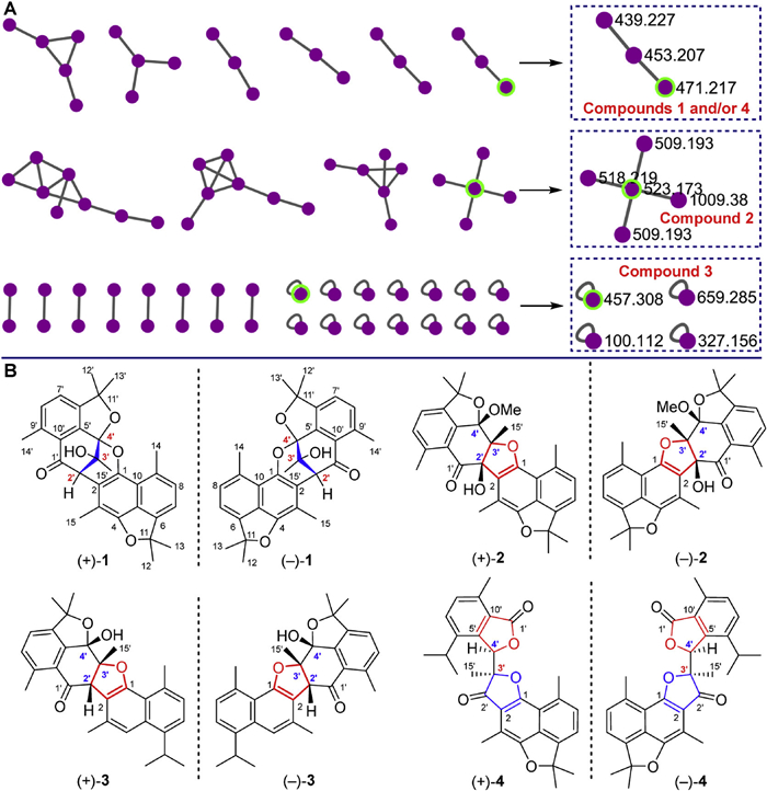
Figure 1.
Molecular network (A) and structures of compounds 1–4 (B).

Unprecedented anti-adipogenic cadinane dimers from Stahlianthus involucratus: Discovery via an MS-MN-based activity-structure strategy
Xiu-Qing Song , Xi-Yu Zheng , Qin-Qin Song , Ting Tang , Peng Sun , Jian-Mei Lu , Yao-Yue Fan , Kai-Long Ji


The discovery of novel bioactive natural products is a critical frontier in pharmaceutical development, particularly for treating chronic metabolic disorders. Obesity, characterized by pathological adipose tissue expansion, represents an urgent global health challenge. The limited availability of effective anti-obesity therapies necessitates the urgent exploration of innovative treatments [1]. In this context, traditional Chinese medicine (TCM) offers a rich source of candidates with documented efficacy in weight management. Recent studies have identified several plant-derived bioactive compounds with unique anti-obesity mechanisms, including celastrol [2], hyperforin [3], and puerarin [4]. These findings highlight TCM-derived small molecules as a promising reservoir for developing new anti-obesity leads.
As part of our ongoing research to discover lipid-lowering compounds from TCM [5–9], we focused on Zingiberaceae plants due to their rich metabolite diversity and well-documented bioactivities [10–17]. Stahlianthus involucratus (Zingiberaceae), a perennial herb, is traditionally used to treat trauma, inflammatory pain, and envenomation [18], with pharmacological studies confirming significant anti-inflammatory, analgesic, and antioxidant activities in its extracts [19,20]. Phytochemical analyses have further identified sesquiterpenoids as characteristic constituents of this species [21–24]. Our preliminary screening using 3T3-L1 adipocytes revealed that ethyl acetate (EtOAc)-soluble fraction of S. involucratus extract markedly reduced lipid accumulation. Subsequent liquid chromatography-tandem mass spectrometry (LC-MS/MS) analysis and bioactivity-directed fractionation unexpectedly identified four pairs of novel enantiomers, distahlianoids A–D (1–4) (Fig. 1). Compound 1 exhibits a unique 5/6/6/6/6/6/5-fused heptacyclic scaffold with a novel 9-methyl-2-oxabicyclo[3.3.1]nona-3,7‑dien-9-ol core. Compounds 2 and 3 possess unconventional 5/6/6/5/6/6/5 and 5/6/6/5/6/6 polycyclic frameworks, respectively, whereas compound 4 represents a skeleton-rearranged dimer featuring an unprecedented 2-methyl-[2,2′-bifuran]−3,5′(2H,2′H)‑dione motif. Their structures and absolute configurations were unambiguously established through comprehensive nuclear magnetic resonance (NMR) analysis, X-ray crystallography, and electronic circular dichroism (ECD) calculations. Plausible biosynthetic pathways for compounds 1–4 were postulated, and stereoselectivity patterns for their formation were tentatively elucidated. All compounds exhibit antiadipogenic effects in 3T3-L1 adipocytes, with (–)-4 being the most potent.
The ethanol extract of S. involucratus was subjected to liquid-liquid partitioning with EtOAc and water, yielding an EtOAc-soluble fraction (JST-EtOAc). This fraction demonstrated significant antiadipogenic activity (half maximal effective concentration (EC50): 18.25 ± 1.03 μg/mL) (Fig. S1 in Supporting information) in 3T3-L1 adipocytes and was subsequently analyzed by LC-MS/MS Molecular networking, performed using the GNPS platform (Fig. S2 in Supporting information), revealed distinct molecular clusters, indicating the presence of potentially novel compounds (Fig. 1A). Then, the macroporous resin column chromatography of JST-EtOAc produced three corresponding fractions JST-50%, JST-80% and JST-95%, of which JST-80% was the most active subfraction (EC50: 8.24 ± 0.13 μmol/L) (Fig. S1). JST-80% was analyzed by LC-MS/MS, and the resulting molecular network, which featured distinct clusters for compounds 1–4 (Fig. 1B and Fig. S3 in Supporting information), guided the subsequent targeted isolation of these new compounds.
Distahlianoid A (1) was isolated as pale-yellow crystals from methanol with a molecular formula of C30H31O5 based on the high-resolution electrospray ionization mass spectrometry spectrum (HRESIMS) ion peak at m/z 471.2167 [M + H]+ (calcd. 471.2166), possessing 16 degrees of unsaturation (DOUs). Elucidation of its 1D NMR data (Table S1 in Supporting information) coupled with heteronuclear single quantum coherence (HSQC) spectrum revealed a total of 30 carbon signals comprising a conjugated ketone carbonyl (δC 194.8), eight methyls, five methines (including four aromatic), five oxygenated tertiary carbons (including two aromatic), and eleven aromatic quaternary carbons. Considering the double bond equivalents (account for 9 DOUs), a heptacyclic scaffold was deduced for compound 1.
Detailed interpretation of 1D and 2D NMR data (Fig. 2 and Table S1) enable establishment of the gross structure of compound 1. Firstly, a cadinene monomeric unit Ⅰ (red) with an ether linkage between C-4 and C-11 was deduced by proton-proton correlation spectroscopy (1H–1H COSY) correlation of H-7/H-8 and heteronuclear multiple bond correlation (HMBC) cross peaks of H3–15/C-2, C-3 and C-4; H-7/C-5; H-8/C-6 and C-10; H3–14/C-8, C-9 and C-10; H3–12/C-11 and C-13; and H3–13/C-6 and C-3 (Fig. 2A), together with the downfield carbon signals of C-4 (δC 151.7) and C-11 (δC 91.2) [22]. Similarly, the 1H–1H COSY spin coupling system (H-7′/H-8′), and HMBC correlations of H-2′/C-1′ and C-10′; OH-3′/C-2′ and C-3′; H3–15′/C-2′, C-3′ and C-4′; H-7′/C-5′; H-8′/C-6′ and C-10′; H3–14′/C-8′, C-9′ and C-10′; H3–12′/C-11′; H3–13′/C-6′, C-11′ and C-12′, coupled with the oxygenated carbon resonances of C-4′ (δC 107.1) and C-11′ (δC 90.4) constructed the monomeric unit Ⅱ (black). The observed HMBC correlation from H-2′ to C-1 and C-2 indicated the –C2–C2′– connection of two substructures. In addition, molecular formula and the carbon resonances of C1 (δC 142.6) and C4′ (δC 107.1) suggested the presence of a –C1–O–C4′– linkage. The planar structure of compound 1 was thus established as a highly oxygenated cadinane sesquiterpenoid dimer with a rare 9-methyl-2-oxabicyclo[3.3.1]nona-3,7‑dien-9-ol core.
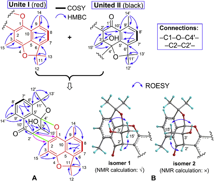
The relative configuration of compound 1 was primarily established through ROESY analysis (Fig. 2B). Key rotating frame overhauser effect spectroscopy (ROESY) correlations of H3–15′/OH-3′ and H-2′ indicated that these protons are co-facial, permitting the tentative assignment of a β-orientation. This finding, combined with spatial structure analysis, enable the assignment of an S*-configuration at C-4′, while the configuration at C-3′ was left unassigned. To resolve this, quantum chemical NMR calculations [25,26] of two possible diastereomers (Fig. 2B and Fig. S9 in Supporting information) were performed and confirmed the complete 2′S*, 3′S*, 4′S* configuration (isomer 1) based on a definitive DP4+ probability of 100% (Figs. S13 and S14 in Supporting information). This assignment was unambiguously confirmed by subsequent X-ray crystallographic analysis (CCDC number: 2475439; Fig. 3), which also revealed the racemic nature of 1. Subsequently chiral separation of 1 afforded two enantiomers (+)- and (–)-1 in the ratio of ca. 1:1 (Fig. S5 in Supporting information), as confirmed by their specific rotations ([α]25D +305.8 and [α]25D −279.7, respectively) and mirror-image electronic circular dichroism (ECD) spectra (Fig. 4). Despite extensive efforts, single crystals of the enantiomers could not be obtained. The absolute configurations of the enantiomers were finally established using time-dependent density functional theory (TDDFT)-ECD calculations. The experimental ECD spectra exhibited good agreement with the theoretical data (Fig. 4), establishing (+)-1 as 2′S, 3′S, 4′S and (−)-1 as 2′R, 3′R, 4′R.
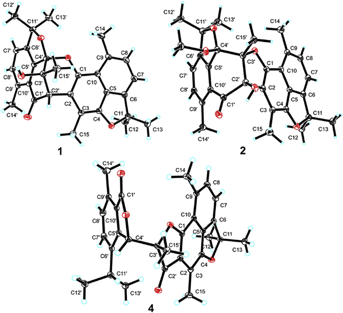
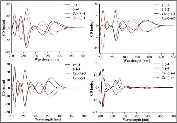
Distahlianoid B (2) was assigned the molecular formula C31H32O6 based on the HRESIMS spectrum. Analysis of the 1D NMR data (Table S1) and distortionless enhancement by polarization transfer (DEPT) spectrum revealed 31 carbons, containing eight methyls, one methoxyl, four aromatic methines, seven oxygenated tertiary carbons (including two aromatic), ten quaternary carbons, and a conjugated ketone carbonyl (δC 196.0). Interpretation of the 2D NMR correlations (Fig. 5) delineated two substructures (units Ⅰ and Ⅱ), analogous to 1 but with a 2′‑hydroxy-4′‑methoxy derivative of unit Ⅱ in 2, as verified by the key HMBC cross peaks of OCH3–4′/C-4′ and OH-2′/C-1′, C-2′ and C-3′ These two substructures were connected via –C2–C2′– and –C1–O–C3′– bonds by the HMBC correlation of OH-2′/C-2 and molecular formula, establishing a 6/5/6/5/6/6/5-fused heptacyclic cadinane dimer for compound 2. ROESY correlations of OCH3–4′/H3–15′ indicated a co-facial arrangement between OCH3–4′ and H3–15′, suggesting a tentative β-orientation, while the configuration of OH-2′ remained undefined. The following quantum chemical NMR calculations of two possible diastereomers (Fig. S10 in Supporting information) unequivocally established the 2′R*, 3′R*, 4′S* configuration, supported by a definitive DP4+ probability of 100% (Figs. S15 and S16 in Supporting information). This assignment was further confirmed by X-ray crystallography (CCDC number: 2475441; Fig. 3), which also revealed racemic nature. Further chiral separation gave two enantiomers (+)-2 ([α]23D +141.3) and (–)-2 ([α]23D −91.0) in a ratio of approximately 1:1 (Fig. S6 in Supporting information), consistent with their mirrored ECD spectra (Fig. 4). The calculated ECD spectra closely matched with experimental data, establishing the absolute configurations as 2′R, 3′R, 4′S for (+)-2 and 2′S, 3′S, 4′R for (–)-2.
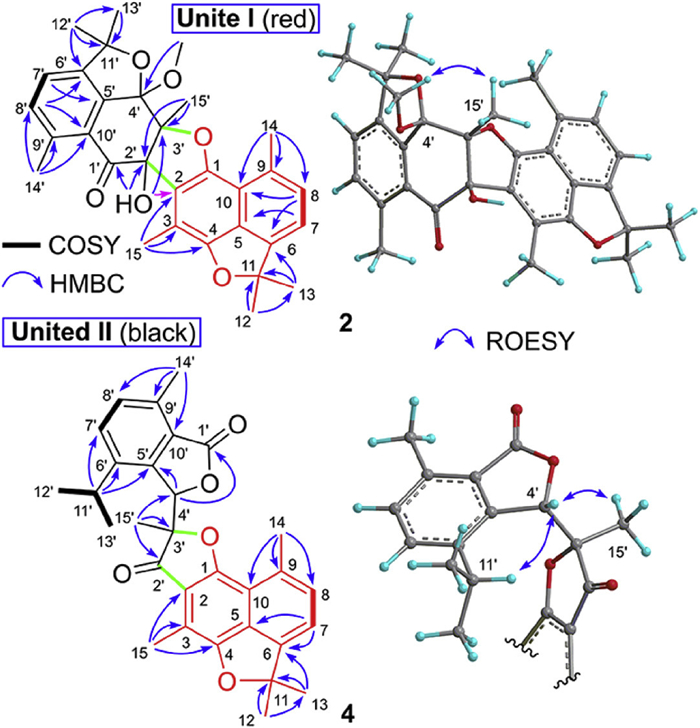
Distahlianoid C (3) has a molecular formula C30H32O4, showing 44 mass units less than that 2. Comparative analysis of their 1D NMR data (Tables S1 and S2 in Supporting information) revealed that compound 3 is a structural analog of 2, with the appearance of two additional methine signals (δH 7.42 s and 3.56 m; δC 117.4 and 28.8), and the absence of two oxygenated tertiary carbons (C4 and C-11) and a methoxy group. The lack of -C4-O-C1- ether linkage in unite Ⅰ was supported by the key 1H–1H COSY correlations from H3–12 to H-11 and from H-11 to H3–13, and HMBC cross peaks from H-4 to C-2, C-5 and C-10; and from H3–15 to C-4 (Fig. S4 in Supporting information). In addition, a 2′-dehydroxy-4′‑hydroxy derivative of unit Ⅱ was identified by the HMBC correlation from H-2′ to C-1, C-2, C-1′ and C-10′, from H3–15′ to C-2′, and from OH-4′ to C-3′, C-4′ and C-5′. ROESY correlations between H-2′ and H3–15′ suggested a tentative β-orientation, while the configuration of OH-4′ was unresolved. The complete relative configuration (2′S*, 3′R*, 4′S*) was ultimately determined via quantum chemical NMR calculations (Fig. S11 in Supporting information), which provided definitive evidence through a DP4+ probability of 100% (Figs. S17 and S18 in Supporting information). Furthermore, the racemic nature was indicated by the zero optical rotation. Chiral separation of 3 also yielded two enantiomers (+)- and (–)-3 in approximately a 1:1 ratio (Fig. S7 in Supporting information), as evidenced by their specific rotations ([α]28D +156.6 and [α]27D −186.4, respectively) and the opposite ECD curves (Fig. 4). Theoretical ECD calculations ultimately assigned the absolute configurations as 2′S, 3′R, 4′S for (+)-3 and 2′R, 3′S, 4′R for (–)-3.
Distahlianoid D (4) possessed the molecular formula C30H30O5, showing 16 DOUs, on account of the HRESIMS spectrum ion peak at m/z 493.1985 [M + Na]+ (calcd. for C30H30O5Na, 493.1985). Analysis of 1D NMR data (Table S2) and HSQC spectrum revealed characteristic resonances for eight methyls and four aromotic protons, and eighteen carbons including a conjugated ketone (δC 201.8) and a carboxyl (δC 170.4) groups, two methine (one oxygenated), four oxygenated tertiary carbons, and ten quaternary carbons. These functionalities account for 10 DOUs, suggesting a hexacyclic system for 4.
Comprehensive 2D NMR analysis (Fig. 5) confirmed that substructure unite Ⅰ (red) in 4 is identical to those in compounds 1 and 3. The unit Ⅱ (black) was established to be a novel monomer generated via C1′–C2′ bond cleavage, as evidenced by 1H–1H COSY correlations of H3–12′/H-11′/H3–13′ and H-7′/H-8′ and key HMBC cross peaks of H3–15′/C-2′ (δC 201.8), C-3′ and C-4′; H-4′/C-1′ and C-10′; H-11′/C-5′, C-6′ and C-7′; H3–14′/C-8′, C-9′ and C-10′ The connectivity between the two subunits, formed through both C2—C2′ and C1—O—C3′ linkages, was unequivocally confirmed by the single-crystal X-ray diffraction analysis (CCDC number: 2475443; Fig. 3), which also revealed the racemic character of 4 and established the relative configuration as 3′R*, 4′R* and/or 3′S*, 4′S*. The key ROESY interactions of H-4′/H3–15′ and quantum chemical NMR calculations (a definitive DP4+ probability of 100%, Figs. S12, S19 and S20 in Supporting information) provided further confirmation of the above structural assignment. Compound 4 was collectively defined as a rearranged cadinane dimer featuring a 2-methyl-[2,2′-bifuran]−3,5′(2H,2′H)‑dione motif. Preparative chiral resolution yielded enantiomers (+)-4 ([α]27D +56.7) and (–)-4 ([α]28D −29.3) in approximately 1:1 ratio (Fig. S7), exhibiting mirror-image ECD profiles (Fig. 4). Comparative TDDFT-ECD calculations and experimental data definitively established the absolute configurations as 3′R, 4′R for (+)-4 and 3′S, 4′S for (–)-4.
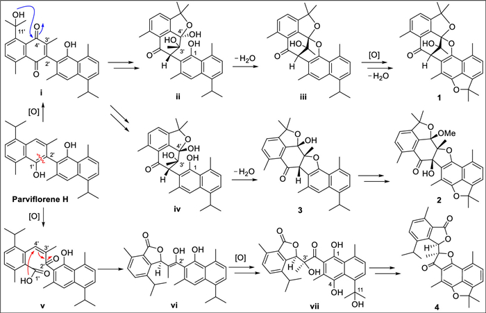
The plausible biosynthetic pathways of 1–4 was proposed in Scheme 1. Parviflorene H, a known compound [27], was treated as the likely precursor. Sequential oxidation of parviflorene H would generate a naphthoquinone-like intermediate ⅰ, which could undergo hemiketalization and further oxidation to produce C-4′ stereochemically distinct intermediates ⅱ and ⅳ. Dehydration of ⅱ between C1-OH and C4′-OH would yield intermediate ⅲ, while subsequent oxidation and dehydration of ⅲ would furnish 1. Parallel dehydration of ⅳ (C1-OH/C3′-OH) forms 3, which undergoes analogous reactions to generate 2. In addition, Δ1′(2′) oxidative cleavage of parviflorene H could produce intermediate ⅴ, which might engage in a Michael addition to form ⅵ, ultimately yielding 4 through oxidation and dehydration. As delineated in Scheme 1, the achiral dimeric precursor parviflorene H undergoes transformations without stereoselective control to yield intermediates ⅱ, ⅳ, and ⅵ, ultimately leading to the formation of the final products as racemic mixtures. The isolation of four enantiomeric pairs, combined with prior reports of diastereomeric sesquiterpenoid dimers [23,24,27–38], prompted our investigation into stereochemical determinants governing enantiomer vs. diastereomer formation in cadinane-type dimerization. Comprehensive analysis revealed that monomers bearing a chiral isopropyl group exclusively form diastereomeric dimers (homodimers or heterodimers) via stereoselective dimerization, while fully achiral monomers invariably produce enantiomeric dimers.
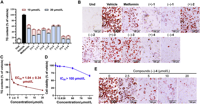
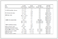
Building on our ongoing research into lipid-lowering TCM compounds from Zingiberaceae plants and motivated by the preliminary anti-adipogenic activity of the S. involucratus extract (Fig. S8 in Supporting information), we assessed the effects of compounds 1–4 in 3T3-L1 adipocytes using oil red O (ORO) staining and triglyceride (TG) analysis (Fig. 6) [5,39,40]. All compounds exhibited the anti-adipogenic activity compared to the vehicle group (differentiation cell group) at concentrations of 10 and 20 μmol/L (Figs. 6A and B). Notably, compounds 1, 3 and 4 demonstrated the significant TG inhibition with EC50 values ranging from 1.04 μmol/L to 12.05 μmol/L, while showing low cytotoxicity (half maximal inhibitory concentration (IC50) > 19.02 μmol/L) (Table 1). Particularly, (–)-4 emerged as the best suppressed potential, exhibiting dose-dependent TG inhibition with an EC50 of 1.04 ± 0.34 μmol/L and an exceptional selectivity index (SI) of 96.15 (Figs. 6C‒E and Table 1). Interestingly, the (−)-enantiomers of 1, 3 and 4 consistently showed stronger anti-adipogenic effects than their (+)-counterparts, likely due to different spatial orientations [35].
 DownLoad:
CSV
| Compound | EC50 (μmol/L) | IC50 (μmol/L) | SIa | Compound | EC50 (μmol/L) | IC50 (μmol/L) | SI |
| (+)-1 | 7.78 ± 1.17 | 19.02 ± 0.72 | 2.45 | (–)-3 | 8.03 ± 1.21 | > 100 | > 12.45 |
| (–)-1 | 5.91 ± 1.01 | 25.15 ± 2.95 | 4.26 | (+)-4 | 1.91 ± 0.26 | 83.18 ± 5.22 | 43.55 |
| (+)-2 | > 20b | > 100 | - | (–)-4 | 1.04 ± 0.34 | > 100 | > 96.15 |
| (–)-2 | > 20 | > 100 | - | Metforminc | 19.94 ± 0.73 | 28.43 ± 1.99 | 1.43 |
| (+)-3 | 12.05 ± 1.68 | > 100 | > 8.30 | ||||
| a SI = IC50/EC50. b EC50 > 20 μmol/L was considered as weak inhibition. c Positive control. |
|||||||
In conclusion, our integrated LC-MS/MS profiling and bioassay-guided isolation strategy of S. involucratus yielded four pairs of novel cadinane dimers, distahlianoids A–D (1–4). Comprehensive spectroscopic analysis unequivocally established their structures, revealing three distinct architectural scaffolds, of which compound 1 features an unprecedented heptacyclic systems containing a 9-methyl-2-oxabicyclo[3.3.1]nona-3,7‑dien-9-ol core, 2 and 3 display exceptional 5/6/6/5/6/6/5 and 5/6/6/5/6/6 polycyclic frameworks, respectively, and 4 represents a skeletally rearranged cadinane dimer. Compounds 1, 3 and 4 exhibited significant triglyceride inhibition, with EC50 values ranging from 1.04 μmol/L to 12.05 μmol/L, while demonstrating minimal cytotoxicity. Our finding not only substantially expands the structural diversity of cadinane-type dimers but also highlights their promise as novel therapeutic candidates for obesity treatment.
Xiu-Qing Song: Validation, Investigation. Xi-Yu Zheng: Validation, Investigation. Qin-Qin Song: Investigation. Ting Tang: Investigation, Funding acquisition. Peng Sun: Software, Investigation. Jian-Mei Lu: Investigation. Yao-Yue Fan: Writing – review & editing, Writing – original draft, Project administration. Kai-Long Ji: Writing – review & editing, Writing – original draft, Supervision, Project administration, Conceptualization.
The authors declare that they have no known competing financial interests or personal relationships that could have appeared to influence the work reported in this paper.
Financial support from Yunnan Fundamental Research Projects (No. 202501AT070248), the Key R&D Program of Shandong Province (No. 2024CXPt029), Taishan Scholars Program (No. tsqn202408315), the Strategic Priority Research Program of the Chinese Academy of Sciences (No. XDB1060000), West Light Foundation of Chinese Academy of Sciences, and China Postdoctoral Science Foundation (No. 2023T160677) are highly acknowledged. We are grateful to the Center for Gardening and Horticulture, Xishuangbanna Tropical Botanical Garden, Chinese Academy of Sciences, for supplying the plant samples.
Supplementary material associated with this article can be found, in the online version, at doi:
X. Wen, B.H. Zhang, B.Y. Wu, et al., Signal Transduct. Tar. Ther. 7 (2022) 298–329. doi: 10.1038/s41392-022-01149-x
J.L. Liu, J. Lee, M.A.S. Hemandez, et al., Cell 161 (2015) 999–1011. doi: 10.1016/j.cell.2015.05.011
S.Z. Chen, X.X. Liu, C. Peng, et al., Cell Metab. 33 (2021) 565–580. doi: 10.1016/j.cmet.2021.02.007
Q.Q. Lyu, W.Z. Xue, R.X. Liu, et al., Nature 634 (2024) 936–943. doi: 10.1038/s41586-024-07929-5
Q.Q. Song, Y. Guo, P. Sun, et al., Chin. J. Chem. 42 (2024) 3211–3218. doi: 10.1002/cjoc.202400749
Z.Y. Zhang, X.H. Gao, Y. Huang, J. Nat. Prod. 87 (2024) 1441–1453. doi: 10.1021/acs.jnatprod.4c00246
Z.Y. Zhang, Y.H. Chen, C.Y. Zheng, et al., Org. Lett. 27 (2025) 2485–2491. doi: 10.1021/acs.orglett.5c00445
X.Q. Song, Y. Guo, X. Wang, et al., J. Nat. Prod. 88 (2025) 1514–1525. doi: 10.1021/acs.jnatprod.5c00390
S.S. Chen, Y.H. Chen, C.Y. Zheng, et al., J. Org. Chem. 90 (2025) 9541–9548. doi: 10.1021/acs.joc.5c01017
Q. Zhao, L.G. Xiao, L.S. Bi, et al., Org. Lett. 24 (2022) 6936–6939. doi: 10.1021/acs.orglett.2c02710
K.L. Ji, M.Z. Wu, C.Y. Huang, J. Agric. Food Chem. 70 (2022) 3989–3999. doi: 10.1021/acs.jafc.1c08038
M.Z. Wu, P.C. GongPan, M.Y. Dai, et al., Tetrahedron 120 (2022) 132901. doi: 10.1016/j.tet.2022.132901
Y. Liu, F. Liu, M.M. Qiao, et al., Org. Lett. 21 (2019) 1197–1201. doi: 10.1021/acs.orglett.9b00149
K.L. Ji, Y.Y. Fan, Z.P. Ge, et al., J. Org. Chem. 84 (2019) 282–288. doi: 10.1021/acs.joc.8b02665
Q. Zhao, J.J. Gao, X.J. Qin, et al., Org. Lett. 20 (2018) 704–707. doi: 10.1021/acs.orglett.7b03836
H. Yin, J.G. Luo, S.M. Shan, et al., Org. Lett. 15 (2013) 1572–1575. doi: 10.1021/ol400348a
C.S. Yang, X.B. Wang, J.S. Wang, et al., Org. Lett. 13 (2011) 3380–3383. doi: 10.1021/ol201137v
X.J. Ma, L.X. Zhang, Y.F. Lin, Chinese Dai Materia Medica (Volume Ⅰ), People's Medical Publishing House, Beijing, 2018, p. 36.
P. Pingsusaen, P. Kunanusorn, P. Khonsung, et al., J. Ethnopharmacol. 162 (2015) 199–206. doi: 10.1016/j.jep.2014.10.060
W.K. Li, B.M. Wu, Y. Wang, et al., BioMed. Res. Int. 2021 (2021) 9490162. doi: 10.1155/2021/9490162
Q.M. Li, J.G. Luo, Y.M. Zhang, et al., Tetrahedron 72 (2016) 6566–6571. doi: 10.1016/j.tet.2016.08.064
Q.M. Li, J.G. Luo, Y.M. Zhang, et al., Chem. Eur. J. 21 (2015) 13206–13209. doi: 10.1002/chem.201502631
Q.M. Li, J.G. Luo, H.J. Zhao, et al., Asian J. Org. Chem. 4 (2015) 1366–1369. doi: 10.1002/ajoc.201500360
Q.M. Li, J.G. Luo, R.Z. Wang, et al., Sci. Rep. 6 (2016) 29744. doi: 10.1038/srep29744
R. Guo, T.M. Lv, F.Y. Han, et al., Chin. Chem. Lett. 31 (2020) 1254–1258. doi: 10.1016/j.cclet.2019.09.042
Y.Y. Feng, S.Q. Zha, H.Q. Zhang, et al., Chin. Chem. Lett. 34 (2023) 107742. doi: 10.1016/j.cclet.2022.107742
K. Toume, M. Sato, T. Koyano, et al., Tetrahedron 61 (2005) 6700–6706. doi: 10.1016/j.tet.2005.05.016
K. Toume, M. Takahashi, K. Yamaguchi, et al., Tetrahedron 60 (2004) 10817–10824. doi: 10.1016/j.tet.2004.09.043
M. Tamaki, S.K. Sadhu, T. Ohtsuki, et al., Heterocycles 72 (2007) 649–654. doi: 10.3987/COM-06-S(K)2
S.K. Sadhu, M. Tamaki, T. Ohtsuki, et al., J. Nat. Prod. 72 (2009) 782–783. doi: 10.1021/np800709k
D.L. Chen, G.X. Ma, E.L. Yang, et al., Bioorg. Chem. 127 (2022) 105982. doi: 10.1016/j.bioorg.2022.105982
A.A. Naini, T. Mayanti, D. Harneti, et al., Phytochemistry 205 (2023) 113477. doi: 10.1016/j.phytochem.2022.113477
X.F. He, W.J. Ma, J. Hu, et al., Signal Transduct. Tar. 8 (2023) 64.
X.F. He, Q.H. Li, T.Z. Li, et al., Org. Chem. Front. 10 (2023) 2635–2641. doi: 10.1039/d3qo00223c
T.B. He, B.C. Yan, Y.F. Zhou, et al., Chem. Sci. 15 (2024) 1260–1270. doi: 10.1039/d3sc05233h
D.L. Chen, J.W. Li, X.D. Xu, et al., Chin. Chem. Lett. 35 (2024) 109451. doi: 10.1016/j.cclet.2023.109451
X.F. He, T.Z. Li, Y.B. Ma, et al., Phytochemistry 226 (2024) 114216. doi: 10.1016/j.phytochem.2024.114216
Y. Wang, T.Z. Li, Y.B. Ma, et al., Chi. J. Chem. 42 (2024) 1493–1508. doi: 10.1002/cjoc.202400032
Q. Tan, R.Z. Fan, W.C. Yang, et al., Chin. Chem. Lett. 35 (2024) 109390. doi: 10.1016/j.cclet.2023.109390
Z.Y. Zhang, X. Wang, Y. Li, et al., Chin. Chem. Lett. 37 (2026) 110951. doi: 10.1016/j.cclet.2025.110951

Figure 3 ORTEP drawing of compounds 1, 2 and 4 (the ellipsoid contour probability levels are 30% for 1, 40% for 2 and 50% for 4).

Figure 6 Antiadipogenic activity of compounds 1 − 4 in 3T3-L1 adipocytes. (A) TG levels under the interference of indicated compounds (10 and 20 μmol/L). The TG level of vehicle group (without compounds treatment) was viewed as 100%. Metformin was used as the positive control. (B) Representative ORO staining microscopy images (× 200) (compounds treatments were 20 μmol/L). (C, E) Compound (−)-4 dose-dependently inhibited TG levels (n = 3). (D) Cytotoxicity assay of compound (−)-4 in 3T3-L1 cells (n = 4). Data are presented as mean ± SD.
Table 1. Antiadipogenic effects and cytotoxicity of compounds 1−4 in 3T3-L1 adipocytes.
| Compound | EC50 (μmol/L) | IC50 (μmol/L) | SIa | Compound | EC50 (μmol/L) | IC50 (μmol/L) | SI |
| (+)-1 | 7.78 ± 1.17 | 19.02 ± 0.72 | 2.45 | (–)-3 | 8.03 ± 1.21 | > 100 | > 12.45 |
| (–)-1 | 5.91 ± 1.01 | 25.15 ± 2.95 | 4.26 | (+)-4 | 1.91 ± 0.26 | 83.18 ± 5.22 | 43.55 |
| (+)-2 | > 20b | > 100 | - | (–)-4 | 1.04 ± 0.34 | > 100 | > 96.15 |
| (–)-2 | > 20 | > 100 | - | Metforminc | 19.94 ± 0.73 | 28.43 ± 1.99 | 1.43 |
| (+)-3 | 12.05 ± 1.68 | > 100 | > 8.30 | ||||
| a SI = IC50/EC50. b EC50 > 20 μmol/L was considered as weak inhibition. c Positive control. |
|||||||
 下载: 导出CSV

 扫一扫看文章


扫一扫关注我们


 下载: