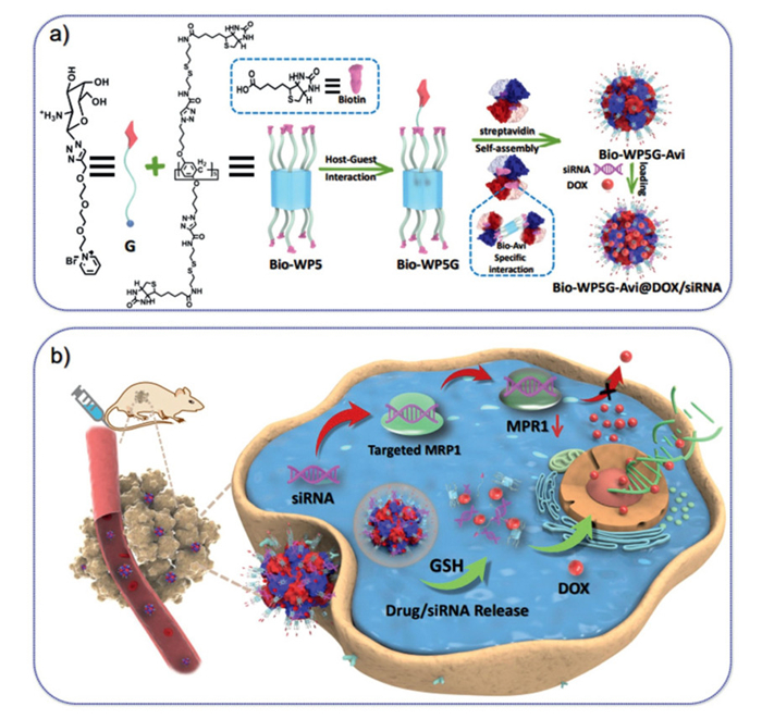
Scheme 1.
Design strategy of supramolecular glycosyl-nanoproteins and their response mechanisms to HepG2/ADR tumors.

Supramolecular glycosyl-nanoproteins based on host-guest chemistry for targeted co-delivery in vivo
Yinghua Lv , Yuchao Lu , Chenxi Hou , Jun Li , Zelong Chen , Aihua Zhen , Jiaming Ren , Yuxin Pei , Tony D James , Lan Chen , Yanjie Jia , Danhong Shang , Pengkun He , Zhongfu Wang , Zhichao Pei


Chemical reconstitution of living matter is one of the most direct and effective means to construct bio-functional materials using various biomolecules (protein, nucleic acid, carbohydrate, etc.) through biomimetic technology (biohybrid, bioinspired, bioengineered, etc.), which can significantly improve the biocompatibility of the material and reduce toxicity [1–4]. Among them, nanoproteins, as a classic biohybrid material, have become an important choice for constructing bio-functional materials via covalent coupling or non-covalent interaction, which have been widely used in drug delivery, biological imaging, tissue engineering, etc. [5–7]. In particular, multifunctional nanoproteins constructed by non-covalent interactions have achieved precise, controllable, and orderly self-assembly, showing great potential for efficient drug delivery [8–10].
Over recent years, the supramolecular nanoproteins fabricated from host-guest chemistry exhibit some extraordinary properties arising from the introduction of macrocyclic hosts, exhibiting promising potential in biomedical applications [11]. Among them, host-guest interactions have become an attractive non-covalent interaction for the design of functional nanoproteins, where macrocyclic host molecules (cyclodextrin, cucurbit[n]urine, calix[n]arene and pillar[n]arene, etc.) with unique cavities can interact with various functional guest molecules to regulate the assembly of protein molecular units, which provide an efficient method for fabricating functional nanoproteins [12–19]. In particular, the construction of supramolecular nanoproteins based on host-guest chemistry can integrate different therapeutic functions into a single nano delivery system to effectively overcome the limitations (drug side effects on normal cells and drug resistance of cancer cells) of chemotherapy, and has been successfully used to construct multifunctional drug delivery systems [20–23].
It is well known that carbohydrates are an indispensable biological substance in living species, and they have been widely used to construct drug delivery systems to improve hydrophilicity and biocompatibility [24–31]. Among them, glycosyl-nanocarriers can also utilize specific interactions with carbohydrate-binding proteins that are targeting ligands to galectin overexpressing cancer cells and achieve targeted drug delivery [32–36]. In particular, previous studies have shown that glycosyl supramolecular drug delivery systems based on functional carbohydrate derivatives can simultaneously exhibit targeting ability, responsiveness to stimuli and cationic properties [37,38]. Systems with such properties could effectively overcome the limitations of chemotherapy and are of great significance for cancer treatments [39–41]. However, there have been no reports on the construction of biohybrid targeted drug delivery platforms using supramolecular glycosyl nanoproteins [42–44].
Meanwhile, there has been a growing interest in exploring this non-covalent interaction in nanoscale drug delivery platforms for pharmaceutical agents. Biotin is known to bind streptavidin to form a stable biotin-(strept)avidin system through the strongest non-covalent interaction known to date [45,46]. The stable biotin-(strept)avidin system has been served as the basis for the developing many techniques ranging from labeling cells for isolation, tagging proteins for purification, and tumor diagnosis and therapy [47–49]. Hence, we have constructed a biohybrid supramolecular glycosyl nanoprotein Bio-WP5G-Avi for co-delivering drug/gene therapy to target HepG2/ADR tumor-bearing nude mice; the system self-assembled as a biotinylated glycosyl supra-molecule complex (Bio-WP5G) with streptavidin to realize targeting, responsiveness, and co-delivery properties as part of a single nanostructure (Scheme 1). Meanwhile, the tumor targeting, biological safety, and enhanced antitumor efficacy of Bio-WP5G-Avi@DOX/siRNA were verified by HepG2/ADR tumor-bearing nude mice in vivo.
In this study, A host molecule of biotinylated pillar[5]arene (Bio-WP5) and a guest molecule (G) of galactosamine pyridine salt derivative were synthesized (Fig. S1a in Supporting information). All experimental details and structural parameters can be found in Supporting information (Figs. S1–S17 in Supporting information). Subsequently, biohybrid supramolecular glycosyl-nanoproteins (Bio-WP5G-Avi) with targeted/GSH responsiveness were successfully prepared using host-guest interactions (Bio-WP5 and G) and molecular interaction (biotin and streptavidin). The spontaneous self-assembly of Bio-WP5G Avi formed nano-proteins with an average diameter of 90 nm (Fig. S1b, inset), which elicited a clear tyndall effect (Fig. S1d). The critical aggregation concentration of Bio-WP5G-Avi in water was calculated to be 50.88 µmol/L (Fig. S1c). In order to investigate the biocompatibility of Bio-WP5G-Avi, the human liver cell line HL7702 was incubated with Bio-WP5G-Avi at 10–100 µmol/L for 72 h. Methyl thiazole tetrazolium (MTT) cell survival assay indicated that the cell viability of HL7702 cells remained above 85% (Fig. S18 in Supporting information), indicating that Bio-WP5G-Avi exhibits good biocompatibility.
To further evaluate the biosafety of Bio-WP5G-Avi for intravenous administration, its blood compatibility was investigated. Hemolysis assays were performed on the key components (Bio-WP5, Bio-WP5G, Bio-WP5G-Avi) as well as the final drug-loaded formulation (Bio-WP5G-Avi@DOX/siRNA). The results showed that all components induced negligible hemolytic activity (hemolysis rate < 5%) even at the maximum tested concentration (Fig. S19 in Supporting information), demonstrating excellent blood compatibility. Furthermore, the stability of the Bio-WP5G-Avi nanoparticles was assessed by monitoring the size distribution and polydispersity index (PDI) over 4 days. The nanoparticles maintained their structural integrity without significant aggregation or size change (Fig. S20 in Supporting information), confirming their good stability, which is crucial for storage and biomedical application. These collective results affirm the excellent hemocompatibility and stability of our Bio-WP5G-Avi nanocarrier system.
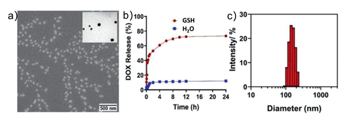
To investigate the drug carrier capability of Bio-WP5G-Avi, firstly, a mixture of doxorubicin (DOX) (model drug) and Bio-WP5G-Avi was treated with ultrasound (30 min). The nanostructure of the DOX-loaded delivery system was characterized by scanning electron microscopy (SEM) and transmission electron microscopy (TEM) (Fig. 1a and inset right), which indicated that the average diameter of Bio-WP5G-Avi@DOX as detected by dynamic light scattering (DLS) was around 153 nm (Fig. 1c). Then, the results of ultraviolet-visible (UV–vis) absorption spectra showed that Bio-WP5G-Avi had good drug-loading capability, and the encapsulation rate of DOX was 88%. As shown in Figs. S21a and S22b (Supporting information), a Tyndall effect of the Bio-WP5G-Avi and Bio-WP5G-Avi@DOX was clearly observed, suggesting the existence of abundant nanospecies. However, the Tyndall effect disappeared upon the addition of excess GSH (Fig. S21b in Supporting information), indicating the destruction of nanostructures. Meanwhile, Bio-WP5G-Avi could no longer be found in the SEM image (Fig. S23 in Supporting information). In addition, only about 12% of DOX was released from Bio-WP5G-Avi@DOX when incubated for 4 h in H2O. In contrast, in the presence of 10 mmol/L GSH, DOX is quickly released from Bio-WP5G-Avi@DOX, reaching ~60.8% at 4 h (Fig. 1b). These results indicate that the Bio-WP5G-Avi@DOX possesses good responsiveness to glutathione (GSH). The rapid release behavior stimulated by GSH makes Bio-WP5G-Avi@DOX an ideal biohybrid carrier for drug delivery to reduce side effects.
The realization of targeted and controllable co-delivery of drugs/siRNA in a safe and effective carrier is an important approach towards overcoming drug resistance and achieving optimal efficacy of cancer therapy. However, avoiding the degradation of siRNA by nucleases, and overcoming the problem associated with high-molecular weight, negatively charged, and hydrophilic siRNA that cannot cross the biofilm through passive diffusion, and improving its transfection efficiency are significant real challenges for the systematic delivery of siRNA. Previous research indicates that polycations can effectively inhibit the degradation of siRNA by enzymes, improve the transfection efficiency, and provide an alternative for the loading, protection and transportation of siRNA. Therefore, we used cationic supramolecular glycosyl-nanoproteins (Bio-WP5G-Avi) as polycations to complex with siRNA and to form Bio-WP5G-Avi@siRNA through electrostatic interactions. As shown in Fig. S24c (Supporting information), an apparent change in zeta potential to −6.539 mV (Bio-WP5G-Avi@siRNA) resulting from the siRNA on Bio-WP5G-Avi also demonstrated that the Bio-WP5G-Avi successfully loading siRNA.
To evaluate the siRNA delivery efficiency, Bio-WP5G-Avi was loaded with FAM-labeled siRNA and applied to transfect HepG2 cells. Flow cytometry and confocal laser scanning microscopy (CLSM) were used to study the cellular uptake and intracellular distribution. As shown in Fig. 2a, more than 75% cellular internalization was achieved with Bio-WP5G-Avi@siRNA, much higher than Lipofectamine 2000 (49%). CLSM imaging showed intense green fluorescence distributed intracellularly (Fig. 2b), providing evidence for successful siRNA transport.
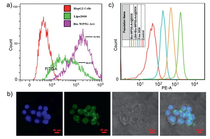
The targeting ability of Bio-WP5G-Avi@DOX was tested using HepG2/ADR cells precultured with lactose to block ASGPR. Fig. 2c shows the strongest fluorescence intensity in cells incubated with Bio-WP5G-Avi@DOX (green curve), while lactose pretreatment caused a clear decrease (yellow curve). In contrast, no significant difference in fluorescence intensity was observed between the Bio-WP5G-Avi@DOX and free DOX groups in HL7702 cells (Fig. S25 in Supporting information), indicating that cellular uptake occurs primarily through ASGPR-mediated endocytosis due to surface galactose moieties. Next, Bio-WP5G-Avi@DOX/siRNA nanoproteins were prepared by ultrasonic treatment of a mixed solution of Bio-WP5G, Streptavidin, DOX, and multidrug resistance-associated protein 1 (MRP1) siRNA, with successful formation characterized by SEM/TEM (Figs. S24a and b in Supporting information). As shown in Fig. S26 (Supporting information), both green and red fluorescence were observed in HepG2/ADR cells after incubation with Bio-WP5G-Avi@DOX/siRNA, proving co-internalization of DOX and siRNA.
The anticancer efficacy was further evaluated by incubating HepG2 and HL7702 cells with Bio-WP5G-Avi@DOX and free DOX for 24, 48, and 72 h. MTT assays showed that the relative survival rate of HepG2 cells incubated with WP5G-Avi@DOX was significantly lower than that of free DOX after 72 h (Fig. S27a; 24 and 48 h, see Fig. S28 in Supporting information). In contrast, the relative survival rate of HL7702 cells incubated with WP5G-Avi@DOX was higher than that of free DOX (Fig. S27b; 24 and 48 h, see Fig. S29 in Supporting information), suggesting that encapsulation enhances efficacy on cancer cells while reducing side effects on normal cells.
Furthermore, to further support the platform's functionality to silence MRP1 and overcome multidrug resistance through coordinated gene-drug action, we quantitatively analyzed MRP1 protein expression using enzyme-linked immunosorbent assay (ELISA). As shown in Fig. S30 (Supporting information), the Bio-WP5G-Avi@DOX/siRNA group demonstrated significant reduction in MRP1 protein levels (P < 0.001 vs. control); effective reversal of chemoresistance phenotype. Moreover, to test the cytotoxic effects of the multidrug resistance gene MRP1 gene silencing in HepG2/ADR cells after Bio-WP5G-Avi@DOX/siRNA treatment. As shown in Fig. 3a and Fig. S31 (Supporting information), Annexin V conjugate staining analysis showed that treatment with Bio-WP5G-Avi@DOX/siRNA increased the proportion of apoptotic and dead HepG2/ADR cells to 72.6%, compared to 49.5% in the negative control. Moreover, Western blot analysis revealed that treatment with Bio-WP5G-Avi@DOX/siRNA for 48 h significantly increased BCL-2-associated X protein (BAX) (pro-apoptotic) expression while decreasing B-cell lymphoma 2 (BCL2) (anti-apoptotic) expression in HepG2/ADR cells (Fig. 3b).
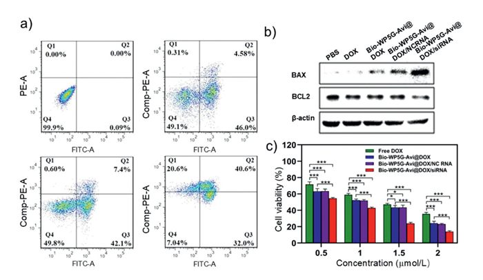
Next, as shown in Fig. S32 (Supporting information), to compare the combined anticancer efficacy of Bio-WP5G-Avi@DOX/siRNA, HepG2-ADR cells were exposed to free DOX, Bio-WP5G-Avi@DOX, Bio-WP5G-Avi@DOX/NC siRNA and Bio-WP5G-Avi@DOX/ siRNA for 24 h (Fig. S32a), 48 h (Fig. S32b) and 72 h (Fig. 3c), respectively. The cell viability incubated with Bio-WP5G-Avi@DOX/siRNA was much lower than that incubated with free DOX and Bio-WP5G-Avi@DOX (Fig. 3c), indicating that Bio-WP5G-Avi@DOX/siRNA could enhance the anti-cancer efficacy and exert a combined effect on cancer cells. Collectively, these observations suggest that Bio-WP5G-Avi@DOX/siRNA is a promising co-carrier to overcome cancer cell resistance to DOX and enhance chemotherapeutic efficacy.
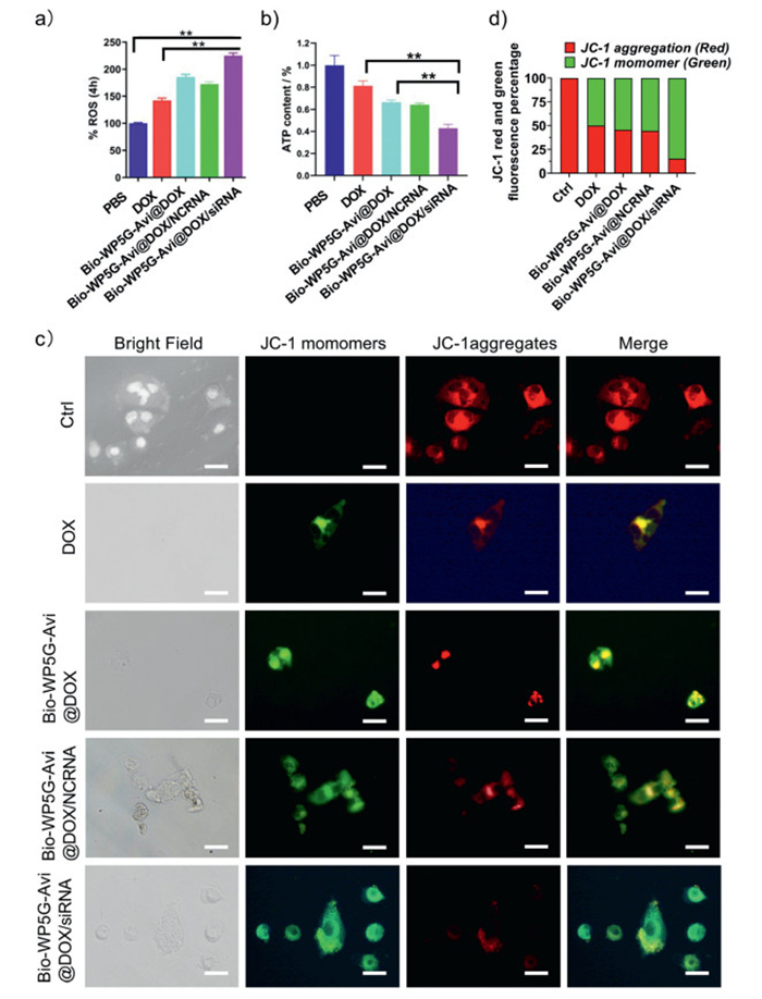
Given that tumor cells maintain their capacity for rapid proliferation and invasion through abnormal metabolism, we investigated the ability of Bio-WP5G-Avi drug carriers to accumulate in multiple organelles. Mitochondria, as the key energy generating units of tumor cells, serve as the main sites for ATP production and metabolism. They play a crucial part in regulating cell survival or death by controlling intracellular reactive oxygen species (ROS), the ATP level, signal transduction, etc. Specific damage to mitochondria can directly disrupt biosynthesis and trigger tumor cell apoptosis. The mitochondrial respiratory chain is also a significant source of destructive ROS. ROS contribute to oxidative stress, which is associated with various diseases and disorders. Treatment with Bio-WP5G-Avi led to an accumulation of ROS (Fig. 4a), along with concurrent reductions in intracellular ATP concentrations (Fig. 4b) and mitochondrial membrane potential (MMP) (JC-1) (Figs. 4c and d). These findings suggest that Bio-WP5G-Avi, when used as a nano-drug carrier, could facilitate the delivery of DOX and enhance DOX inducing mitochondrial dysfunction and subsequent tumor cell death.
We therefore explored the capacity of Bio-WP5G-Avi to enter the mitochondria. As shown in Fig. S33a (Supporting information), the microscopy images of HepG2/ADR cells treated with Bio-WP5G-Avi@DOX/siRNA show colocalization (yellow in merged images) of DOX fluorescence (red) with mitochondria (green), indicating mitochondrial accumulation of DOX. The overlapping fluorescence in all Bio-WP5G-Avi nano-carrying drug treatment groups was stronger than that in the DOX-only group. These observations suggest that the Bio-WP5G-Avi nano-loading platform enhances the ability of DOX to accumulate in the mitochondria. In addition, lysosomes are organelles that decompose biological macromolecules such as proteins, nucleic acids and polysaccharides. Most drugs ingested through endocytosis and phagocytosis are usually sent to lysosomes and are then transported into the cytoplasm to induce a pharmacological effect. Therefore, to examine the Bio-WP5G-Avi lysosome targeting ability, we performed lysosomal staining. Through the co-localization of DOX (red) and lysosome (green), we found that Bio-WP5G-Avi-carrying drugs could accumulate in lysosomes (Fig. S33b in Supporting information). Collectively, these observations suggest that Bio-WP5G-Avi-can deliver therapeutic drugs to multiple organelles, including mitochondria and lysosomes, resulting in drug accumulation and eventually leading to tumor cell death.
An ongoing theme in this field of drug delivery is how to improve therapeutic efficacy, which ultimately relies on the ability of anticancer agents to reach their final site of therapeutic action. In order to prove that our Bio-WP5G-Avi-carrying drugs can target the tumor and enrich at the tumor site, Bio-WP5G-Avi@DOX/siRNA was injected into HepG2/ADR tumor model mice through tail-vein injection. Immunodeficient BALB/C athymic nude mice were purchased from Hunan Slake Laboratory Animal Co., Ltd. (Changsha, China). All animal experiments were approved by the Animal Care and Use Committee of Northwest A&F University (approval No. NWAFU-314020038). Following 4 and 24 h of drug administration, significant drug fluorescence signals were detected at the tumor site using the PerkinElmer IVIS in vivo imaging system (Fig. 5a). Furthermore, the mice were administered injections at three-day intervals, with the treatment concluding after 24 days. Thereafter, the mice were euthanized to excise the tumors and organs. As shown in Fig. 5b and Fig. S34 (Supporting information), a significant intracellular drug fluorescence was detected by the Bio-WP5G-Avi@DOX/siRNA treatment. In contrast, no significant fluorescence was observed in the major organs, indicating that the Bio-WP5G-Avi carrying drugs (DOX and DOX/siRNA) are capable of targeting the tumor and accumulating at the tumor site, without clear aggregation in the major organs.
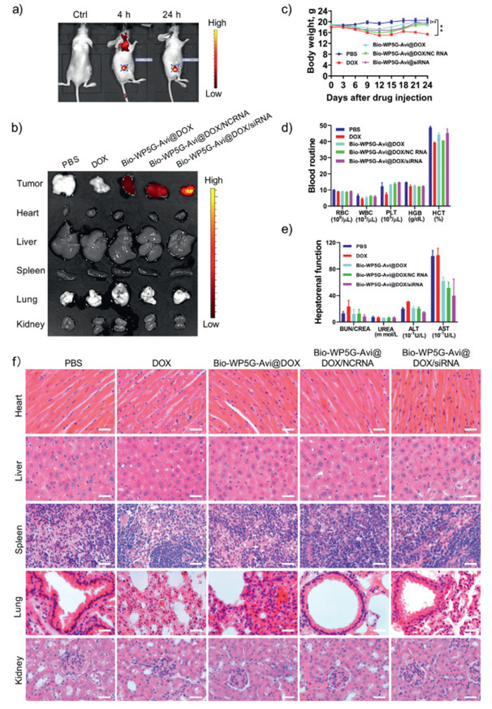
To further demonstrate that Bio-WP5G-Avi@DOX/siRNA has a high potential for biological application, we evaluated the long-term cytotoxicity of Bio-WP5G-Avi@DOX/siRNA in vivo. The mean body weights of the mice were evaluated. No statistically significant difference was observed between the Bio-WP5G-Avi group and the control group after 24 days of treatment. In contrast, treatment with DOX resulted in a significant reduction in body weight (Fig. 5c). In addition, we examined the blood routine, including red blood cell count (RBC), white blood cell count (WBC), platelets (PLT), hemoglobin (HGB), and hematocrit (HCT) and blood biochemical-related indices, including the blood urea nitrogen to creatinine ratio (BUN/CREA), urea (UREA), alanine aminotransferase (ALT), and aspartate aminotransferase (AST) of nude mice in each group to further explore the biosafety of Bio-WP5G-Avi carrying drugs. As shown in Fig. 5d, a decrease of both WBC and HCT was observed in the DOX-treated group, while the Bio-WP5G-Avi group was within the normal range compared to the control group. For the hepatorenal function of mice, the levels of ALT and AST after DOX treatment were higher, which is mainly due to the toxic side effects of DOX on the liver, while in the Bio-WP5G-Avi group, the concentrations of these enzymes were lower than those in the DOX group and were comparable to those in the control group (Fig. 5e), indicating that Bio-WP5G-Avi can effectively reduce the toxic side effects of DOX on the main organs, thus corroborating the fact that our Bio-WP5G-Avi nanocarrier has a good biosafety in vivo.
Hematoxylin and eosin (HE) staining of paraffin sections of the main organs in nude mice also indicated that Bio-WP5G-Avi@DOX/siRNA did not cause any significant damage to the main organs of mice. These observations indicated the potential of Bio-WP5G-Avi as a biodegradable and versatile drug delivery nanocarrier in vivo (Fig. 5f). In light of the aforementioned evidence, it can be concluded that Bio-WP5G-Avi exhibits good biosafety and can reduce the systemic toxicity of chemotherapy drugs, which provides a promising biological hybrid nano-platform for efficient tumor-targeted co-drug (DOX/siRNA) delivery.
To further evaluate the tumor-inhibitory effect of Bio-WP5G-Avi@ DOX/siRNA on solid tumors in mice, BALBC nude mice-bearing HepG2/ADR tumors were randomly divided into 5 groups: (1) The PBS group (control); (2) the DOX group (naked DOX); (3) the Bio-WP5G-Avi@DOX group (Bio-WP5G-Avi-loaded DOX); (4) the Bio-WP5G-Avi@DOX/NCRNA group (Bio-WP5G-Avi-loaded DOX and negative control siRNA); (5) the Bio-WP5G-Avi@DOX /siRNA group (Bio-WP5G-Avi loaded DOX and MRP1 siRNA). After 24 days of treatment with intravenous injections of different drug formulations, the mice were euthanized, and the tumor was removed surgically, photographed, and the tumor size was compared. As shown in Fig. 6a and Fig. S35 (Supporting information), although the administration of DOX resulted in a reduction in tumor size in comparison with the PBS control group, the tumor continued to grow. This was confirmed by the volume and weight of the tumor (Figs. 6b and c). The reason why naked DOX treatment did not inhibit tumor growth might be due to the major transmembrane transporter P-glycoprotein (P-gp) encoded by the human multidrug resistance gene (MRP1), which plays an important role in multidrug resistance in cancer cells, resulting in the failure of DOX to inhibit HepG2/ADR tumors in vivo.
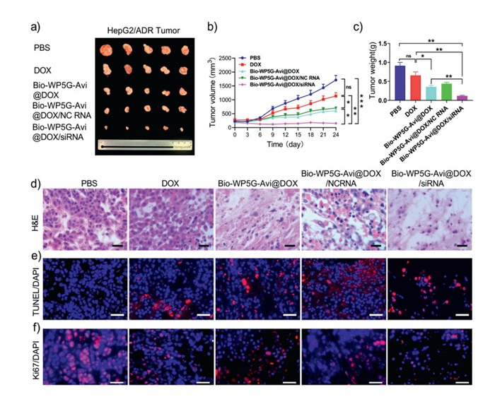
To curtail the chemoresistance caused by MRP1, we used Bio-WP5G-Avi as a nano-drug carrier platform loaded with DOX and MRP1 siRNA. We observed the tumor size, monitored the tumor growth curve, calculated the tumor weight after 24 days of treatment, and identified that Bio-WP5G-Avi@DOX and Bio-WP5G-Avi@DOX/siRNA therapy, promoting the effective uptake of drugs in tumor cells, enriching drug concentration, and inhibiting drug efflux in tumor tissues. As shown in Fig. 6c, WP5G-Avi@DOX/siRNA treatment could effectively inhibit tumor growth compared to the control and DOX groups. In particular, Bio-WP5G-Avi@DOX/siRNA treatment effectively decreased the tumor weight and exhibited the best antitumor effect compared to the other four groups (Fig. 6c). This superior tumor-suppression effect may be the result of Bio-WP5G-Avi augmenting the combination therapy of chemotherapy and gene interference therapy, promoting the effective uptake of drugs in tumor cells, enriching drug concentration, and inhibiting drug efflux in tumor tissues.
In addition, we also carried out a histological analysis of the tumor sections by means of HE staining. In comparison with the PBS control and the DOX groups, the synergistic treatment of DOX chemotherapy and MRP1 siRNA gene therapy from Bio-WP5G-Avi@DOX/siRNA treatment obviously destroyed the tumor histological structure, devastated the tumor nest, enlarged the distance between the tumor cells, loosened the tumor tissue and significantly reduced the number of tumor cells (Fig. 6d).
Furthermore, the terminal deoxynucleotidyl transferase (TdT) dUTP nick-end labeling (TUNEL) assay confirmed that Bio-WP5G-Avi@DOX/siRNA significantly induced irreversible apoptosis in a large number of tumor cells (Fig. 6e). Moreover, we investigated the proliferation of tumor cells by Ki67 cell immunofluorescence examination. As shown in Fig. 6f, Bio-WP5G-Avi@DOX/siRNA significantly inhibited the proliferation of HepG2/ADR tumor cells, thus inhibiting tumor growth, which confirmed that Bio-WP5G-Avi@DOX/siRNA had excellent antitumor effects. All these results confirmed that our biosafe Bio-WP5G-Avi nano platform that enables co-delivery of DOX and MRP1 siRNA significantly curtailed chemotherapeutic resistance, facilitated chemotherapeutic drug penetration and enrichment in tumor tissue, inhibited drug efflux, and destroyed the histological structure of tumor tissues. As such, our system provides a new means for cancer treatment that inhibits the proliferation of tumors, effectively induces apoptosis, and enables good synergistic therapeutic effects to enhance therapeutic outcomes while reducing toxicity.
To summarize, a biohybrid supramolecular glycosyl-nanoprotein co-delivery system of Bio-WP5G-Avi was successfully developed through self-assembly of biotin functionalized glycosyl-supramolecule complex (Bio-WP5G) with streptavidin, where Bio-WP5G based on the multi-functional host-guest complex between Bio-WP5 and G endowed the biohybrid supramolecular glycosyl-nanoproteins with several desirable functionalities, including GSH responsiveness, tumor targeting specificity, and efficient siRNA/DOX co-delivery in vitro and in vivo. Unlike many other multifunctional nanoproteins based on multistep fabrication or sophisticated components, Bio-WP5G-Avi fabricated in this work was constructed via the simple self-assembly of Bio-WP5G with streptavidin. Thus, our unique strategy offers a novel insights into the simple design of multifunctional nanoproteins that can synergistically enhance the cytotoxicity of targeted combination therapy. We hope that this biohybrid supramolecular system will expand the landscape of nanocarriers for targeted co-delivery, offering a robust strategy to improve clinical outcomes in drug resistant cancers.
Yinghua Lv: Writing – original draft, Visualization, Investigation, Formal analysis, Data curation. Yuchao Lu: Writing – original draft, Funding acquisition, Formal analysis, Conceptualization. Chenxi Hou: Methodology, Investigation, Formal analysis, Data curation. Jun Li: Investigation, Formal analysis. Zelong Chen: Validation. Aihua Zhen: Methodology. Jiaming Ren: Validation. Yuxin Pei: Writing – review & editing, Supervision, Project administration, Funding acquisition. Tony D James: Resources. Lan Chen: Validation. Yanjie Jia: Validation. Danhong Shang: Validation. Pengkun He: Validation. Zhongfu Wang: Resources. Zhichao Pei: Writing – review & editing, Supervision, Project administration, Funding acquisition.
The authors declare that they have no known competing financial interests or personal relationships that could have appeared to influence the work reported in this paper.
This work was supported by the National Natural Science Foundation of China (Nos. 22571257, 22171230 and 22577103), the Doctoral Research Foundation Project of Changzhi Medical College (No. 2024BS04), the Scientific and Technological Innovation Programs of Higher Education Institutions in Shanxi (No. 2024L278), the Fundamental Research Program of Shanxi Province (No. 202403021221212), the Research Foundation Project of Changzhi Medical College (HZZD202406), the Fundamental Research Funds for the Provincial Universities of Hebei (Nos. KY2022055, KY2023011 and KY2023022), the Key Research and Development Program of Shaanxi (No. 2023-YBSF-337). The authors thank Ms. Xiaona Hu (College of Chemistry & Pharmacy), Ms. Zhenzhen Ma (Crop Biology Innovation Centre, College of Agriculture), Mr. Kerang Huang (Life Science Research Core Services), Northwest A&F University, Yangling, China, for assistance with the laser confocal microscopy experiments and compound characterizations.
Supplementary material associated with this article can be found, in the online version, at doi:
J.W. Yoo, D.J. Irvine, D.E. Discher, S. Mitragotri, Nat. Rev. Drug Discov. 10 (2011) 521–535. doi: 10.1038/nrd3499
M. Wang, S. Sun, C.I. Neufeld, B. Perez-Ramirez, Angew. Chem. Int. Ed. 53 (2014) 13444–13448. doi: 10.1002/anie.201407234
X. Ge, H. Wen, Y. Fei, et al., Biomaterials 299 (2023) 122184. doi: 10.1016/j.biomaterials.2023.122184
Z. Lyu, L. Ding, A. Tintaru, L. Peng, Acc. Chem. Res. 53 (2020) 2936–2949. doi: 10.1021/acs.accounts.0c00589
J. Wen, Y. Lv, Y. Xu, et al., Acta Biomater. 83 (2019) 359–371. doi: 10.1016/j.actbio.2018.11.009
M. Yang, K. Yang, B. Gao, et al., Chem. Commun. 58 (2022) 11147–11150. doi: 10.1039/d2cc04238j
W. Jin, Z. Chen, Y. Wang, et al., Chin. Chem. Lett. 35 (2024) 109328. doi: 10.1016/j.cclet.2023.109328
Q. Luo, Z. Dong, C. Hou, J. Liu, Chem. Commun. 50 (2014) 9997–10007. doi: 10.1039/C4CC03143A
R. Cai, C. Chen, Adv. Mater. 31 (2019) e1805740. doi: 10.1002/adma.201805740
K.O. Ramberg, S. Engilberge, T. Skorek, P.B. Crowley, J. Am. Chem. Soc. 143 (2021) 1896–1907. doi: 10.1021/jacs.0c10697
J. Mo, L. Wang, X. Huang, et al., Nanoscale 9 (2017) 13142–13152. doi: 10.1039/C7NR04473A
L. Zhao, S. Qiao, J. Liu, Sci. China Chem. 59 (2016) 1531–1540. doi: 10.1007/s11426-016-0231-3
C. Yuvienco, H.T. More, J.S. Haghpanah, R.S. Tu, J.K. Montclare, Biomacromolecules 13 (2012) 2273–2278. doi: 10.1021/bm3005116
S. van Dun, C. Ottmann, L.G. Milroy, L. Brunsveld, J. Am. Chem. Soc. 139 (2017) 13960–13968. doi: 10.1021/jacs.7b01979
K. Yang, S. Qi, X. Yu, et al., Angew. Chem. Int. Ed. 61 (2022) e202203786. doi: 10.1002/anie.202203786
Y. Bai, Q. Luo, J. Liu, Chem. Soc. Rev. 45 (2016) 2756–2767. doi: 10.1039/C6CS00004E
Y. Ding, C. Kwong, S. Li, et al., Biomater. Sci. 9 (2021) 3804–3813. doi: 10.1039/d1bm00426c
J.A. Jones, A.S. Cristie-David, M.P. Andreas, T.W. Giessen, Angew. Chem. Int. Ed. 60 (2021) 25034–25041. doi: 10.1002/anie.202110318
D.Y. Ng, Y. Wu, S.L. Kuan, T. Weil, Acc. Chem. Res. 47 (2014) 3471–3480. doi: 10.1021/ar5002445
W. Cao, X. Qin, Y. Wang, et al., Angew. Chem. Int. Ed. 60 (2021) 11196–11200. doi: 10.1002/anie.202100916
H. Zhu, H. Wang, B. Shi, et al., Nat. Commun. 10 (2019) 2412. doi: 10.1038/s41467-019-10385-9
L. Wu, Y. Zhang, Z. Li, et al., J. Am. Chem. Soc. 139 (2017) 14684–14692. doi: 10.1021/jacs.7b07768
J. Xie, Y. Lu, B. Yu, J. Wu, J. Liu, Chin. Chem. Lett. 31 (2020) 1173–1177. doi: 10.1016/j.cclet.2019.10.030
C. Gao, G. Chen, Acc. Chem. Res. 53 (2020) 740–751. doi: 10.1021/acs.accounts.9b00552
B. Nitsa, A. Mangraviti, F. Volpin, Adv. Funct. Mater. 31 (2021) e2100643. doi: 10.1002/adfm.202100643
X. Qiu, Q. Li, Y. Zhou, Chem. Commun. 51 (2015) 4237–4240. doi: 10.1039/C4CC10413G
H. Wang, Z. Liu, C. An, et al., Adv. Sci. 7 (2020) 2001264. doi: 10.1002/advs.202001264
X. Wu, Y. Tan, H.T. Toh, et al., Chem. Sci. 8 (2017) 3980–3988. doi: 10.1039/C6SC05251G
P. Zhao, W. Yin, A. Wu, et al., Adv. Funct. Mater. 27 (2017) 1700403. doi: 10.1002/adfm.201700403
Y. Chang, Y. Lv, P. Wei, et al., Adv. Funct. Mater. 27 (2017) 1703083. doi: 10.1002/adfm.201703083
C. Xian, H. Chen, F. Xiong, et al., Biomater. Sci. 9 (2021) 6023–6036. doi: 10.1039/d1bm00879j
X. Liu, W. Shao, Y. Zheng, et al., Chem. Commun. 53 (2017) 8596–8599. doi: 10.1039/C7CC04932C
P. Sreedevi, J.B. Nair, M.M. Joseph, et al., J. Control. Release 339 (2021) 284–296. doi: 10.1016/j.jconrel.2021.09.038
S. Wan, P.M. Kelly, E. Mahon, et al., ACS Nano 9 (2015) 2157–2166. doi: 10.1021/nn506060q
B. Gao, K. Yang, M. Yang, et al., Chem. Commun. 60 (2024) 8892–8895. doi: 10.1039/d4cc02944e
L. Wang, X. Huang, X. You, et al., Signal Transduct. Target. Ther. 5 (2020) 196. doi: 10.1038/s41392-020-00248-x
Y. Lu, C. Hou, J. Ren, et al., Int. J. Nanomedicine 14 (2019) 3525–3532. doi: 10.2147/ijn.s191256
W. Feng, Y. Lv, Z. Chen, et al., Chem. Eng. J. 417 (2021) 129178. doi: 10.1016/j.cej.2021.129178
Z. Shen, N. Ma, F. Wang, et al., Chin. Chem. Lett. 33 (2022) 4563–4566. doi: 10.1016/j.cclet.2022.01.069
S. Zhang, J. Li, X. Hu, et al., Chin. Chem. Lett. 36 (2025) 110314. doi: 10.1016/j.cclet.2024.110314
Y. Dang, Y. Feng, X. Chen, et al., Chin. Chem. Lett. 35 (2024) 109660. doi: 10.1016/j.cclet.2024.109660
L. Fu, Z. Su, S. Wu, et al., Chin. Chem. Lett. 36 (2025) 110227. doi: 10.1016/j.cclet.2024.110227
Z. Shen, S. Chao, L. Wu, et al., Chem. Soc. Rev. 54 (2025) 8345–8405. doi: 10.1039/d4cs00458b
K. Shang, Y. Lv, Y. Yang, et al., Chin. Chem. Lett. 37 (2026) 111798. doi: 10.1016/j.cclet.2025.111798
A. Jain, K. Cheng, J. Control. Release 245 (2017) 27–40. doi: 10.1016/j.jconrel.2016.11.016
H.P. Lesch, M.U. Kaikkonen, J.T. Pikkarainen, et al., Expert Opin. Drug Deliv. 7 (2010) 551–564. doi: 10.1517/17425241003677749
Y. Jin, Z. Wu, C. Wu, et al., J. Control. Release 320 (2020) 142–158. doi: 10.1016/j.jconrel.2020.01.040
S. Prakash, P.P. Hazari, V.K. Meena, et al., Bioconjug. Chem. 27 (2016) 2780–2790. doi: 10.1021/acs.bioconjchem.6b00576
H. Zhou, J. Liu, J. Xu, S. Zhang, H. Chen, Chem. Soc. Rev. 47 (2018) 1996–2019. doi: 10.1039/c7cs00573c

Scheme 1 Design strategy of supramolecular glycosyl-nanoproteins and their response mechanisms to HepG2/ADR tumors.

Figure 1 (a) The SEM and TEM image. Inset: TEM image (right). (b) Cumulative release profiles of Bio-WP5G-Avi@DOX in H2O, H2O with GSH (10 mmol/L). (c) DLS analysis of Bio-WP5G-Avi@DOX aqueous solution.

Figure 2 (a) Flow cytometry to detect the Bio-WP5G-Avi@siRNA cellular uptake in HepG2 cells for 4 h. (b) The fluorescence of siRNA in HepG2/ADR cells was observed with confocal laser scanning microscopy (CLSM) (from left to right: 4′, 6-diamidino-2-phenylindole (DAPI), Bio-WP5G-Avi@siRNA, bright field, overlap). Scale bar: 40 µm. (c) Flow cytometry to detect the targeting ability of Bio-WP5G-Avi with or without LBA to HepG2/ADR cells.

Figure 3 (a) The survival rate of HepG2/ADR cells under different treatments (from left to right: Ctrl, Bio-WP5G-Avi@DOX, Bio-WP5G-Avi@DOX/NC siRNA and Bio-WP5G-Avi@DOX/siRNA). (b) Expression levels of apoptosis-related genes in HepG2/ADR cells cultured with different treatment for 48 h. (c) Detection of Bio-WP5G-Avi@DOX/siRNA's effect on eliminating the drug resistance of HepG2/ADR cells by the MTT assay at 72 h. Data are presented as means ± SD (n = 3). *P < 0.05, ***P < 0.001.

Figure 4 (a) Intracellular ROS levels. (b) ATP release. (c) MMP assessed by JC-1 staining (red: JC-1 aggregates; green: JC-1 monomers). Scale bar: 5 µm. (d) Quantitative analysis of JC-1 red/green fluorescence intensity. Data are presented as means ± SD (n = 3). **P < 0.01.

Figure 5 (a) In vivo fluorescence imaging of HepG2/ADR tumor-bearing BALB/c nude mice after intravenous injection of Bio-WP5GAvi@DOX/siRNA at different time points. (b) Ex vivo fluorescence imaging of tumors and organs. (c) Animal body weights of HepG2/ADR tumor-bearing BALB/c nude mice. Data are presented as means ± SD (n = 5). **P < 0.01. ns, not significant (P > 0.05). (d) Mice blood routine. (e) Hepatorenal function. (f) Histological examination of major organs. Paraffin section thickness: 5 µm. Scale bar: 20 µm.

Figure 6 (a) Tumor morphology. (b) Tumor volume. (c) Tumor weight. (d) Tumor histology. (e) TUNEL staining and (f) Ki67 staining for tumor slices. Pink signals represent TUNEL-stained apoptotic cells and Ki67 positive cells, respectively. Blue signals represent DAPI-stained cell nuclei. Paraffin slices thickness: 5 µm. Scale bar: 50 µm. All data are presented as means ± SD (n = 5). *P < 0.05, **P < 0.01, ***P < 0.001.

 扫一扫看文章


扫一扫关注我们
 DownLoad:
 下载:
 下载: