
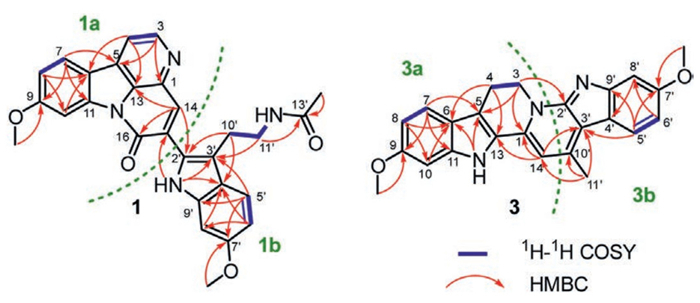
Figure 1.
The structures of compounds 1–3 and peganumium B.

Pecanthines A–C, unprecedented dimeric β-carboline alkaloids from the seeds of Peganum harmala
Qing Tang , Kai Zhao , Hui-Hui Zhu , Qiang Lin , Hai-Yue Zhao , Wei-Xuan Zheng , Zhong-Nan Wu , Hao Wang , Guo-Cai Wang , Yu-Bo Zhang


DNA topoisomerase Ⅰ (Topo Ⅰ), a pivotal enzyme orchestrating DNA replication and transcription, is a validated drug target for cancer therapy [1,2]. Topo Ⅰ inhibitors exert their therapeutic effects by stabilizing the DNA-Topo Ⅰ cleavable complex, thereby preventing DNA religation and subsequently inducing DNA damage and tumor cell apoptosis [3–5]. Currently, the clinical use of Topo Ⅰ inhibitors is largely restricted to camptothecin derivatives such as irinotecan and topotecan [6,7]. Although these agents demonstrate significant efficacy against various malignancies, their clinical utility is substantially limited by dose-limiting toxicities and acquired drug resistance, which severely compromise long-term therapeutic outcomes [8–11]. Therefore, there is an urgent need for Topo Ⅰ inhibitors with novel chemical scaffolds.
β-Carboline alkaloids are nitrogen-containing heterocycles biosynthesized from tryptophan with a unique tricyclic pyrido[3,4-b]indole skeleton [12]. This structure is associated with a range of pharmacological activities, with its notable antitumor potential attracting significant attention [12–15]. Among natural sources, plants of the Peganum genus have been identified as exceptionally rich repositories of structurally diverse β-carboline derivatives [16]. Critically, many of these compounds, such as harmine and its analogs, have been identified as Topo Ⅰ inhibitors, making them a key focus for phytochemists and medicinal chemists [17–20].
The seeds of Peganum harmala have been traditionally employed in Chinese folk medicine for the treatment of various ailments, including cancer, rheumatism, cough, and asthma [21–24]. In a previous study, we reported the isolation of new β-carboline alkaloids [24,25]. As a part of our ongoing research, three novel long conjugated β-carboline alkaloids, pecanthines A–C (1−3), were isolated from the seeds of P. harmala (Fig. 1). Their structures were elucidated by spectroscopic analyses and X-ray diffraction. Compounds 1 and 2 represent the first heterodimeric alkaloids merging canthin-6-one and melatonin-type scaffolds. More strikingly, the structure of peganumium B reported from the seeds of P. harmala was revised to be compound 3 (Fig. 1) [26]. Herein, we report the isolation, structural elucidation, hypothetical biogenetic pathway, and biological evaluation of 1–3. Meanwhile, the first gram-scale synthesis of 3 was achieved, further confirming the correctness of the structure.
Pecanthine A (1) was isolated as pink needle-shaped crystals. The molecular formula of 1 was established to be C28H24N4O4 from its high-resolution electrospray ionization mass spectrometry (HR-ESI-MS) data (m/z 481.1872 [M + H]+, calcd. for C28H25N4O4 481.1870). The 1H and 13C nuclear magnetic resonance (NMR) spectra data showed the presence of two AMX spin systems [δH 8.32 (1H, d, J = 8.7 Hz), δH 8.16 (1H, d, J = 2.4 Hz), δH 7.56 (1H, d, J = 8.7 Hz), δH 7.24 (1H, dd, J = 8.7, 2.4 Hz), δH 6.97 (1H, d, J = 2.3 Hz), δH 6.73 (1H, dd, J = 8.7, 2.3 Hz)], three aromatic protons [δH 8.83 (1H, d, J = 5.0 Hz), δH 8.20 (1H, d, J = 5.0 Hz), δH 8.15 (1H, s)], two methoxys [δH 3.94 and 3.80 (each 3H, s); δC 55.9 and 55.2], a methyl [δH 1.71 (3H, s); δC 22.6], and an indole NH signal [δH 11.14 (1H, s)]. The above data indicated that 1 was a dimeric β-carboline alkaloid. The 1H and 13C NMR data of 1 were definitively assigned by integrating 2D NMR experiments, as documented in Table S1 (Supporting information).
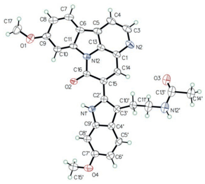
In the 1H–1H COSY spectrum, the correlations between H-3 and H-4, between H-7 and H-8, between H-5′ and H-6′, and between H2–11′ (δH 3.34) and H2–10′ (δH 2.98)/NH-12′ (δH 8.00) indicated the presence of four spin systems (C-3 to C-4, C-7 to C-8, C-5′ to C-6′, and C-10′ to N-12′) in 1 (Fig. 2). In the heteronuclear multiple bond correlation (HMBC) spectrum, correlations between H-3 and C-1/C-5, between H-4 and C-6/C-13, between H-7 and C-5/C-9/C-11, between H-8 and C-6/C-10, between H-10 and C-6, between H-14 and C-13/C-16, as well as between 9–OCH3 and C-9 suggested the existence of a 9-methoxycanthin-6-one alkaloid unit 1a (Fig. 2). The HMBC correlations between H-5′ and C-3′/C-7′/C-9′, between H-6′ and C-4′/C-8′, between H-8′ and C-4′, between H2–10′ and C-2′/C-4′, between H2–11′ and C-3′/C-13′, H3–14′ and C-13′, between 7′–OCH3 and C-7′, as well as between NH-1′ and C-3′/C-4′ suggested the presence of a melatonin-type unit 1b (Fig. 2). Furthermore, the HMBC correlations between H-14 and C-2′, and between NH-1′ and C-15 implied that the substructures unit 1a and unit 1b were connected through C-15–C-2′ bonds. Subsequently, crystals of 1 for single-crystal X-ray diffraction were acquired from CH3OH–CH2Cl2 (1:1). Thus, compound 1 was defined as an unprecedented heterodimer alkaloid and further determined by single-crystal X-ray diffraction (Cu Kα) analysis (Fig. 3).
The molecular formula of 2 was determined to be C27H22N4O4 based on its HR-ESI-MS data (m/z 467.1711 [M + H]+, calcd. for C27H23N4O4, 467.1714). Analysis of the 1H and 13C NMR spectra of 2 was similar to those of 1, except for the absence of a methyl signal (δC 22.6, C-14′). The structure was supported by the 1H–1H correlation spectroscopy (COSY) correlation between NH-12′ and H-13′, as well as the HMBC correlations between H-13′ and C-11′, between H2–11′ and C-13′ (Fig. S1 in Supporting information). Thus, compound 2 was elucidated and named pecanthine B.
Pecanthine C (3) was yielded as yellow powder. The molecular formula of 3 was deduced to be C24H21N3O2 on the basis of its HR-ESI-MS at m/z 384.1709 [M + H]+ (calcd. for C24H22N3O2: 384.1707). The UV spectrum of 3 exhibited absorption maxima at 205, 223, 280, 310 and 432 nm. Analysis of the 1H and 13C NMR spectra of 3 revealed the presence of two AMX spin systems [δH 7.91 (1H, d, J = 8.6 Hz), δH 7.53 (1H, d, J = 8.7 Hz), δH 7.10 (1H, d, J = 2.3 Hz), δH 6.92 (1H, d, J = 2.2 Hz), δH 6.77 (1H, dd, J = 8.7, 2.2 Hz), δH 6.74 (1H, dd, J = 8.6, 2.3 Hz)], two methoxyls [δH 3.84 and 3.83 (each 3H, s); δH 55.1 and 55.3], a methyl [δH 2.82 (3H, s); δH 19.6], and an indole NH signal [δH 11.73 (1H, s)]. Comprehensive analysis of 1H–1H COSY, heteronuclear single quantum coherence (HSQC), and HMBC spectra of 3 resulted in the unambiguous assignment of its 1H and 13C NMR signals as shown in Table S3.
In the 1H–1H COSY spectrum, the correlations between H2–3 (δH 4.92) and H2–4 (δH 3.26), between H-7 and H-8, and between H-5′ and H-6′ indicated the presence of three spin systems (C-3 to C-4, C-7 to C-8, and C-5′ to C-6′) in 3 (Fig. 2). In the HMBC spectrum, correlations between H2–3 and C-1/C-5, between H2–4 and C-6/C-13, between H-7 and C-5/C-9/C-11, between H-8 and C-6/C-10, between H-10 and C-6, between H-14 and C-1/C-13, between 9–OCH3 and C-9, as well as between 12-NH and C-5/C-6 suggested the existence of a harmaline unit 3a (Fig. 2). The HMBC correlations between H-5′ and C-3′/C-7′/C-9′, between H-6′ and C-8′, between H-8′ and C-4′, between H3–11′ and C-3′/C-10′, as well as between 7′–OCH3 and C-7′ suggested the presence of a 3-ethylidene-indole unit 3b (Fig. 2). Furthermore, the HMBC correlations between H2–3 and C-2′, between H-14 and C-3′, and between H3–11′ and C-14 indicated that the substructures 3a and 3b were connected through N-2–C-2′ and C-14–C-10′ bonds. Therefore, the planar structure of 3 was established as a unique long conjugated 6/5/6/6/5/6 fused six-ring system.
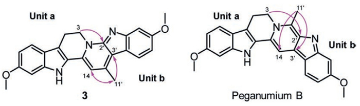
Peganumium B, recently reported from the seeds of P. harmala as a β-carboline dimer with one novel long conjugated structure, has attracted our attention [26]. Interestingly, compound 3 and peganumium B share identical molecular weights and NMR data, prompting us to re-evaluate peganumium B’s structure. In the original literature, the authors deduced peganumium B as a β-carboline dimer based on NMR analysis. They further identified unit a as harmaline through HMBC spectroscopy, which is consistent with our analysis of 3 in this study. Subsequently, the linkage between units a and b was inferred from chemical shifts and HMBC correlations. However, we have found that the HMBC spectrum of peganumium B exhibits strong cross-peaks for H2–3/C-2′, H-14/C-11′, and H3–11′/C-14 and C-3′ (Fig. S2 in Supporting information), which were not reported in the original study [26]. These correlations perfectly match those of 3, where each pair of atoms is separated by three bonds, whereas in peganumium B, they would be separated by four or five bonds (Fig. 4). Thus, a comprehensive re-evaluation of the spectroscopic data led to the revision of peganumium B as structure 3.
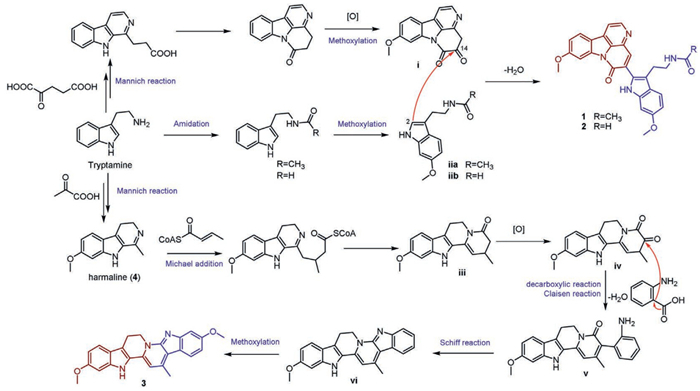
Compounds 1–3 represent two novel classes of dimeric β-carboline alkaloid analogues. The plausible biogenetic pathway of 1–3 could be proposed based on the findings of key biogenetic precursors in the plant (Scheme 1). The biosynthesis initiates with a Mannich reaction between tryptamine and 2-ketoglutaric acid, followed by successive oxidation, methoxylation, and ring condensation to afford the pivotal precursor ⅰ [27,28]. Dimerization of ⅰ with intermediate ⅱa/ⅱb through nucleophilic addition-elimination accounts for the formation of 1 and 2. As for compound 3, its source can be traced back to its precursor, harmaline. First, harmaline (4) undergoes a Michael addition with crotonyl-CoA, followed by intramolecular condensation to afford the key intermediate ⅲ [29,30]. Subsequent oxidation of ⅲ yields intermediate ⅳ, which then engages in a Claisen reaction with anthranilic acid to generate the dehydrated intermediate ⅴ [31]. Finally, 3 is formed through Schiff reaction and methoxylation.
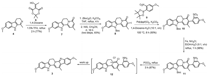
Following the structural revision of peganumium B to 3 and unsuccessful attempts to obtain X-ray-quality crystals, we designed a biomimetic synthesis of 3 according to the biogenic pathway (Scheme 2). The synthesis commenced with harmaline (4) from P. harmala seeds. Using commercially available Meldrum’s acid derivative 6 as an acylketene source, the key intermediate 7 was synthesized in 77% yield [32]. Subsequent Boc protection of the indole NH followed by iodination at the α-position of the carbonyl group afforded intermediate 8 in 83% yield over two steps. Next, a Suzuki-Miyaura coupling of 8 with boronic ester 9 was performed using Pd(dppf)Cl2 and K2CO3 in 1,4-dioxane at 100 ℃, affording the desired 10 in 90% yield. Subsequent reduction with iron powder and NH4Cl cleanly afforded intermediate 11 in 96% yield. The final transformation involved POCl3-mediated intramolecular cyclization under reflux conditions, simultaneously effecting N-Boc deprotection. Ammonia workup delivered 3 in 87% yield on gram scale. The 1H and 13C NMR spectra of synthetic 3 matched those of the natural isolate.
β-Carboline alkaloids have attracted considerable attention due to their notable neuroprotective and antitumor activities. Our study demonstrated that compound 1 exhibited dose-dependent neuroprotective effects in an L-glutamate-induced HT-22 cell model (Fig. S3 in Supporting information). Meanwhile, compound 3 displayed significant cytotoxicity against PC-9 and H1299 cells, with the half maximal inhibitory concentration (IC50) values of 0.856 ± 0.13 and 1.312 ± 0.08 µmol/L, respectively (Fig. 5a). Colony formation assays (Figs. 5b and c) and EdU proliferation assays (Figs. S4a and b in Supporting information) revealed that compound 3 effectively suppressed tumor cell clonogenicity and DNA synthesis. Furthermore, wound healing and transwell invasion assays confirmed that 3 significantly inhibited the migratory and invasive capabilities of PC-9 cells (Figs. S4c–g in Supporting information).
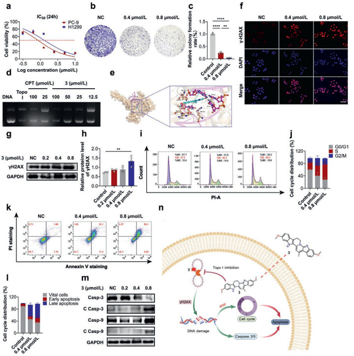
To elucidate the anti-tumor molecular mechanism of compound 3, we focused on Topo Ⅰ, a classic anti-tumor target. The results of relaxation experiments showed that 3 had significantly better inhibitory activity against Topo Ⅰ than the positive control drug camptothecin (Fig. 5d). Molecular docking analysis revealed that 3 formed hydrogen bond interactions with Topo Ⅰ and produced π-π stacking effects with DNA base pairs (Fig. 5e). These molecular interactions were the structural basis for its inhibitory activity against Topo Ⅰ. Given that compound 3 has potent Topo Ⅰ inhibition and antiproliferative activity, we further examined its effect on DNA damage. Immunofluorescence detection and Western blot revealed that the expression level of γ-H2AX, a DNA damage marker, was significantly increased in PC-9 cells treated with 3 (Figs. 5f–h). Flow cytometry analysis showed that 3 could arrest the PC-9 cell cycle at the S phase (Figs. 5i and j) and significantly induce cell apoptosis (Figs. 5k–m).
In summary, three novel long-conjugated β-carboline alkaloids, pecanthines A–C (1–3), were isolated from the seeds of P. harmala. Compounds 1 and 2 represent the first heterodimeric alkaloids merging canthin-6-one and melatonin-type scaffolds. The structure of peganumium B reported from the seeds of P. harmala was revised to be compound 3. Meanwhile, a gram-scale synthesis of 3 was achieved in six steps, leveraging a key acyl-ketene imine condensation and Suzuki coupling, further corroborating its revised structure based on NMR analysis. The bioassays revealed that compound 1 exhibited dose-dependent neuroprotective effects in an L-glutamate-induced HT-22 cell model. Compound 3 displayed significant cytotoxicity against PC-9 and H1299 cells, with IC50 values of 0.856 ± 0.13 and 1.312 ± 0.08 µmol/L, respectively. Significantly, compound 3 inhibits tumor cell proliferation by suppressing Topo Ⅰ activity, inducing DNA damage, arresting the cell cycle, and promoting apoptosis (Fig. 5n). This finding provides a new structural template for the development of Topo Ⅰ inhibitors.
Qing Tang: Writing – original draft, Software, Investigation, Data curation, Conceptualization. Kai Zhao: Validation, Resources, Investigation, Data curation. Hui-Hui Zhu: Software, Investigation, Formal analysis. Qiang Lin: Resources. Hai-Yue Zhao: Validation. Wei-Xuan Zheng: Software. Zhong-Nan Wu: Software, Resources. Hao Wang: Methodology, Investigation. Guo-Cai Wang: Writing – review & editing, Supervision, Project administration, Investigation, Funding acquisition, Data curation. Yu-Bo Zhang: Writing – review & editing, Writing – original draft, Visualization, Project administration.
The authors declare that they have no known competing financial interest or personal relationships that could have appeared to influence the work reported in this paper.
This work was supported by the National Natural Science Foundation of China (Nos. 82273803, 82104015, and 82173695), the Guangdong Basic and Applied Basic Research Foundation (Nos. 2023A1515011896 and 2020A1515110453), Guangzhou Basic and Applied Basic Research (Nos. SL2024A04J0113 and 202102080022), Fundamental Research Funds for the Central Universities (No. 21623224), the Characteristic Innovation Project of General Universities in Guangdong Province (No. 2024KTSCX167), and the high-performance public computing service platform of Jinan University. The authors gratefully acknowledge the assistance of Huanyong Li, Lin Wang, and Wen Li from the Analytical and Testing Center of Jinan University for the single crystal characterizations, NMR, and HRMS analysis, respectively.
Supplementary material associated with this article can be found, in the online version, at doi:
Y. Pommier, Chem. Rev. 109 (2009) 2894–2902. doi: 10.1021/cr900097c
Y. Pommier, A. Nussenzweig, S. Takeda, et al., Nat. Rev. Mol. Cell. Biol. 23 (2022) 407–427. doi: 10.1038/s41580-022-00452-3
K. Hevener, T.A. Verstak, K.E. Lutat, et al., Acta. Pharm. Sin. B 8 (2018) 844–861. doi: 10.1016/j.apsb.2018.07.008
A.M. Elshazly, P.A. Wright, J. Xu, et al., Autophagy Rep 2 (2023) 1–16. doi: 10.1080/27694127.2022.2155904
F. Lei, Y. Xiong, Y. Wang, et al., J. Med. Chem. 65 (2022) 7975–7992. doi: 10.1021/acs.jmedchem.2c00520
X. Deng, Y. Wang, X. Yang, et al., J. Med. Chem. 68 (2025) 3518–3546. doi: 10.1021/acs.jmedchem.4c02689
H. Wang, X. Bai, Y. Huang, et al., Chin. Chem. Lett. 34 (2023) 107671. doi: 10.1016/j.cclet.2022.07.014
X. Wei, Z.W. Luo, G.Q. Zhang, et al., Chin. Chem. Lett. 37 (2026) 111048. doi: 10.1016/j.cclet.2025.111048
S. Kumar, M.Y. Sherman, Int. J. Mol. Sci. 24 (2023) 7233. doi: 10.3390/ijms24087233
B. Wang, S. Wu, S. Jia, et al., J. Med. Chem. 67 (2024) 14155–14174. doi: 10.1021/acs.jmedchem.4c00982
M. Cushman, J. Med. Chem. 64 (2021) 17572–17600. doi: 10.1021/acs.jmedchem.1c01491
A. Beato, A. Gori, B. Boucherle, et al., J. Med. Chem. 64 (2021) 1392–1422. doi: 10.1021/acs.jmedchem.0c01887
L. Yu, N. Shen, J. Ren, et al., Fitoterapia 180 (2025) 106326. doi: 10.1016/j.fitote.2024.106326
Q. Liu, C. Yang, J. Qi, et al., Chem. Biodivers. 22 (2025) e202402953. doi: 10.1002/cbdv.202402953
M.U. Rehman, Y. Zuo, N. Tu, et al., Eur. J. Med. Chem. 287 (2025) 117350.
F.A. Khan, A. Maalik, Z. Iqbal, et al., Eur. J. Pharmacol. 721 (2013) 391–394. doi: 10.1016/j.ejphar.2013.05.003
Y.L. Guo, J.W. Yu, Y. Cao, et al., Eur. J. Med. Chem. 265 (2024) 116061. doi: 10.1016/j.ejmech.2023.116061
R. Cao, W. Peng, H. Chen, et al., Biochem. Biophys. Res. Commun. 338 (2005) 1557–1563. doi: 10.1016/j.bbrc.2005.10.121
K.B. Wang, Y.T. Di, Y. Bao, et al., Org. Lett. 16 (2014) 4028–4031. doi: 10.1021/ol501856v
K.B. Wang, D.H. Li, P. Hu, et al., Org. Lett. 18 (2016) 3398–3401. doi: 10.1021/acs.orglett.6b01560
R. Cao, W. Peng, Z. Wang, et al., Curr. Med. Chem. 14 (2007) 479–500. doi: 10.2174/092986707779940998
K.B. Wang, D.H. Li, Y. Bao, et al., J. Nat. Prod. 80 (2017) 551–559. doi: 10.1021/acs.jnatprod.6b01146
S. Li, X. Cheng, C. Wang, J. Ethnopharmacol. 203 (2017) 127–162.
Z.N. Wu, N.H. Chen, Q. Tang, et al., Org. Lett. 22 (2020) 7310–7314. doi: 10.1021/acs.orglett.0c02650
Z.N. Wu, Y.B. Zhang, G.C. Wang, et al., Fitoterapia 179 (2024) 106237. doi: 10.1016/j.fitote.2024.106237
Y. Liu, C. Liu, H. Guo, et al., Chin. Chem. Lett. 36 (2025) 110558.
G. Cebrián-Torrejón, N. Mackiewicz, R.P. Vázquez-Manrique, et al., Eur. J. Org. Chem. 2013 (2013) 5821–5828. doi: 10.1002/ejoc.201300770
J. Dai, N. Li, J. Wang, et al., Molecules 21 (2016) 493. doi: 10.3390/molecules21040493
D. Scharnagel, J. Goller, N. Deibl, Angew. Chem. Int. Ed. 57 (2018) 2432–2435. doi: 10.1002/anie.201712852
X. Zhang, Nat. Prod. Rep. 41 (2024) 784–812. doi: 10.1039/d3np00048f
T.M. Lv, D.L. Chen, J.J. Liang, et al., Org. Lett. 23 (2021) 7231–7235. doi: 10.1021/acs.orglett.1c02626
N. Pemberton, L. Jakobsson, F. Almqvist, Org. Lett. 8 (2006) 935–938. doi: 10.1021/ol052998e

Figure 4 This unobserved HMBC correlations in Liu et al.’s study represents a three-bond coupling in 3, but a four- to five-bond coupling in peganumium B.

Figure 5 Biological activity test results of 3. (a) The cytotoxicity of 3 against PC-9 and H1299 cell lines was evaluated by MTT assay for 24 h. (b) The inhibitory effect of 3 on colony formation in PC-9 cells was assessed by clonogenic assay. (c) Colony formation rates. (d) Compound 3 inhibited Topo Ⅰ at concentration gradient. (e) Docking of 3 with the Topo Ⅰ/DNA cleavage complex (PDB: 1T8I). Hydrogen bonds and π–π stacking interactions are represented by yellow and blue dashed lines, respectively. (f) γ-H2AX fluorescence was measured using fluorescence microscopy. Scale bar: 100 µm. (g) The protein expression levels of γ-H2AX. (h) Statistical analysis of the protein expression levels of γ-H2AX. (i) Cell cycle arrest induced by 3 in PC-9 cells. (j) Statistical analysis of cell cycle distribution. (k) Apoptosis induced by compound 3 in PC-9 cells. (l) Statistical analysis of apoptosis distribution. (m) The expression of apoptosis-related proteins in PC-9 cells treated with 3 was detected by Western blot. (n) Working model: compound 3 inhibits Topo Ⅰ activity, which results in DNA damage, S-phase cell cycle arrest, and ultimately, apoptosis in tumor cells. The results were the mean ± SD of three independent experiments. **P < 0.01, ****P < 0.0001 vs. the control group.

 扫一扫看文章


扫一扫关注我们
 DownLoad:

 下载:
下载:


 下载:
下载: