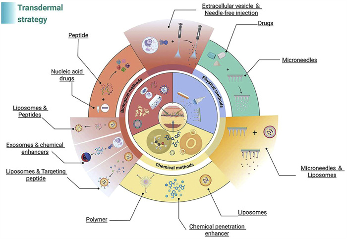
Figure 1.
Three major strategies for enhancing skin permeation: Biological, chemical, and physical approaches. The combined application of these strategies can significantly improve the efficiency of transdermal drug transport.

Transdermal drugs and delivery strategies: History, advances and future prospects
Yefeng Wang , Siwen Wu , Jiyu Chen , Siyi Yang , Chenrui Wu , Qingying Zhao , Heqi Zhang , Kaiting Wei , Jiani Zhang , Rui Zhang , Li Yang


The first transdermal drug delivery system (TDDS) approved by the U.S. Food and Drug Administration (FDA) was the scopolamine patch in 1982. Its success, followed by the introduction of nicotine patches a decade later, markedly increased public and clinical interest in TDDS. Owing to its key advantages, painless use, sustained release (e.g., the three-day scopolamine patch), avoidance of first-pass hepatic metabolism [1], low cost, non-invasive and targeted delivery, suitability for self-administration, and reduced dosing frequency [2]. TDDS has become an appealing alternative to conventional routes such as oral, intravenous, and subcutaneous administration.
To date, approximately 23 transdermal drug formulations, classified by their active chemical components, have been approved by the U.S. FDA (Table S1 in Supporting information), including estradiol, nicotine, and asenapine, in forms such as creams, sprays, paints, foams, and microsponges [3]. Clinical trials of these drugs were reviewed, and their indications, dosage forms, trade names, approval dates, and targets are summarized in Table S1. Despite these advances, TDDS is limited to certain drugs due to the skin’s unique structure. Innovations in delivery strategies have expanded its applicability, including for biomacromolecules such as nucleic acids, proteins, and peptides [4].
In this review, we first analyzed skin structure to delineate the barrier that TDDS must overcome and compiled clinical trial data on FDA—approved transdermal drugs to anticipate future development prospects. We then examined current physical, biological, and chemical strategies advancing TDDS (Fig. 1) and summarized ongoing clinical development and experimental-stage approaches, highlighting their current and potential future impact in medicine.
Challenges in TDDS arise from the skin’s complex structure, making understanding skin anatomy essential. The skin consists of three main layers: The epidermis (30–150 µm), dermis (1–2 mm), and subcutaneous tissue [5–7], with the epidermis and dermis being most relevant. The epidermis includes the stratum corneum (SC), lucidum, granular layer, stratum spinosum, and basal layer; except for the SC, the other four layers form the active epidermis [8]. The SC, composed of 6–10 keratinocyte layers, provides protection, waterproofing, and antibacterial effects, while the lucidum regulates moisture [9]. The dermis contains collagen and elastic fibers, capillaries, lymphocytes, nerve plexuses, and accessory organs such as hair follicles, sweat glands, and sebaceous glands [10]. The skin maintains homeostasis and protects against physical, chemical, and biological insults [11–13], but this barrier limits drug penetration. The basal layer generates keratinocytes that migrate outward to form the SC [14,15]. Its intercellular matrix, consisting of fatty acids, ceramides, cholesterol, and keratin fibers, is the primary “brick-and-mortar” barrier to most drugs [16,17], while the basal membrane forms a secondary barrier, restricting drugs >400 Da from systemic absorption [18–20]. Transdermal absorption occurs via two main pathways: The epidermal route, subdivided into cellular (through keratinocytes) and intercellular (through lipid layers) paths [6,16,21], and the appendageal route via pores and sweat glands, suitable for ionic or highly polar macromolecules. However, hair follicles and sweat glands occupy only ~0.1% and 0.01% of skin area, with all appendages covering ~1% [22,23], limiting this pathway. Overcoming these barriers is essential, and advances have led to physical, chemical, and biological strategies to enhance transdermal absorption, as summarized in this review.
To assess advances in medicine, clinical trials of 23 FDA-approved transdermal drugs were analyzed (Fig. 2a). Estradiol, fentanyl, nicotine, rotigotine, testosterone, buprenorphine, and rivastigmine account for over 90% of these trials, with phase 3 and 4 studies representing >50% of their total. 1172 trials were compiled from all currently available percutaneous routes (as of December 2024) and summarized their indications. Major clinical applications include pain, mental disorders, infectious diseases, inflammation, endocrine disorders, cancer, nicotine-related conditions, and nervous system diseases (Fig. 2b), indicating that transdermal delivery is increasingly targeting systemic, not just local diseases. Notably, in mental and neurological disorders such as Alzheimer’s and Parkinson’s diseases, transdermal drug delivery (TDD) offers convenience for patients with limited self-care abilities and benefits medical staff in administration.
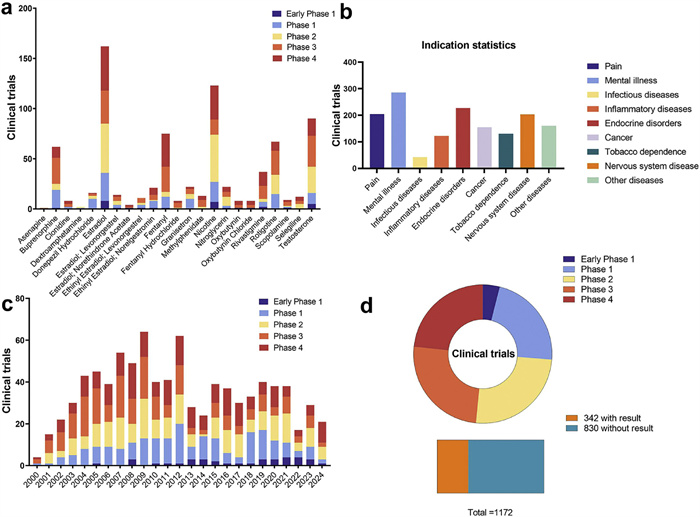
Analysis of the 1172 transdermal-related clinical trials (Fig. 2c) shows that the number of studies increased steadily after 2000 and peaked in 2009. This surge coincides with the market introduction of several transdermal formulations (e.g., nitroglycerin, ketoprofen, fentanyl, estradiol) by major pharmaceutical companies (e.g., ALZA Group, Jiuguang Pharmaceutical), which significantly boosted research activity. After 2010, the overall development of TDD declined. As summarized in Table S1, only a limited number of transdermal drugs, primarily two estradiol combination formulations, have been approved since then. Furthermore, Fig. 2d shows that 441 ongoing clinical trials are in phases Ⅲ–Ⅳ, and 830 have reported definitive outcomes, indicating a high level of maturity in current transdermal drug research. Consequently, expanding the pipeline of new transdermal drugs and advancing emerging technologies such as microneedles and iontophoresis into clinical practice will be essential for driving future progress in TDD.
The 1172 trials were classified by drug type: Small-molecule drugs in late-stage trials over the past five years (Table S2 in Supporting information) and biomacromolecule drugs entering clinical trials (Table S3 in Supporting information). For small molecules, only active or completed phase Ⅲ–Ⅳ trials were included to enhance table reliability. For macromolecules, trials labeled “withdrawn”, “unknown”, or “terminated” were excluded to maintain timeliness. The progression of ~11 biomacromolecule drugs into clinical trials marks a significant TDD advancement. The SC presents a major barrier, making epidermal penetration and subcutaneous delivery challenging. Five macromolecules have reached late-stage trials. Notably, insulin transdermal delivery has matured, with six phase Ⅳ and two phase Ⅲ studies, demonstrating that skin absorption of biomacromolecules is feasible and highlighting promising strategies for future TDD development.
The 23 approved transdermal drugs showing substantial clinical progress primarily include estradiol, nicotine, testosterone, and fentanyl, with most research reaching maturity. Clinical trends indicate rapid growth peaking between 2000 and 2010, followed by a slowdown due to limited new agents. Recent trials focus on systemic diseases, including mental disorders and cancer. Innovations such as microneedles and iontophoresis are increasingly overcoming the skin barrier, enabling biomacromolecule delivery and expanding transdermal therapy.
Future efforts should prioritize novel therapeutic agents, biopharmaceutical breakthroughs, and clinical translation of advanced absorption strategies. These directions underscore the main challenges and opportunities for next-generation TDDS and naturally lead to examining the primary strategies, biological, chemical, and physical, used to enhance transdermal delivery.
We charted the timeline of TDD development from the 20th century to the present (Fig. 3) [24–28]. Between the 1970s and 1990s, strategies to enhance skin permeation emerged, including microneedles, electroporation, iontophoresis, and chemical penetration enhancers (CPEs), alongside the launch of the first transdermal drug, the scopolamine patch, followed by a wave of approvals. After 2000, microneedle vaccine strategies appeared, and prior studies had explored iontophoresis for peptide delivery, marking the expansion from small molecules to biologics. Over the next decade, these strategies matured toward clinical application. Following 2010, the slowdown in new transdermal drugs led to a stable period, until updates and iterations in delivery strategies revitalized the field.
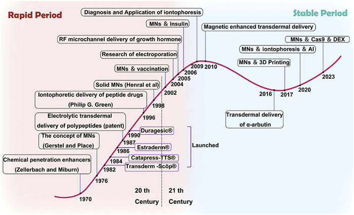
Post-2015, clinical demands exceeded the capabilities of individual strategies, prompting their combination to enhance penetration. After 2020, efforts shifted toward intelligent drug delivery, aiming to improve permeation while enabling devices to adjust drug release according to clinical needs for long-term, convenient administration. Intelligent drug delivery is poised to become a key focus in TDD development. Below, we summarize the development and recent breakthroughs of nine TDD strategies, categorized as biological, chemical, and physical.
Recently, biological strategies have been applied to enhance TDDS, including viruses, exosomes and framework nucleic acids (FNAs) [29]. These approaches have expanded TDDS applicability from lipophilic small molecules to large molecules such as nucleic acids and protein vaccines. Despite rapid progress, their use is limited by biosafety, preparation, and biocompatibility concerns, with most remaining at the preclinical stage.
Viral vectors have been investigated for TDD since the 1990s, leveraging the skin’s rich capillaries and immune factors to deliver macromolecules such as microRNA (miRNA), small interfering RNA (siRNA), peptides, and proteins, with the TDDS offering painlessness and improved compliance [30–32]. Early studies demonstrated proof-of-concept: Hallek et al. used recombinant adeno-associated virus (rAAV) encoding β-galactosidase or green fluorescent protein to transfect keratinocytes in vitro, achieving up to 70% efficiency within 48 h, with stable expression for 50 days and inheritance in daughter cells [33]. A decade later, Spector et al. compared adenovirus, adeno-associated virus, and lentivirus vectors in dermal cell lines, finding >90% induction with lentivirus and stable inheritance, whereas adenovirus expression declined rapidly [34]. Marinkovich et al. utilized beremagene geperpavec (B-VEC), an engineered, non-replicating collagen, type Ⅶ, alpha 1 (COL7A1) containing herpes simplex virus type 1 (HSV-1) vector, to treat recessive dystrophic epidermolysis bullosa (RDEB) skin lesions, showing effective wound healing with no grade ≥2 adverse effects after 12 weeks [35].
Despite these advances, transdermal delivery remains challenging due to the skin barrier, limiting viral absorption. Laboratory vectors such as adenovirus and lentivirus can transfect skin cell lines, but systemic TDD is still difficult. Strategies to enhance viral penetration include microneedles, electroporation, thermal ablation, and CPEs, which create temporary microchannels through the SC, enabling viral vectors to reach the dermis or subcutaneous tissue. Future directions involve engineering viral vectors with intrinsic transdermal capability via surface protein modification or artificial intelligence-guided design. Overall, viral vectors hold substantial potential for TDD, and emerging methods are expected to overcome current limitations.
EVs, membrane-derived nanovesicles 40–160 nm in size, are rich in proteins, nucleic acids, and lipids, and hold potential for skin wound repair and anti-aging therapies (Fig. 4a). EVs also enable multiparameter diagnostic detection in liquid biopsies [36], with applications in cardiovascular, central nervous system, and cancer diagnostics, and expanding use in other organs such as the liver [37]. In therapy, EVs can specifically target diseased cells, exhibit low cytotoxicity, and show prolonged in vivo retention, making them ideal carriers to enhance drug bioavailability and reduce toxicity to normal cells [38]. For example, EVs loaded with doxorubicin have demonstrated targeted efficacy against glioblastoma multiforme [39].
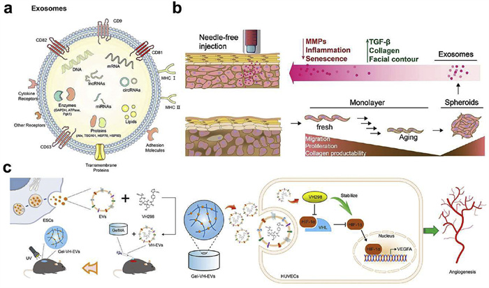
EVs can also be utilized in TDDS. Endogenous EVs regulate skin physiological and pathological conditions, whereas exogenous EVs, such as those from stem cells, provide novel therapeutic options for repairing, regenerating, and revitalizing skin tissue [40]. Park et al. developed EVs from human adipose stem cells delivered via hyaluronic acid-based dissolvable microneedles, enabling precise and sustained dermal delivery [41]. Cheng et al. used EVs derived from 3D- and 2D-cultured human dermal fibroblasts (HDFs) for needle-free treatment and prevention of skin aging. EVs from 3D-cultured HDFs showed higher tissue inhibitor of metalloproteinases-1 (TIMP-1) expression than 2D-derived EVs, promoting type Ⅰ procollagen synthesis via downregulation of tumor necrosis factor-alpha (TNF-α) and upregulation of transforming growth factor-beta (TGF-β) (Fig. 4b), validated in vitro and in nude mouse photoaging models [42]. Fu et al. loaded EVs from epidermal stem cells (ESC) with VH-298 in gelatin methacryloyl (GelMA) hydrogels (Gel-VH-EVs) to enhance local blood supply and angiogenesis via the hypoxia-inducible factor-1 alpha (HIF-1α)/vascular endothelial growth factor A (VEGFA) signaling pathway, promoting wound healing in diabetic mice (Fig. 4c) [43]. Luo et al. generated nuclear factor kappa B (NF-κB) siRNA-enriched EVs from reprogrammed adipose-derived mesenchymal stem cells, which accelerated skin repair and reduced inflammatory factor expression in mice with skin injuries [44].
EVs offer intrinsic advantages for TDDS due to their endogenous origin and lipid bilayer, which confer excellent biocompatibility, low immunogenicity, and strong affinity for skin lipids, enabling SC penetration via fusion or lipid exchange. Surface adhesion molecules and membrane proteins facilitate targeted uptake by keratinocytes and fibroblasts, while their vesicular structure protects cargos—including nucleic acids, peptides, and small molecules—from enzymatic degradation. Stem- or immune cell-derived EVs can also actively modulate inflammation, tissue regeneration, and immune responses, making them a biologically responsive platform for next-generation TDDS. However, poor skin penetration remains a key limitation. Microneedle-assisted delivery can improve transdermal efficiency, targeting, release control, and cargo stability, but broader application is constrained by low storage stability, production and isolation yields, batch heterogeneity, and limited cargo loading [41]. Future efforts should focus on enhancing skin penetration, engineering EVs for targeted delivery, standardizing and scaling production, improving cargo loading, developing long-term stable formulations, integrating artificial vesicles, and addressing regulatory and safety requirements to fully realize their therapeutic potential.
CPEs are an extension of traditional TDDS [45], incorporating amphiphilic molecules to disrupt SC lipid packing. Solvents and surfactants can induce nanoscale defects in the epidermal barrier, facilitating drug penetration through the skin’s “brick-and-mortar” structure [2]. Chemical enhancers can be classified by origin or chemical nature, but the most accepted method categorizes them by functional groups such as hydrocarbons, aldehydes, alcohols, esters, amines, carboxylic acids, sulfoxides, and amides [46]. Alcohol enhancers are commonly divided into short- and long-chain types, with ethanol and isopropanol as typical representatives [47]. Liu et al. showed that the flux of saturated estradiol changes with ethanol concentration in both symmetric and asymmetric models, consistent with ethanol concentration–dependent drug transport [48]. Azone, the first compound designed for skin penetration, was widely studied in the 1980s–1990s [49–53]. With a polar seven-membered ring and a C12 hydrophobic chain, it shows strong enhancing effects at 0.5%–2.5%. Other frequently used enhancers include ethyl acetate [54,55], octyl salicylate [56], isopropyl myristate [57], fatty acids [58], and dimethylsulfoxide (DMSO) [59]. Combining different CPEs often yields superior performance; for example, a choline/geranic acid deep eutectic solvent (DES) significantly improved the solubility and transdermal penetration of amphotericin B (AmB) [60].
The application of CPEs, however, is limited by safety and performance issues. Many enhancers cause irritation or toxicity by disrupting the SC lipid matrix, and prolonged use may damage barrier integrity [61]. Their non-selective enhancement increases the risk of unintended systemic exposure, particularly in individuals with compromised skin. Some CPEs also exhibit inconsistent reversibility, with certain formulations potentially leading to irreversible barrier changes [62]. In addition, their efficacy strongly depends on drug physicochemical properties, making universal enhancer systems difficult to develop [63], while inter-individual variations in skin type, age, hydration, and skin conditions further contribute to inconsistent outcomes [64]. An ideal penetration enhancer should therefore meet the following criteria [65]: (1) Induce only reversible barrier disruption; (2) exhibit minimal off-target activity; (3) be non-toxic and non-irritating; (4) provide reproducible effects; and (5) preserve drug activity [46].
Future CPEs development should prioritize both safety and efficacy. Biocompatible enhancers derived from natural or bioengineered materials may improve skin tolerance and reduce adverse effects [66]. Stimuli-responsive systems, activated by pH, temperature, or light, enable precise control over drug permeation, minimizing unnecessary exposure [67]. Combining CPEs with advanced transdermal techniques, such as microneedles [68], iontophoresis [69], or sonophoresis, can enhance delivery while lowering enhancer concentrations and irritation risk [70]. Personalized formulations tailored to individual skin properties may further optimize treatment efficacy. Strategies to design reversible or self-limiting enhancers, which are rapidly deactivated after action, help restore the skin barrier and reduce long-term damage [66,67,70]. Incorporating additional therapeutic functions, such as anti-inflammatory or antimicrobial activity, may further improve outcomes while facilitating drug penetration.
“Liposome” derives from the Greek lipo and soma, meaning “fat” and “body” [71]. British hematologist Bangham and collaborators at the Babraham Institute, University of Cambridge, first discovered liposomes in 1960 and published their initial study in 1964 [72]. Unlike the broader class of lipid nanoparticles (LNPs), liposomes are bilayers of amphiphilic lipids, such as phospholipids, with polar ends facing aqueous interfaces, forming closed vesicles with an internal hydrophilic core [73]. LNPs do not necessarily have such a core. By incorporating ionizable lipids, LNPs encapsulate nucleic acid drugs (RNA, DNA) via electrostatic interactions and include higher cholesterol content for stability. Liposomes can deliver drugs of diverse properties: Hydrophilic drugs occupy the aqueous core, lipophilic drugs the lipid bilayer, and amphiphilic drugs at the water/lipid interface (Fig. S1a in Supporting information). They can also carry nucleic acids, proteins, and peptides [61]. Over decades, liposomes have become popular vehicles for cancer chemotherapy, ocular and colon-specific drug delivery, and other applications [74]. Their widespread use in transdermal delivery stems from simple preparation and efficient encapsulation. Key factors affecting transdermal performance include liposome size, zeta potential, cargo oil–water partition coefficient, and interactions with skin surface lipids [75].
Elastic liposomes, a novel class, differ from traditional liposomes by their high membrane elasticity, fluidity, and bending energy, enabling compression and twisting to penetrate intercellular lipids [76]. Tyagi et al. encapsulated the carboxyl-terminal 19 kDa fragment of Plasmodium falciparum merozoite surface protein-1 (PfMSP-119) in elastic liposomes for transdermal delivery, achieving enhanced encapsulation efficiency and permeability [77]. Kajimoto et al. combined iontophoresis with liposomes for non-invasive hair follicle delivery of insulin in type Ⅰ diabetic rats, reducing blood glucose to 20% of baseline within 18 h and maintaining it for 24 h, with detectable plasma insulin [78]. Liposomes also enhance delivery of conventional drugs; Sacha et al. showed that a 1% diclofenac sodium liposomal gel exhibited higher permeation flux (69.3 ± 14.4 × 10⁻⁸ cm/s) than 1% (34.9 ± 9.1 × 10⁻⁸) and 2% (47.1 ± 9.5 × 10⁻⁸) commercial emulsions [79]. Niosomes, composed of nonionic surfactants, offer greater flexibility and stability than liposomes, enhancing transdermal drug release [80,81]. Recent applications include improved dermal drug delivery, sustained release, and storage stability [81,82]. Therapeutic uses span hormonal therapy, diabetes management, pain control, wound healing with bioactive compounds, psoriasis, mycoses, and anesthesia (Fig. S1b in Supporting information) [83].
Numerous studies indicate that traditional liposomes cannot fully penetrate the skin barrier to reach the viable epidermis or dermis [84]. Only a small fraction enters the subcutaneous layer via accessory organs, while most accumulate in the SC [85]. Kirjavainen et al. reported that aqueous phospholipid liposome solutions fail to penetrate human skin [86], and Hasan et al. observed no rhodamine B fluorescence in the circulatory system of hairless mice following liposome application [87]. These findings suggest that liposomes adhere to the SC, causing phospholipid bond rupture and premature drug leakage [88]. Consequently, contemporary TDD research focuses on developing composite materials, functional modifications, and combining liposomes with other strategies to create next-generation formulations.
Cell-penetrating peptides (CPPs) in TDD are classified into two types: Peptides with intrinsic therapeutic effects, such as anti-wrinkle peptides [89], and those that carry or enhance drugs. CPPs are typically positively charged, enabling them to neutralize the negatively charged cell membrane and facilitate drug translocation [90]. Since their discovery in the 1980s, CPPs have been explored in preclinical and clinical studies for diagnostics and therapy, mainly via passive translocation and active endocytosis [91,92]. Preclinical studies show CPPs as pro-absorbent agents for cancer, anesthesia, anti-inflammatory therapy, atopic dermatitis (AD), and gene delivery [93], with common CPPs like TAT and P28 advancing to clinical trials (phases Ⅰ–Ⅲ) [94–96]. Unlike viral vectors, CPPs are not limited to nucleic acids and have been applied to chemical drugs; for instance, Mo et al. developed a paclitaxel-encapsulated CPPs-modified transfersomes/gelatin (PTX-CTs/Gel) patch for transdermal melanoma therapy, enhancing skin and tumor penetration and effectively delaying tumor growth in mice when combined with systemic chemotherapy (Figs. S2a and b in Supporting information) [97].
To date, over 1800 CPPs with diverse functions have been identified, and preclinical and clinical development is rapid, though none have received FDA approval [98]. Key challenges include poor stability, limited targeting and efficacy, potential immunogenicity, enzymatic degradation, endosomal trapping, and clearance by metabolic organs, with attempts to increase dosage or frequency risking non-selective uptake, toxicity, and local irritation [99,100]. Combining CPPs with nanomaterials such as microneedles, liposomes, and nanometallic particles can partially overcome these limitations [101]. Stability can be enhanced via encapsulation or hydrogel incorporation, and targeting improved through artificial modifications or coupling with targeting peptides, as shown by Hu et al. delivering Smad4 mRNA to integrin-expressing colorectal cancer cells [102]. Skin-penetrating peptides (SKPs), a key CPPs branch, bypass the SC by disrupting lipid bilayers or interacting with keratin and tight junctions, enhancing macromolecule absorption with low skin toxicity [103–105]. SKPs are applied via drug conjugation, skin pretreatment, or co-administration [103,106–108], and can be further optimized through sequence refinement, physicochemical modifications, in silico validation, or conjugation to nanocarriers, such as TAT-coated systems shown to improve penetration in vitro and in vivo [109–113]. Combining SKPs with other permeation strategies offers additional potential for effective transdermal drug delivery.
Chemical nanomaterials for TDDS encompass nanoemulsions, polymer carriers, nanomicelles, dendritic macromolecules, metal nanoparticles (NPs), silicon NPs, metal-organic frameworks, and liposomes, offering broad and expanding applications [114–120]. Nanoemulsions are particularly promising due to their small droplet size (20–400 nm), fluidity, protective interface, and interaction with skin cells, which improve penetration and bioavailability [121–128]. They are classified as oil-in-water, water-in-oil, or multiple emulsions, stabilized by emulsifiers and co-emulsifiers, and enhance solubility for both hydrophilic and lipophilic drugs, with several formulations such as cyclosporine (Neoral, Gengraf), saquinavir (Fortovase), and ritonavir (Norvir) receiving FDA approval [123–129]. Polymer nanocarriers, including polymer NPs, polymer micelles, and dendrimers, similarly provide high versatility and improved drug loading and stability compared to lipid carriers [130,131]. Biodegradable polymers such as poly(lactic-co-glycolic acid) (PLGA)-PEG, gelatin, chitosan, hyaluronic acid, and albumin have been applied, with chitosan-coated donepezil hydrochloride-PLGA NPs achieving 2.2-fold higher subcutaneous donepezil concentrations than uncoated NPs, enhancing follicular delivery and skin accumulation [132–134]. Inorganic NPs, including silver, gold, and silica NPs, offer tunable structures and surface properties for efficient drug loading and cargo protection [135–138]. Notably, copper sulfide (CuS) NPs generate photothermal effects under near-infrared irradiation to disrupt the SC barrier [139], and silica NPs (~70 nm) have demonstrated epidermal penetration and lymph node transport in mice within three days, confirming in vivo delivery potential [140].
To overcome the skin barrier, strategies such as chemical penetration enhancers [141], liposomes [142], iontophoresis [143], and ultrasound [144] have been developed. In recent years, microneedles have attracted attention as an effective approach to bypass the SC [145]. The concept was first introduced by Gerstel and Place in their patent “drug delivery device” [146]. Microneedles are arrays of micron-scale projections (typically 25–2000 µm) that penetrate the SC without causing pain or bleeding [147]. They possess sufficient mechanical strength to form temporary microchannels on the skin, with trauma being reversible [148–150]. Designed to reach the epidermis, microneedles avoid contact with dermal blood vessels and nerve fibers, offering a minimally invasive and painless drug delivery method [151,152]. Over five decades, microneedles have shown efficacy in treating superficial tumors, scars, psoriasis, hair loss, and certain immune disorders [68]. Based on their properties, microneedles are classified into five types: Solid, coated, hollow, dissolving, and hydrogel microneedles (Fig. 5) [68].
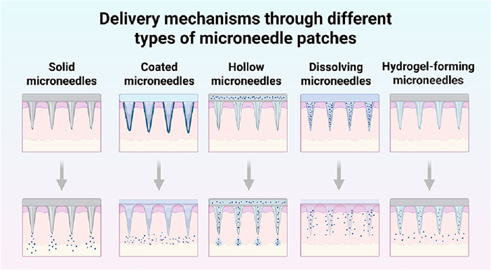
Microneedle strategies for TDDS leveraged distinct mechanisms to bypass the SC and enhance drug penetration [147,153–165]. Solid microneedles, fabricated from mechanically robust materials such as silicon or titanium via precision machining, create microchannels for passive diffusion of topically applied drugs [153–155]. Coated microneedles carry a thin drug layer on the needle surface, releasing the payload upon insertion without secondary administration [147,156], while hollow microneedles feature internal cavities for higher drug loading and controlled release via flow rate and pressure adjustments, exemplified by 3M’s commercial hollow microstructure system [150,157,158]. Dissolving microneedles, composed of biodegradable materials such as chitosan, hyaluronic acid, or polylactic acid, release encapsulated drugs as the matrix degrades; for instance, polymer-encapsulated Cas9 ribonucleoprotein and dexamethasone-loaded dissolving microneedles demonstrated superior therapeutic efficacy in a mouse model of AD induced by 1‑chloro-2, 4-dini-trochlorobenzene (DNCB) [28,159–162]. Hydrogel microneedles function similarly but swell upon absorbing interstitial fluid, forming porous microchannels that enable controlled release and prevent pore closure [163–165]. To meet the demands of precision medicine, research has focused on controlled- or sustained-release microneedles, including polymeric, core–shell polymeric, nanoparticle- or nanosuspension-loaded, swellable, and stimuli-responsive designs [166–168]. Polymers such as polyvinyl alcohol (PVA), PLGA and polyvinylpyrrolidone (PVP) are commonly employed, with strategies like core–shell structuring or nanocarrier incorporation enabling programmed, delayed, or sustained drug release. Optimization efforts target carrier properties, drug loading, polymer ratios, and matrix composition to achieve precise permeation and therapeutic profiles [166].
Looking forward, microneedles are expected to evolve toward intelligent and integrated systems to meet the requirements of precision medicine. While traditional microneedles revolutionized transdermal delivery, they often lack the specificity and adaptability needed for individualized treatments. Physical penetration techniques such as microneedles, iontophoresis, and electrophoresis have demonstrated significant potential for enhancing transdermal delivery of macromolecules [169].
Electroporation, initially developed for transfecting gene drugs such as DNA, plasmids, and mRNA, has been adapted for TDD by temporarily creating aqueous pores in the SC through applied electrical pulses [170–172]. As early as 1993, Prausnitz et al. demonstrated in vitro and in vivo that millisecond- to microsecond-level pulses increase SC permeability, enhancing drug flux in excised and live skin [171]. The mechanism mirrors cellular electroporation, with voltage and pulse duration dictating permeability; the SC, composed of ~20 keratinized layers, contributes most of skin resistance, so transient electrical breakdown allows drug diffusion [172]. Electroporation has increased transdermal flux for drugs including cyclosporine, heparin, fentanyl, metoprolol, and flurbiprofen, often using 75–100 V [173–179]. Electrophoretic forces also assist, enabling drug transport within 10–30 min post-pulse to equal or exceed that during the pulse [180–182]. Nanohydrogel-based systems combining drug reservoir and electrode have further improved uptake in mouse skin [183]. Despite its efficacy, electroporation can induce muscle contractions, pain, and irritation [184,185], making milder iontophoresis, which directly drives charged drugs without SC disruption, preferable in some cases [186].
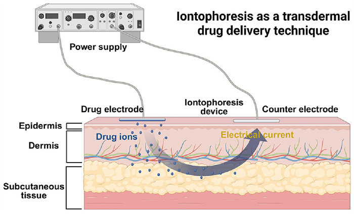
Iontophoresis enhances transdermal drug delivery by applying a mild electric current (<0.5 mA/cm2) to drive ions through the epidermis and dermis, with preferential transport via skin appendages such as pores and hair follicles (Fig. 6) [187,188]. Its primary mechanisms are electric repulsion and enhanced permeation, and efficiency depends on ion composition, pH, and molecular properties: Cations penetrate more readily than anions, and anions of 35–500 Da show decreasing transport with size, while uncharged compounds <400 Da permeate independently of lipophilicity [189–192]. Studies demonstrate effective iontophoresis delivery of lidocaine and bupivacaine (>95% released, 40% higher than traditional patches) [193] and rapid subcutaneous accumulation of naloxone when combined with microneedles, achieving sixfold higher uptake within 1 h [69]. Iontophoresis also enables reverse iontophoresis for biomarker extraction, transporting cations and neutral molecules to the cathode and anions to the anode, allowing non-invasive monitoring such as blood glucose detection [194,195].
Future developments in electroporation and iontophoresis will focus on improving drug permeability, precision, and safety. Electroporation may expand to larger biomolecules like proteins and nucleic acids, integrating smart systems for controlled, personalized delivery while minimizing skin damage and combining with other methods for enhanced efficiency. Iontophoresis will continue to optimize delivery of small charged drugs, aiming for precise dosing and improved patient compliance. Both strategies are likely to evolve into hybrid systems, leveraging their strengths to advance personalized transdermal therapy and therapeutic outcomes.
Ultrasound enhances TDD by generating mechanical waves (>20 kHz) that induce molecular displacements, with cavitation—oscillation or collapse of bubbles under ultrasonic pressure—being a primary mechanism [196–199]. At low pressures, stable cavitation oscillates bubbles around an equilibrium radius, whereas inertial cavitation at higher pressures rapidly collapses bubbles, creating channels in the SC to facilitate hydrophilic molecule transport [2,200]. Additional effects include thermal heating and acoustic streaming, which can be bulk or microstreaming, altering membrane permeability and cellular activity [201]. Ho et al. demonstrated that citric acid–NaHCO3 microbubbles coupled with Evans blue and fluorescein isothiocyanate (FITC)-hyaluronic acid increased penetration in rat abdominal skin by 2.4× and 2.1×, respectively, with Evans blue reaching the epidermis, while mitigating typical cavitation damage (Figs. S3a–d in Supporting information) [202]. These advances indicate that cavitation ultrasound provides a safe and versatile approach to enhance TDD beyond small lipophilic drugs.
Future developments in ultrasound-based TDD will focus on enhancing drug permeation, patient compliance, and broadening applications. Ultrasound can create temporary microchannels, enabling efficient delivery of larger and hydrophilic molecules, including proteins and nucleic acids [203]. Research aims to optimize parameters such as frequency, intensity, and duration to maximize absorption. Combining ultrasound with other transdermal methods, like microneedles or iontophoresis, may produce synergistic effects and enable targeted delivery [204]. Advancements are also expected in device miniaturization, portability, and usability, improving patient compliance and facilitating home administration [144]. Integration of sensor technology can allow real-time monitoring of drug delivery and skin response, allowing for personalized adjustments and enhancing therapeutic outcomes [202]. Overall, ultrasound holds strong potential to improve the efficacy, safety, and convenience of transdermal therapies.
Insulin, discovered by Banting and Best in 1922 [205], remains the primary therapy for type 1 and advanced type 2 diabetes. Its large molecular size necessitates subcutaneous injection, which can cause pain, needle phobia, infection, tissue injury, and nerve damage [206–211]. Transdermal delivery offers advantages including avoidance of first-pass metabolism, reduced gastrointestinal degradation, minimized liver and kidney stress, improved compliance, continuous delivery, and lower toxicity [212–214]. Existing alternatives, such as insulin pumps, mimic physiological insulin release but face limitations in cost, device complexity, and patient burden [215,216]. Consequently, microneedle patches and needle-free strategies are being developed to enhance convenience, reduce dosing frequency, and improve quality of life [217].
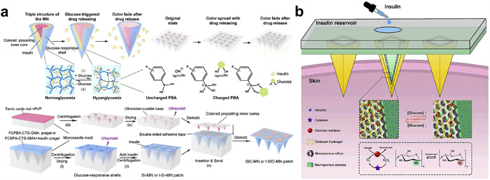
Microneedle-based systems have emerged as a promising transdermal route. He et al. designed a triple-structure microneedle with a glucose-responsive shell, internal insulin reservoir, and colored propulsion core, achieving closed-loop delivery and a 24 h hypoglycemic effect in diabetic mice with a 0.3 cm2 patch (Fig. 7a) [218]. Similarly, Gholami et al. developed a glucose-responsive porous microneedle array with nanopore gates and chitosan-mediated expansion/contraction, enabling cyclic insulin release in response to glucose levels (Fig. 7b) [219]. These strategies improve delivery intelligence, reduce administration frequency, minimize skin damage and infection risk, and enhance long-term efficacy.
Qi et al. combined microneedles with electrical stimulation to create an electrically responsive insulin delivery system based on sulfurized silk fibroin protein. Electrical stimulation reduces disulfide bonds to thiol groups, expanding the microneedle and triggering insulin release. Upon power removal, disulfide bonds reform, slowing insulin release and enabling controllable delivery [220]. This integration provides a novel approach for precise transdermal insulin administration. Insulin formulation also impacts clinical efficacy. Sanofi’s Lantus (Insulin glargine) was FDA-approved in 2015 as a higher-concentration, long-acting insulin compared to the 2000 version [221,222]. Novo Nordisk’s Tresiba (Insulin degludec), approved in 2015, is the longest-acting insulin currently available, lasting up to 42 h [223,224]. Although administered by injection, these formulations improve in vitro insulin stability, supporting transdermal strategies [225]. Transdermal insulin delivery offers a less invasive to injections. Nevertheless, long-term use, distribution efficiency, and reliability require further study. Chemical enhancers and micro-/nanoparticles must be evaluated for short- and long-term safety. Clinical research should assess irradiation and inflammatory risks, and optimal dosing requires understanding consistent delivery across patients and skin sites. Pharmacokinetic characterization is essential for determining transport efficiency [226].
Cholinesterase inhibitors are currently the main drugs used in the clinical treatment of Alzheimer's disease. However, oral administration is often inefficient, making transdermal donepezil clinically valuable (Fig. S4a in Supporting information) [227]. Currently, four main types of Alzheimer's disease drugs are FDA-approved. Memantine, a N-methyl-D-aspartate (NMDA) receptor antagonist, is primarily used for moderate to severe Alzheimer's disease. The remaining two are acetylcholinesterase inhibitors: Galantamine, used for mild to moderate Alzheimer's disease, is oral-only, while donepezil, used for severe Alzheimer's disease, is available in both oral and transdermal formulations [228].
The FDA approved the first transdermal donepezil system in 2022, following roughly ten years of development [229]. Penetration enhancers remain the most common method to improve transdermal absorption. Choi et al. investigated the effects of fatty acids on donepezil free base (DPB) and hydrochloride salt (DPH) using hairless mice and human cadaver skin. Oleic acid increased DPB penetration by 11.85-fold, while palmitoleic acid enhanced DPH penetration 105-fold. The bioavailability of oleic acid–DPB was ~10 times higher than palmitic acid–DPH [230]. Nano lipid carriers are another approach: In 2019, DPB-loaded nanoparticles (DPB-NP) were prepared using stearic acid, oleic acid, lecithin, and sodium taurocholate via a microemulsion method, doubling DPB penetration in pig skin compared to DPB carbomer gel (Figs. S4b–e in Supporting information) [231].
Iontophoresis and microneedles significantly improve donepezil transport. Combined, they can achieve up to 1000 µg/cm2 within 24 h, whereas iontophoresis and microneedles alone reach 623 and 282 µg/cm2, respectively [232]. Despite the potential for portable patches, clinical application is limited: Only three FDA-approved devices exist, and none are marketed, with one withdrawn due to burns and scarring [227]. A novel method employs ionic liquids, which react with donepezil and dicarboxylic acids to form stable salts with enhanced stratum corneum permeability. Dinh et al. screened multiple combinations in hairless mice, with one achieving a 50-fold increase in penetration compared to DPB patches [233]. A major challenge is low drug loading, necessitating large patches and high costs. The oral maximum donepezil dose is 23 mg, but a 5 mg patch requires 8.3 × 10.8 cm, and a 10 mg patch 10.8 × 14.4 cm [227]. Monthly costs are ~$500 for the patch, versus $70 for the maximum oral dose and $15 for lower doses. The rivastigmine patch, with similar efficacy, costs ~$150 per month [234].
Cannabinoids are natural or synthetic compounds that interact with the endocannabinoid system. The primary phytocannabinoid is Δ9-tetrahydrocannabinol (THC), the main psychoactive component of cannabis, while cannabidiol (CBD), cannabinol (CBN), and cannabigerol (CBG) are largely non-psychoactive. Increasing evidence indicates therapeutic potential, particularly in neuropsychiatric disorders [235], including brain cancer (glioblastoma multiforme) [236], chronic pain [237], mental illness [238], and peripheral and central nervous system pain [239]. Conventional administration routes, oral, pulmonary, intranasal, rectal, and intravenous, often suffer from invasiveness, low compliance, poor bioavailability, and first-pass metabolism [240,241], highlighting the potential of transdermal delivery.
Topical administration is mainly applied for dermatological conditions (eczema, acne, psoriasis) via ointments, creams, emulsions, and gels [242], but systemic therapeutic effects require improved transdermal strategies [243,244]. Research focuses on two aspects: Optimizing formulations (using penetration enhancers [245], excipients [246], or crystal structure optimization [247]) and exploring carriers or transport strategies such as liposomes and nanovesicles (ethosomes, transfersomes, niosomes) [242,248,249]. CPEs are particularly effective; for instance, Diethylene glycol monoethyl ether (Transcutol) combined with oleic acid enhanced THC penetration in rat skin, confirmed via autoradiography after 24 h [250], while ethanol disrupts SC lipid packing to promote penetration [245,251].
Nanovesicles improve transdermal delivery by enhancing drug accumulation and efficacy. Lodzki et al. applied a CBD ethosome (3% w/w CBD, 40% w/w ethanol) to ICR mice abdominal skin for 72 h, achieving significant subcutaneous and muscle tissue uptake by 24 h and mitigating inflammation and edema induced by carrageenan [252]. Prodrug strategies offer another mature approach, covalently linking cannabinoids to chemical moieties to overcome solubility, stability, bioavailability, targeting, and adverse-effect limitations [253–255]. Gelest Vertasil, the only FDA-approved CBD prodrug, is a trisilane derivative with improved solubility and compatibility with silicon-based materials [242].
TDDS offer multiple advantages, including simple and convenient administration that enables self-use by patients, thereby reducing the workload of healthcare professionals. Compared with oral administration, TDDS bypasses gastrointestinal irritation and first-pass metabolism, enhancing bioavailability. Relative to injection, it is a non-invasive and painless approach that improves patient compliance and minimizes the risk of pathogen transmission. Nevertheless, TDDS still faces significant limitations. The SC acts as a major barrier to drug permeation, restricting effective dosage and therapeutic efficacy. High local drug concentrations may cause irritation, allergic reactions, or tissue damage. Moreover, drugs can undergo hydrolysis or enzymatic degradation during the relatively slow process of transdermal absorption, further reducing their stability.
In this review, we summarized recent clinical progress and categorized current TDDS strategies into biological, chemical, and physical approaches. Among these, physical strategies such as microneedle systems have demonstrated remarkable potential. The evolution from solid to dissolving microneedles [162] has effectively reduced infection risks and improved biocompatibility. Integration with strategies such as micro-pumps [256,257], iontophoresis [258], or intelligent control systems [219] further enhances delivery precision and stability, accelerating clinical translation.
Representative drugs, including insulin, donepezil, and cannabinoids, exemplify the expanding scope of TDDS from dermatology to broader therapeutic areas such as oncology, anesthesia, and gene therapy. These cases highlight both the versatility and technical challenges of achieving efficient, stable, and patient-friendly transdermal formulations. Looking forward, the development of TDDS is expected to move toward intelligent, personalized, and multifunctional platforms. Smart patches capable of real-time monitoring and feedback-controlled release may offer dynamic dose adjustment according to individual physiological signals. Nanostrategy-based carriers, such as LNPs and peptide-modified vesicles, will further improve permeability and targeting accuracy. In addition, combination strategies integrating biological enhancers, physical penetration strategies, and controlled-release systems may break through current barriers of low efficiency and instability. Ultimately, future progress will depend on cross-disciplinary collaboration among materials science, pharmaceutical chemistry, bioengineering, and clinical medicine. Such integration will not only improve the therapeutic efficiency and safety of TDDS but also broaden its application to precision medicine and next-generation therapeutics.
Yefeng Wang: Writing – review & editing, Writing – original draft, Investigation. Siwen Wu: Investigation, Funding acquisition. Jiyu Chen: Writing – original draft, Resources. Siyi Yang: Supervision, Resources. Chenrui Wu: Validation. Qingying Zhao: Investigation. Heqi Zhang: Supervision. Kaiting Wei: Supervision. Jiani Zhang: Investigation. Rui Zhang: Conceptualization. Li Yang: Writing – original draft, Investigation, Funding acquisition.
The authors declare that they have no known competing financial interests or personal relationships that could have appeared to influence the work reported in this paper.
This work was supported by the 1.3.5 Project for Disciplines of Excellence, West China Hospital, Sichuan University (No. ZYGD23008) and National Natural Science Foundation of China (No. 82073366). China Postdoctoral Science Foundation (No. 2024M762231). the Post-doctor Research Fund of West China Hospital, Sichuan University (No. 2024HXBH148). Graphic materials: Biorender.
Supplementary material associated with this article can be found, in the online version, at doi:
S. Zhang, P. Xin, Q. Ou, et al., J. Mater. Chem. B 6 (2018) 6723–6730. doi: 10.1039/c8tb01466c
M.R. Prausnitz, R. Langer, Nat. Biotechnol. 26 (2008) 1261–1268. doi: 10.1038/nbt.1504
W. Zhang, Y. Jiao, Z. Zhang, et al., J. Control. Release 371 (2024) 516–529. doi: 10.1016/j.jconrel.2024.06.013
X. Chen, Adv. Drug Deliv. Rev. 127 (2018) 85–105. doi: 10.1016/j.addr.2017.12.014
M. Huzaira, F. Rius, M. Rajadhyaksha, et al., J. Invest. Dermatol. 116 (2001) 846–852. doi: 10.1046/j.0022-202x.2001.01337.x
S. Jain, N. Patel, M.K. Shah, P. Khatri, N. Vora, J. Pharm. Sci. 106 (2017) 423–445. doi: 10.1016/j.xphs.2016.10.001
P. Volz, A. Boreham, A. Wolf, et al., Int. J. Mol. Sci. 16 (2015) 6960–6977. doi: 10.3390/ijms16046960
R.S. Moreci, T. Lechler, Curr. Biol. 30 (2020) R144–R149. doi: 10.1016/j.cub.2020.01.004
B. Zhang, P. Luo, J. Sun, et al., J. Invest. Surg. 36 (2023) 2146318. doi: 10.1080/08941939.2022.2146318
N. Rutter, Semin. Neonatol. 5 (2000) 297–302. doi: 10.1053/siny.2000.0016
E. Proksch, J.M. Brandner, J.M. Jensen, Exp. Dermatol. 17 (2008) 1063–1072. doi: 10.1111/j.1600-0625.2008.00786.x
I.H. Blank, J. Invest. Dermatol. 45 (1965) 249–256. doi: 10.1038/jid.1965.125
M. Köberle, Y. Amar, I.M. Hölge, S. Kaesler, T. Biedermann, Handb. Exp. Pharmacol. 268 (2022) 43–52.
V.G. Wilson, Methods. Mol. Biol. 1195 (2014) 33–41.
B. Russo, N.C. Brembilla, C. Chizzolini, Curr. Opin. Rheumatol. 34 (2022) 337–342. doi: 10.1097/bor.0000000000000895
G.K. Menon, G.W. Cleary, M.E. Lane, Int. J. Pharm. 435 (2012) 3–9. doi: 10.1016/j.ijpharm.2012.06.005
H. Xing, Z. Zhao, X. Zhu, et al., MedComm Biomater. Appl. 4 (2025) e70023.
K. Bäsler, S. Bergmann, M. Heisig, et al., J. Control. Release 242 (2016) 105–118. doi: 10.1016/j.jconrel.2016.08.007
K. Kabashima, T. Honda, F. Ginhoux, G. Egawa, Nat. Rev. Immunol. 19 (2019) 19–30. doi: 10.1038/s41577-018-0084-5
Y. Zhang, C. Liu, J. Wang, et al., Chin. Chem. Lett. 34 (2023) 107631. doi: 10.1016/j.cclet.2022.06.054
F. Erdő, N. Hashimoto, G. Karvaly, et al., J. Control. Release 233 (2016) 147–161. doi: 10.1016/j.jconrel.2016.05.035
R.J. Scheuplein, J. Invest. Dermatol. 48 (1967) 79–88. doi: 10.1038/jid.1967.11
S.S. Szabo, J. Eng. Mech. Div. 88 (1962), doi: 10.1061/JMCEA3.0000308.
D. Ramadon, M.T.C. McCrudden, A.J. Courtenay, R.F. Donnelly, Drug Deliv. Transl. Res. 12 (2022) 758–791. doi: 10.1007/s13346-021-00909-6
N. Joshi, S. Azizi Machekposhti, R.J. Narayan, JID. Innov. 3 (2023) 100225. doi: 10.1016/j.xjidi.2023.100225
P.G. Green, J. Control. Release 41 (1996) 33–48. doi: 10.1016/0168-3659(96)01354-5
R.S. Vaseem, A. D’Cruz, S. Shetty, et al., Adv. Pharm. Bull. 14 (2024) 67–85.
T. Wan, Q. Pan, Y. Ping, Sci. Adv. 7 (2021) eabf7199.
D. Xiao, T. Chen, T. Zhang, et al., Chin. Chem. Lett. 35 (2024) 108602. doi: 10.1016/j.cclet.2023.108602
R.S. Gibbs, M.S. Amstey, D.C. Lezotte, JAMa 270 (1993) 94–95. doi: 10.1001/jama.1993.03510010100039
B. Roizman, Proc. Natl. Acad. Sci. U. S. A. 93 (1996) 11307–11312. doi: 10.1073/pnas.93.21.11307
E. Gorell, N. Nguyen, A. Lane, Z. Siprashvili, Cold Spring Harbor Perspect 4 (2014) a015149. doi: 10.1101/cshperspect.a015149
M. Braun-Falco, A. Doenecke, H. Smola, M. Hallek, Gene Ther. 6 (1999) 432–441. doi: 10.1038/sj.gt.3300815
E.H. Teo, K.J. Cross, E.D. Bomsztyk, D.C. Lyden, J.A. Spector, Ann. Plast. Surg. 62 (2009) 576–580. doi: 10.1097/SAP.0b013e31819fafbd
I. Gurevich, P. Agarwal, P. Zhang, et al., Nat. Med. 28 (2022) 780–788. doi: 10.1038/s41591-022-01737-y
F.J. Verweij, L. Balaj, C.M. Boulanger, et al., Nat. Methods 18 (2021) 1013–1026. doi: 10.1038/s41592-021-01206-3
Y. Wang, J. Liu, J. Ma, et al., Mol. Cancer 18 (2019) 116. doi: 10.1186/s12943-019-1041-z
R. Raguraman, D. Bhavsar, D. Kim, et al., Cancer Lett. 558 (2023) 216093. doi: 10.1016/j.canlet.2023.216093
J. Liu, Y. Sun, X. Zeng, et al., Adv. Mater. 35 (2023) 41–55.
M. Xiong, Q. Zhang, W. Hu, et al., Pharmacol. Res. 166 (2021) 105490. doi: 10.1016/j.phrs.2021.105490
V.D. Bui, S. Son, W. Xavier, et al., Biomaterials 287 (2022) 121644. doi: 10.1016/j.biomaterials.2022.121644
S. Hu, Z. Li, J. Cores, et al., ACS Nano 13 (2019) 11273–11282. doi: 10.1021/acsnano.9b04384
Y. Wang, Z. Cao, Q. Wei, et al., Acta Biomater. 147 (2022) 342–355. doi: 10.1016/j.actbio.2022.05.018
W. Lu, J. Zhang, Y. Wu, et al., Front. Immunol. 14 (2023) 1109381. doi: 10.3389/fimmu.2023.1109381
Y. Yang, R. Zhou, Y. Wang, et al., Angew. Chem. Int. Ed. 62 (2023) e202214795. doi: 10.1002/anie.202214795
A. Kovácik, M. Kopečná, K. Vávrová, Expert. Opin. Drug. Deliv. 17 (2020) 145–155. doi: 10.1080/17425247.2020.1713087
M.E. Lane, Int. J. Pharm. 447 (2013) 12–21. doi: 10.1016/j.ijpharm.2013.02.040
P. Liu, T. Kurihara-Bergstrom, W.R. Good, Pharm. Res. 8 (1991) 938–944. doi: 10.1023/A:1015876117627
D. Southwell, B.W. Barry, J. Investig. Dermatol. 80 (1983) 507–514. doi: 10.1111/1523-1747.ep12535090
J.W. Wiechers, B.F. Drenth, J.H. Jonkman, R.A. de Zeeuw, Pharm. Res. 4 (1987) 519–523. doi: 10.1023/A:1016439908270
J.E. Harrison, A.C. Watkinson, D.M. Green, J. Hadgraft, K. Brain, Pharm. Res. 13 (1996) 542–546. doi: 10.1023/A:1016037803128
J. Engblom, S. Engström, B. Jönsson, J. Control. Release 52 (1998) 271–280. doi: 10.1016/S0168-3659(97)00219-8
I.T. Degim, A. Uslu, J. Hadgraft, et al., Int. J. Pharm. 179 (1999) 21–25. doi: 10.1016/S0378-5173(98)00353-6
D.R. Friend, S.J. Phillips, J.R. Hill, Food. Chem. Toxicol. 29 (1991) 639–646. doi: 10.1016/0278-6915(91)90147-Y
D.R. Friend, Med. Res. Rev. 11 (1991) 49–80. doi: 10.1002/med.2610110105
P. Santos, A.C. Watkinson, J. Hadgraft, M.E. Lane, Int. J. Pharm. 384 (2010) 67–72. doi: 10.1016/j.ijpharm.2009.09.043
C.S. Leopold, B.C. Lippold, J. Pharm. Pharmacol. 47 (1995) 276–281. doi: 10.1111/j.2042-7158.1995.tb05795.x
M. Artusi, S. Nicoli, P. Colombo, et al., J. Pharm. Sci. 93 (2004) 2431–2438. doi: 10.1002/jps.20152
R.B. Stoughton, Arch. Dermatol. 91 (1965) 657–660. doi: 10.1001/archderm.1965.01600120089022
B. Xie, Q. Jiang, F. Zhu, et al., Chin. Chem. Lett. 36 (2025) 110508. doi: 10.1016/j.cclet.2024.110508
V. Phatale, K.K. Vaiphei, S. Jha, et al., J. Control. Release 351 (2022) 361–380. doi: 10.1016/j.jconrel.2022.09.025
X. Zhou, Y. Hao, L. Yuan, et al., Chin. Chem. Lett. 29 (2018) 1713–1724. doi: 10.1016/j.cclet.2018.10.037
M.S. Roberts, H.S. Cheruvu, S.E. Mangion, et al., Adv. Drug Deliv. Rev. 177 (2021) 113929. doi: 10.1016/j.addr.2021.113929
S. Münch, J. Wohlrab, R.H.H. Neubert, Eur. J. Pharm. Biopharm. 119 (2017) 235–242. doi: 10.1016/j.ejpb.2017.06.019
A.C. Williams, B.W. Barry, Pharm. Res. 8 (1991) 17–24. doi: 10.1023/A:1015813803205
M. Sala, R. Diab, A. Elaissari, H. Fessi, Int. J. Pharm. 535 (2018) 1–17.
M.S. Roberts, Y. Mohammed, M.N. Pastore, et al., J. Control. Release 247 (2017) 86–105. doi: 10.1016/j.jconrel.2016.12.022
D. Yang, M. Chen, Y. Sun, et al., Acta Biomater. 121 (2021) 119–133. doi: 10.1016/j.actbio.2020.12.004
A.O. Tijani, D. Connors, C. Schiavone, et al., Int. J. Pharm. 660 (2024) 124289. doi: 10.1016/j.ijpharm.2024.124289
E. Abd, S. Namjoshi, Y.H. Mohammed, M.S. Roberts, J.E. Grice, J. Pharm. Sci. 105 (2016) 212–220. doi: 10.1002/jps.24699
R. Sen, S.K. Sahoo, S. Satpathy, Int. J. Res. Pharm. Sci. 5 (2014) 309–321.
A.D. Bangham, R.W. Horne, J. Mol. Biol. 8 (1964) 660-IN10. doi: 10.1016/S0022-2836(64)80115-7
R. Nisini, N. Poerio, S. Mariotti, F. De Santis, M. Fraziano, Front. Immunol. 9 (2018) 00155. doi: 10.3389/fimmu.2018.00155
S. Wang, Y. Chen, J. Guo, Q. Huang, Int. J. Mol. Sci. 24 (2023) 3. doi: 10.32347/2786-7269.2023.5.3-11
G. Bozzuto, A. Molinari, Int. J. Nanomed. 10 (2015) 975–999.
G. Cevc, G. Blume, Biochim. Biophys. Acta Biomembr. 1104 (1992) 226–232. doi: 10.1016/0005-2736(92)90154-E
R.K. Tyagi, N.K. Garg, R. Jadon, et al., Vaccine 33 (2015) 4630–4638. doi: 10.1016/j.vaccine.2015.06.054
K. Kajimoto, M. Yamamoto, M. Watanabe, et al., Int. J. Pharm. 403 (2011) 57–65. doi: 10.1016/j.ijpharm.2010.10.021
M. Sacha, L. Faucon, E. Hamon, I. Ly, E. Haltner-Ukomadu, Biomed. Pharmacother. 111 (2019) 785–790. doi: 10.1016/j.biopha.2018.12.079
L. Li, C. Guo, Y. Fang, et al., Chin. Chem. Lett. 36 (2025) 110839. doi: 10.1016/j.cclet.2025.110839
S. Ghanbarzadeh, A. Khorrami, S. Arami, Front. Immunol. 22 (2015) 1071–1077. doi: 10.3109/10717544.2013.873837
K.K. Patel, P. Kumar, H.P. Thakkar, AAPS PharmSciTech 13 (2012) 1502–1510. doi: 10.1208/s12249-012-9871-7
L. Antonara, E. Triantafyllopoulou, M. Chountoulesi, et al., Curr. Opin. Biomed. Eng. 35 (2025) 100603. doi: 10.1016/j.cobme.2025.100603
J. Lasch, R. Laub, W. Wohlrab, J. Control. Release 18 (1992) 55–58. doi: 10.1016/0168-3659(92)90211-9
M. Ashtikar, K. Nagarsekar, A. Fahr, J. Control. Release 242 (2016) 126–140. doi: 10.1016/j.jconrel.2016.09.008
M. Kirjavainen, A. Urtti, R. Valjakka-Koskela, J. Kiesvaara, J. Mönkkönen, European J. Pharm. Sci. 7 (1999) 279–286. doi: 10.1016/S0928-0987(98)00037-2
M. Hasan, A. Khatun, T. Fukuta, K. Kogure, Adv. Drug Deliv. Rev. 154-155 (2020) 227–235. doi: 10.1504/ijbaaf.2020.106712
R. Jijie, A. Barras, R. Boukherroub, S. Szunerits, J. Mater. Chem. B 5 (2017) 8653–8675. doi: 10.1039/C7TB02529G
S.M. Mortazavi, H.R. Moghimi, Int. J. Cosmet. Sci. 44 (2022) 232–248. doi: 10.1111/ics.12770
N.A. Alhakamy, U.A. Fahmy, PLoS One 14 (2019) e0226639. doi: 10.1371/journal.pone.0226639
J. Xie, Y. Bi, H. Zhang, et al., Front. Pharmacol 11 (2020) 697. doi: 10.3389/fphar.2020.00697
I. Szabó, M. Yousef, D. Soltész, et al., Pharmaceutics 14 (2022) 907. doi: 10.3390/pharmaceutics14050907
L. Chablani, V. Singh, AAPS PharmSciTech 23 (2022) 266. doi: 10.1208/s12249-022-02424-4
C. Chiquet, F. Aptel, C. Creuzot-Garcher, et al., Am. J. Ophthalmol. 174 (2017) 76–84. doi: 10.1016/j.ajo.2016.10.012
R.R. Lulla, S. Goldman, T. Yamada, et al., Neuro-Oncology 18 (2016) 1319–1325. doi: 10.1093/neuonc/now047
H. Staecker, G. Jokovic, S. Karpishchenko, et al., Otol. Neurotol. 40 (2019) 584–594. doi: 10.1097/mao.0000000000002229
T. Jiang, T. Wang, T. Li, et al., ACS Nano 12 (2018) 9693–9701. doi: 10.1021/acsnano.8b03800
G.C. Kim, D.H. Cheon, Y. Lee, Biochim. Biophys. Acta Proteins Proteom. 1869 (2021) 140604. doi: 10.1016/j.bbapap.2021.140604
A.Erazo Oliveras, N. Muthukrishnan, R. Baker, T.Y. Wang, J.P. Pellois, Pharmaceuticals 5 (2012) 1177–1209. doi: 10.3390/ph5111177
K. Saar, M. Lindgren, M. Hansen, et al., Anal. Biochem. 345 (2005) 55–65. doi: 10.1016/j.ab.2005.07.033
T. Zhang, X. Luo, K. Xu, W. Zhong, Adv. Drug Deliv. Rev. 203 (2023) 115139. doi: 10.1016/j.addr.2023.115139
M. Hu, C. Feng, Q. Yuan, et al., Nat. Commun. 14 (2023) 1307. doi: 10.1038/s41467-023-37020-y
J.M. Lim, M.Y. Chang, S.G. Park, et al., J. Cosmet. Sci. 54 (2003) 483–491.
S.A. Nasrollahi, C. Taghibiglou, E. Azizi, E.S. Farboud, Chem. Biol. Drug Des. 80 (2012) 639–646. doi: 10.1111/cbdd.12008
S. Kumar, M. Zakrewsky, M. Chen, et al., J. Control. Release 199 (2015) 168–178. doi: 10.1016/j.jconrel.2014.12.006
J.B. Rothbard, S. Garlington, Q. Lin, et al., Nat. Med. 6 (2000) 1253–1257. doi: 10.1038/81359
S. Kumar, P. Sahdev, O. Perumal, H. Tummala, Mol. Pharm. 9 (2012) 1320–1330. doi: 10.1021/mp200594z
Y. Chen, Y. Shen, X. Guo, et al., Nat. Biotechnol. 24 (2006) 455–460. doi: 10.1038/nbt1193
O. Al Musaimi, L. Lombardi, D.R. Williams, F. Albericio, Pharmaceuticals 15 (2022) 1283. doi: 10.3390/ph15101283
D. Kalafatovic, E. Giralt, Molecules 22 (2017) 1929. doi: 10.3390/molecules22111929
L. Chang, A. Mondal, A. Perez, Front. Bioinform. 2 (2022) 1046493. doi: 10.3389/fbinf.2022.1046493
P. Desai, R.R. Patlolla, M. Singh, Mol. Membr. Biol. 27 (2010) 247–259. doi: 10.3109/09687688.2010.522203
M.J. Kang, J.Y. Eum, M.S. Jeong, et al., Biol. Pharm. Bull. 33 (2010) 100–106. doi: 10.1248/bpb.33.100
Y. Singh, J.G. Meher, K. Raval, et al., J. Control. Release 252 (2017) 28–49. doi: 10.1016/j.jconrel.2017.03.008
F. Sabbagh, B.S. Kim, J. Control. Release 341 (2022) 132–146. doi: 10.1016/j.jconrel.2021.11.025
J. Cui, X. Wang, J. Li, et al., ACS Nano 17 (2023) 1464–1484. doi: 10.1021/acsnano.2c10219
H. Ding, P. Tan, S. Fu, et al., J. Control. Release 348 (2022) 206–238. doi: 10.1016/j.jconrel.2022.05.056
O.A. Kuchur, S.A. Tsymbal, M.V. Shestovskaya, et al., J. Inorg. Biochem. 209 (2020) 111117. doi: 10.1016/j.jinorgbio.2020.111117
L. Mahawar, K.P. Ramasamy, M. Suhel, et al., Environ. Res. 232 (2023) 116292. doi: 10.1016/j.envres.2023.116292
T. Scattolin, G. Tonon, E. Botter, et al., J. Mater. Chem. B 12 (2024) 3807–3839. doi: 10.1039/d4tb00312h
N. Ghalandarlaki, A.M. Alizadeh, S. Ashkani-Esfahani, Biomed. Res. Int. 2014 (2014) 394264.
F. Shakeel, S. Shafiq, N. Haq, F.K. Alanazi, I.A. Alsarra, Expert. Opin. Drug Deliv. 9 (2012) 953–974. doi: 10.1517/17425247.2012.696605
P. Sinha, S. Srivastava, N. Mishra, et al., Drug. Dev. Ind. Pharm. 42 (2016) 1434–1445. doi: 10.3109/03639045.2016.1141931
Z. Ding, Y. Zhang, N. Wen, et al., J. Control. Release 213 (2015) e11. doi: 10.1242/dev.129791
E. Sigward, N. Mignet, P. Rat, et al., Int. J. Nanomed. 8 (2013) 611–625.
S. Sood, K. Jain, K. Gowthamarajan, Colloids Surf. B: Biointerfaces 113 (2014) 330–337. doi: 10.1016/j.colsurfb.2013.09.030
M. Jaworska, E. Sikora, M. Zielina, J. Ogonowski, Acta Biochim. Pol. 60 (2013) 779–782.
Y. Baspinar, H.H. Borchert, Int. J. Pharm. 430 (2012) 247–252. doi: 10.1016/j.ijpharm.2012.03.040
L. Zhao, Y. Wei, Y. Huang, et al., Int. J. Nanomed. 8 (2013) 3769–3779.
F. Rancan, U. Blume-Peytavi, A. Vogt, Clinical, Clin. Cosmet. Investig. Dermatol. 7 (2014) 23–34. doi: 10.2147/CCID.S39559
L. Liu, W. Zhao, Q. Ma, et al., Nanoscale Adv. 5 (2023) 1527–1558. doi: 10.1039/d2na00530a
R. Karnik, F. Gu, P. Basto, et al., Nano Lett. 8 (2008) 2906–2912. doi: 10.1021/nl801736q
M. Cui, C. Wiraja, S.W.T. Chew, C. Xu, Mol. Pharmaceutics 18 (2021) 491–505. doi: 10.1021/acs.molpharmaceut.0c00154
I. Takeuchi, T. Takeshita, T. Suzuki, K. Makino, Colloids Surf. B: Biointerfaces 160 (2017) 520–526. doi: 10.1016/j.colsurfb.2017.10.011
S.A. Oleyaei, Y. Zahedi, B. Ghanbarzadeh, A.A. Moayedi, Int. J. Biol. Macromol. 89 (2016) 256–264. doi: 10.1016/j.ijbiomac.2016.04.078
A.K. Ramanunny, S. Wadhwa, M. Gulati, et al., Eur. J. Pharmacol. 890 (2021) 173691. doi: 10.1016/j.ejphar.2020.173691
C.O. Silva, P. Rijo, J. Molpeceres, et al., Ther. Deliv. 7 (2016) 287–304. doi: 10.4155/tde-2015-0011
X.D. Wang, Z.X. Shen, T. Sang, et al., J. Colloid Interface Sci. 341 (2010) 23–29. doi: 10.1016/j.jcis.2009.09.018
H. Shen, Y. Qiao, C. Zhang, et al., Chin. Chem. Lett. 35 (2024) 110283. doi: 10.1016/j.cclet.2024.110283
T. Hirai, T. Yoshikawa, H. Nabeshi, et al., Pharmazie 67 (2012) 742–743. doi: 10.31083/ph.2012.1853
L. Dao, Y. Dong, L. Song, C. Sa, Curr. Drug Deliv. 21 (2024) 697–708. doi: 10.2174/1567201820666230509101602
H. Xing, X. Pan, Y. Hu, et al., Acta Biomater. 182 (2024) 171–187. doi: 10.1016/j.actbio.2024.05.026
P. Batheja, R. Thakur, B. Michniak, Expert. Opin. Drug Deliv. 3 (2006) 127–138. doi: 10.1517/17425247.3.1.127
G. Merino, Y.N. Kalia, R.H. Guy, J. Pharm. Sci. 92 (2003) 1125–1137. doi: 10.1002/jps.10369
S.W.A. Shah, X. Li, H. Yuan, et al., Med. Comm. Biomater. Appl. 4 (2025) e70020.
M.S. Gerstel, V.A. Place, Patent, US-3964482-A, 1976.
T.M. Tuan-Mahmood, M.T.C. McCrudden, B.M. Torrisi, et al., Eur. J. Pharm. Sci. 50 (2013) 623–637. doi: 10.1016/j.ejps.2013.05.005
O. Olatunji, D.B. Das, M.J. Garland, L. Belaid, R.F. Donnelly, J. Pharm. Sci. 102 (2013) 1209–1221. doi: 10.1002/jps.23439
M. Kaur, K.B. Ita, I.E. Popova, S.J. Parikh, D.A. Bair, Eur. J. Pharm. Biopharm. 86 (2014) 284–291. doi: 10.1016/j.ejpb.2013.10.007
K. Cheung, T. Han, D.B. Das, J. Diabetes Sci. Technol. 8 (2014) 444–452. doi: 10.1177/1932296813519720
G. Ma, C. Wu, J. Control. Release 251 (2017) 11–23. doi: 10.1016/j.jconrel.2017.02.011
S.M. Bal, J. Caussin, S. Pavel, J.A. Bouwstra, Eur. J. Pharm. Sci. 35 (2008) 193–202. doi: 10.1016/j.ejps.2008.06.016
R. Wang, W. Wang, Z. Li, Sensors 16 (2016) 10.
O. Khandan, A. Famili, M.Y. Kahook, M.P. Rao, Annu. Int. Conf. IEEE Eng. Med. Biol. Soc. 28 (2012) 6572–6575. doi: 10.1109/EMBC.2012.6347500
R.J. Narayan, J. Funct. Biomater. 10 (2014) 2244–2260. doi: 10.1166/jbn.2014.1976
H.S. Gill, M.R. Prausnitz, Pharm. Res. 24 (2007) 1369–1380. doi: 10.1007/s11095-007-9286-4
T. Waghule, G. Singhvi, S.K. Dubey, et al., Biomed. Pharmacother. 109 (2019) 1249–1258. doi: 10.1016/j.biopha.2018.10.078
S.A. Burton, C.Y. Ng, R. Simmers, et al., Pharm. Res. 28 (2011) 31–40. doi: 10.1007/s11095-010-0177-8
Z. Liu, Y. Wang, Y. Zhang, et al., Bioact. Mater. 33 (2024) 497–505.
W. Ma, X. Zhang, Y. Liu, et al., Adv. Sci. 9 (2022) e2103317. doi: 10.1002/advs.202103317
M.W. Wang, J.H. Jeng, Polym. Plast. Technol. Eng. 48 (2009) 730–735. doi: 10.1080/03602550902824614
K.J. Lee, S.S. Jeong, D.H. Roh, et al., Int. J. Pharm. 573 (2020) 118778. doi: 10.1016/j.ijpharm.2019.118778
R.F. Donnelly, D.I. Morrow, M.T. McCrudden, et al., Photochem. Photobiol. 90 (2014) 641–647. doi: 10.1111/php.12209
A.J. Courtenay, E. McAlister, M.T.C. McCrudden, et al., J. Control. Release 322 (2020) 177–186. doi: 10.1016/j.jconrel.2020.03.026
C.A. Dreiss, Curr. Opin. Colloid Interface Sci. 48 (2020) 1–17.
T. Karve, A. Dandekar, V. Agrahari, et al., Adv. Drug Deliv. Rev. 210 (2024) 115326. doi: 10.1016/j.addr.2024.115326
A.A. Dandekar, H.T. Garimella, C.L. German, A.K. Banga, Pharm. Res. 40 (2023) 735–747. doi: 10.1007/s11095-022-03190-5
H.X. Nguyen, A.K. Banga, Pharm. Res. 35 (2018) 68. doi: 10.1007/s11095-018-2369-6
J. Guo, Q. Ping, L. Zhang, Drug Deliv. 7 (2000) 113–116. doi: 10.1080/107175400266687
P. Kumar, A. Nagarajan, P.D. Uchil, Cold Spring Harb. Protoc. (2019) 7 2019.
M.R. Prausnitz, V.G. Bose, J.C.Weaver R.Langer, Proc. Natl. Acad. Sci. U. S. A. 90 (1993) 10504–10508. doi: 10.1073/pnas.90.22.10504
U. Pliquett, S. Gallo, S.W. Hui, C. Gusbeth, E. Neumann, Bioelectrochemistry 67 (2005) 37–46. doi: 10.1016/j.bioelechem.2004.12.003
H. Inada, A.H. Ghanem, W.I. Higuchi, Pharm. Res. 11 (1994) 687–697. doi: 10.1023/A:1018924228916
S. Wang, M. Kara, T.R. Krishnan, J. Control. Release 50 (1998) 61–70. doi: 10.1016/S0168-3659(97)00117-X
M.R. Prausnitz, E.R. Edelman, J.A. Gimm, R. Langer, J.C. Weaver, Biotechnology 13 (1995) 1205–1209.
R. Vanbever, E. LeBoulengé, V. Préat, Pharm. Res. 13 (1996) 559–565. doi: 10.1023/A:1016093920875
R. Vanbever, N. Lecouturier, V. Préat, Pharm. Res. 11 (1994) 1657–1662. doi: 10.1023/A:1018930425591
M.J. Choi, H.I. Maibach, Skin. Pharmacol. Appl. Skin. Physiol. 16 (2003) 271–282. doi: 10.1159/000072067
J.Y. Fang, Y.B. Huang, H.Y. Wang, Y.H. Tsai, Biol. Pharm. Bull. 28 (2005) 1695–1701. doi: 10.1248/bpb.28.1695
S.N. Murthy, A. Sen, Y.L. Zhao, S.W. Hui, J. Control. Release 93 (2003) 49–57. doi: 10.1016/j.jconrel.2003.08.002
Q. Hu, W. Liang, J. Bao, Q. Ping, Int. J. Pharm. 202 (2000) 121–124. doi: 10.1016/S0378-5173(00)00432-4
D.B. Bommannan, J. Tamada, L. Leung, R.O. Potts, Pharm. Res. 11 (1994) 1809–1814. doi: 10.1023/A:1018983804635
G. Kougkolos, L. Laudebat, S. Dinculescu, et al., J. Control. Release 367 (2024) 235–247. doi: 10.1016/j.jconrel.2024.01.036
A.R. Denet, R. Vanbever, V. Préat, Adv. Drug Deliv. Rev. 56 (2004) 659–674. doi: 10.1016/j.addr.2003.10.027
J.J. Escobar-Chávez, D. Bonilla-Martínez, M.A. Villegas-González, A.L. Revilla-Vázquez, J. Clin. Pharmacol. 49 (2009) 1262–1283. doi: 10.1177/0091270009344984
S. Lakshmanan, G.K. Gupta, P. Avci, et al., Adv. Drug Deliv. Rev. 71 (2014) 98–114. doi: 10.1016/j.addr.2013.05.010
R.J. Babu, M.B. Chougule, M.J.C.P.D. Aldawsari, Curr. Pharm. Des. 21 (2015) 4594–4605. doi: 10.2174/1381612821666150911093606
R.H. Guy, J. Pharm. Pharmacol. 50 (1998) 371–374. doi: 10.1111/j.2042-7158.1998.tb06875.x
R. Panchagnula, O. Pillai, V.B. Nair, P. Ramarao, Curr. Opin. Chem. Biol. 4 (2000) 468–473. doi: 10.1016/S1367-5931(00)00111-3
P.M. Lai, M.S. Roberts, J. Control. Release 58 (1999) 323–333. doi: 10.1016/S0168-3659(98)00172-2
G. Ridout, R.S. Hinz, J.J. Hostynek, et al., Fundam. Appl. Toxicol. 16 (1991) 41–50. doi: 10.1093/toxsci/16.1.41
A. Kim, P.G. Green, G. Rao, R.H. Guy, Pharm. Res. 10 (1993) 1315–1320. doi: 10.1023/A:1018969713547
R. Morarad, K. Uerpairojkit, P. Chalermkitpanit, A. Sirivat, Drug Deliv. Transl. Res. 15 (2024) 688–699.
H. Zheng, Z. Pu, H. Wu, et al., Biosens. Bioelectron. 223 (2023) 115036. doi: 10.1016/j.bios.2022.115036
T.K. Giri, S. Chakrabarty, B. Ghosh, J. Control. Release 246 (2017) 30–38. doi: 10.1016/j.jconrel.2016.12.007
N.B. Smith, Expert. Opin. Drug. Deliv. 5 (2008) 1107–1120. doi: 10.1517/17425247.5.10.1107
R.E. Apfel, Br. J. Cancer Suppl. 5 (1982) 140–146.
J. Wu, W.L. Nyborg, Adv. Drug Deliv. Rev. 60 (2008) 1103–1116. doi: 10.1016/j.addr.2008.03.009
B. Krasovitski, V. Frenkel, S. Shoham, E. Kimmel, Proc. Natl. Acad. Sci. U. S. A. 108 (2011) 3258–3263. doi: 10.1073/pnas.1015771108
G.K. Menon, D.B. Bommannan, P.M. Elias, Skin Pharmacol. 7 (1994) 130–139.
K.G. Baker, V.J. Robertson, F.A. Duck, Phys. Ther. 81 (2001) 1351–1358. doi: 10.1093/ptj/81.7.1351
Y.J. Ho, H.C. Hsu, S.T. Kang, et al., ACS Appl. Bio Mater. 3 (2020) 1968–1975. doi: 10.1021/acsabm.9b01126
W. Beaubien-Souligny, P. Rola, K. Haycock, et al., Ultrasound. J. 12 (2020) 16. doi: 10.1186/s13089-020-00163-w
G.C. Collins, A. Sarma, Z.L. Bercu, J.P. Desai, B.D. Lindsey, IEEE Trans. Biomed. Eng. 68 (2021) 2222–2232. doi: 10.1109/tbme.2020.3042115
F. Sanger, H. Tuppy, Biochem. J. 49 (1951) 463–481. doi: 10.1042/bj0490463
L.F. Santos, I.J. Correia, A.S. Silva, J.F. Mano, Eur. J. Pharm. Sci. 118 (2018) 49–66. doi: 10.1016/j.ejps.2018.03.020
M.L. Misso, K.J. Egberts, M. Page, et al., Evid. Based Child Health 5 (2010) 1726–1867. doi: 10.1002/ebch.623
H.S. Gill, D.D. Denson, B.A. Burris, M.R. Prausnitz, Clin. J. Pain. 24 (2008) 585–594. doi: 10.1097/AJP.0b013e31816778f9
R.F. Donnelly, T.R.R. Singh, M.M. Tunney, et al., Pharm. Res. 26 (2009) 2513–2522. doi: 10.1007/s11095-009-9967-2
A.D. Morris, D.I.R. Boyle, A.D. McMahon, et al., Lancet 350 (1997) 1505–1510. doi: 10.1016/S0140-6736(97)06234-X
J. Pickup, H. Keen, Diabetes Care 25 (2002) 593–598. doi: 10.2337/diacare.25.3.593
F. Sabbagh, I.I. Muhamad, R. Niazmand, P.K. Dikshit, B.S. Kim, Int. J. Biol. Macromol. 203 (2022) 222–243. doi: 10.1016/j.ijbiomac.2022.01.134
C.R. Gordijo, A.J. Shuhendler, X.Y. Wu, Adv. Funct. Mater. 20 (2010) 1404–1412. doi: 10.1002/adfm.200901581
S. Karmakar, M. Bhowmik, B. Laha, S. Manna, Med. Nov. Technol. Devices 19 (2023) 100253. doi: 10.1016/j.medntd.2023.100253
C. Bétry, S. Lablanche, M. Carvalho, et al., Diabetes Res. Clin. Pract. 200 (2023) 110698. doi: 10.1016/j.diabres.2023.110698
K.P. Madsen, K.R. Olsen, K. Rytter, et al., Diabetes Res. Clin. Pract. 196 (2023) 110225. doi: 10.1016/j.diabres.2022.110225
G. Iyer, S. Dyawanapelly, R. Jain, P. Dandekar, Int. J. Biol. Macromol. 208 (2022) 565–585. doi: 10.1016/j.ijbiomac.2022.03.144
Y. He, N. Chen, M. Zang, et al., J. Control. Release 368 (2024) 430–443. doi: 10.3390/drones8090430
S. Gholami, I. Zarkesh, M.H. Ghanian, et al., Chem. Eng. J. 421 (2021) 127823. doi: 10.1016/j.cej.2020.127823
Z. Qi, X. Tao, G. Tan, et al., Int. J. Biol. Macromol. 242 (2023) 124684. doi: 10.1016/j.ijbiomac.2023.124684
J. Rosenstock, S.L. Schwartz, C.M. Clark Jr., et al., Diabetes Care 24 (2001) 631–636. doi: 10.2337/diacare.24.4.631
C.M. George, A. Byun, A.H. Thompson, Am. J. Med. 131 (2018) 752–754. doi: 10.1016/j.amjmed.2018.01.049
S. Korsatko, S. Deller, G. Koehler, et al., Clin. Drug Investig. 33 (2013) 515–521. doi: 10.1007/s40261-013-0096-7
M.F. Sun, Y. Xu, J.J. Yuan, W.J. Fang, Eur. J. Pharm. Biopharm. 177 (2022) 147–156. doi: 10.1016/j.ejpb.2022.06.011
H. Li, Y. Shi, X. Ding, et al., Int. J. Biol. Macromol. 274 (2024) 133452. doi: 10.1016/j.ijbiomac.2024.133452
Z. Jan, M. Mohammadi, S. Mollazadeh, et al., J. Drug Deliv. Sci. Technol. 111 (2025) 107128. doi: 10.1016/j.jddst.2025.107128
A. Buck, K. Rezaei, A. Quazi, et al., Expert Rev. Neurother. 24 (2024) 607–614. doi: 10.1080/14737175.2024.2355981
H.A. Alhazmi, M. Albratty, Saudi Pharm. J. 30 (2022) 1755–1764. doi: 10.1016/j.jsps.2022.10.004
L. Sutthapitaksakul, C.R. Dass, P. Sriamornsak, J. Drug Deliv. Sci. Technol. 63 (2021) 102549. doi: 10.1016/j.jddst.2021.102549
J. Choi, M.K. Choi, S. Chong, et al., Int. J. Pharm. 422 (2012) 83–90. doi: 10.1016/j.ijpharm.2011.10.031
I.T. Mendes, A.L.M. Ruela, F.C. Carvalho, et al., Colloids Surf. B Biointerfaces 177 (2019) 274–281. doi: 10.1016/j.colsurfb.2019.02.007
M. Kale, T. Kipping, A.K. Banga, Int. J. Pharm. 589 (2020) 119853. doi: 10.1016/j.ijpharm.2020.119853
L. Dinh, S. Lee, S.M. Abuzar, H. Park, S.J. Hwang, Pharmaceutics 14 (2022) 1. doi: 10.1109/vtc2022-spring54318.2022.9860872
J. Guo, Z. Wang, R. Liu, et al., Brain Behav. 10 (2020) e01831. doi: 10.1002/brb3.1831
T. Rajapakse, W.J. Davenport, CNS Drugs 33 (2019) 399–415. doi: 10.1007/s40263-018-0597-2
M. Guzmán, M.J. Duarte, C. Blázquez, et al., Br. J. Cancer 95 (2006) 197–203. doi: 10.1038/sj.bjc.6603236
M.A. Ware, T. Wang, S. Shapiro, et al., J. Pain. 16 (2015) 1233–1242. doi: 10.1016/j.jpain.2015.07.014
P. McGuire, P. Robson, W.J. Cubala, et al., Am. J. Psychiatry 175 (2018) 225–231. doi: 10.1176/appi.ajp.2017.17030325
M. Serpell, S. Ratcliffe, J. Hovorka, et al., Eur. J. Pain 18 (2014) 999–1012. doi: 10.1002/j.1532-2149.2013.00445.x
K.S. Paudel, M. Milewski, C.L. Swadley, et al., Ther. Deliv. 1 (2010) 109–131. doi: 10.4155/tde.10.16
K.S. Paudel, D.C. Hammell, R.U. Agu, S. Valiveti, A.L. Stinchcomb, Drug Dev. Ind. Pharm. 36 (2010) 1088–1097. doi: 10.3109/03639041003657295
A. Torabi, F.B. Madsen, A.L. Skov, Cannabis. Cannabinoid. Res. 9 (2024) 449–463. doi: 10.1089/can.2023.0130
B. Stella, F. Baratta, C. Della Pepa, et al., Drugs 81 (2021) 1513–1557. doi: 10.1007/s40265-021-01579-x
M. Palrasu, L. Wright, M. Patel, et al., Med. Cannab. Cannabinoids 5 (2022) 102–119. doi: 10.1159/000525629
R. Gupta, Y. Badhe, B. Rai, S. Mitragotri, RSC Adv. 10 (2020) 12234–12248. doi: 10.1039/d0ra01692f
D.W. Sullivan, S.C. Gad, M. Julien, Food Chem. Toxicol. 72 (2014) 40–50. doi: 10.1016/j.fct.2014.06.028
S.A. Millar, R.F. Maguire, A.S. Yates, S. E.O’Sullivan, Pharmaceuticals 13 (2020) 219. doi: 10.3390/ph13090219
B. Almeida, O.K. Nag, K.E. Rogers, J.B. Delehanty, Molecules 25 (2020) 23. doi: 10.14195/1647-7723_28-1_2
E. Eljarrat-Binstock, A.J. Domb, J. Control. Release 110 (2006) 479–489. doi: 10.1016/j.jconrel.2005.09.049
B. Fabin, E. Touitou, Int. J. Pharm. 74 (1991) 59–65. doi: 10.1016/0378-5173(91)90408-G
V.R. Sinha, M.P. Kaur, Drug Dev. Ind. Pharm. 26 (2000) 1131–1140. doi: 10.1081/DDC-100100984
M. Lodzki, B. Godin, L. Rakou, et al., J. Control. Release 93 (2003) 377–387. doi: 10.1016/j.jconrel.2003.09.001
J. Rautio, H. Kumpulainen, T. Heimbach, et al., Nat. Rev. Drug Discov. 7 (2008) 255–270. doi: 10.1038/nrd2468
Y.H. Yang, H. Aloysius, D. Inoyama, Y. Chen, L.Q. Hu, Acta Pharm. Sin. B 1 (2011) 143–159. doi: 10.1007/978-94-007-1500-4_7
P.W. Hsieh, C.F. Hung, J.Y. Fang, Curr. Pharm. Des. 15 (2009) 2236–2250. doi: 10.2174/138161209788682523
A.P. Attiguppe, D. Chatterjee, A. DasGupta, Micromachines 14 (2023) 71.
M. Sedky, A. Ali, M. Abdel Mottaleb, M. Serry, Sens. Actuators B: Chem. 408 (2024) 135549. doi: 10.1016/j.snb.2024.135549
D. Park, H. Park, J. Seo, S. Lee, Ultrasonics 54 (2014) 56–65. doi: 10.1016/j.ultras.2013.07.007

Figure 1 Three major strategies for enhancing skin permeation: Biological, chemical, and physical approaches. The combined application of these strategies can significantly improve the efficiency of transdermal drug transport.

Figure 2 Clinical landscape of FDA-approved transdermal drugs. (a) Clinical trial phases of transdermal drugs approved by FDA, including preclinical, phase Ⅰ, phase Ⅱ, phase Ⅲ, and phase Ⅳ stages. (b) Distribution of clinical trial indications for transdermal drugs. (c) Annual number of transdermal clinical trials conducted from 2000 to 2024. (d) Summary of transdermal clinical trial phases and corresponding outcomes.

Figure 4 Schematic illustrations of EVs structures and applications in transdermal delivery. (a) Composition and structural organization of EVs. Reproduced with permission [40]. Copyright 2021, Elsevier. (b) Schematic representation of HDF-derived EVs preparation and treatment process. Copied with permission [42]. Copyright 2019, American Chemical Society. (c) Schematic illustration of the preparation, treatment, and molecular mechanism of VH298-loaded EVs. Reproduced with permission [43]. Copyright 2022, Elsevier.

Figure 7 Schematic illustrations of microneedle-based insulin delivery systems. (a) Structure of triple microneedles and their insulin release mechanism. Reproduced with permission [218]. Copyright 2024, Elsevier. (b) Intelligent insulin delivery using nanogated microneedles. Copied with permission [219]. Copyright 2021, Elsevier.

 扫一扫看文章


扫一扫关注我们
 DownLoad:
 下载:
下载:


 下载:
下载: