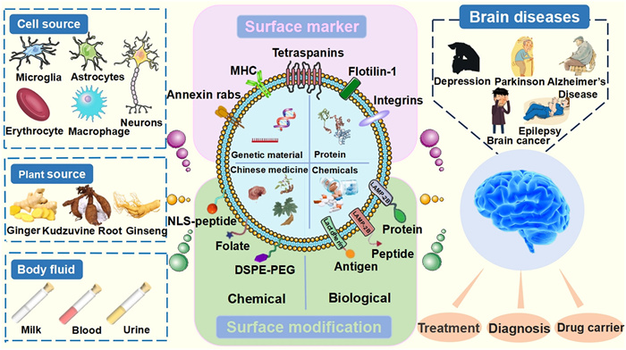
Figure 1.
Illustration of the origin, the surface modification of exosomes and their application in the diagnosis and treatment of brain diseases.

Perspective and application advancements of chemically modified exosomes as novel tools for precise targeting in brain diseases: A strategic appraisal
Xinying Wang , Yuhang Fan , Xiyao Dong , Han Wu , Qingxiang Guan


Brain diseases, including brain tumors, depression, epilepsy, Alzheimer’s disease (AD), and Parkinson’s disease (PD), rank among the most intricate and devastating conditions in the medical realm. The World Health Organization predicts brain diseases will surpass cancer as the second leading cause of death in 2040 [1]. A variety of potential therapeutic drugs have been developed. Nevertheless, these drugs still encounter a high failure rate in clinical applications. The difficult effective delivery of drugs into the brain and the constant risk of adverse effects within the central nervous system (CNS) represent significant obstacles that hinder the treatment of these diseases. Herein, we pay attention to two well-known yet extremely challenging issues: BBB crossing and targeted delivery [2].
Blood-brain barrier (BBB) poses a significant barrier and blocks the entry of approximately 99% of administered small molecules and biotherapeutic drugs [3,4]. The highly invasive techniques of direct intrathecal or intraventricular injection have significantly augmented treatment complexity, costs, and the risk of infection. The burgeoning development of nanotechnology offers a beacon of hope for cross-BBB transport. A diverse array of nanoparticles involving liposomes, micelles, dendrimers, carbon quantum dots, and various inorganic materials have emerged as effective noninvasive drug carriers for the treatment of brain diseases [5–7]. It is inevitably encumbered by concerns regarding biocompatibility, safety, and complexity. However, exosomes supplied with natural trans-BBB capabilities, higher biocompatibility, and low immunogenicity have garnered significant interest in the pharmaceutical field [8].
Exosomes, which are 30–150 nanometer-sized vesicles secreted by animal or plant cells, are widely present in various biological fluids, plant sap, and extracts. Exosomes from different sources exhibit distinct characteristics and application potentials. Exosomes derived from cells show high precision and efficiency in immune regulation and tissue repair due to their homology with the host and abundant functional molecules. Plant-derived exosomes are renowned for their excellent safety profiles. They can simultaneously deliver the unique active components of the plants, possessing both pharmacological activity and carrier functions. Milk-derived exosomes, due to their inherent acid-resistant and enzyme-resistant properties, are able to maintain structural integrity in the harsh environment of the gastrointestinal tract. In brain-targeted therapy, they also eliminate the inconvenience associated with traditional injection methods, significantly enhancing patients’ medication compliance [9]. Exosomes have gradually and extensively functioned as highly promising biomarkers for disease diagnosis, disease treatment, and drug delivery [10–13]. Additionally, a large number of applications have been carried out in the fields of biomedicine, pharmacy, neuroscience, etc. [14,15]. However, exosomes tend to exhibit non-specific global distribution upon entering the brain. They fail to accurately recognize and accumulate in specific lesion areas. This leads to insufficient effective concentration of therapeutic active ingredients at the target site, thereby reducing therapeutic efficacy. Apart from this, nonspecific interactions with normal brain tissues may also trigger potential biological side effects. Consequently, the clinical translation and application of unmodified exosomes in the precise treatment of brain diseases are severely limited. Against this backdrop, precise modification of exosomes has emerged as a key strategy to break through the aforementioned bottlenecks. By introducing chemical groups or molecular fragments with lesion-targeting recognition functions, exosomes can be endowed with efficient disease site-specific targeting capabilities. After penetrating the BBB, they are able to actively home in on and colonize the brain lesion microenvironment via mechanisms such as receptor-ligand specific binding. This significantly enhances both the efficiency of drug delivery to the target site and the precision of treatment. Furthermore, it effectively reduces the nonspecific accumulation of exosomes in healthy tissues, lowering the risk of side effects from a mechanistic perspective. This provides core technical support for advancing the translation of exosome carrier systems into clinical therapeutic applications for brain diseases.
The modification of exosomes includes chemical modification and genetic engineering modification. Chemical modification involves the attachment of target proteins to the exosome surface via chemical reactions using lipid-binding proteins, membrane-bound proteins, or lipid–lipid interactions. Genetic engineering is to genetically modify parental cells with an exosomal secretory capacity to express the target protein on the surface of their cell membranes, which in turn is stably displayed on the surface of exosomes secreted by the cell. Chemical methods allow for the precise regulation of the type, quantity, and spatial distribution of functional groups on the exosome surface. The site-specific conjugation techniques can accurately link ligands or antibodies, ensuring consistent targeting efficiency. Additionally, chemical modification strategies are characterized by remarkable flexibility and efficiency. Functional molecules can be selectively conjugated according to the specific requirements of different diseases, enabling seamless adaptation to diverse pathological conditions [16,17]. For instance, a study first combined a reactive dibenzylcyclooctyne group with amine-containing molecules on exosomes from mesenchymal stem cells (MSCs). The cyclo(Arg-Gly-Asp-D-Tyr-Lys) peptide [c(RGDyK)] was modified with the azide group on lysine, and the azide group on c(RGDyK) was then bonded to the amine group by click chemistry, thus modifying c(RGDyK) to the exosome surface. The c(RGDyK)-conjugated exosomes (cRGD-Exo) then targeted ischemic brain lesion regions in a mouse model of ischemia–reperfusion [18]. Chemical modification also circumvents ethical issues and technical limitations associated with genetic manipulation. It eliminates the risk of insertion mutations and immune responses caused by viral vectors in biological modification, thereby offering higher safety for clinical applications [19,20]. Despite the fact that chemical modification requires strict control to prevent exosomes from being exposed to excessive reagents, high temperature or pressure, etc., it remains the most delightful way of embellishment.
Herein, a comprehensive overview of the latest research progress related to exosomes for achieving precise delivery in the brain is performed to supplement the partial information or gaps in the literature. As shown in Fig. 1, beginning with the biogenesis and function of exosomes, the limitations posed by the challenges of the “BBB” and targeted delivery are detailed. It also emphasized the unique advantages and mechanisms of chemical modification in constructing highly heterogeneous brain disease-targeting delivery systems. Furthermore, a comprehensive summary of examples of targeting strategies from leading journals worldwide is analyzed to offer a more cutting-edge and authoritative perspective. Finally, special emphasis on the recent applications of exosomes in the diagnosis, early detection, and treatment of brain diseases is presented. Additionally, the insights into current barriers, challenges, and future directions for achieving effective treatment outcomes are also proposed. It is hoped to create a platform that stimulates researchers’ interest in exosomes for the treatment of brain diseases and promotes the use of exosomes to better serve human health.
The biogenesis and secretion of exosomes generated through a continuous process of membrane double invagination and the formation of intracellular vesicles are shown in Fig. 2 [21]. Extracellular components such as proteins, lipids, metabolites, small molecules, and ions enter cells along with cell surface proteins through the invagination of the plasma membrane [22]. Afterward, early sorting endosome (ESE) are formed on the inner side of the cell and then fuse with the endoplasmic reticulum, trans-Golgi network, or preexisting ESE to further mature into late sorting endosome (LSE) [23]. As the LSE membrane invaginates, its various proteins, nucleic acids, lipids, and other substances are randomly mixed and wrapped, forming multiple intracavitary vesicles (ILVs) with different contents and sizes [24]. The remaining portion of the LSE membrane invaginates and acts as the outer membrane, concentrating the formed ILVs in the LSE lumen. Namely, LSE further forms intracellular multivesicular bodies (MVBs). MVBs can fuse and degrade with autophagosomes or lysosomes. The degradation products can be recycled and utilized by cells. In addition, MVBs can also be transported to the plasma membrane through the cytoskeleton and microtubule networks. MVBs fuse with the plasma membrane and secrete ILVs outside cells with the help of MVBs docking proteins as exosomes through exocytosis [25].

Exosomes that are typically loaded with genetic and proteomic information act as component transporters through three mechanisms [26,27]. Firstly, the downstream signals are transmitted through the binding of specific ligands to the surface receptors of target cells. Secondly, release the contents by fusing with the target cell membrane. Thirdly, the target cells endocytose and release their contents, depending on lipid rafts, cage proteins, or caveolin proteins. The uptake of exosomes by receptor cells displays a higher cell specificity depending on the interaction between the surface molecules of receptor cells and the exosomes.
The BBB is a physiological barrier that is composed mainly of brain capillary endothelial cells, pericytes, astrocytes, and other cellular components. It is regulated by neurovascular units, limiting the free exchange of substances between blood and the brain tissue, thereby providing a nutrient supply and preventing the damage caused by potentially harmful substances in the brain environment. The unique filtering function of the BBB is related to two key characteristics of brain endothelial cells. As shown in Fig. 3, there is a tight junction complex between endothelial cells and highly restricted intercellular transport [28,29]. The tight junctions of endothelial cells reduce paracellular diffusion and limit transcellular activity, strictly controlling nonspecific inflow or outflow of the BBB. Second, intercellular adhesion connections regulate the permeability of the BBB in a dynamic opening and closing manner [30]. In addition, vascular endothelium-cadherin can control endothelial permeability, which is crucial for the maintenance of BBB function [31]. This tightly connected structure directly adapts to small blood vessel-sized molecules and gas exchange. In contrast, macromolecules are selectively transported by specific transport systems [32]. This selective behavior of the BBB prevents the penetration of most chemotherapeutic or biotech drugs, limiting their therapeutic effectiveness against brain diseases [33]. Another feature of the BBB is that endothelial cells have multiple strict intercellular transport pathways, including solute carriers, peptide and protein receptors, and ion transporter-mediated intercellular transport. However, window pore structures and phagocytic vesicles that can directly penetrate the BBB are lacking [34]. The vesicle density of endothelial cells in the CNS is much lower than that in the periphery, which limits the intercellular movement mediated by CNS vesicles [35]. Multiple strict restrictions on specific intercellular transport play crucial roles in effectively clearing brain endotoxins and ensuring the input of energy and nutrients [36].
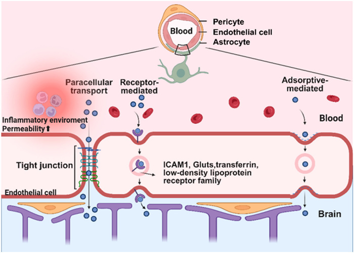
The mechanism of exosomes penetrating the BBB involves multiple pathways: First, BBB penetration through specific binding of exosome surface ligands to brain endothelial cell receptors is the main mechanism by which exosomes cross the BBB. Compared with other somatic cells, the BBB expresses more cellular receptors, including transferrin, the low-density lipoprotein receptor family, intracellular adhesion molecule 1 (ICAM1), and glucose receptors (GLUTs). Exosomes targeting these receptors enhance targeted drug delivery to the brain. For instance, the surface of macrophage-derived exosomes contains lymphocyte function-related antigen-1 inherited from the parent cells, which can interact with intercellular adhesion molecule-1 on the surface of microvascular endothelial cells to cross the BBB. Similarly, unmodified blood-derived exosomes also possess a natural brain-targeting ability, successfully delivering dopamine to the brain through the interaction of transferrin and transferrin receptors. A recent study suggested that CD46 is one of the main receptors for internalizing cancer cell-derived exosomes through the BBB into the brain and promoting tumor metastasis [37]. Similarly, exosomes derived from neural stem cells interact with endothelial cells via heparin sulfate proteoglycans (HSPGs) receptors for endocytosis, transporting cargo to the BBB [38]. Secondly, exosomes attach to the surface of the BBB endothelial cells through non-specific interactions such as surface charges and hydrophobic interactions. They are then internalized and subsequently released via exocytosis. Third, exosomes may also cross the barrier through the tight junction gaps between endothelial cells, diffusing beside the cells and transporting the carried biologically active substances (nucleic acids or proteins) to the brain tissue.
The permeability of exosomes to the BBB varies to some extent. For example, the upregulation of the inflammatory factor ICAM-1 increases the uptake of Mφ exosomes by the BBB. Pharmacokinetic data revealed that the accumulation of Mφ exosomes in the inflammatory region of the brain was 5.8 times greater than that in the healthy brain [39]. A study revealed that 10 different types of exosomes isolated from different species (mice and humans) and different cell types (tumor cells and healthy cells) have the ability to cross the BBB. However, the penetration rate of different types of exosomes varies greatly, even up to 10 times [40]. Lipopolysaccharide (LPS) can enhance the passage of most types of exosomes across the BBB. Wheat germ lectin regulates the transport of most exosomes. These data indicate that the ability of exosomes to cross the BBB is closely related to surface markers, size and shape, source cell type, and pathological conditions (inflammation or injury), etc.
Exosomes are naturally stable, double-membrane nanovesicles, and their structural integrity stems from three key molecular components. The protein framework, particularly cytoskeletal proteins and tetraspanins, forms their architectural basis, while the lipid bilayer reinforces membrane stability and even facilitates cellular signaling. Additionally, encapsulated nucleic acids contribute to functional diversity [41]. This sophisticated composition enables exosomes to maintain remarkable stability across various physiological conditions, making them exceptionally suitable as drug carriers for targeted delivery applications requiring sustained circulation and precise biodistribution. Exosome production remains limited, hindering clinical use. Strategies to boost yield include genetic engineering (e.g., CD9/TSPAN6 overexpression, STEAP3/syndecan-4/NadB coexpression), chemical induction (e.g., N-methyl dopamine, norepinephrine), environmental stimulation (hypoxia, glucose deprivation), and 3D culture systems. 3D culture and bioreactors can increase secretion 5–100-fold. Physical methods like ultrasound also enhance production 8–10-fold via calcium signaling. These approaches improve quantity while generally preserving exosome properties, though cell specificity and potential compositional changes require consideration [42].
The regulatory framework for exosome-based therapy remains in its early stages, with comprehensive guidelines for production, characterization, and quality control still under development. Exosomes are generally regarded to have low immunogenicity. The potential for immune reactions, particularly when using allogeneic exosomes, requires careful evaluation. It is vitally essential for rigorous long-term safety assessments and immunogenicity studies to identify and mitigate potential risks associated with clinic therapy [43]. Multiple recent clinical trials have shown mesenchymal stem cell-derived exosomes (MSCs-Exo) exhibit superior tolerability with no reported serious adverse effects in treating AD, treatment-resistant depression, and post-stroke dementia (NCT04388982, NCT04202770, NCT05326724). Targeted delivery strategies combining exosomes with focused ultrasound (FUS) and endogenous exosome induction therapies further reduce immunological risks, confirming the safety advantages of exosomes as non-invasive therapeutic carriers (NCT04202783). However, the larger-scale validation is still needed. Current data provide a reliable safety foundation for the clinical application of exosomes in neurological disorders.
Exosome-based therapies exhibit significant promise for targeted brain delivery, yet their clinical application faces stringent stability and safety challenges that should be addressed. Regulatory agencies, including the Food and Drug Administration (FDA) and European Medicines Agency (EMA), require well-defined guidelines and quality standards to ensure the development of safe and effective exosome therapeutics. A critical hurdle is the lack of precise, standardized methods for tracking exosome biodistribution, pharmacokinetics, and stability. Current labeling techniques, such as lipophilic dyes and donor-cell genetic modifications, suffer from limited accuracy and clinical relevance, underscoring the urgent need for non-invasive, compliant imaging technologies. Ethical concerns regarding exosome sourcing remain paramount. MSCs-Exo are favored for their safety and therapeutic potential, but tumor-derived exosomes carry oncogenic risks. Immunogenicity is another major consideration. Exosomes are generally biocompatible, yet surface engineering or foreign cargo loading may trigger immune responses, particularly with repeated dosing. Furthermore, the complete cargo composition and potential off-target effects should be rigorously evaluated, especially in long-term clinical applications.
It is a key obstacle arising from the unpredictable biodistribution of exosomes. Despite surface-targeting strategies, accumulation in off-target organs often occurs, reducing brain delivery efficiency. Additionally, scalable and standardized manufacturing remains a major bottleneck. Conventional isolation methods like ultracentrifugation are inefficient and poorly reproducible at large scales, while the absence of universally accepted quality control and potency assays continues to impede regulatory approval and clinical adoption.
Despite the considerable advantages of exosomes as brain drug delivery vehicles, including their low immunogenicity, biodegradability, and encapsulation of bioactive molecules to maintain their stability and ability to cross the BBB. The therapeutic potential of exosomes can still be achieved through a variety of techniques, such as chemical modification or genetic modification, to enhance target recognition and biological distribution, thereby reducing toxicity and adverse reactions and maximizing therapeutic effectiveness. Chemical modification refers to the use of chemical reactions involving lipid-binding proteins, membrane-binding proteins, or lipid interactions to bind peptides, proteins, lipids, ligands, or polymers to exosomes for brain-targeted delivery. Compared with gene modification, the chemical modification of exosomes shows the significant advantages of simple operation and no need for complex gene editing tools, which greatly reduces the technical threshold. Moreover, the modification process avoids the introduction of foreign genes, greatly reducing the risk of immune response and potential safety hazards. Table 1 [44–57] lists the latest research progress on the use of various chemically modified exosomes that cross the BBB for the treatment of brain diseases.

 DownLoad:
CSV
DownLoad:
CSV
| Targeting ligand | Targeting mechanism | Brain ailment | Exosome origin | Route of administration | Payload type | Loading method | Ref. |
| Ginsenoside Rg3 | Targeted to the GLUT1 on BBB and GBM cells | GBM | GL261 | Intravenous | arsenic trioxide (Arsenic Trioxide) and chlorin e6 | Sonication and incubation (room temperature, 1.5 h) | [44] |
| Sialic acid analogues with N-acyl side chains | Targeted to the microglia in CNS | MS | RAW264.7 | Intranasal | Resveratrol | Sonication and incubation (37 ℃, 1 h) | [45] |
| DSPE-PEG-RVG | Targeted to DA neurons | PD | Pueraria lobata | Intranasal | — | — | [46] |
| DSPE-PEG-HSSP | Targeted to the HMOX-1 overexpressed in TMZ resistant glioma cells | GBM | BMSC | Intravenous | TMZ and STAT3 targeted siRNA | Sonication | [47] |
| RGDyK peptide | Targeted to the brain tumor cell surface receptor integrins | GBM | ReNcell VM | Intravenous | PD-L1 siRNA | Incubation | [48] |
| Tf-SPION nanoparticles | Placed a magnet in rats’ brain and targeted to the brain by magnetism | Many kinds of brain disease | MSCs | Intravenous | Doxorubicin | Incubation (4 ℃, 4 h and 37 ℃, 30 min) | [49] |
| DSPE-PEG-T7 and CAR | Targeted to the LRP1 in brain | HER2+ BCBM | NK-92 | Intravenous | PEG-TK-Ce6@RSL3 | Coextrusion | [50] |
| sicPLA2 | Targeted to the mitochondrion in GBM cells | GBM | Blood | Intravenous | metformin (Glucophage) | Electroporation | [51] |
| RVG29 peptide | Targeted to the nAchR expressed in neuronal cells | TBI | Microglia cell | Intravenous | NR2B9c | Sonication | [52] |
| ANG peptide | Targeted to the LRP1 expressed in brain microvascular endothelial cells | CNS-TB | BMSC | Intravenous | Rifampin | Electroporation and incubation (37 ℃, 1 h) | [53] |
| Ang-2 and CD133 targeted peptide | Targeted to the receptor expressed in the tumor cells | GBM | U251 | Intravenous | Temozolomide and doxorubicin | Sonication and incubation (37 ℃, 1 h) | [54] |
| Antibody against GAP43 | Targeted to the GAP43 expressed in damaged neurons | IRI | Blood | Intravenous | Quercetin | Incubation under ultrasonic shaking in an ice water | [55] |
| CD22 antigen fragments | Targeted to the CD22 antigen expressed on the membrane of malignant mature B lymphocytes | PCNSL | 293T | Intravenous | Doxorubicin | Incubation (37 ℃, 1 h) | [56] |
| iRGD peptide | Targeted to αvβ3 integrins and neuropilin-1 receptors in neovasculature and glioblastoma | GBM | U87 | Intravenous | HCQ@ZnS nanoparticles | Thin film hydration followed by membrane extrusion | [57] |
Chemical modifications can be divided into covalent modifications and noncovalent modifications. Click chemistry is the core technology of covalent modification [58]. Click chemical reactions are highly efficient and can be performed well in organic solvents and aqueous buffers with short reaction times. In addition, noncovalent modifications include electrostatic interactions, ligand-receptor interactions, and hydrophobic interactions.
Covalent modification techniques hold significant value in the engineering of exosomes. Currently, covalent modification methods primarily rely on click chemistry and include the following three types: Copper-catalyzed azide-alkyne cycloaddition (CuAAC), strain-promoted copper-free azide-alkyne cycloaddition (SPAAC), and other bioorthogonal click chemistry reactions.
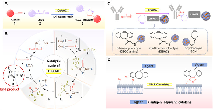
The CuAAC reaction enables rapid and specific conjugation to preserve exosomal cellular uptake capabilities [59]. Notably, this reaction demonstrates exceptional compatibility, proceeding efficiently in aqueous and organic environments. The proposed mechanism for CuAAC is illustrated in Figs. 4A and B. The catalytic cycle begins with the formation of copper acetylide intermediate Ⅰ from the initial Cu-alkyne complex. Subsequent coordination expansion and reaction with azide 2 yields intermediate Ⅱ, where nucleophilic attack by the terminal N-3 at the C-4 position generates a stabilized spatial configuration. This transforms intermediate Ⅱ into intermediate Ⅲ, then undergoes rearrangement via a six-membered transition state. During this stage, N-1′s lone pair attacks C-5 within the metallacycle, ultimately forming the more rigid and thermodynamically favorable cyclic structure Ⅳ [60].
Among all click chemistry reactions, CuAAC remains the most widely utilized. Recent advances in exosome engineering have leveraged click chemistry for precise functionalization, enabling enhanced targeting and therapeutic delivery. Glioma-targeting exosomes were engineered by conjugating the exosomal membrane with a neuropilin-1-binding peptide (RGE) via click chemistry. Through EDC/NHS-mediated pre-activation of phosphatidylethanolamine to introduce alkyne groups, which optimized conjugation efficiency. These modified exosomes exhibited enhanced BBB penetration and prolonged tumor retention, demonstrating their potential for image-guided glioblastoma treatment [61]. In a similar approach, CuAAC click chemistry was used to functionalize exosomes. Alkynyl-modified COFs were conjugated with azide-tagged DNA through Cu(Ⅰ)-catalyzed cycloaddition to create DNA-COF complexes. These complexes were loaded with methylene blue (MB) as signal reporters and functionalized with cholesterol-DNA (Chol-DNA) for exosome membrane targeting. Combined with CD63 aptamer-modified electrodes, this approach established a sandwich-type detection system. The method demonstrates stable covalent conjugation, efficient signal amplification, and programmable target recognition through interchangeable DNA sequences, offering a sensitive and specific platform for exosome detection [62]. These studies exemplify how click chemistry-mediated exosome engineering can enhance the precision of drug delivery, enabling both diagnostic and therapeutic applications in challenging disease contexts.
However, its biomedical applications are significantly limited by the intrinsic cytotoxicity of copper ions [63]. Cu(Ⅰ) easily chelates with natural amino acid residues, thereby disrupting the structure and function of proteins. Cu(Ⅰ) can also induce the formation of reactive oxygen species (ROS) [64]. In vitro studies demonstrate that Escherichia coli subjected to surface CuAAC labeling lose replicative capacity even when returned to nutrient-rich medium [65]. Notably, copper concentrations exceeding micromolar levels induce severe cellular damage [66] and eventual apoptosis [63]. Furthermore, in the presence of Cu(Ⅱ), elevated triazole product concentrations may destabilize biomolecular complexes, such as causing cowpea mosaic virus (CPMV) capsid dissociation [67]. The inherent cytotoxicity associated with copper catalysts in CuAAC reactions has prompted the development of ligand-optimized systems (THPTA, BTTAA) to mitigate copper-induced toxicity. However, empirical evidence suggests these modifications provide only partial remediation, as residual bioincompatibility persists even with optimized chelation protocols [68].
The Bertozzi group pioneered a solution to Cu(Ⅰ) ion toxicity through the development of SPAAC, a copper-free bioorthogonal reaction designed to circumvent the limitations of traditional CuAAC chemistry [69]. Especially, copper-free click chemistry offers a powerful strategy for precisely functionalizing plasma extracellular vesicle membranes, allowing exosomes to be tailored for specific therapeutic or diagnostic applications. This precise membrane engineering further enhances the stability of exosomes, improves targeting efficiency, and optimizes cargo delivery sites, ultimately unlocking their full potential as versatile and high-performance vehicles for drug delivery or diagnostic tools. The evolution of SPAAC reactivity has been primarily achieved through the rational design of cycloalkyne architectures. Its structural parameters, including ring strain, lipophilicity, and molecular geometry, collectively determine reaction dynamics [70]. This structure-activity relationship has motivated the development of strained alkynes such as dibenzocyclooctyne (DBCO) derivatives, dibenzoazacyclooctyne (DIBAC), and bicyclononyne (BCN), as illustrated in Fig. 4C [71]. The strategically incorporated sp2-hybridized carbons induce sufficient ring strain to achieve rapid, selective bioorthogonal conjugation without the cytotoxicity concerns inherent to copper-catalyzed alternatives [60].
SPAAC has been increasingly recognized for its copper-free nature, low toxicity, and superior biocompatibility, making it an attractive alternative in biomedical applications. A study developed a copper-free click chemistry-based method for fluorescently labeling exosomes, enabling efficient tracking of their uptake in pancreatic cancer (PC) models. Using design of experiments (DoE), the authors identified time- and dose-dependent uptake kinetics, with PC-derived exosomes (PANC-1) showing preferential internalization by homologous PC cells, particularly at longer incubation times and higher doses. In vivo biodistribution studies further demonstrated selective accumulation of PANC-1 exosomes in pancreatic tumors over more vascularized melanoma tumors, suggesting intrinsic homing properties. The work provides a reliable labeling strategy and mechanistic insights for optimizing exosome-based drug delivery in PC therapy [72]. Azide-modified exosomes (N3-Exo) were covalently conjugated with DBCO–Cy5 (a dibenzocyclooctyne-bearing fluorescent dye) via SPAAC, forming stable triazole linkages. This catalyst-free reaction eliminated copper ion toxicity while preserving exosomal integrity [45].
CuAAC offers more rapid reaction kinetics (~1–10 L mol−1 s−1) and higher labeling efficiency, but its reliance on cytotoxic Cu(Ⅰ) limits its in vivo use due to copper-induced oxidative stress and potential damage to exosome membranes. In contrast, copper-free click chemistry eliminates metal toxicity, enabling safer in vivo applications, albeit with slower reaction rates (~0.1–1 L mol−1 s−1). It is suitable for CuAAC in vitro exosome engineering as optimized with copper chelators. In contrast, the copper-free method is preferred for clinical application, as evidenced by its compatibility with FDA-approved reagents and superior retention of exosome integrity [66,73]. The reaction mechanisms and conditions are compared in Table 2 [69,74].
DownLoad:
CSV
| Parameter | CuAAC | Copper-free (e.g., SPAAC) |
| Reactive groups | Azide (−N3) + terminal alkyne (−C≡CH) | Azide (−N3) + cyclooctyne (e.g., DBCO) |
| Catalyst | Cu(Ⅰ) required (e.g., CuSO4/sodium ascorbate) | Metal-free (spontaneous) |
| Reaction rate (k, L mol−1 s–1) | ~1–10 (Cu-dependent) | ~0.1–1 (strain-promoted) |
| Reaction conditions | RT/37 ℃, pH 7–8 | RT/37 ℃, pH 7–9 |
| Bioorthogonality | Moderate (cellular Cu interference) | High (no metal interference) |
Emerging therapeutic paradigms require precision strategies for spatially and temporally controlled drug activation at pathological sites. The bioorthogonal chemistry platform has appeared as a transformative solution, enabling “on-demand” therapeutic release through reactions that maintain both biocompatibility and orthogonality to native biological processes [75]. This approach offers two fundamental advantages involving exquisite selectivity for synthetic reactants over endogenous biomolecules and preserved functionality in physiological environments without perturbing innate biochemistry [76,77]. The characteristics position bioorthogonal systems as versatile tools for targeted intervention across diverse disease states where localized therapeutic action is paramount [78].
First, introduce specific bioorthogonal groups (such as azide groups, N3) into the target biomolecules through chemical, metabolic, or genetic means. Subsequently, introduce exogenous complementary probe molecules with functional modifications, such as DBCO and BCN [79]. Under physiological conditions, these two groups achieve covalent linkage through an efficient [3 + 2] cycloaddition reaction—azide (N3) and alkyne (C≡C) form a stable 1,2,3-triazole bond under the action of a copper catalyst (Cu+) (CuAAC reaction). The bioorthogonality of this reaction stems from the fact that azide and alkyne have almost no reactivity in natural biological systems but can selectively combine under the catalysis of exogenous Cu+, thus enabling the precise labeling and functionalization of target molecules (Fig. 4D).
Bioorthogonal reactions offer some unique advantages in biomarking applications, with their high selectivity, minimal interference, and biocompatibility making them ideal tools for in vivo labeling. In exosome labeling studies, metabolic glycoengineering (such as Ac4ManNAz) enables efficient incorporation of azide groups (-N3) into cell surface glycoproteins, followed by SPAAC to achieve specific covalent conjugation with DBCO–Cy5, representing a mainstream approach for precise exosome tagging. Some studies employ a one-step labeling strategy, preserving the native characteristics and functionalities of exosomes as well as overcoming the toxicity, inefficiency, and background interference associated with conventional membrane dyes [80]. Alternatively, isolated exosomes are subsequently labeled via copper-free click chemistry by reacting the azide-modified glycans with DBCO-conjugated fluorescent probes. This bioorthogonal labeling is combined with epithelial cell adhesion molecule (EpCAM)-specific fluorescent aptamers to enable fluorescence resonance energy transfer (FRET)-based imaging, allowing high-resolution visualization of both EpCAM localization and its glycosylation status [81]. Collectively, this demonstrates that bioorthogonal reactions enable efficient and non-disruptive exosome labeling, providing a robust methodology for nanocarrier tracing, drug delivery monitoring, and intercellular communication studies. Another reported strategy involves a more complex active chemical conjugation approach. In this method, targeting peptides (e.g., RGD) and thiol groups are first introduced into the exosomal membrane via donor cell membrane modification. Then, gold nanorods (AuNRs) are conjugated to the thiol groups through Au–S covalent bonds, while chemotherapeutic drugs are simultaneously loaded, resulting in multifunctional engineered exosomes termed FA-AuNR@RGD-DOX-Exos. This system not only enables dual-ligand active targeting but also utilizes the photothermal effect of AuNRs to achieve on-demand drug release, demonstrating a synergistic combination of photothermal therapy and chemotherapy [82]. Additionally, an indirect regulatory strategy has been developed, in which macrophages are loaded with polymer nanoparticles containing o-nitrobenzyl groups (UV-responsive) and upconverting nanoparticles (UCNPs). Under 980 nm near-infrared light excitation, the UCNPs emit UV light intracellularly, triggering the depolymerization of the nanoparticles and stimulating the macrophages to secrete large quantities of drug-loaded exosomes. This process enables light-controlled drug release and enhances deep tumor penetration [83].
The non-covalent modification of exosomes refers to the attachment of functional molecules to the surface or interior of exosomes through non-covalent bonding. It encompasses various strategies, including electrostatic and hydrophobic interactions as well as ligand-receptor interactions. The modified exosome via electrostatic interactions enables efficient tissue-specific delivery by leveraging the negative surface charge of exosomes and the positive charge of modifying molecules. The cationized pullulan modified with spermine acquires positive charges and electrostatically binds to the negatively charged exosome membranes, facilitating specific targeting to hepatocyte asialoglycoprotein receptors (ASGPR) and significantly enhancing anti-inflammatory effects [84]. Similarly, RVG peptide conjugation via the 1,2-dioleoyl-sn‑glycero-3-phosphoethanolamine-N-hydroxysuccinimide (DOPE-NHS) linker enables exosomes to cross the blood-brain barrier and target neurons, thereby improving Aβ clearance in AD models [85]. In cartilage delivery, cationic peptides or ε-polylysine neutralize the exosome surface charges, overcoming electrostatic repulsion with negatively charged glycosaminoglycans (GAGs) and enabling deep tissue penetration through Donnan equilibrium effects, thereby facilitating efficient nucleic acid delivery in osteoarthritis treatment [86]. For ocular applications, arginine-rich CPC modifications optimize exosome charge properties, enhancing corneal penetration and vitreous diffusion to achieve 3-fold higher mRNA transfection efficiency in the retina without compromising tissue safety [87]. Collectively, these studies suggest precise modulation of electrostatic interactions can tailor exosome delivery efficiency to specific tissue microenvironments, offering innovative therapeutic solutions for diverse diseases.
Hydrophobic interactions in non-covalent modification of exosomes supply an efficient and mild loading strategy for drug delivery, with applications mainly demonstrated in two aspects. On one hand, drugs can achieve efficient loading through the hydrophobic interaction between their hydrophobic groups and the membrane lipid layer of genetically engineered exosomes. AMO181a-chol loaded onto exosome surfaces offers high loading efficiency of 24.75% by leveraging hydrophobic interactions between cholesterol’s hydrophobic groups and the exosomal lipid bilayer [88]. The result demonstrates significant gene silencing and therapeutic effects in cerebral ischemia models. On the other hand, hydrophobic interactions are also applicable for small-molecule drug loading. Curcumin, a hydrophobic drug, can achieve ultra-high drug loading of 106–107 molecules/vesicle through physical adsorption, which might affect exosomal physicochemical properties. In contrast, cholesterol-anchored hydrophilic ligands, CMR19-chol, enable more controlled drug loading (104–105 molecules/vesicle) via membrane insertion while better preserving exosomal functionality [89]. These studies systematically compare the advantages and disadvantages of different loading approaches through advanced characterization techniques, providing critical guidance for optimizing the design of exosomal drug delivery systems. In the development of photoresponsive chemical modification, one approach involves hydrophobic interactions that utilize the hydrophobic nature of the photosensitizer chlorin e6 (Ce6). Through incubation, Ce6 is directly loaded into the lipid bilayer of milk-derived exosomes (mExos), successfully constructing Ce6@mExo complexes. This strategy maximally preserves the natural membrane structure and targeting functions of the exosomes, enabling them to efficiently cross biological barriers and achieve oral delivery to the brain for photodynamic therapy. Gao et al. further advanced the encapsulation strategy by using a membrane fusion technique to combine Ce6-loaded liposomes with exosomes derived from γδ-T cells, creating hybrid exosomes. Under 650 nm red light irradiation, these hybrid exosomes generate ROS, achieving a synergistic effect between photodynamic therapy and the inherent immune activity of the exosomes [90].
In the non-covalent modification of exosomes, ligand-receptor interactions are widely employed to achieve targeted delivery and efficient drug loading. The molecular conjugate of bovine lactoferrin with poly-L-lysine electrostatically interacts with negatively charged siRNA while leveraging the specific ligand-receptor interaction between lactoferrin and glyceraldehyde-3-phosphate dehydrogenase (GAPDH) on exosome membranes, enabling the natural loading of siRNA [91]. An innovative ligand-receptor recognition strategy for precisely modifying blood exosomes has been successfully designed. The key breakthrough involves conjugating transferrin (Tf) with superparamagnetic nanoparticles (M-Tf), specifically anchoring to exosome membranes through native Tf-TfR interactions, creating magnetically targeted SMNC-Exo [92]. Meanwhile, Tf can be utilized as a bridging molecule to achieve magnetic modification of exosomes. Specifically, polyethylene glycol-coated superparamagnetic iron oxide nanoparticles (PEG-SPIONs) are first synthesized and then conjugated with transferrin to form Tf-SPIONs. By leveraging the abundant transferrin receptors on the exosomal surface, Tf-SPIONs can efficiently bind to exosomes, resulting in the formation of magnetic exosomes (Ex-SPIONs). This complex exhibits both superparamagnetism and the ability to cross the blood-brain barrier, enabling targeted delivery to the brain under an external magnetic field [49]. On the whole, the natural ligand-receptor pairing ensured precise and stable modification while enabling functional expansion.
In summary, non-covalent modification of exosomes requires no disruption of the membrane structure. It includes electrostatic interactions, hydrophobic interactions, and ligand-receptor interactions. These three modification approaches each exhibit distinct advantages, thus offering a mild modification pathway for the application of exosomes in fields including drug delivery and targeted therapy.
The analysis indicates that covalent modifications (e.g., CuAAC, SPAAC) offer advantages such as high efficiency and strong stability, but their toxicity highly depends on the reaction type (CuAAC exhibits significant toxicity due to copper catalysts, while SPAAC demonstrates good biocompatibility). In contrast, non-covalent modifications are recognized for their operational simplicity, mild conditions, and low toxicity, though they suffer from relatively poor stability and variable efficiency. Ultimately, the choice of strategy depends on balancing stability, biosafety, and operational simplicity according to the specific application scenario.
Due to the high biological relevance of exosomes, their contents can reflect the microenvironment of their original cells and ongoing physiological processes, providing relevant information about the nature, extent, and affected sites of brain diseases. Accordingly, exosomes emerge as key advantages over traditional peripheral blood biomarkers. Furthermore, exosomes are minimally invasive liquid biopsy samples that are easy to capture and enrich. They can be used for comprehensive multiparameter detection and longitudinal tracking of disease progression and have good clinical diagnostic value. Some of the deficiencies in peripheral blood biomarkers can be remedied by the detection and diagnosis of centrally derived exosomes [93]. In addition, exosome treatment has received much attention [94]. The ability of exosomes to naturally cross the BBB and their low immunogenicity encourage them to be excellent therapeutic agents for the efficient delivery of various functional molecules [95,96]. The engineered exosomes for the treatment of brain diseases have been reported and the delivery mechanism of therapeutic exosomes has been extensively investigated. Currently, exosomes are extensively used to treat various brain diseases [97–100]. The diagnosis and treatment of depression are illustrated in Fig. 5.
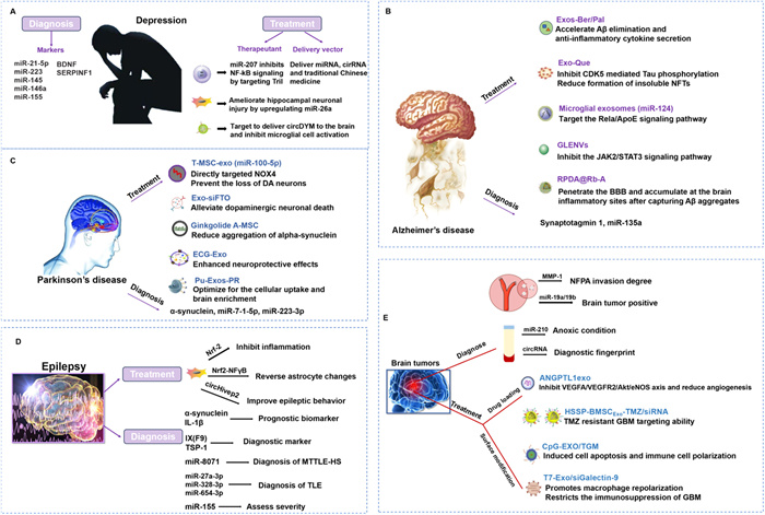
Depression is a chronic mental disorder characterized by brain lesions. Its pathogenesis mainly involves changes in synaptic plasticity, oxidative stress, neuroinflammation, the gut microbiota, and changes in neurotransmitter metabolism. Currently, the main treatment methods are oral antidepressants supplemented with psychotherapy or physical therapy. However, common oral antidepressants still only emerge therapeutic effects on some patients. In addition, material exchange and information transmission from the periphery to the brain are hindered by the BBB. This phenomenon poses challenges for the treatment of this brain disease. As important carriers of intercellular communication, exosomes can alter the functional status of receptor cells through the transfer of miRNAs and mRNAs and play important roles in the occurrence and development of depression [101–103]. The diagnosis and treatment of depression are illustrated in Fig. 5A.
The diagnosis of depression relies primarily on clinical symptomatology and lacks objective biochemical indicators. Therefore, exploring specific molecular markers is highly important for the early diagnosis and treatment evaluation of depression. Since exosomes can cross the BBB, the circulating of exosomes in the blood may be involved in pathophysiological mechanisms of brain diseases. A previous case-control study revealed the levels of several biomarkers related to the neuronal status (such as tumor necrosis factor receptor 1 (TNFR1) and synaptophysin (SYP)) in peripheral blood were significantly correlated with the exosome surface marker CD81 [104]. These findings indirectly indicate neuron-derived exosomes can enter the peripheral blood circulation through the BBB and reflect the functional status of the CNS. Although this study did not directly obtain neuron-derived exosomes, it provides a new perspective for the study of brain-derived exosomes in patients with depression. Subsequently, Gómez-Molina conducts a mass spectrometry proteomic analysis on serum exosomes from depressed rats subjected to restraint stress and discovers the stress-specific protein aldehyde C. The results are confirmed to originate from the brain through fluorescence labeling [105]. This finding also suggests the brain can transmit disease states and disease-related protein molecules to the peripheral blood circulation through exosomes.
At present, research is dedicated to analyzing the pathophysiological differences between depressed patients and healthy individuals via nonbrain tissue biopsy. Noninvasive methods of exosome acquisition and composition analysis have become a new direction in the study of molecular markers of depression. Among the various types of molecular markers, noncoding RNAs, mainly miRNAs, have become powerful tools for diagnosing depression in patients and predicting drug treatment efficacy. Studies have shown that the levels of miR-21–5p, miR-223, miR-145, miR-146a, and miR-155 in the serum exosomes of patients with depression increase significantly after antidepressant drug treatment and may be potential biomarkers for predicting the efficacy of antidepressant drug treatment [106]. In addition to RNA, protein molecular markers carried by exosomes are also emerging. It is reported that the content of brain-derived neurotrophic factor (BDNF) in the serum exosomes of depressed patients was lower than that in healthy controls [107]. A significant decrease in SERPINF1 expression in the peripheral blood exosomes of patients with depression and SERPINF1 expression is shown to be regulated by miR-186–5p [108]. This finding was also confirmed in mice subjected to chronic unpredictable mild stress. These results also suggest that SERPINF1 in peripheral exosomes can serve as a reliable biomarker for the occurrence and development of depression, and miR-186–5p can also serve as a potential therapeutic target for depression. In summary, exosomes and their derivatives are closely related to the occurrence and development of depression and can serve as important components for the diagnosis and treatment of depression.
A study discovered that miR-207 derived from natural killer (NK) cells can alleviate depressive symptoms in mice by targeting Tril to inhibit NF-κB signaling in astrocytes. This suggests that miRNAs in exosomes have great potential value in the treatment of depression [109]. With the development of stem cell regenerative medicine in recent years, stem cell-derived exosomes have also received much more attention in the treatment of depression. Exosomes derived from bone marrow mesenchymal stem cells (BMSCs) can ameliorate hippocampal neuronal damage in depressed rats by upregulating miR-26a. This finding provides a new direction for the study of stem cell-derived exosomes in the field of depression [110]. Exosomes themselves can act as drug carriers to enter the CNS to exert antidepressant effects and also alleviate depressive symptoms by transporting active substances. Microglia-derived exosomes carrying miR-146a-5p play important roles in the regulation of neurogenesis in depressed rats and may be used to develop new antidepressant drugs [111]. Engineered RVG-extracellular vesicles (RVG-EVs) can be targeted to deliver circDYM to the mouse brain and inhibit microglial cell activation, significantly improving depressive-like behaviors in mice [112].
AD is the most common neurodegenerative disease and is accompanied by impairments in memory, judgment, and attention span. To date, drugs, e.g., donepezil, rivastigmine, and galantamine, that target cholinergic or glutamatergic neurotransmission are the main strategies used to treat AD. However, these drugs only relieve symptoms and fail to serve a curative effect. This is ascribed to affecting only the consequences of AD rather than the cause [113]. Currently, exosomes appear to play a relevant role in neurodegenerative disease pathogenesis [114]. Exosomes can carry therapeutic proteins and help the disease spread in the brain, and they appear to have the ability to induce astrocyte apoptosis, which impairs neuronal function during disease progression. Besides, exosome-based drug delivery is attracting attention to accelerate anti-AD activity. Moreover, it is more sensitive for exosomes to be diagnosis indicators and reliable to monitor the progression of disease. The diagnosis and treatment of AD are illustrated in Fig. 5B.
Exosomes are an emerging candidate for biomarkers of AD for they can pass through the BBB and can be detected in peripheral blood or in cerebrospinal fluid. miRNA sequencing is performed on exosomes isolated from the plasma of AD patients, and some miRNAs are differentially expressed between AD patients and healthy individuals. The miRNAs in whole blood, serum, plasma, exosomes, cerebrospinal fluid, and extracellular fluid can serve as potential diagnostic and therapeutic biomarkers for AD. It is demonstrated that 22 and 21 miRNAs in AD plasma exosomes are upregulated and downregulated, respectively [115]. Another study found that microRNA-135a in ABCA1-labeled exosome is a serum biomarker candidate for AD [116]. In addition to miRNA differences, lncRNAs also show higher accuracy in identifying AD, which can be considered promising biomarkers for disease diagnosis through a meta-analysis [117]. There is proof indicating that proteins can be biomarkers for AD. The concentration of protruding binding protein 1 in the neuron-derived exosomes of AD patients is closely related to the concentration of synaptic binding protein 1 in cerebrospinal fluid. The finding makes the early diagnosis of AD possible [118].
Numerous studies have validated exosomes as carriers to treat AD. A study suggested that the combination of berberine and palmatine (Ber/Pal) ameliorated Aβ pathological symptoms, achieved through promoting phagocytosis of Aβ plaques by microglia and inhibiting neuroinflammation. The data also indicated defects in BBB permeability via absorption, distribution, metabolism, excretion, and toxicity (ADMET) computational prediction. To improve the BBB permeation and targeting ability, microglia-derived exosomes were utilized as drug carriers. Meanwhile, the Ber/Pal coloaded microglia-derived exosomes (Exos-Ber/Pal) system could enhance the therapeutic effects [119]. The exosomes loaded with quercetin (Exo-Que) relieved the symptoms of AD by inhibiting cyclin-dependent kinase 5-mediated Tau phosphorylation and lessening the formation of insoluble neurofibrillary tangles [120]. The miRNAs in exosomes constitute important elements involved in intercellular communication [121]. The exosomal miR-124 boosts cognitive function and alleviates neurodegeneration by targeting the Rela/ApoE signaling pathway after repetitive mild traumatic brain injury (rmTBI) [122]. Another study has designed a new drug delivery system to address the low permeability of BBB and the lack of selectivity for the action sites of AD. By hybridizing the membranes of exosomes from brain microvascular endothelial cells with those of macrophage exosomes and combining dopamine nanoparticles, resveratrol, and Aβ-targeting aptamers, an engineered exosome with multiple targeting capabilities (RPDA@Rb-A) was constructed. RPDA@Rb-A intervened in the clearance of Aβ and regulated the dysfunction of microglia. Based on the homing effect of exosomes from brain microvascular endothelial cells and the natural inflammatory targeting ability of macrophage exosomes, RPDA@Rb-A can easily penetrate the BBB and accumulate at the brain inflammatory sites after capturing Aβ aggregates [123]. In addition, plant exosomes also play a significant role in the treatment of AD. Intranasal administration allowed G. lucidum contained exosome-like nanovesicles (GLENVs) to penetrate the BBB to exert their effects directly. The GLENVs treatment reduced Aβ deposition in the cortex and hippocampus of AD mice, overactivated microglia, reactive astrocytes, and pro-inflammatory factors, and inhibited the Janus kinase 2 (JAK2)/Signal transducer and activator of transcription 3 (STAT3) signaling pathway. Therefore, GLENVs may be a promising candidate for AD treatment [124].
PD is the second most common neurodegenerative disease and is characterized by a tremor that is maximal at rest, retropulsion, rigidity, a stooped posture, slowness of voluntary movements, and a masklike expression [125]. Current treatments involve the administration of drugs, including dopamine precursors, activated dopamine agonists, and drugs modulating dopamine metabolism. However, these drugs can’t prevent ongoing dopaminergic damage because of their poor ability to cross the BBB [126]. Moreover, an asymptomatic phase occurs during the progression of PD, and clinical symptoms do not appear until 70%–80% of the dopaminergic terminus in the striatum and 50% of the dopaminergic neurons in the substantia nigra are lost, which highlights the importance of early detection and intervention. Considering the advantages of BBB penetrability, metabolic stability, and target specificity, exosomes have been successfully loaded with enzymes, mRNA, and small interfering RNA for PD treatment and illustrate the desired therapeutic effect. Exosomes from cerebrospinal fluid, plasma, serum, saliva, and urine are valuable biomarkers for PD diagnosis. The diagnosis and treatment of PD are illustrated in Fig. 5C.
Exosomes are differentially expressed in patients with PD. Different types and contents of exosomes can be used as biomarkers for PD. According to a meta-analysis, α-synuclein (α-Syn) in L1CAM exosomes derived from blood can be used for PD diagnosis [127]. α-Syn in blood exosomes immunoprecipitated using antibodies against neuronal and oligodendroglial markers can distinguish PD from multiple system atrophy [128]. It is possible for miR-7–1–5p and miR-223–3p in exosomes to be biomarkers for PD. Its concentrations are correlated with the levodopa equivalent daily dosage (LEDD) [129]. In addition to α-Syn and GCase, long noncoding RNAs (lncRNAs) in L1CAM exosomes have also been reported to be potential biomarkers [130].
Exosomes delivering medicinal compounds, siRNAs, and proteins to the brain hold therapeutic potential for PD. Bioinformatics analysis and the luciferase report showed that the NADPH oxidase 4 (Nox4) gene was a target for miR100–5p, which acts on Nox4 to regulate Nox4 expression. miR-100–5p-enriched T-MSCs-Exo protect against the loss of dopaminergic (DA) neurons, maintain nigro-striatal system function, ameliorate motor deficits, and reduce oxidative stress through the Nox4-ROS-Nrf2 axis, suggesting broad neuroprotective properties of T-MSCs-Exo [131]. The Exo-siFTO system, encapsulating the m6A demethylase fat mass and obesity-associated protein (FTO)-targeted siRNAs into MSC-Exos is developed. The results reveal MSC-Exo delivery of si-FTO can alleviate dopaminergic neuronal death in individuals with PD [132]. In addition to RNA exosome therapy, exosomes carrying natural compounds are also designed. A ginkgolide A–MSCs system is developed. It is reported exosomes derived from the system cause the diminution in α-Syn levels, which may be a potential therapeutic candidate for PD. In addition, epicatechin gallate (ECG) was loaded into Exo and its neuroprotective effects were detected in vitro. It was speculated that ECG-Exo exerted neuroprotective effects against PD mainly through antiapoptosis and antimitophagy. The neuroprotective effects of ECG were much improved when loaded into Exo. In vivo experiments to verify whether Exo can deliver drugs precisely to the target site will be conducted in the future [133]. BMSC exosomes containing glioma-associated oncogene homolog 1 (Gli1) alleviate inflammatory damage and apoptosis in PD by directly binding to the Sp1 promoter to inhibit the LRRK2 signaling pathway. Moreover, the expression of Gli1 in BMSC-derived exosomes may be an effective therapeutic strategy for PD [134]. Another study showed that Pueraria lobata derived exosomes (Pu-Exos) engineered with DSPE-PEG-RVG could be further optimized for the cellular uptake and brain enrichment in vivo, therefore excellently promoting the survival of dopaminergic neurons [46].
Epilepsy is one of the most intractable diseases of the CNS. Its incidence is relatively higher in neonates and elderly individuals. It is caused by a variety of factors, including trauma and disorders. The symptoms usually present as unprovoked seizures. It is often thought to be related to sudden abnormal discharges between neurons in the brain. The prognosis of patients with epilepsy is poor, and drug treatment options are limited. Currently, it is unavailable to obtain a better treatment method in clinical practice. In recent years, a series of novel therapies, e.g., gene therapy, cell therapy, and exosome therapy, have been applied to the treatment of epilepsy. Some progress has been made [135]. The diagnosis and treatment of epilepsy are illustrated in Fig. 5D.
In the determination of biomarkers of epilepsy, exosomes have the same potential value. A study revealed that the α-Syn and interleukin-1β (IL-1β) levels in serum exosomes from children with epilepsy were significantly correlated with drug resistance in these children [136]. Thereby, exosomes may serve as potential prognostic biomarkers to identify disease severity in children with epilepsy. It is demonstrated that 76 proteins expressed in serum exosomes differ between epileptic patients and healthy people through tandem mass labeling (TMT). Among them, the serum exosomal proteins coagulation factor Ⅸ and thrombospondin-1 are differentially expressed, which might serve as promising biomarkers for the diagnosis of epilepsy [137]. miRNAs in exosomes have received particular attention as biomarkers for epilepsy. The expression and function of exosomal miRNAs in mesial temporal lobe epilepsy with hippocampal sclerosis (mTLE-HS) via bioinformatics analysis are explored. The miR-8071 has higher diagnostic value for mTLE-HS [138]. It is also revealed that three miRNAs involving miR-27a–3p, miR-328–3p, and miR-654–3p in exosomes have the potential to be biomarkers for temporal lobe epilepsy (TLE). These findings provide a reference for the establishment of a clinical diagnostic tool for the hematology of temporal lobe epilepsy [139]. Furthermore, the relative expression of miR-155 in serum exosomes is significantly correlated with the duration of the disease and the degree of EEG abnormality. It has the potential to be used as a biomarker for the diagnosis and assessment of the severity of epilepsy. It plays critical roles for miRNAs in the onset and development of epilepsy [140]. It is an important target for epilepsy diagnosis and treatment. Both the therapeutic potential and mechanism of action are expected to be further explored.
A vicious cycle of "seizure-neuroinflammation-epilepsy susceptibility" between epilepsy and neuroinflammation makes epilepsy difficult to cure [141]. The treatment of neuroinflammation may play an important role in the treatment of epilepsy. Some proteins, lipids, mRNAs, miRNAs, and other substances in exosomes can be used for the control and treatment of epilepsy. It is shown exosomes play an important role in the regeneration and repair processes of the nervous system, providing a theoretical possibility for the use of exosomes as antiepileptic therapies. Exosomes from MSCs show immunoreactivity, which can increase the level of anti-inflammatory factors, decrease the level of proinflammatory factors, and induce an M2-like phenotype in monocytes. Moreover, CD4 T cells are induced to differentiate into regulatory T cells, regulating the immune response and reducing inflammation [142]. Clearly, exosomes have therapeutic effects on neuroinflammation-associated epilepsy. Indeed, several studies support this hypothesis [110]. Research has shown that exosomes isolated from IL-1-treated MSCs mediate the inhibition of inflammatory responses in LPS-treated hippocampal astrocytes and status epilepticus (SE) mice via the Nrf-2 signaling pathway [143]. It is also shown MSC-derived exosomes alone can modulate the Nrf2-NFγB signaling pathway and ameliorate inflammation-induced astrocyte alterations to exert a therapeutic effect on corticosteroid-induced status epilepticus as well as restore the expression of relevant proteins to levels comparable to those of the control group [144]. It is also revealed exosomes secreted by MSCs can strengthen functional recovery with spinal cord injury by decreasing tissue damage and inflammatory vesicle-associated pyroptosis through the delivery of circ_003564. Besides, the expression of circHivep2 is significantly downregulated in the hippocampal tissues and BV-2 microglia of kainic acid (KA)-treated mice with seizures. Consequently, circHivep2+ exosomes derived from adipose-derived stem cells (ADSCs) have been used to significantly improve the behavior of KA-induced epileptic mice [145]. Despite the fact that exosomes have been shown to have superior therapeutic potential in epilepsy models, studies related to exosome therapy for epilepsy are currently scarce. The therapeutic effect of exosomes on epilepsy and the therapeutic mechanism need to be further studied and elucidated.
Brain tumors, despite accounting for only 1%–2% of all types of tumors, are extremely destructive and deadly. Among them, glioblastoma multiforme (GBM) is the most common and highly aggressive adult brain tumor with a median survival rate of no more than two years [146,147]. The main detection methods are imaging examinations and lumbar puncture. A head CT examination can determine whether brain lesions are present and the preliminary nature of the lesions. Head magnetic resonance imaging is the most important auxiliary examination method for the diagnosis of GBM. According to the results, the lesions can be essentially determined as tumors or inflammation, the precise location of the lesions can be identified, and whether the tumor has spread can be determined. It is difficult for patients to diagnose through magnetic resonance imaging, further PET examination is needed. For patients who cannot be diagnosed by the above methods, pathological tissue needs to be sampled for a pathological examination. The standard treatment for GBM is surgical resection, but a particular challenge for GBM is the invasive growth of tumor cells with unclear boundaries, which can easily spread to healthy brain tissue. Therefore, achieving the maximum safe range of resection is a major challenge. The surgery is often accompanied by radiation therapy and chemotherapy. To date, temozolomide (TMZ) is one of the most commonly used chemotherapy drugs approved by the FDA for the treatment of GBM. All of these chemotherapy drugs moderately prolong the survival time of GBM patients. Unfortunately, the bioavailability, frequent drug resistance, and side effects of chemotherapy drugs in brain tissue hinder their efficacy against GBM. Targeted drugs have also failed to show desired efficacy in clinical practice. Therefore, GBM therapy based on nanotechnology is urgently needed. Biocompatible nanomaterials such as exosomes have the potential to transition from experimental research to clinical studies. The diagnosis and treatment of brain tumors are illustrated in Fig. 5E.
Data indicate that the composition of exosomal contents in the cerebrospinal fluid or blood of patients with brain tumors often changes, which provides the possibility for exosomes to serve as a basis for the clinical diagnosis of brain tumors. An exosome-based GBM examination has become a part of the new generation of liquid biopsies, playing a diagnostic role in several brain tumors [148]. It is demonstrated miR-21 is a potential biomarker for GBM. Overexpression of miR-21 in GBM increases cell proliferation by inhibiting insulin-like growth factor binding protein 3. Multiple studies have analyzed the miR-21 content in cerebrospinal fluid-derived exosomes. The results revealed miR-21 is correlated with the tumor grade. This suggests it can reduce the survival rate of GBM patients. Isocitrate dehydrogenase-1 (IDH-1) is an enzyme involved in the citric acid cycle. IDH-1 mutations are present in 80% of secondary GBM cases. Exosomes are isolated from the cerebrospinal fluid of GBM patients, and IDH-1-mutant tumors are detected via ultrasensitive PCR technology. The results showed exosomes can be isolated from the plasma of GBM patients with IDH-1 mutations. IDH-1 mutations can be detected using plasma exosomes alone.
The miRNA content in exosomes reflects the pathological state of abnormal tissues. The miR-210 level in the serum of glioma patients is detected by quantitative PCR. The increase in miR-210 expression in serum is positively correlated with high levels of hypoxia-inducible factor 1α, which can reflect the hypoxia status in glioma patients [149]. The dysregulation of miR-19a, miR-19b, and PTEN gene expression in exosomes is significantly correlated with brain tumors and displays better potential for the clinical diagnosis of brain tumors [150]. The statistical models for guiding the clinical diagnosis are also developed. It uses machine algorithms and bioinformatics methods to establish a universal risk score based on exosomal genomics. It can independently predict the prognosis of patients with gliomas and provide useful statistical models for predicting the overall survival time of patients and guiding clinical immunotherapy [151]. The composition of circRNAs in the serum exosomes of GBM patients and control subjects can be detected. The most diagnostic circRNA combinations are screened, which can be used as a diagnostic "fingerprint" for GBM [152].
Exosomes also show broad prospects in the treatment of brain tumors. Exosomes derived from allogeneic BMSCs decorated with heme oxygenase-1-specific short peptides carrying TMZ and a small interfering RNA for the treatment of TMZ-resistant glioblastoma. It restored the sensitivity of GBM to TMZ and was even highly effective at killing GBM [47]. The immune exosomes loaded with self-assembled nanovesicles of traditional Chinese medicine (CpG-EXO/TGM) are developed, which enable them to efficiently cross the BBB and exert potent anti-GBM effects by inducing apoptosis and immune cell polarization. CpG-EXOs/TGMs in combination with TMZ also prevent postoperative recurrence [153]. The T7 peptide-modified exosomes efficiently deliver AMO-2 into glioblastoma. It is possible to effectively reduce miR-21 levels in glioblastoma, induce PDCD4 and PTEN expression in tumors, and reduce the tumor size. T7 peptide-modified exosomes can also support the ability of a galectin-9 siRNA to exert an anti-GBM effect. This novel drug promotes macrophage repolarization and limits GBM immunosuppression [154]. Exosomes derived from BMSCs that encapsulate ANGPTL1 can reduce GBM angiogenesis by inhibiting the vascular endothelial growth factor A/vascular endothelial growth factor receptor 2/protein kinase B/endothelial nitric oxide synthase (VEGFA/VEGFR2/Akt/eNOS) axis, thereby inhibiting tumor progression [155]. It is reported the nanoplatform can accurately target GBM tumor cells to achieve specific resistance without toxicity to normal brain cells. These findings are promising for application [156]. The combination of exosomes and magnetic particles can effectively target GBM tumor cells and enrich them via magnetic localization to achieve fixed-point targeting of GBM cells and coordinate iron oxidation therapy. It is a promising therapeutic approach [157]. In addition, the use of silica nanoparticles encapsulating catalase and indocyanine green-coated exosomes effectively solved the problems of BBB transport and hypoxia in tumor acoustic kinetic therapy and has translational potential for use as a clinical adjuvant drug in acoustic kinetic therapy [158].
Over the past few decades, both academia and industry have witnessed a boom in exosome research. This enthusiasm is driven by their potential to transform our understanding of disease biology, diagnosis, and therapeutic interventions. Given the advantages of the biological origin of exosomes and their crucial role in intercellular communication, they pave the way for cell proliferation, angiogenesis, and the traversal of physiological barriers such as the BBB. Exosomes are considered promising candidates for the diagnosis and treatment of neurological disorders [159–162]. The fields of exosome biology, technology, and application are intricately interconnected, influencing and promoting one another. However, it is undeniable that the number of FDA-approved exosome-based in vitro diagnostics and therapies remains limited. This situation underscores the lengthy journey toward full clinical application and highlights several unanswered questions that must be addressed in future research. Quality control, efficient production and purification methods, and optimized engineering solutions are all major challenges to be addressed [163–165]. Product application development and production processes also require breakthroughs in technology. However, technological improvement and innovation rely on a deeper understanding of exosomes.
Currently, exosome modification often faces problems such as high randomness of reaction sites and strong heterogeneity of modification products [166,167]. However, site-specific click chemistry, with its high selectivity and low interference, can achieve the directional coupling of modification molecules to the membrane protein or lipid regions of exosomes through reactions designed to precisely match specific targets on the exosome membrane surface (such as specific amino acid residues of high-abundance membrane proteins or active functional groups of lipid molecules). At the same time, the precise regulation of reaction conditions during modification is the key support to ensure the efficiency and stability of modification. Exosomes, as natural biological carriers, are extremely sensitive to changes in the external environment. High temperatures may lead to membrane structure damage or leakage of contents, pH fluctuations may affect the spatial conformation of membrane proteins, and excessively long reaction times may trigger non-specific side reactions or excessive coupling of modification molecules. Therefore, a multi-parameter coordinated regulation system needs to be established by optimizing the reaction temperature to the physiologically compatible range, reducing interference to the fluidity of the cell membrane, precisely regulating the pH value of the reaction system to match the chemical environment of the target site, improving reaction specificity, and combining real-time monitoring technology to dynamically adjust the reaction time, ensuring that the modification reaches the expected threshold while avoiding excessive reaction. This "precise site targeting + multi-condition coordinated regulation" strategy can not only significantly improve the uniformity and reproducibility of exosome chemical modification and reduce the interference of batch-to-batch differences on subsequent functional research but also lay the foundation for the functional standardization of modified exosomes. For example, in the treatment of brain diseases, this technology can achieve uniform modification of surface-targeting peptides on exosomes, ensuring the stability and controllability of their efficiency in crossing the blood-brain barrier, providing reliable technical support for subsequent clinical translation of dose optimization and efficacy evaluation. With the rapid development of related technologies and the broad prospects for interdisciplinary cooperation, by integrating the fields of translational medicine, precision medicine, and regenerative medicine, the path of exosomes to technological innovation and practical application has become clearer, providing hope for breakthroughs in addressing various brain diseases and medical challenges. With ongoing efforts focused on biomarker validation and regulatory compliance, the future approval of more exosome-based diagnostic and therapeutic products appears promising. These advancements have the potential to revolutionize the prevention, diagnosis, and treatment of brain diseases through the implementation of precise and effective interventions. In summary, the prospects for exosome applications are encouraging and warrant further research in the future. However, sustained and focused efforts are needed to successfully navigate the challenges that lie ahead.
Xinying Wang: Writing – original draft, Investigation, Formal analysis, Data curation. Yuhang Fan: Writing – review & editing, Data curation. Xiyao Dong: Writing – review & editing. Han Wu: Writing – review & editing. Qingxiang Guan: Writing – review & editing, Resources, Funding acquisition, Formal analysis.
The authors declare that they have no known competing financial interests or personal relationships that could have appeared to influence the work reported in this paper.
S. Yu, S. Liu, N. Wang, et al., Phytomedicine 93 (2021) 153792. doi: 10.1016/j.phymed.2021.153792
Y. Jiao, L. Yang, R. Wang, et al., Pharmaceutics 16 (2024) 1611. doi: 10.3390/pharmaceutics16121611
S. Bárbara, G. Isabel, B. Marival, G. Marta, Int. J. Pharm. 636 (2023) 122759. doi: 10.1016/j.ijpharm.2023.122759
W. Zhang, Blood-brain barrier (BBB)-crossing strategies for improved treatment of CNS disorders, in: M. Schäfer-Korting, U.S. Schubert (Eds.), Drug Delivery and Targeting, Handbook of Experimental Pharmacology, Springer, Cham, 2023, pp. 213–230.
S. Xie, H. Liu, K. Yang, et al., Nano Biomed. Eng. 17 (2025) 91–110. doi: 10.26599/nbe.2025.9290117
S. Mo, Z. Wang, D. Ding, et al., Chin. Chem. Lett. 36 (2025) 110190. doi: 10.1016/j.cclet.2024.110190
H. Wu, Y. Wang, Z. Ren, et al., Chin. Chem. Lett. 36 (2025) 109996. doi: 10.1016/j.cclet.2024.109996
J. Chen, S. Shan, B. Xia, L. Zhang, X. Liang, Adv. Funct. Mater. 33 (2023) 2302378. doi: 10.1002/adfm.202302378
P. Farkhondeh, A. Sahar, H. Behnam, T. Shahla, B. Azam, Ther. Deliv. 14 (2023) 775–794. doi: 10.4155/tde-2023-0131
H. Geonhee, K. Hyosuk, J. Hochung, et al., Bioact. Mater. 34 (2024) 138–149.
P. Li, Y. Xie, J. Wang, et al., Bioact. Mater. 34 (2024) 466–481. doi: 10.3390/electronics13030466
S. Shi, Z. Yu, J. Jia, Int. J. Mol. Sci. 24 (2023) 1968. doi: 10.3390/ijms24031968
L. Zheng, B. Hu, D. Zhao, et al., Chin. Chem. Lett. 35 (2024) 108647. doi: 10.1016/j.cclet.2023.108647
Z. Guo, W. Liu, T. Liu, et al., Chin. Chem. Lett. 35 (2024) 109060. doi: 10.1016/j.cclet.2023.109060
Y. Xu, L. Ma, Y. Wang, C. Shi, Chin. Chem. Lett. 36 (2025) 109766. doi: 10.1016/j.cclet.2024.109766
N. Kucuk, M. Primožič, Z. Knez, L.M. Leitgeb, Int. J. Mol. Sci. 22 (2021) 9543. doi: 10.3390/ijms22179543
I. Gaurav, A. Thakur, A. Iyaswamy, et al., Molecules 26 (2021) 1544. doi: 10.3390/molecules26061544
T. Tian, H.X. Zhang, C.P. He, et al., Biomaterials 150 (2018) 137–149. doi: 10.1016/j.biomaterials.2017.10.012
C. Hu, K. Zhang, F. Jiang, H. Wang, Q. Shao, Clin. Epigenetics 13 (2021) 210. doi: 10.1186/s13148-021-01197-0
S. Fu, Y. Wang, X. Xia, J. Zheng, NanoImpact 20 (2020) 100261. doi: 10.1016/j.impact.2020.100261
Y. Li, Z. Zhu, S. Li, et al., J. Nanobiotechnol. 22 (2024) 398. doi: 10.1186/s12951-024-02684-1
M. Bahadorani, M. Nasiri, K. Dellinger, et al., Int. J. Nanomed. 19 (2024) 7137–7164. doi: 10.2147/ijn.s464249
C. Mahmood, A. Taqdees, M. Ruhma, T. David, Biomolecules 14 (2024) 792. doi: 10.3390/biom14070792
T. Gong, Y. Liu, J. Fan, Mil. Med. Res. 11 (2024) 24.
Y.J. Lee, K.J. Shin, Y.C. Chae, Exp. Mol. Med. 56 (2024) 877–889. doi: 10.1038/s12276-024-01209-y
A. Shojaeian, T. Naeimi, M. Parsapasand, et al., Discov. Oncol. 15 (2024) 162. doi: 10.1007/s12672-024-01024-x
D. Moataz, Neurochem. Int. 179 (2024) 105835. doi: 10.1016/j.neuint.2024.105835
R. Md Ataur, J. Maroua, Y. Mahesh, et al., Cancers 17 (2025) 701. doi: 10.3390/cancers17040701
S. Sonia, T. Domenico, C. Nicole, et al., Int. J. Mol. Sci. 26 (2025) 1345. doi: 10.3390/ijms26031345
Y. Yang, C. Li, S. Yang, et al., Aging 16 (2024) 5905–5915.
D. Vestweber, Arterioscler. Thromb. Vasc. Biol. 28 (2008) 223–232. doi: 10.1161/atvbaha.107.158014
Q. Luo, J. Yang, M. Yang, et al., Mater. Today Bio 31 (2025) 101457. doi: 10.1016/j.mtbio.2025.101457
Z. Sadat Razavi, S. Sina Alizadeh, F. Sadat Razavi, et al., Int. J. Pharm. 670 (2025) 125186. doi: 10.1016/j.ijpharm.2025.125186
M. Tajes, E. Ramos-Fernández, X. Weng-Jiang, et al., Mol. Pharm. 31 (2014) 152–167. doi: 10.3109/09687688.2014.937468
M. De Bock, V. Van Haver, R. Vandenbroucke, et al., Glia 64 (2016) 1097–1123. doi: 10.1002/glia.22960
K. Nathalie, P. Michelle E, D. Claire B, et al., Nat. Commun. 16 (2025) 1822. doi: 10.1038/s41467-025-57108-x
H. Kuroda, M. Tachikawa, Y. Yagi, Mol. Pharm. 16 (2019) 292–304. doi: 10.1021/acs.molpharmaceut.8b00985
B. Joshi, I. Zuhorn, Eur. J. Neurosci. 53 (2020) 706–719. doi: 10.1080/00295450.2019.1674581
D. Yuan, Y. Zhao, W.A. Banks, et al., Biomaterials 142 (2017) 1–12.
A. William, S. Priyanka, M. Kristin, et al., Int. J. Mol. Sci. 21 (2020) 4407. doi: 10.3390/ijms21124407
R. Kalluri, V. LeBleu, Science 367 (2020) eaau6977. doi: 10.1126/science.aau6977
J. Huang, H. Chen, N. Li, et al., Bioact. Mater. 52 (2025) 338–365.
J. Li, J. Song, L. Jia, et al., Biomolecules 14 (2024) 1519. doi: 10.3390/biom14121519
J. Li, X. Wang, Y. Guo, et al., Chem. Eng. J. 471 (2023) 144692. doi: 10.1016/j.cej.2023.144692
X. Zheng, K. Sun, Y. Liu, et al., J. Control. Release 353 (2023) 675–684. doi: 10.1016/j.jconrel.2022.12.026
Y. Xu, G. Yan, J. Zhao, et al., Nano Today 58 (2024) 102438. doi: 10.1016/j.nantod.2024.102438
R.F. Ur, L. Yang, Y. Qingshan, et al., J. Control. Release 345 (2022) 696–708. doi: 10.1016/j.jconrel.2022.03.036
T. Tian, R. Liang, G. Erel-Akbaba, et al., ACS Nano 16 (2022) 1940–1953. doi: 10.1021/acsnano.1c05505
J. Wang, X. Zhu, C. Li, et al., Mater. Today Chem. 23 (2022) 100637. doi: 10.1016/j.mtchem.2021.100637
B. Tao, R. Du, X. Zhang, et al., J. Control. Release 363 (2023) 692–706. doi: 10.1016/j.jconrel.2023.10.007
Q. Zhan, K. Yi, X. Cui, et al., Neuro Oncol. 24 (2022) 1871–1883. doi: 10.1093/neuonc/noac071
K. Haroon, H. Zheng, S. Wu, et al., Int. J. Pharm. 649 (2024) 123656. doi: 10.1016/j.ijpharm.2023.123656
H. Li, Y. Ding, J. Huang, et al., Int. J. Nanomed. 18 (2023) 489–503. doi: 10.2147/ijn.s395246
Y. Zhou, L. Wang, L. Chen, et al., Nano Res. 16 (2023) 13283–13293. doi: 10.1007/s12274-023-5921-6
L. Guo, Z. Huang, L. Huang, et al., J. Nanobiotechnol. 19 (2021) 141. doi: 10.1109/aeees51875.2021.9403156
X. Liu, T. Xia, Y. Fang, et al., Nanomed. Nanotechnol. Biol. Med. 41 (2022) 102523. doi: 10.1016/j.nano.2022.102523
W. Liu, L. Wei, M. Li, J. Mo, Mater. Today Adv. 17 (2023) 100327. doi: 10.1016/j.mtadv.2022.100327
Y. An, T. Jin, Y. Zhu, F. Zhang, P. He, Biosens. Bioelectron. 142 (2019) 111503. doi: 10.1016/j.bios.2019.111503
T. Smyth, K. Petrova, N.M. Payton, et al., Bioconjug. Chem. 25 (2014) 1777–1784. doi: 10.1021/bc500291r
S.S. Ragab, RSC Adv. 15 (2025) 10583–10601. doi: 10.1039/d5ra01196e
G. Jia, Y. Han, Y. An, et al., Biomaterials 178 (2018) 302–316. doi: 10.1016/j.biomaterials.2018.06.029
J. Lu, M. Wang, Y. Han, et al., Anal. Chem. 94 (2022) 5055–5061. doi: 10.1021/acs.analchem.1c05222
D.C. Kennedy, C.S. McKay, M.C. Legault, et al., J. Am. Chem. Soc. 133 (2011) 17993–18001. doi: 10.1021/ja2083027
G.J. Brewer, Chem. Res. Toxicol. 23 (2010) 319–326. doi: 10.1021/tx900338d
A.J. Link, D.A. Tirrell, J. Am. Chem. Soc. 125 (2003) 11164–11165. doi: 10.1021/ja036765z
C. Besanceney-Webler, H. Jiang, T. Zheng, et al., Angew. Chem. Int. Ed. 50 (2011) 8051–8056. doi: 10.1002/anie.201101817
Q. Wang, T.R. Chan, R. Hilgraf, et al., J. Am. Chem. Soc. 125 (2003) 3192–3193. doi: 10.1021/ja021381e
C.D. Hein, X.M. Liu, D. Wang, Pharm. Res. 25 (2008) 2216–2230. doi: 10.1007/s11095-008-9616-1
N.J. Agard, J.A. Prescher, C.R. Bertozzi, J. Am. Chem. Soc. 126 (2004) 15046–15047. doi: 10.1021/ja044996f
A. Amiri, S. Abedanzadeh, B. Davaeil, A. Shaabani, A. MoosaviMovahedi, Quarterly Rev. Biophys. 57 (2024) e6. doi: 10.1017/S0033583524000027
D. Svatunek, A. Murnauer, Z. Tan, et al., Chem. Sci. 15 (2024) 2229–2235. doi: 10.1039/d3sc05789e
L. Xu, F. Faruqu, R. LiamOr, et al., J. Extracell. Vesicles 9 (2020) 1779458. doi: 10.1080/20013078.2020.1779458
S.A.A. Kooijmans, L.A.L. Fliervoet, R. van der Meel, et al., J. Control. Release 224 (2016) 77–85. doi: 10.1016/j.jconrel.2016.01.009
H.C. Kolb, M.G. Finn, K.B. Sharpless, Angew. Chem. Int. Ed. 40 (2001) 2004–2021. doi: 10.1002/1521-3773(20010601)40:11<2004::AID-ANIE2004>3.0.CO;2-5
L. Lin, L. Jiang, E. Ren, et al., Front. Chem. Sci. Eng. 17 (2023) 483–489. doi: 10.1007/s11705-022-2227-2
L. Taiariol, C. Chaix, C. Farre, et al., Chem. Rev. 122 (2022) 340–384. doi: 10.1021/acs.chemrev.1c00484
R.E. Bird, S.A. Lemmel, X. Yu, et al., Bioconjug. Chem. 32 (2021) 2457–2479. doi: 10.1021/acs.bioconjchem.1c00461
T. Deb, J. Tu, R.M. Franzini, Chem. Rev. 121 (2021) 6850–6914. doi: 10.1021/acs.chemrev.0c01013
H. Wang, D.J. Mooney, Nat. Chem. 12 (2020) 1102–1114. doi: 10.1038/s41557-020-00587-w
S. Song, M.K. Shim, S. Lim, et al., Bioconjug. Chem. 31 (2020) 1562–1574. doi: 10.1021/acs.bioconjchem.0c00216
Z. Li, Y. Liu, F. Liu, et al., Anal. Chim. Acta 1339 (2025) 343623. doi: 10.1016/j.aca.2025.343623
J. Wang, Y. Dong, Y. Li, et al., Adv. Funct. Mater. 28 (2018) 1707360. doi: 10.1002/adfm.201707360
J. Liu, H. Yang, X. Sang, et al., Acta Pharm. Sin. B 14 (2024) 5053–5068. doi: 10.1016/j.apsb.2024.08.033
R. Tamura, S. Uemoto, Y. Tabata, Acta Biomater. 57 (2017) 274–284. doi: 10.1016/j.actbio.2017.05.013
G. Cui, H. Guo, H. Li, et al., Immun. Ageing 16 (2019) 10. doi: 10.1186/s12979-019-0150-2
A. Selvadoss, H.M. Baby, H. Zhang, et al., Nanoscale 16 (2024) 19174–19191. doi: 10.1039/d4nr02792b
H.A. Millán Cotto, T.V. Pathrikar, B. Hakim, et al., J. Mater. Chem. B 12 (2024) 7384–7400. doi: 10.1039/d4tb00849a
M. Kim, Y. Lee, M. Lee, Nanoscale 13 (2021) 14166–14178. doi: 10.1039/d0nr07516g
G. Tréton, C. Sayer, M. Schürz, et al., J. Control. Release 361 (2023) 694–716. doi: 10.1016/j.jconrel.2023.08.010
Y. Gao, J. Liu, M. Wu, et al., ACS Nano 19 (2025) 4251–4268. doi: 10.1021/acsnano.4c11024
S. Shandilya, P. Rani, S.K. Onteru, et al., J. Biotechnol. 318 (2020) 1–9.
Q. Zhan, K. Yi, H. Qi, et al., Theranostics 10 (2020) 7889–7905. doi: 10.7150/thno.45028
B. Abdullah E, A. Amira, S. Maha M, M. Tarek M, Cancer Cell Int. 25 (2025) 150. doi: 10.1186/s12935-025-03731-z
P. Bindal, D. Kumar, S. Dhamnaskar, et al., Naunyn Schmiedebergs Arch. Pharmacol. 398 (2025) 14899–14916. doi: 10.1007/s00210-025-04288-4
X. Zhang, A. Nichole, S. Dennis A, et al., Biochim. Biophys. Acta 1880 (2025) 189300.
J.Q. Ng, N.A.A. Yazid, S.C. Tan, et al., Asian J. Pharm. Sci. 20 (2025) 101043.
C. Pang, J. Zhang, Y. Gu, Q. Zhang, Y. Zhao, J. Pharm. Anal. 15 (2025) 101131. doi: 10.1016/j.jpha.2024.101131
T. Premchandani, A. Tatode, J. Taksande, et al., Drug Candidat 4 (2025) 19. doi: 10.3390/ddc4020019
R. Tandon, S. Kumar, M. Handa, et al., Ther. Deliv. 16 (2025) 11–12. doi: 10.1108/dlo-04-2024-0105
M. Nakashima, N. Suga, A. Fukumoto, et al., Discov. Med. 36 (2024) 2132–2142. doi: 10.24976/discov.med.202436190.196
Y. Zhang, Y. Zhao, C. Tian, et al., Eur. J. Psy. 32 (2018) 105–112. doi: 10.1016/j.ejpsy.2017.10.002
C.Y. Chen, Y.F. Wang, L. Lei, et al., Prog. Neuropsychopharmacol. Biol. Psychiatry 136 (2025) 111186. doi: 10.1016/j.pnpbp.2024.111186
F. Ying, C. Min, Z. Jinqiang, et al., Crit. Rev. Ther. Drug Carrier Syst. 35 (2018) 433–467. doi: 10.1615/CritRevTherDrugCarrierSyst.2018024697
N. Kuwano, T.A. Kato, M. Mitsuhashi, et al., J. Affect. Disord. 240 (2018) 88–98. doi: 10.1016/j.jad.2018.07.040
C. Gómez-Molina, M. Sandoval, R. Henzi, Int. J. Neuropsychopharmacol. 22 (2019) 232–246. doi: 10.1093/ijnp/pyy098
Y. Hung, C. Chou, Y.C. Yang, et al., Biomedicines 9 (2021) 1428. doi: 10.3390/biomedicines9101428
G. Thibaut, S.R. Altine, P. Brigitte, et al., Prog. Neuropsychopharmacol. Biol. Psychiatry 109 (2020) 110229.
M. Jiang, Y. Gu, J. Cai, et al., Neuroscience 479 (2021) 48–59. doi: 10.1016/j.neuroscience.2021.10.005
D. Li, Y. Wang, X. Jin, et al., J. Neuroinflammation 17 (2020) 126. doi: 10.1186/s12974-020-01787-4
Y. Zhao, Y. Chen, Z. Wang, et al., Mol. Neurobiol. 59 (2022) 6771–6789. doi: 10.1007/s12035-022-03006-y
C. Fan, Y. Li, T. Lan, et al., Mol. Ther. 30 (2022) 1300–1314. doi: 10.1016/j.ymthe.2021.11.006
X. Yu, Y. Bai, B. Han, et al., J. Extracell. Ves. 11 (2022) 12185. doi: 10.1002/jev2.12185
S. Khan, K.H. Barve, M.S. Kumar, Curr. Neuropharmacol. 18 (2020) 1106–1125. doi: 10.2174/1570159x18666200528142429
C.E. Muolokwu, B. Chaulagain, A. Gothwal, et al., Front. Pharmacol. 15 (2024) 1405423. doi: 10.3389/fphar.2024.1405423
C. Nie, Y. Sun, H. Zheng, Front. Neurosci. 14 (2020) 438. doi: 10.3389/fnins.2020.00438
C.G. Liu, S. Meng, Y. Li, et al., Biomed. Environ. Sci. 34 (2021) 19–28.
S.M. Gheibihayat, P. Shobeiri, S. Alilou, et al., PLoS One 18 (2023) e0281784. doi: 10.1371/journal.pone.0281784
L. Jia, M. Zhu, C. Kong, et al., Alzheim. Dementia 17 (2020) 49–60.
X. Zhao, P. Ge, S. Lei, Drug Des. Devel. Ther. 17 (2023) 2401–2420. doi: 10.2147/dddt.s417465
Q. Yao, G. Lin, J. Yibing, et al., Drug Deliv. 27 (2020) 745–755. doi: 10.1080/10717544.2020.1762262
Y. Yu, K. Hou, T. Ji, et al., Mol. Cell. Biochem. 476 (2021) 2111–2124. doi: 10.1007/s11010-021-04053-0
X. Ge, M. Guo, T. Hu, et al., Mol. Ther. 28 (2020) 503–522. doi: 10.1016/j.ymthe.2019.11.017
B. Du, Q. Zou, X. Wang, et al., Biomaterials 322 (2025) 123403. doi: 10.1016/j.biomaterials.2025.123403
X. Mi, X. Ruan, R. Lin, et al., Front. Pharmacol. 16 (2025) 1572771. doi: 10.3389/fphar.2025.1572771
J. Prasuhn, N. Brüggemann, Genes 12 (2021) 1840. doi: 10.3390/genes12111840
S. Luo, L. Du, Y. Cui, Mol. Pharm. 17 (2020) 1447–1457. doi: 10.1021/acs.molpharmaceut.0c00195
I.S. Nila, D.M. Sumsuzzman, Z.A. Khan, et al., Ageing Res. Rev. 82 (2022) 101764. doi: 10.1016/j.arr.2022.101764
S. Dutta, S. Hornung, A. Kruayatidee, et al., Acta Neuropathol. 142 (2021) 495–511. doi: 10.1007/s00401-021-02324-0
C.L. Agostino, M. Roberta, A. Simone, et al., Biomolecules 13 (2023) 865. doi: 10.3390/biom13050865
Z. Jing, G. Yue, W. Lei, et al., Neurotherapeutics 17 (2020) 1104–1119. doi: 10.1002/2211-5463.12860
S. He, Q. Wang, L. Chen, et al., J. Transl. Med. 21 (2023) 747. doi: 10.1007/s11269-022-03401-z
Y. Gong, X. Long, Y. Zhang, et al., J. Transl. Med. 21 (2023) 652. doi: 10.1186/s12967-023-04461-4
S. Luo, X. Sun, M. Huang, et al., J. Agric. Food Chem. 69 (2021) 5134–5143. doi: 10.1021/acs.jafc.0c07658
Y. Cai, M. Zhang, M. Wang, Z. Jiang, Z. Tan, J. Neuropathol. Exp. Neurol. 81 (2022) 522–534. doi: 10.1093/jnen/nlac037
A. Mehdizadeh, M. Barzegar, S. Negargar, et al., Acta Neurol. Belg. 119 (2019) 155–162. doi: 10.1007/s13760-019-01120-8
J. Choi, S.Y. Kim, H. Kim, et al., BMC Neurol. 20 (2020) 85. doi: 10.1186/s12883-020-01662-y
Z. Lin, Y. Gu, R. Zhou, et al., Front. Neurosci. 14 (2020) 737. doi: 10.3389/fnins.2020.00737
S. Yan, H. Zhang, W. Xie, et al., Oncotarget 8 (2017) 4136–4146. doi: 10.18632/oncotarget.13744
R. Raoof, S. Bauer, H.E. Naggar, et al., eBioMedicine 38 (2018) 127–141. doi: 10.1016/j.ebiom.2018.10.068
Y. Liu, G. Yu, Y. Ding, et al., Dis. Markers 2022 (2022) 7979500.
D. Cukovic, S. Bagla, D. Ukasik, et al., Noncoding RNA 7 (2021) 40. doi: 10.3390/ncrna7030040
B. Zhang, Y. Yin, R. Lai, et al., Stem Cells Dev 23 (2014) 1233–1244. doi: 10.1089/scd.2013.0479
K. Liu, G. Cai, Z. Zhuang, et al., Int. J. Nanomed. 16 (2021) 1423–1434. doi: 10.2147/ijn.s289914
P. Xian, Y. Hei, R. Wang, et al., Theranostics 9 (2019) 5956–5975. doi: 10.7150/thno.33872
X. Gao, G. Mian, J. Liu, et al., J. Cell. Mol. Med. 24 (2020) 12980–12993. doi: 10.1111/jcmm.15894
S. Quader, K. Kataoka, H. Cabral, Adv. Drug Del. Rev. 182 (2022) 114115. doi: 10.1016/j.addr.2022.114115
W. Tang, W. Fan, J. Lau, et al., Chem. Soc. Rev. 48 (2019) 2967–3014. doi: 10.1039/c8cs00805a
S. Nag, B. Bhattacharya, S. Dutta, et al., ACS Biomater. Sci. Eng. 9 (2023) 5205–5221. doi: 10.1021/acsbiomaterials.3c00212
F. Lan, X. Yue, T. Xia, Oncol. Lett. 19 (2020) 1967–1974.
N.S. Ashraf, I. Mahjabeen, M.Z. Hussain, et al., Future Oncol. 19 (2023) 1563–1576. doi: 10.2217/fon-2023-0234
M. Ding, Q. Xu, X. Jin, et al., Front. Immunol. 14 (2023) 1071023. doi: 10.3389/fimmu.2023.1071023
D. Xia, X. Gu, Aging 13 (2021) 19575–19586. doi: 10.18632/aging.203368
J. Cui, X. Wang, J. Li, ACS Nano 17 (2023) 1464–1484. doi: 10.1021/acsnano.2c10219
C. Li, N. Guan, F. Liu, J. Neurooncol. 162 (2023) 93–108. doi: 10.1007/s11060-023-04257-y
D. Wang, H. Li, T. Zeng, et al., J. Neuroimmunol. 387 (2024) 578266. doi: 10.1016/j.jneuroim.2023.578266
H. Lee, K. Bae, A. Baek, et al., Pharmaceutics 14 (2022) 1002. doi: 10.3390/pharmaceutics14051002
M. Abdelmonem, N. Saad, H.F. Teh, et al., Nano Biomed. Eng. 16 (2024) 386–401. doi: 10.26599/nbe.2024.9290099
T. Wu, Y. Liu, Y. Cao, Z. Liu, Adv. Mat. 34 (2022) e2110364. doi: 10.1002/adma.202110364
C. Li, A.M. Javad, L.J. Liu, Meta-Radiology 1 (2023) 100006. doi: 10.1016/j.metrad.2023.100006
H. Khatami Seyyed, N. Karami, M. TaheriAnganeh, et al., Mol. Neurobiol. 60 (2023) 4659–4678. doi: 10.1007/s12035-023-03365-0
H. Li, Y. Yuan, Q. Xie, et al., J. Transl. Med. 22 (2024) 115. doi: 10.1186/s12967-024-04893-6
M.H. Mathieu, R. Grace W, S. Julia L, et al., Philos. Trans. R. Soc. London, Ser. B 379 (2024) 20220387. doi: 10.1098/rstb.2022.0387
K.I. KimizGebologlu, S. Oncel, J. Control. Release 347 (2022) 533–543. doi: 10.1016/j.jconrel.2022.05.027
X. Qian, F. Xie, D. Cui, BioMed Res. Int. 2023 (2023) 2336536. doi: 10.1155/2023/2336536
E. P.K, M. Eliana, L. H.J, et al., Anal. Bioanal. Chem. 406 (2014) 7855–7866. doi: 10.1007/s00216-014-8040-0
M. Nobendu, S. Swarup, Clin. Transl. Discovery 4 (2024) e335. doi: 10.1002/ctd2.335
M. Nobendu, M. Swastika, K. Mandeep, et al., Chem. Eng. J. 512 (2025) 160915. doi: 10.1016/j.cej.2025.160915

Figure 1 Illustration of the origin, the surface modification of exosomes and their application in the diagnosis and treatment of brain diseases.

Figure 4 Application of the three covalent modification strategies. (A) The reaction process of CuAAC. (B) Plausible mechanism of CuAAC. (C) SPAAC reaction and the cycloalkynes used for this strategy. (D) Bioorthogonal click chemistry in tracking drugs.

Figure 5 Diagnosis and treatment of (A) depression, (B) AD, (C) PD, (D) epilepsy, (E) brain tumors.
Table 1. Representative examples of chemical modifications for exosomes in brain diseases.
| Targeting ligand | Targeting mechanism | Brain ailment | Exosome origin | Route of administration | Payload type | Loading method | Ref. |
| Ginsenoside Rg3 | Targeted to the GLUT1 on BBB and GBM cells | GBM | GL261 | Intravenous | arsenic trioxide (Arsenic Trioxide) and chlorin e6 | Sonication and incubation (room temperature, 1.5 h) | [44] |
| Sialic acid analogues with N-acyl side chains | Targeted to the microglia in CNS | MS | RAW264.7 | Intranasal | Resveratrol | Sonication and incubation (37 ℃, 1 h) | [45] |
| DSPE-PEG-RVG | Targeted to DA neurons | PD | Pueraria lobata | Intranasal | — | — | [46] |
| DSPE-PEG-HSSP | Targeted to the HMOX-1 overexpressed in TMZ resistant glioma cells | GBM | BMSC | Intravenous | TMZ and STAT3 targeted siRNA | Sonication | [47] |
| RGDyK peptide | Targeted to the brain tumor cell surface receptor integrins | GBM | ReNcell VM | Intravenous | PD-L1 siRNA | Incubation | [48] |
| Tf-SPION nanoparticles | Placed a magnet in rats’ brain and targeted to the brain by magnetism | Many kinds of brain disease | MSCs | Intravenous | Doxorubicin | Incubation (4 ℃, 4 h and 37 ℃, 30 min) | [49] |
| DSPE-PEG-T7 and CAR | Targeted to the LRP1 in brain | HER2+ BCBM | NK-92 | Intravenous | PEG-TK-Ce6@RSL3 | Coextrusion | [50] |
| sicPLA2 | Targeted to the mitochondrion in GBM cells | GBM | Blood | Intravenous | metformin (Glucophage) | Electroporation | [51] |
| RVG29 peptide | Targeted to the nAchR expressed in neuronal cells | TBI | Microglia cell | Intravenous | NR2B9c | Sonication | [52] |
| ANG peptide | Targeted to the LRP1 expressed in brain microvascular endothelial cells | CNS-TB | BMSC | Intravenous | Rifampin | Electroporation and incubation (37 ℃, 1 h) | [53] |
| Ang-2 and CD133 targeted peptide | Targeted to the receptor expressed in the tumor cells | GBM | U251 | Intravenous | Temozolomide and doxorubicin | Sonication and incubation (37 ℃, 1 h) | [54] |
| Antibody against GAP43 | Targeted to the GAP43 expressed in damaged neurons | IRI | Blood | Intravenous | Quercetin | Incubation under ultrasonic shaking in an ice water | [55] |
| CD22 antigen fragments | Targeted to the CD22 antigen expressed on the membrane of malignant mature B lymphocytes | PCNSL | 293T | Intravenous | Doxorubicin | Incubation (37 ℃, 1 h) | [56] |
| iRGD peptide | Targeted to αvβ3 integrins and neuropilin-1 receptors in neovasculature and glioblastoma | GBM | U87 | Intravenous | HCQ@ZnS nanoparticles | Thin film hydration followed by membrane extrusion | [57] |
 下载: 导出CSV
Table 2. Reaction mechanisms and conditions comparison.
| Parameter | CuAAC | Copper-free (e.g., SPAAC) |
| Reactive groups | Azide (−N3) + terminal alkyne (−C≡CH) | Azide (−N3) + cyclooctyne (e.g., DBCO) |
| Catalyst | Cu(Ⅰ) required (e.g., CuSO4/sodium ascorbate) | Metal-free (spontaneous) |
| Reaction rate (k, L mol−1 s–1) | ~1–10 (Cu-dependent) | ~0.1–1 (strain-promoted) |
| Reaction conditions | RT/37 ℃, pH 7–8 | RT/37 ℃, pH 7–9 |
| Bioorthogonality | Moderate (cellular Cu interference) | High (no metal interference) |
下载: 导出CSV

 扫一扫看文章


扫一扫关注我们


 下载:
下载: