Citation:
Ran Wang, Chang Tian, Entong Ji, Qixiang Wu, Jie Wang, Tao Xu. Research progress of LNP-based mRNA delivery system in the treatment of liver disease[J]. Chinese Chemical Letters,
2026, 37(10): 112138.
doi:
10.1016/j.cclet.2025.112138
Research progress of LNP-based mRNA delivery system in the treatment of liver disease
English
Research progress of LNP-based mRNA delivery system in the treatment of liver disease
Inflammation and Immune Mediated Diseases Laboratory of Anhui Province, School of Pharmaceutical Sciences, Anhui Medical University, Hefei 230032, China
b.
Department of Orthopedics, The Second Affiliated Hospital of Anhui Medical University, Hefei 230601, China
c.
Tongling People’s Hospital, Tongling 244000, China
xutao@ahmu.edu.cn (T. Xu). 1 These authors contributed equally to this work.
Received Date:
24 August 2025 Accepted Date:
19 November 2025 Revised Date:
18 November 2025 Available Online:
15 October 2026
Abstract:
Liver disease remains one of the most prevalent health concerns, attracting significant attention for its treatment options. As an emerging technology, messenger RNA (mRNA) technology has shown remarkable potential in the biomedical field, with RNA drugs offering distinct advantages such as rapid onset, safety, multiple target options, and ease of clinical translation. However, the inherent instability and susceptibility to degradation of mRNA molecules pose significant challenges for efficient and targeted delivery. Nanotechnology offers innovative solutions to these challenges, not only overcoming physiological barriers but also significantly enhancing the efficacy of mRNA drugs. As an ideal strategy for treating liver diseases, lipid nanoparticle (LNP)-encapsulated mRNA can activate regenerative pathways in hepatocytes within a short time frame, facilitating the treatment of liver injuries from various causes. This review highlights recent advancements in LNP-based mRNA delivery systems for treating different liver diseases. It begins with an introduction to mRNA drugs and their delivery systems, followed by a discussion on the targeting capabilities of LNP. It also emphasizes the application of LNP-based mRNA delivery systems in treating liver diseases. Finally, It outlines the current challenges faced by LNP-based mRNA delivery systems in liver disease treatment and anticipates future applications in addressing liver-related disorders. It is hoped that these insights presented here will help to further improve the application of mRNA-LNP technology in disease treatment. This review aims to provide a reference for researchers and clinicians in related fields and promotes the further development of this technology.
Liver diseases account for 4% of global annual mortality, with 2 million deaths worldwide attributed to hepatic disorders [1]. Notably, viral hepatitis ranks as the second leading infectious cause of mortality, claiming 1.3 million lives annually [2]. The escalating burden of liver disease has intensified research into innovative therapies, with mRNA-based strategies emerging as a transformative approach. Clinical and preclinical studies demonstrate mRNA's therapeutic potential in hepatology due to its inherent advantages: Cytoplasmic activity without nuclear integration (eliminating insertional mutagenesis risks) [3], rapid GMP-compliant production [4], and adaptability for diverse clinical applications including cancer immunotherapy [5]. Nevertheless, translational success hinges on advanced nanomedicine platforms to overcome challenges in systemic stability and targeted delivery [6].
Lipid nanoparticles (LNP) currently represent the gold-standard delivery system for hepatic mRNA delivery, achieving efficient transfection with favorable safety profiles [7]. Their clinical validation stems from optimized physicochemical properties enabling hepatocyte-selective uptake through apolipoprotein-E-mediated endocytosis [8]. While LNP-mRNA formulations have revolutionized vaccine development, therapeutic applications require further refinement to address three critical limitations: (1) Sequence-dependent immunogenicity mediated by Toll-like receptor (TLR) activation, (2) off-target biodistribution potentially triggering inflammatory cascades, and (3) transient expression kinetics unsuitable for chronic conditions [9]. Current strategies to mitigate these challenges include nucleoside modifications (e.g., N1-methylpseudouridine), codon optimization with humanized sequences, and rational design of untranslated regions (UTRs) [10]. Moreover, next-generation LNP incorporating ionizable lipids with pH-dependent charge transitions enhance endosomal escape while reducing cationic lipid-associated cytotoxicity [11]. The therapeutic urgency is underscored by unmet needs in prevalent and rare liver diseases. Metabolic dysfunction-associated steatotic liver disease (MASLD), affecting 25% globally, lacks disease-modifying therapies despite its progression risk to cirrhosis and hepatocellular carcinoma (HCC) [12]. For monogenic disorders like argininosuccinic aciduria, mRNA therapy offers potential for enzymatic restoration without viral vector limitations [13]. Preclinical success in delivering hepatotropic mRNA encoding functional proteins (e.g., low density lipoprotein (LDL) receptor in familial hypercholesterolemia) validates this paradigm. However, clinical translation requires resolution of dose-limiting inflammatory responses characterized by transient elevations in interleukin-6 (IL-6) and C-reactive protein. Emerging solutions combine mRNA chemical modifications with anti-inflammatory nanoparticle coatings, demonstrating reduced cytokine release in primate models.
The present review systematically examines the transformative role of LNP-mediated mRNA delivery in hepatology. We first delineate the molecular mechanisms and classification of RNA therapeutics, emphasizing mRNA’s unique advantages. Next, we dissect cutting-edge LNP design strategies, including hybrid systems, exosome-based vectors, and liquid-liquid phase separation (LLPS) technologies. Translational applications in end-stage liver disease, hepatitis B virus (HBV) eradication, and HCC immunotherapy are critically evaluated, with a focus on preclinical and clinical validation. Finally, we discuss unresolved challenges in nanoparticle engineering and propose AI-driven combinatorial approaches to advance precision hepatology.
2.
RNA drugs
RNA drugs constitute a class of nucleic acid-based therapeutics capable of modulating gene expression via diverse molecular mechanisms [14]. These drugs are widely classified and used for the treatment of various diseases, and their development and application are constantly improving. In recent years, RNA-based therapy has become a focus of modern life science research, and many RNA based drugs are undergoing clinical trials [15]. The classification of RNA drugs can be broadly divided into several major categories, including antisense oligonucleotides (ASOs) [16,17], small interfering RNA (siRNA) drugs [18,19], microRNA (miRNA) [20], small activating RNA (saRNA) drugs [21], mRNA drugs [22–24], and circular RNA (circRNA) (Table S1 in Supporting information) [25,26].
Notably, among various RNA drugs, mRNA exhibits the highest potential for development. mRNA therapy constitutes a novel biologic treatment technology, which involves synthesizing functional mRNA molecules in vitro and subsequently delivering these mRNA molecules to target cells in the human body [27]. Once delivered, the mRNA guides the synthesis of specific therapeutic proteins, thereby interfering with pathological processes underlying diseases [28,29]. As the largest protein-synthesizing organ and metabolic hub in the human body, the liver possesses inherent biological traits that naturally position it as an ideal target for mRNA therapy. Historically, the evolution of mRNA therapy in liver diseases has centered on two pivotal areas of advancement: Enhancing mRNA stability and optimizing delivery systems [30]. Correspondingly, this developmental journey can be delineated into three key phases: 1990 to 2010, 2010 to 2020, and 2020 to the present. Firstly, the characteristics of the early exploration stage (from 1990 to 2010) were the discovery of problems related to mRNA, including poor stability, rapid degradation of the ribozyme secreted by hepatic sinusoidal endothelial cells in the body, and strong immunogenicity, which could easily trigger local inflammatory responses in the liver. Subsequently, during the second phase (2010–2020), significant technological breakthroughs were achieved, among which the emergence of nucleoside modification technology significantly reduced the activation of Kupffer cells in the liver by mRNA, solving the problem of mRNA instability [31]. Meanwhile, the new generation of delivery systems enabled liver-targeted enrichment. Collectively, these major technological innovations played a key role in promoting mRNA to enter the clinical application field. The period from 2020 to the present can be regarded as the clinical explosion stage [32]. Finally, the third phase (2020–the present), can be regarded as the clinical expansion stage. Significantly, the emergence of the coronavirus disease 2019 (COVID-19) pandemic was a major driving force for the development of mRNA technology. The advent of mRNA vaccines against the novel coronavirus (severe acute respiratory syndrome coronavirus 2 (SARS-CoV-2)) confirmed the safety, effectiveness, and large-scale production capacity of this specific technology platform. Since then, mRNA therapy has rapidly expanded into areas such as liver infection prevention and control, liver genetic disease treatment, and liver cancer immunotherapy [33]. The development of mRNA is shown in Fig. S1 (Supporting information).
3.
The mRNA delivery system
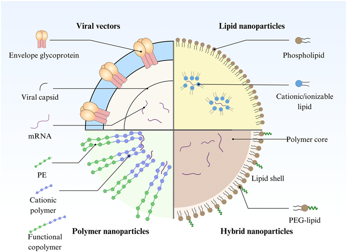
The application of mRNA therapy in the treatment of various liver diseases is due to its unique advantages over other RNA therapies, such as highly efficient targeting of liver delivery, rapid clinical translation, and controllable safety [34]. Importantly, these advantages rely on the emergence of mRNA delivery systems [35]. The mRNA delivery systems, through technical design, overcome the inherent defects of mRNA and become a crucial bridge connecting the enhancement of mRNA advantages with the improvement of clinical efficacy [36,37]. As illustrated in Fig. 1, the mRNA delivery system vectors can be broadly classified into two categories: Viral and non-viral. Compared with viral systems, non-viral delivery systems exhibit distinct advantages, including the absence of viral genome integration, dependence on synthetic materials or natural biological carriers, and the capacity to circumvent the immunogenicity and genomic risks associated with viral vectors [38]. Moreover, within the category of non-viral delivery systems, the mRNA hybrid nanoparticle system, the exosome system, and the LNP system are three representative platforms [39]. Among these, the liver-targeting capability of LNP, the biocompatibility of exosomes, and the multi-functionality of hybrid nanoparticles collectively offer safe and effective delivery strategies for liver disease therapy [40].
Figure 1
Figure 1.
The core classification framework of the mRNA delivery system: Based on the different sources and structures of the carriers, mRNA delivery carriers can be divided into two major categories: Viral carriers and non-viral carriers. Among them, viral carriers rely on the natural infection mechanism of the viral capsid, while non-viral carriers (such as LNP, cationic polymers, nanoparticles and mixed nanoparticles) achieve targeted delivery by optimizing their physical and chemical properties.
mRNA hybrid nanoparticle system is an advanced carrier technology for mRNA delivery. Encapsulating mRNA in nanoparticles improves its stability and delivery efficiency, and is widely used in vaccine development, cancer treatment, and gene editing [41]. LNP, which are composed of ionizable lipids, phospholipids, cholesterol, and pegylated lipids, are the most common mRNA delivery vehicles [42]. Ionizable lipids coat negatively charged mRNA electrostatically, phospholipids and cholesterol maintain structural stability, and pegylated lipids provide stability and reduce immune recognition. Polymer nanoparticles, such as polylactic acid nanoparticles, etc., can protect mRNA from degradation and promote its entry into cells by binding to mRNA to form complexes [43]. Inorganic nanoparticles, such as mesoporous silica nanoparticles (MSNS), have the advantages of uniform pore channels and high specific surface area, which can load large amounts of mRNA and achieve targeted delivery through surface modification [44]. The application of mRNA-mixed nanoparticle systems in vaccine development, cancer therapy, and gene editing is promising, and significant progress has been made, such as mRNA COVID-19 vaccine: mRNA is delivered by LNP to express viral antigens in cells and activate immune responses. For example, the mRNA COVID-19 vaccines from Moderna and Pfizer/BioNTech use LNP delivery systems. In addition, recent studies have explored LNP systems that form virus-like structures by surface-decorating spike proteins, capable of specifically enhancing mRNA expression and immune responses. In addition to COVID-19 vaccines, mRNA vaccines are being developed for other infectious diseases [45]. For example, mRNA vaccines against respiratory syncytial virus (RSV) are also under investigation to improve vaccine stability and delivery efficiency by optimizing the formulation of LNP. Recent studies have developed LNP for the delivery of nucleoside-modified mRNA-encoded immunomodulatory proteins such as IL-21, IL-7, and 4–1BBL, creating a triple-combination therapy. This therapy can significantly increase the frequency and function of tumor-infiltrating CD8+ T cells, leading to tumor clearance and the formation of long-term immune memory [46]. Several companies are developing mRNA tumor therapeutic vaccines against various solid tumors, including personalized tumor therapeutic vaccines (PCVS) and tumor-associated antigen tumor therapeutic vaccines. These vaccines activate the immune system to attack tumor cells by delivering mRNAs encoding tumor-specific antigens [47]. The clustered regularly interspaced shortpalindromic repeats/CRISPR-associated systems9 (RISPR/Cas9) system uses LNP to achieve precise gene editing by delivering mRNA and single guide RNA (sgRNA) encoding Cas9 protein [48]. For example, recent studies have shown that LNP target gene editing of dendritic cells (DC) to enhance antitumor immune responses. Despite the high efficiency and specificity of the CRISPR/Cas9 system, its delivery in vivo still faces challenges, such as off-target effects and immune responses [49]. By optimizing the composition and surface modification of nanoparticles, the efficiency and safety of gene editing can be improved. mRNA-mixed nanoparticle systems have shown great potential in vaccine development, cancer therapy, and gene editing. Delivery systems such as LNP can effectively improve the stability and delivery efficiency of mRNA, activate immune responses, or achieve precise gene editing [50]. In the future, with the continuous optimization of technology and the expansion of clinical applications, mRNA hybrid nanoparticle systems are expected to play an important role in more fields.
3.2
Exosomes system
Exosomes are nanoscale vesicles secreted by cells with a single-layer lipid bilayer structure, which can encase biomolecules such as proteins, lipids, and nucleic acids [51,52]. As mRNA delivery vectors, exosomes are natural extracellular vesicles with low immunogenicity. They can effectively avoid the clearance of the immune system and penetrate biological barriers, such as the blood-brain barrier (BBB), which has unique advantages in the treatment of central nervous system diseases [53]. In addition, the lipid bilayer structure of exosomes is able to protect mRNA from nuclease degradation and prolong its circulation time in vivo [54]. Exosomes can efficiently deliver anti-tumor drugs and genes and reduce the damage to normal tissues [55]. For example, exosomes loaded with chemotherapeutic drugs show better tumor penetration and lower systemic toxicity in mouse models. Through surface modification, exosomes can specifically target nerve cells to deliver mRNA for the treatment of neurodegenerative diseases [56]. For example, exosomes modified with RVG peptide can effectively deliver neurotrophic factor to the area of ischemic brain injury, reduce inflammation, and promote neural repair [57]. Exosomes can be used to deliver gene editing tools such as CRISPR/Cas9 to achieve precise gene therapy. Studies have shown that exosomes can efficiently deliver gene editing tools to leukemia stem cells to achieve precise genetic modification [58]. Despite the great potential of exosomes in mRNA delivery, there are still some challenges [59,60]. The components of exosomes are naturally heterogeneous and the yield of exosomes extracted from cells is relatively low, which limits their large-scale production and clinical application [61]. The targeting efficiency is low, and it is still challenging to achieve accurate targeting of exosomes. Exosomes may be recognized and eliminated by the immune system, affecting their delivery efficiency. Engineering exosome surfaces to make them harder to recognize by the immune system is being explored.
3.3
LNP system
LNP represents a class of nano-scale delivery systems formed through the self-assembly of lipid molecules. With a typical diameter ranging from 80 nm to 100 nm, LNP are designed to efficiently encapsulate and deliver nucleic acid therapeutics. Structurally, the architecture of LNP comprises four essential components: (1) Ionizable lipids, which exhibit pH-dependent charge characteristics, protonating under acidic endosomal conditions to facilitate nucleic acid complexation and endosomal escape; (2) helper lipids, such as distearoylphosphatidylcholine (DSPC), which contribute to bilayer stability and integrity; (3) cholesterol, enhancing membrane rigidity and promoting fusion with endosomal membranes; and (4) polyethylene glycol (PEG)-modified lipids, which prolong circulation half-life and reduce opsonization and immune recognition. Functionally, a defining advantage of LNP lies in their non-viral nature, which confers low immunogenicity, biodegradability, scalability for manufacturing, and the capacity for tissue-specific targeting. Collectively, these attributes have established LNP as the current "gold standard" platform for mRNA vaccine development and gene therapies. Of note, central to LNP functionality are ionizable lipids, often regarded as the “soul” of the system due to their ability to undergo protonation in the acidic endosomal compartment. This facilitates electrostatic interaction with nucleic acids, promotes endosomal disruption, and enables cytosolic release. Specifically, at physiological pH, these lipids remain neutral, thereby minimizing nonspecific cytotoxicity. For instance, SM-102, an ionizable lipid employed in Moderna's COVID-19 vaccine, exemplifies how optimized alkyl chain length and amine architecture can markedly enhance mRNA delivery efficiency. Recently, research have focused on incorporating functional moieties such as vitamin E or siloxane derivatives into lipid designs, yielding novel LNP with tailored immunomodulatory or organ-targeting capabilities. Furthermore, the successful global deployment of COVID-19 mRNA vaccines (e.g., those developed by BioNTech/Pfizer and Moderna) has validated the clinical scalability, safety, and efficacy of LNP technology. In summary, billions of doses have been administered worldwide, demonstrating significant protection against severe disease. Beyond SARS-CoV-2, LNP are being leveraged in vaccine development against influenza, HIV, and oncology applications, including Moderna’s personalized cancer vaccine, which has advanced to Phase Ⅲ clinical trials.
Among these three non-viral delivery systems, LNP has emerged as the mainstream choice for mRNA delivery due to its efficient packaging and protective capabilities, as well as its excellent intracellular delivery efficiency [62]. Overall, LNP is particularly well-suited for liver targeting and holds great potential in the treatment of liver disease.
4.
LNP and liver
The anatomical and functional characteristics of the liver, which match the delivery requirements of LNP, have made it one of the organs most easily and precisely enriched by LNP. Specifically, the hepatic sinusoidal endothelial cells of the liver have pores with diameters of 80–100 nm. The particle size of LNP is usually 50–100 nm, which can quickly reach the liver with the blood flow and directly contact the liver cells through the pores without penetrating the complete vascular wall, reducing the delivery resistance [63,64]. Furthermore, the surface of liver cells highly expresses ASGPR (sialic acid-containing glycoprotein receptor), and LNP can specifically bind to ASGPR through modification with galactose and other ligands, triggering receptor-mediated endocytosis. Moreover, even without modifying the targeting ligand, the ionizable lipids of LNP are neutral under physiological pH and enter the liver, where they become positively charged due to endosomal acidification and can fuse with the negatively charged endosomal membranes of liver cells through electrostatic interactions, achieving efficient uptake [65]. Additionally, as the metabolic center, the liver has extremely high requirements for the safety of delivery vehicles, and the structural optimization of LNP can avoid potential damage to the liver [66]. Notably, the core components of LNP (ionizable lipids, phospholipids, cholesterol) are mostly substances that can be metabolized by the human body [67,68]. For example, cholesterol can participate in liver lipid metabolism, and phospholipids can be decomposed by liver cells into fatty acids for energy supply, avoiding the accumulation of carrier components in the liver and causing metabolic burden. Furthermore, the mRNA delivered by LNP in liver cells is "transiently expressed" and subsequently degraded by hepatic nucleases after translation, without integrating into the genome. This avoids potential liver genome damage caused by gene editing or viral vectors [69]. Currently, the association between LNP and the liver has been verified by a large number of clinical studies. For instance, the large-scale application of the COVID-19 mRNA vaccine has indirectly verified the safety of LNP in liver metabolism, laying the foundation for its long-term application in the treatment of liver diseases. Besides, approximately 80% of liver-targeted mRNA therapies currently in clinical trials worldwide employ LNP as the delivery vehicle. In addition to mRNA, LNP can also deliver other nucleic acid drugs such as siRNA for liver disease treatment. For example, the siRNA-LNP therapy that has been on the market is used for the treatment of liver diseases caused by hereditary transthyretin amyloidosis [70].
In summary, the physiological characteristics of the liver provide natural targeting conditions for LNP, and LNP has been optimized in structure to become a key tool for addressing liver diseases [71]. The synergy of the two promotes the clinical transformation of liver-targeted nucleic acid therapy and serves as the core technical support for mRNA therapy of liver diseases.
5.
Targeting of LNP in liver disease
LNP serve as the core delivery vector for mRNA and other therapeutic agents in liver disease treatment, which can be targeted by the rich blood supply of the liver and the natural uptake tendency of hepatocytes. Different targeting strategies can make LNP more accurately adapt to different liver diseases.
5.1
Liver targeting strategies for LNP
The liver-targeting property of LNP can be achieved through various design strategies (Fig. S2 in Supporting information), including passive targeting and active targeting, which are the two core designs [72]. These strategies respectively utilize the liver's physiological characteristics and molecular recognition mechanisms to enhance the enrichment efficiency and specificity of LNP in liver cells. They contribute to improved drug efficacy while reducing side effects, making them a major focus of ongoing research (Table S2 in Supporting information) [73,74].
5.1.1
Passive targeting strategies
Passive targeting does not require additional ligand modifications, only by optimizing the lipid composition and proportion of LNP, and using the anatomical and cellular characteristics of the liver to achieve targeted enrichment, which is the most mature design for clinical transformation at present [75].
LNP consist of ionizable lipids, cholesterol, phospholipids, and PEG. Ionizable lipids are critical for passive targeting, as their pH sensitivity determines the interaction of LNP with hepatocytes [76]. At physiological pH (7.4), these lipids are electrically neutral, preventing clearance by negatively charged albumin or immune cells in the blood. Upon reaching the liver, LNP are endocytosed by hepatocytes to form endosomes. After the pH decreases to 5.0–6.0, LNP can ionize lipid protonation with positive charge, attract and fuse with the negatively charged endosomal membrane, and release mRNA to the cytoplasm [77]. This "pH-triggered fusion" mechanism enables LNP to achieve hepatocyte enrichment of 50%–70% without the need for ligand modification. Cholesterol and phospholipids play essential roles in enhancing the stability of LNP, ensuring efficient liver delivery [78]. Cholesterol and phospholipids: Enhanced stability for efficient liver delivery. Cholesterol can be inserted into the LNP lipid bilayer to improve membrane rigidity and reduce the degradation of mRNA by RNase in the blood [79]. Phospholipids such as DSPC can optimize the fusion efficiency of LNP and hepatocyte endosomal membrane by regulating membrane fluidity, and they work together to prolong the half-life of LNP in the blood. This extended circulation time allows the LNP to follow the liver's rich blood flow, reach the hepatic sinusoids, and interact with hepatocytes via the fenestrations in the hepatic sinusoidal endothelial cells [80,81]. PEG modification is key to balancing "long circulation" with "hepatic uptake". PEG coupled with lipid molecules, such as PEG2000-DMG, form a hydrated layer on the surface of LNP, reducing the clearance of splenic macrophages and prolonging blood circulation. Optimizing the content of PEG in total lipids can achieve a balance between "long circulation" and "hepatic uptake". Reducing excessive PEG modification will hinder the effect of hepatocytes on LNP endocytosis, and further improve the efficiency of passive targeting [82–84].
5.1.2
Active targeting strategies
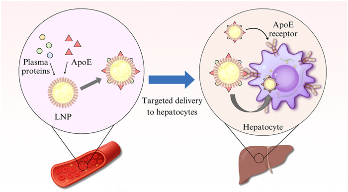
Active targeting of LNP involves the modification of ligands that can bind to liver-specific receptors on the surface of LNP, thereby enhancing targeting specificity through "receptor-mediated endocytosis". This approach is particularly suitable for the precise treatment of liver tumors [85]. Active targeting can be divided into three main strategies: ASGPR ligand modification, peptide/antibody fragment modification, and ligand coupling. ASGPR (asialoglycoprotein receptor) ligand modification is a classic strategy to adapt highly expressed receptors on hepatocytes. ASGPR is specifically expressed on the surface of hepatocytes and can recognize molecules containing glycans, such as galactose and lactose. Then, form galactose derivatives (such as galactosylceramide, N-acetylgalactosamine (GalNAc), N-acetylgalactosamine) on the surface of LNP [86]. Furthermore, ASGPR-mediated endocytosis was used to precisely deliver LNP to hepatocytes. This strategy has been shown to increase hepatocyte enrichment to over 80%, while reducing non-target organs (such as spleen and lung) by >50% [87]. At present, this strategy has entered the late preclinical stage, with GalNAc-modified LNP for siRNA delivery having entered phase Ⅱ clinical trials for liver diseases treatment. Peptide and antibody fragment modification is a targeted design for specific liver cell types. LNP can be modified with ligands that bind to specific receptors of liver cancer cells. For example, peptides like YIGSR can target the glypican-3 (GPC3) receptor on the surface of liver cancer cells, and single-chain variable fragments (scFv) can target epidermal growth factor receptor (EGFR). Such modifications can enable LNP to cross the physical barrier of the tumor microenvironment and be precisely enriched in liver cancer cells [88]. Reduce the damage to normal liver cells. Ligand coupling mode can ensure the compatibility of "targeting activity" with "LNP stability". Typically, ligands are usually coupled to the surface of LNP in a "lipid-linker-ligand" manner. The linkers (such as disulfide bonds and ester bonds) should be biocompatible and do not destroy the lipid bilayer structure of LNP. At the same time, the density of ligand modification (usually 1–5 ligands per 100 lipid molecules) should be controlled to avoid the steric hindrance between ligands affecting the receptor binding activity [89]. Apolipoprotein E (ApoE) is an example of a ligand used in active targeting strategies. Its core mechanism involves the specific binding of ApoE to the LDL receptor (LDLR) on liver cells (Fig. 2) [90]. The N-terminal domain of ApoE (containing Arg/Lys 136–158 rich region) is a high-affinity binding site for LDLR. This binding is receptor-dependent and can be competitively inhibited by anti-LDLR antibodies or excess ApoE, a key characteristic of ligand-receptor interactions. There are two main ways to achieve ApoE ligand coupling: Nonspecific adsorption and active conjugation. Nonspecific adsorption is the passive adsorption of ApoE from blood on the surface of LNP due to the residual charge or hydrophobic region of ionizable lipids [91]. For example, although ionizable lipids are neutral at physiological pH, their tertiary amine groups can interact weakly with ApoE. The active conjugation design is to covalently link ApoE or its receptor-binding peptides, such as N-terminal amino acids 136–158, to the surface of LNP by chemical cross-linking. The ApoE peptide, for example, direct coupling to the LNP, targeted to enhance specificity. ApoE confers a high degree of liver specificity to LNP through an active targeting mechanism mediated by LDLR [92]. Further studies are needed to explore the dynamic interaction between ApoE and LNP and to investigate the application potential of non-LDLR pathways in liver diseases [93].
Figure 2
Figure 2.
LNP forms a "protein cap" on its surface together with ApoE in the plasma. The LDLR abundantly expressed on the surface of the liver cell basement membrane can specifically recognize and bind to the ApoE component in the protein cap. After the binding of ApoE and LDLR, LNP is encapsulated into the cell and forms an endosome. After the endosome is acidified, the encapsulated therapeutic mRNA is released into the cytoplasm of the liver cell, and then translated to generate the required functional protein.
Passive targeting has become the first choice for the scenarios requiring systemic drug delivery such as liver genetic diseases and chronic liver diseases due to its advantages of "simple process, good mass production and controllable cost". On the other hand, active targeting enhances specificity through ligand modification, making it ideal for scenarios requiring a "precise attack", such as liver tumors and local viral infections. It is the core direction for the development of LNP liver targeting to "individualized therapy" in the future. Together, they support LNP to become a multifunctional platform for liver-targeted delivery and promote the application of nucleic acid drugs such as mRNA in the treatment of liver diseases [94].
5.2
LNP-mediated mRNA delivery system to treat liver disease
Liver diseases pose a major global health challenge, and existing treatments have many limitations [95]. The nucleotide-modified mRNA-LNP (nucleotide-modified mRNA-encapsulated LNP) technology has generated a lot of interest with the introduction of the new coronavirus mRNA vaccine. This technology can target the liver and deliver the substance precisely. mRNA delivery systems mediated by LNP have demonstrated great potential and advantages in the treatment of end-stage liver disease (ESLD), HCC, and hepatitis B.
5.2.1
ESLD
ESLD is a clinical syndrome characterized by abnormal liver function and systemic manifestations, including decompensated cirrhosis, severe hepatitis, and advanced liver cancer [96]. Liver transplantation is the only treatment for ESLD, but this treatment is severely limited by the shortage of organ donations. The liver has a strong capacity for regeneration, but in the case of severe acute hepatocyte death or chronic ESLD, the proliferative capacity of mature liver cells is exhausted due to the continuous intensification of steatosis, inflammation, fibrosis, and cirrhosis [97].
Vascular endothelial growth factor (VEGF) is a multifunctional factor that can specifically act on vascular endothelial cells, stimulate the division and proliferation of these cells, and induce the formation of blood vessels. In the liver, VEGF is mainly involved in tumor angiogenesis and regulation of tumor angiogenesis by binding to its receptor VEGFR [98]. The VEGF family includes a variety of members, such as VEGFA, VEGFB, and VEGFC, and their roles in liver diseases are different [99]. VEGFA, for example, can promote new blood vessels and increase blood vessel permeability formation [100]. Although VEGF's mRNA drug for ESLD has shown potential to promote liver regeneration by activating liver stem cells, enabling them to proliferate and generate new healthy hepatocytes, the functional cells of the liver, there are some drawbacks [101]. For example, efficient delivery of mRNA to target cells remains a challenge in a complex in vivo environment. In addition, due to the poor stability of mRNA in vivo and its easy degradation, special delivery systems are required [102]. In these cases, delivery of nucleoside-modified mRNA by LNP rapidly activates regenerative pathways in hepatocytes, thereby accelerating liver regeneration after injury [103]. In 2023, the research group of Valerie Gouon-Evans at Boston University in the United States proposed to use VEGFA as a treatment to accelerate the transformation of bile duct epithelial cells (BEC) into hepatocytes [104]. Studies in zebrafish confirm that blockade of VEGFR attenuates bile duct epithelial-driven liver repair, whereas overexpression of VEGFA promotes liver repair. Delivery of VEGFA by a nonintegrated, safe nucleoside-modified mRNA encapsulated in LNP (mRNA-LNP) in acutely or chronically injured mouse liver induced the potent transformation of BEC into hepatocytes and the elimination of steatosis and fibrosis [105]. In both human and mouse diseased livers, the investigators further identified BEC expressing the VEGFA receptor KDR associated with KDR-expressing cell-derived hepatocytes [106]. This study defined KDR-expressing cells (most likely BEC) as facultative hepatic progenitor cells [107]. The team from Boston University successfully induced the transdifferentiation of bile duct cells and reversed fibrosis in the mouse model using VEGFA mRNA-LNP. This strategy for promoting liver regeneration involves encapsulating mRNA of VEGF-A, HGF or Wnt pathway agonists, and targeting liver cells through GalNAc-LNP, or targeting liver progenitor cells through VEGFR2/KDR to promote the transdifferentiation of BEC into liver cells and the proliferation of parenchymal cells [108]. The safety of mRNA-LNP has been widely verified in COVID-19 vaccines [109]. The research team at Peking University developed an organ- and cell- specific mRNA-LNP technology, which allows precise delivery to the lung, liver, and spleen by combining targeted delivery of LNP and controlled expression of mRNA sequences, providing a new strategy for precision tumor therapy [110]. In the treatment of ESLD, LNP-mediated mRNA therapeutic strategies can be designed to target a wide range of liver diseases (Fig. S3 in Supporting information).
For instance, the regeneration and repair of liver cells can be facilitated by the delivery of mRNAs encoding specific growth or repair factors. Furthermore, LNP delivery systems can be employed to achieve mRNA therapy targeting specific liver diseases, including the delivery of mRNAs encoding enzymes used for the treatment of inherited metabolic diseases [111]. However, the application of LNP-mediated mRNA therapy in ESLD is confronted with several challenges. Firstly, the natural barrier effect of the liver may prove an obstacle to the effective delivery of LNP [112]. Secondly, the stability and biocompatibility of LNP in vivo must also be taken into consideration [113]. Furthermore, ensuring the effective expression and functional recovery of mRNA in liver cells represents a crucial avenue of research [114]. Notwithstanding these challenges, recent research has demonstrated that the delivery efficiency and therapeutic efficacy of LNP in the liver can be enhanced by optimizing its composition and structure. For instance, the capacity of LNP to target liver cells can be augmented by modifying the lipid composition of LNP or incorporating specific targeting ligands [78,115]. Furthermore, the biocompatibility and therapeutic window of LNP delivery systems can be enhanced by utilizing novel liposomes with improved stability and an extended half-life [116].
In conclusion, LNP-mediated mRNA therapy represents a promising therapeutic strategy for ESLD. Through further research and optimization, it is anticipated that more effective therapies will be developed to bring hope to ESLD patients.
5.2.2
HBV
Hepatitis B is a liver disease caused by the HBV [117]. The HBV genome is a relaxed circular DNA (rcDNA) approximately 3.2 bases long. After infection, HBV reverse transcriptase converts loose circular DNA (cDNA) into covalently closed circular DNA [118]. This form of DNA is stable as microchromosomes in the nucleus and difficult to remove. This process indicates the fundamental cause of HBV recurrence and the difficulty of virus eradication. In some cases, the disease results in chronic infection, which can lead to the development of cirrhosis or primary liver cancer [119]. Because a complete cure (cccDNA and intracellular HBV clearance) is not attainable, the objective of therapeutic intervention in chronic hepatitis B is the attainment of HBsAg seroclearance for a defined period of treatment, that is to say, a 'functional cure' [120].
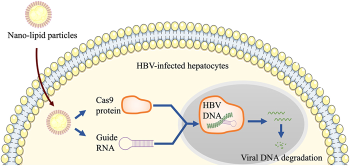
The utilization of LNP for the treatment of the HBV has demonstrated the potential of this technology in inhibiting HBV replication and clearance. In particular, the delivery of anti-HBsAg antibodies and IL-21 via LNP has shown clearance of HBV in a mouse model. First, the use of mRNA-LNP expressing anti-HBsAg antibodies (G12-scFv, G12-scFv-Fc, and G12-IgG) produced sustained effects on HBsAg seroclearance in an adeno-associated virus (AAV)/HBV mouse model. The mRNA-LNP encoded by these antibodies significantly reduced HBsAg levels in mouse serum up to 30 days post-treatment, whereas the exogenous antibodies lost their effect in reducing HBsAg or HBV DNA levels 9 days after administration [121]. This suggests that anti-HBsAg antibodies encoded by mRNA-LNP have the potential to clear HBsAg in the long term. Second, IL-21-encoded mRNA delivered via LNP induced clearance of HBV antigen and DNA in a mouse model of HBV persistence, which correlated with HBV-specific humoral and cellular immune responses [122]. These findings suggest that the IL-21-binding mRNA-LNP platform represents an effective and promising strategy for the development of new therapies against chronic HBV infection. In addition, other studies have explored the use of LNP in HBV therapy, such as the use of LNP-encapsulated PLK1-targeted siRNAs to show antiviral activity in HBV-infected hepatocytes and the use of CRISPR/Cas9 to efficiently knockdown targeted LNP mRNAs for the treatment of HBV in HBV-infected mice via a novel pH-sensitive cationic liposome delivery system. Clinical trial progress is actively underway (Fig. 3) [123,124]. A team of researchers from Fudan University has developed a novel siHBV+ mRNA combination therapy designed to enhance the therapeutic efficacy of chronic hepatitis B. The therapy is designed for the treatment of HBV in mice. The therapy achieves efficient, safe, hepatocyte-targeted delivery of siHBV in vivo by designing a combination of highly efficient, multifunctional, and highly conserved siHBV and combining it with a modified LNP (tLNP) platform [125]. In a variety of hepatitis B-associated cellular and animal models, tLNP/siHBV demonstrated sustained HBV inhibition and HBV-specific immunomodulatory effects. In addition, the study explored a novel strategy for the treatment of chronic hepatitis B based on modified tLNP co-delivery of siRNA in combination with interleukin-2 mRNA (tLNP/siHBV/IL2). This approach inhibits viral transcriptional replication through the RNAi pathway and promotes the proliferation and infiltration of HBV-specific CD8+ and CD4+ T cells through the efficient expression of IL-2, thus achieving both antigenic suppression and immune control of HBV [126]. In another study, Ying's group at Fudan University, in collaboration with Lin's group, used mRNA technology to encode antibodies to the surface antigen of HBV, and the clearance of serum hepatitis B surface antigen induced by a single dose of G12-mRNA-LNP injection in a mouse model of chronic hepatitis B lasted for at least 30 days, significantly reducing serum hepatitis B surface antigen levels. Furthermore, a research team developed LNP targeting hepatic macrophages, encapsulating mRNA encoding CAR and CD24-Siglec-G lacking ITIMs (Siglec-GΔITIMs). This significantly improved the phagocytic function of liver macrophages in HCC mouse models, effectively reducing tumor burden and increasing the survival time of mice [127]. Current therapeutic strategies focus on inhibiting HBV replication and improving liver-related outcomes, while the goal of achieving HBsAg clearance and functional cure remains limited. Although LNP shows potential as a drug delivery platform in HBV treatment, its limitations in hepatic clearance may affect therapeutic efficacy. This new therapy is still in the early clinical trial stage, and its safety and efficacy need to be further validated. In conclusion, although LNP technology shows some potential in HBV therapy, the above limitations still need to be overcome and more effective therapeutic strategies need to be developed to achieve a complete cure for HBV.
Figure 3
Figure 3.
The therapeutic mechanism of LNP delivering CRISPR/Cas9 components (Cas protein and guide RNA) to HBV-infected liver cells: After entering the cells, the Cas9 protein, which, upon binding to the guide RNA, specifically cuts the HBV DNA, promoting the degradation of the viral genome and ultimately achieving the inhibition of HBV infection.
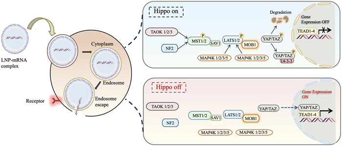
Primary carcinoma of the liver (PLC) refers to cancer that occurs from liver cells or intrahepatic bile duct cells, of which >90% are HCC [128,129]. In 2020, there were 905,677 new cases and 830,180 deaths of liver cancer worldwide, accounting for 4.7% and 8.3% of all cancer cases and deaths, respectively [130]. So far, studies have found that: The occurrence and development of HCC are related to the abnormal activation or inhibition of a variety of signaling pathways, such as the Janus kinase-signal transducer and activator of transcription (JAK/STAT) signaling pathway [131,132], Wnt/β-catenin signaling pathway [133], Hedgehog signaling pathway [134], Hippo signaling pathway [135,136]. The importance of the Hippo signaling pathway has been gradually revealed. The Hippo signaling pathway was discovered in Drosophila melanogaster in 1994, so it is named after the Drosophila Ste20-like kinase Hippo. As shown in Fig. 4, the Hippo signal consists of several core components, including MOBKL1A/B (MOB1A/B), large tumor suppressor kinase 1/2 (LATS1/2), Yes-associated protein 1 (YAP1), transcription regulator 1 (TAZ1) containing the WW domain, and transcription enhancer associated domain family 1 (TEAD1) [137].
Figure 4
Figure 4.
The principal components of the mammalian signaling Hippo pathway are MST1/2, LATS1/2, SAV1, MOB1, YAP or TAZ and TEAD. The pathway is activated by a series of phosphorylation events. MST1/2 can be activated by TAO1/2/3, NF2, or autophosphorylation. Upon activation, MST1/2 binds to SAV1, which in turn phosphorylates and activates LATS1/2. Subsequently, the activated LATS1/2 complex forms with MOB1, which then phosphorylates YAP/TAZ. This results in the sequestration or degradation of YAP/TAZ by 14–3–3 proteins in the cytoplasm.
The Hippo signaling pathway participates in early embryonic development, tissue regeneration, and organ size regulation. Additionally, it regulates the occurrence and development of various tumors, including HCC [138]. Many studies have shown that the Hippo signaling pathway plays an important role in regulating organ size in mammals by regulating cell proliferation and decay. During the development of HCC, this pathway is disabled in tumor-initiating cells, which can overcome the inhibition of surrounding normal tissues and allow for clonal expansion and tumor development [139]. Inhibition or activation of the Hippo signaling pathway can increase or decrease the activity of YAP/TAZ. The process of LNP-delivered mRNA to regulate the Hippo pathway primarily entails encapsulating mRNA in LNP, subsequently utilizing LNP to deliver mRNA into cells, and finally releasing mRNA within cells to regulate the expression of Hippo pathway-related genes. This approach enables the precise control of mRNA expression in cells, subsequently influencing the activity of the Hippo pathway to regulate cell proliferation, apoptosis, and other biological processes. Consequently, LNP -the mediated mRNA delivery system has shown significant potential and prospects in the treatment of liver cancer. In 2015, to investigate the role of YAP in the development and maintenance of HCC and to determine its downstream oncogene expression profile, a series of studies were conducted in genetically engineered mice using genetic manipulation methods and siRNA-LNP technology. It was found that activation of endogenous YAP could disrupt the differentiation process of liver cells and maintain this state in advanced tumors. Inhibition of YAP activity in HCC can reestablish the process of liver cell differentiation and lead to tumor regression [140]. These results suggest that inhibition of the cell differentiation process is a potential therapeutic strategy for the treatment of endothelial tumors, and the significant response induced by siYAP-LNP supports the clinical development of this therapeutic modality.
Furthermore, clinical trials of LNP mRNA for HCC are currently underway. Several preclinical studies have yielded positive results. For example, one study delivered IL-12 mRNA via intravenous LNP to slow the progression of MYC oncogene-driven HCC with no significant animal toxicity [141]. Furthermore, in 2023, LNP targeting liver macrophages was developed, which can encapsulate mRNA encoding cytokine receptors (CAR) and CD24-Siglec-G (Siglec-GΔITIMs) without the ITIM domain. This approach significantly improved the phagocytic function of liver macrophages in HCC mouse models, effectively reducing tumor burden and extending the survival time of mice [142,143]. Concerning clinical trials, although no definitive results have yet been published, mRNA-LNP-based therapeutic strategies are demonstrating considerable promise in the context of cancer treatment. Furthermore, studies have proposed the innovative concept of utilizing circRNA instead of mRNA, in conjunction with LNP, to develop a therapeutic vaccine for the treatment of HCC. This approach combines the advantages and capabilities of circRNA and LNP to provide an adjuvant for the immune response and enhanced stability [144,145].
Although LNP technology has shown great potential in the treatment of HCC, its use still faces some toxicity challenges. For example, LNP may cause cytotoxicity and genotoxicity, which need to be overcome by measures taken at the pre-formulation stage [146]. Future studies should continue to investigate novel therapeutic approaches to further enhance the survival and quality of life of HCC patients.
5.2.4
MASLD
MASLD remains a rapidly growing global health burden [147,148], encompassing a wide spectrum of liver injuries. These range from hepatic steatosis, metabolic dysfunction-associated steatohepatitis (MASH), fibrosis, and cirrhosis to MASLD-associated HCC (MASLD-HCC) [149]. Currently, no effective treatment methods are available in clinical practice [150]. However, LNP technology has shown great potential in the treatment of MASLD. As an efficient delivery vector for nucleic acid drugs such as mRNA and siRNA, LNP can achieve liver-targeted delivery, regulate lipid metabolism within the liver, and alleviate inflammation and fibrosis, thereby providing a promising approach to treat MASLD [151]. In 2024, LNP was developed, which could simultaneously encapsulate siRNA targeting hypoxia-inducible factor-1α (HIF-1α) and silybin, and targets liver tissues through the ApoE-LDLR/VLDLR pathway. In a MASLD mouse model, this LNP can effectively silenced the expression of HIF-1α in liver cells, and working synergistically with SLB to exert anti-inflammatory and anti-fibrotic effects. Furthermore, LNP can act as a carrier to deliver gene editing tools, such as CRISPR-Cas9, to liver cells, enabling the repair or editing of gene mutations related to MASLD [152]. For instance, certain genetic factors may cause abnormal regulation of lipid metabolism in liver cells, and correcting these mutations through gene editing could potentially offer a fundamental treatment for MASLD [153]. In 2024, a study proposed a novel strategy for delivering therapeutic proteins to liver MASLD lesions using mRNA LNP, namely the "fibrosis overexpression and retention (FORT)" process. The core mechanism involves using retinoic acid derivatives (such as ATRA) to enhance the targeting and expression of LNP in fibrotic liver. Additionally, a fusion protein, like CBD, is employed to retain and carry mRNA encoding anti-fibrotic or anti-inflammatory proteins, specifically targeting hepatic stellate cells or the fibrotic ECM. This strategy primarily aims to overcome the challenges of low drug delivery efficiency and short retention time in the fibrotic environment. At the same time, this study has also proposed a multi-targeted combined therapy, where multiple mRNAs encoding different mechanism proteins (such as RLN and IL-10), are delivered either simultaneously or sequentially. This strategy targets multiple liver cells and non-essential cells, aiming to synergistically regulate lipid metabolism, and exhibit anti-fibrotic and anti-inflammatory effects, which is expected to solve the complexity of MASLD diseases [154]. Currently, LNP has certain targeting properties for the liver. However, there remains potential for further optimization of their composition and surface modification. By improving these aspects, LNP can be more precisely targeted to liver cells, reducing their distribution in other tissues and organs. This optimization not only enhances treatment efficacy but also minimize side effects. For example, by modifying the surface ligands of LNP, it can specifically bind to the specific receptors on the surface of liver cells, achieving more efficient liver-targeted delivery [155].
6.
Future expectations
The use of nanoparticles, primarily lipid-based, is crucial for achieving in vivo mRNA delivery, as they help prevent degradation in physiological environments and enhance the uptake and subsequent antigen expression of mRNA-nanoparticle complexes. As the primary delivery carrier for mRNA, LNP are the focus of current research, which emphasizes their design and screening, optimization of internal ratios, surface modification, and selection of drug delivery routes [156]. These optimization measures aim to strengthen the stability of mRNA and intracellular delivery efficiency. To further expand the application prospects of mRNA-LNP technology, researchers are conducting multidimensional design and optimization studies based on the standard LNP formulations of marketed mRNA vaccines [157]. They are continuously developing LNP delivery vehicles with various functional characteristics. For example, an esterase-responsive biodegradable ionizable lipid LNP was developed by Tan’s group at the Hangzhou Institute of Medical Sciences, in collaboration with Zhang's group [158]; and the all-trans retinoic acid-based ATRA LNP developed by Lv's group at Institute of Chemistry, Chinese Academy of Sciences, in collaboration with Miao's group at School of Pharmacy, Peking University [159], are both aimed at improving the cellular uptake and endosomal escape of mRNA-LNP, enhancing the mRNA All of the ATRA LNP are designed to improve the cellular uptake and endosomal escape of mRNA-LNP, enhance the translation efficiency of mRNAs, and have some extra-hepatic tissue targeting properties [160]. LNP-mediated mRNA therapy not only shows promise in conventional cancer treatment but also explores innovative targeting strategies and future directions. Specific surface modifications and functionalization can enable precise localization and efficient delivery to cancer cells. Recently, the research led by Liu, Gu, and Ping’s groups developed a three-component LNP system consisting of nAcx-Cm lipid, a permanent cationic lipid, and a PEG-based lipid. This system facilitates the simultaneous accumulation and translation of mRNA in both the lung and liver. By regulating and simplifying the intrinsic components, this three-component LNP significantly enhances the efficiency of mRNA delivery to target organs, offering a novel strategy for precise mRNA therapy. This approach presents new possibilities for cancer treatment and the prevention of infectious diseases; however, its effectiveness still requires further validation through clinical trials [161].
Through the integration of mRNA technology with CRISPR gene editing and artificial intelligence, it is promoting breakthroughs in many fields of biomedicine [162]. Combined with CRISPR, it can encode and deliver Cas9 systems (such as LNP vectors), which show the advantages of efficient and precise editing in the treatment of genetic diseases such as ATTR. Phase Ⅲ trials have been launched, and the off-target effects are reduced. In the future, it may be expanded to cancer, neurological diseases, and other fields. artificial intelligence (AI) technologies such as the Baidu Linear Design algorithm can optimize mRNA sequences in 11 min, improve stability and expression efficiency, and realize customized medical treatment combined with individual genetic characteristics [32,163]. The synergy of the two technologies can significantly improve the research and development efficiency, accelerate the vaccine/drug development cycle, reduce costs, and provide innovative solutions for personalized therapeutics, rare diseases, and regenerative medicine [164]. COVID-19 vaccines encourage regulatory agencies to establish an efficient approval framework, and the platform technology certification system can reuse the quality/safety data of previous products, significantly shortening the approval cycle of new drugs. By April 2023, 20% of the 416 mRNA clinical trials worldwide were for non-COVID-19 indications, and data accumulation further accelerated the approval process. Relying on the LNP platform technology, enterprises can expand the pipeline of infectious disease vaccines, tumor vaccines, rare disease treatment, and other fields, disperse risks, and expand market space. However, there are also problems such as limited licensing time, "stuck" risk, pressure on development, product cost, continuous innovation, and other negative impacts. However, once successful, companies will have greater autonomy and competitiveness in pipeline advancement, market development, business transformation, etc., and avoid being controlled by others.
7.
Conclusion
In the treatment of liver-specific diseases, although LNP delivery systems have significant liver targeting and the current research and development progress is rapid, their clinical application remains relatively limited [165,166]. Several factors contribute to this issue. First, it is difficult to precisely target specific liver subsets, such as hepatic stellate cells, relying solely on natural metabolic pathways. Moreover, over 50% of LNP are nonspecifically cleared by Kupffer cells [167,168]. The structural changes of liver sinusoid in pathological states such as cirrhosis further hinder the delivery efficiency. Second, immunogenicity poses a key obstacle. The immunogenicity of LNP is not triggered by a single component but rather from the synergistic action of its core components, including ionizable lipids, phospholipids, cholesterol, PEG-lipids, which are recognized by the innate immune recognition pathways. Ionizable lipids are the main effector components of LNP in activating the innate immunity, and their structural differences directly determine the intensity and type of the immune response. Research from the United States Armed Forces Medical University confirmed that commonly used ionizable lipids, such as SM102 and ALC315, activate the MyD88-dependent signaling pathway by binding to TLR4 on immune cells. This binding induces the activation of NF-κB and IRF transcription factors, promoting the release of pro-inflammatory factors such as IL-6 and tumor necrosis factor-alpha (TNF-α) [169]. Furthermore, in a 2022 study, it was found that SM102-type LNP stimulate IL-1β release from human peripheral blood mononuclear cells, while MC3-type LNP does not exhibit this effect [170]. This is closely related to the chain length and charge distribution of the lipid tail. Auxiliary lipids also regulate the "quality" and "quantity" of the immune response by altering the membrane structure and biological distribution of LNP. Negative phospholipids (such as dioleoylphosphatidylserine (DOPS) and dioleoylphosphatidylglycerol (DOPG)) can target secondary lymphoid organs, enhancing uptake by DCs. Specifically, DOPS-LNP can activate CD8⁺ T cells, while DSPC-LNP is more likely to induce humoral immunity [171]. Although PEG modification can prolong the circulation time of LNP, it also triggers anti-PEG immune response, presenting a key challenge for repeated administrations. Pre-existing or repeatedly induced anti-PEG antibodies can bind to the PEG chain on the surface of LNP, activating the complement cascade reaction and leading to phagocytosis and clearance by macrophages. Reducing the proportion of PEG (from 1.5% to 0.5%) can enhance the production of antigen-specific antibodies, but excessive PEG (>2%) may inhibit the uptake of LNP by DCs and weaken the immune activation effect. Repeated administration can accelerate the clearance of anti-PEG antibodies and ionizable lipids, causing endosomal damage and activating the release of pro-inflammatory factors such as IL-6 and TNF-α, which increases the risk of toxicity and tolerance to repeated administration [172]. Finally, the complex production process, large disease heterogeneity, and high long-term safety regulatory requirements have delayed the transformation process from laboratory to clinical practice [173]. Addressing these challenges involves investigating various factors, such as lipid composition, surface modifications, and particle size, all of which can influence the performance and biodistribution of LNP [174]. Furthermore, as research progresses, there is a pressing need for comprehensive in vivo studies to assess the long-term effects and therapeutic outcomes of LNP-based mRNA delivery systems in diverse patient populations [175]. Collaborations between academic institutions and biotechnology companies may accelerate the translation of these findings into clinical settings [176]. Looking ahead, the future of LNP-mediated mRNA therapies for liver disease treatment appears promising [177]. With continued innovation and optimization, these systems could not only improve the management of existing liver diseases but also pave the way for novel therapeutic interventions, including gene editing and personalized medicine approaches. The successful application of LNP delivery systems in clinical practice could revolutionize the treatment landscape for liver diseases and beyond, ultimately enhancing patient outcomes and quality of life. Future studies need to further explore the design of liver disease specific LNP: By adjusting the proportion of selective organ targeting (SORT) molecules or developing novel targeting ligands, such as Ly6C antibody specific for liver sinusoidal endothelial cells, to achieve precise delivery of different cell subsets in the liver. For the microenvironment of chronic liver disease (such as fibrosis barrier), the design of enzyme-responsive LNP can improve the penetration efficiency. In addition, the combination of saRNA technology with LNP is expected to solve the challenge of frequent drug administration in the treatment of chronic diseases. In the field of combination therapy, the strategy of LNP co-delivery of CRISPR components and immunomodulatory mRNAs, such as IL-21, may provide a new paradigm for functional cure of HBV and HCC immunotherapy. In terms of clinical translation, AI-driven formulation screening and freeze-drying process development of LNP will accelerate accessibility, while personalized evaluation models based on organoids and organ-on-a-chip are expected to shorten the research and development cycle.
CRediT authorship contribution statement
Ran Wang: Writing – review & editing, Writing – original draft, Visualization, Conceptualization. Chang Tian: Writing – review & editing, Writing – original draft, Conceptualization. Entong Ji: Writing – review & editing, Writing – original draft, Conceptualization. Qixiang Wu: Writing – review & editing, Writing – original draft, Conceptualization. Jie Wang: Writing – review & editing, Writing – original draft, Funding acquisition, Conceptualization. Tao Xu: Writing – review & editing, Writing – original draft, Supervision, Funding acquisition, Conceptualization.
Declaration of competing interest
The authors declare that they have no known competing financial interests or personal relationships that could have appeared to influence the work reported in this paper.
Acknowledgments
This work was supported by the National Natural Science Foundation of China (No. 82373932); Department of Education of Anhui Province Outstanding Young Teacher Training Project (No. YQZD2023023); Anhui Province University Outstanding Youth Research Project (No. 2024AH020006); Anhui Medical University Youth Shuangpei Program (2024); Natural Science Foundation of Anhui Province (Nos. 2208085MH203, 2408085QH242, 2508085J050); Anhui Provincial Health Commission Scientific Research Project (No. AHWJ2024Aa30231); The Research fund of Anhui Translational Medicine Research Institute (No. 2023zhyx-C10).
Supplementary materials
Supplementary material associated with this article can be found, in the online version, at doi:10.1016/j.cclet.2025.112138.
[1]
H. Devarbhavi, S.K. Asrani, J.P. Arab, et al., J. Hepatol. 79 (2023) 516–537. doi: 10.1016/j.jhep.2023.03.017
Z. Tang, F. Yu, J.C. Hsu, et al., Adv. Mater. 36 (2024) e2302901. doi: 10.1002/adma.202302901
[176]
D. Kim, C.H. Whang, J. Hong, et al., Adv. Mater. 36 (2024) e2311283. doi: 10.1002/adma.202311283
[177]
M.G. Alameh, I. Tombácz, E. Bettini, et al., Immunity 54 (2021) 2877–2892 e2877. doi: 10.1016/j.immuni.2021.11.001
Figure 1
The core classification framework of the mRNA delivery system: Based on the different sources and structures of the carriers, mRNA delivery carriers can be divided into two major categories: Viral carriers and non-viral carriers. Among them, viral carriers rely on the natural infection mechanism of the viral capsid, while non-viral carriers (such as LNP, cationic polymers, nanoparticles and mixed nanoparticles) achieve targeted delivery by optimizing their physical and chemical properties.
Figure 2
LNP forms a "protein cap" on its surface together with ApoE in the plasma. The LDLR abundantly expressed on the surface of the liver cell basement membrane can specifically recognize and bind to the ApoE component in the protein cap. After the binding of ApoE and LDLR, LNP is encapsulated into the cell and forms an endosome. After the endosome is acidified, the encapsulated therapeutic mRNA is released into the cytoplasm of the liver cell, and then translated to generate the required functional protein.
Figure 3
The therapeutic mechanism of LNP delivering CRISPR/Cas9 components (Cas protein and guide RNA) to HBV-infected liver cells: After entering the cells, the Cas9 protein, which, upon binding to the guide RNA, specifically cuts the HBV DNA, promoting the degradation of the viral genome and ultimately achieving the inhibition of HBV infection.
Figure 4
The principal components of the mammalian signaling Hippo pathway are MST1/2, LATS1/2, SAV1, MOB1, YAP or TAZ and TEAD. The pathway is activated by a series of phosphorylation events. MST1/2 can be activated by TAO1/2/3, NF2, or autophosphorylation. Upon activation, MST1/2 binds to SAV1, which in turn phosphorylates and activates LATS1/2. Subsequently, the activated LATS1/2 complex forms with MOB1, which then phosphorylates YAP/TAZ. This results in the sequestration or degradation of YAP/TAZ by 14–3–3 proteins in the cytoplasm.


 DownLoad:

 下载:
下载:


 下载:
下载:

