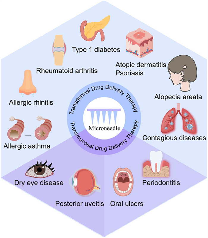
Figure 1.
Schematic illustration of MN-mediated transdermal and transmucosal drug deliver for treating diverse IMIDs.

Microneedle-mediated precision immunomodulation: A next-generation therapeutic paradigm for immune disorders
Chunxian Zhou , Mingyu Gong , Jubo Jian , Huanhuan Pan , Wanshan Hu , Zeshi Jiang , Chao Lu , Guilan Quan , Chuanbin Wu , Xin Pan , Junhuang Jiang , Tingting Peng


Immune-mediated inflammatory diseases (IMIDs) are systemic disorders characterized by dysregulated innate and adaptive immune responses, often leading to end-organ damage [1,2]. Their etiologies are multifactorial, involving genetic predisposition and environmental triggers such as trauma, infections, and allergens [3,4]. IMIDs affect 5%–7% of populations in developed countries and are increasingly prevalent worldwide [5]. These conditions impair multiple organ systems, reducing quality of life and life expectancy. Major IMIDs include allergic, autoimmune, and inflammatory skin diseases, as well as infection-related, oral, and ocular disorders. Effective management requires strategies that suppress inflammation and restore immune homeostasis while minimizing systemic toxicity.
Chronic inflammation bridges disease onset and clinical manifestations, emphasizing the importance of early immune regulation for improved outcomes. Current treatments, including glucocorticoids, immunosuppressants, biologics, and small-molecule inhibitors, have improved precision in immune modulation but remain limited by poor bioavailability, systemic side effects, and low patient compliance [6–8]. Oral drugs face enzymatic degradation and first-pass metabolism, injections cause systemic toxicity and pain, while topical formulations often fail to penetrate the stratum corneum or mucosal barriers. Consequently, new drug delivery systems with enhanced targeting and convenience are urgently needed.
Microneedles (MNs) have emerged as a promising platform for minimally invasive, localized, and controlled drug delivery to cutaneous and mucosal tissues [9]. By forming transient microchannels, MNs achieve high local drug concentrations with minimal systemic exposure [10]. They can deliver diverse agents, including small molecules, proteins, nucleic acids, and nanoparticles, and be engineered for sustained or stimuli-responsive release [11,12]. In immune disorders, MNs exploit abundant skin- and mucosa-resident immune cells to modulate local or systemic immune responses, showing potential in diseases such as psoriasis, rheumatoid arthritis (RA), and allergic asthma, as well as localized conditions like oral ulcers and dry eye disease (DED).
Recent advances highlight combination approaches, such as integrating MNs with nanozymes [13], nanovesicle [14,15], probiotics [16], cell therapy [17] and antigen inoculation [18], to enhance efficacy and safety. However, a systematic summary of MN applications in IMIDs remains lacking. This review discusses recent progress in MN-based immunotherapies for IMIDs, emphasizing transdermal and transmucosal delivery, clinical translation challenges, and future directions for precision immunotherapy (Fig. 1).
MNs have evolved through several technological generations, from early solid and hollow prototypes to advanced stimuli-responsive systems. The major MN types include solid, coated, hollow, dissolving, and hydrogel MNs, each offering distinct drug delivery mechanisms and clinical applications [19]. Recent advancements have further yielded smart MN systems capable of responding to pathological cues such as pH, glucose, reactive oxygen species (ROS), or light irradiation, enabling on-demand therapeutic release and precise therapy.
A comprehensive classification of MN systems (including detailed characteristics, fabrication methods, material compositions, and comparative analysis of all MN types) is provided in Section S1 (Supporting information). This section contains complete schematic illustrations (Fig. S1 in Supporting information), detailed comparison tables (Tables S1 and S2 in Supporting information), and thorough technical descriptions of each MN category and its respective applications in IMIDs [20–77].
The skin represents an attractive route for drug administration, as transdermal delivery not only bypasses gastrointestinal degradation and hepatic first-pass metabolism but also enables localized treatment of inflammatory skin conditions while minimizing systemic side effects. Furthermore, the skin contains abundant immune cells, such as dermal dendritic cells, macrophages, resident memory T cells, and intraepithelial γδ T cells, that play key roles in local immune surveillance. With the aid of MNs, antigens or immunomodulators can be precisely delivered into the epidermal and dermal layers, enhancing their uptake and processing by Langerhans cells (LCs). Activated LCs then migrate to draining lymph nodes, where they promote the polarization of antigen-specific T cells and initiate adaptive immune responses. Additionally, the mild microtrauma caused by MN application recruit immune cells to the site, creating a transient yet immunologically active microenvironment that supports immunotherapy [78].
Anaphylaxis disorder refers to a spectrum of clinical conditions induced by an aberrant immune response to typically harmless exogenous substances or allergens, such as pollen, dust, tobacco smoke, and animal dander [79,80]. The pathogenesis involves IgE-mediated degranulation of mast cells and basophils upon antigen exposure, leading to the release of inflammatory mediators such as histamine, leukotrienes, and platelet-activating factor, which ultimately induces a type I hypersensitivity reaction [81]. Both allergic rhinitis and allergic asthma are common type I hypersensitivity-mediated disorders. Allergen-specific immunotherapy (AIT) represents a disease-modifying therapeutic regimen that alters the immune system’s response to allergens [82]. Although subcutaneous and sublingual immunotherapy are widely used AIT modalities, their clinical utility is frequently constrained by drawbacks including injection-related pain, low patient adherence, and variable therapeutic efficacy [83]. In this context, MN-mediated AIT has emerged as a promising minimally invasive alternative, garnering substantial attention in recent research.
Allergic rhinitis is clinically characterized by nasal obstruction, paroxysmal sneezing, watery rhinorrhea, and postnasal drip, significantly impairing patients' quality of life. For individuals who do not respond adequately to pharmacotherapy, AIT presents a disease-modifying alternative [84]. To overcome the limitations of transdermal delivery in epicutaneous AIT, Choi et al. [85] developed a coated MN system loaded with house dust mite (HDM) antigen for allergic rhinitis treatment. In vitro skin permeation studies demonstrated efficient allergen deposition within the skin layer by bypassing the stratum corneum, while pharmacodynamic studies in mice showed reduced IgE levels and nasal eosinophil infiltration after administration of 0.1–0.5 µg HDM-loaded MNs. These findings suggest that MN-mediated AIT could represent a promising therapeutic strategy for allergic rhinitis. In addition to transdermal administration, investigated for the treatment of allergic rhinitis, providing an alternative mucosal route for localized immunotherapy [86].
Allergic asthma is characterized by heightened airway responsiveness to various allergens and environmental irritants, manifesting as recurrent wheezing, shortness of breath, chest tightness, and coughing. Conventional subcutaneous AIT faces two major limitations: The risk of systemic hypersensitivity and suboptimal patient compliance. To overcome these challenges, Park et al. [87] developed biodegradable MNs loaded with HDM allergens for asthma treatment. In murine studies, transdermal delivery of HDM via MNs demonstrated superior therapeutic efficacy to subcutaneous injection, significantly reducing HDM-specific IgE levels while promoting regulatory T-cell (Treg) responses and Th1 cytokine induction. Safety assessments confirmed excellent cutaneous tolerability with no systemic adverse effects, suggesting that MN-based AIT could be a promising therapeutic option for asthma. Similarly, Shakya et al. [88] fabricated MNs coated with ovalbumin and cytosine-phosphodiester-guanosine (CpG) for airway allergy immunotherapy. Pharmacodynamic evaluations in murine airway allergy models demonstrated that MN-mediated immunotherapy significantly increased the levels of ovalbumin-specific IgG and interleukin 10 (IL-10), while reducing the levels of IL-5/IL-13 cytokines and pulmonary eosinophil infiltration, producing comparative immunoregulatory effects to subcutaneous administration. Taken together, these findings highlight the dual advantages of MN-based AIT in effective immunomodulation and enhanced treatment adherence, positioning it as a clinically viable, safer alternative to conventional allergy management strategies.
RA is a chronic autoimmune disease characterized by symmetrical polyarticular inflammation, progressive joint destruction, and systemic complications. Clinically, it manifests as persistent joint swelling, pain, and irreversible damage to articular cartilage and subchondral bone, often leading to significant functional impairment and reduced quality of life [89]. While the exact etiology remains elusive, current understanding implicates dysregulated adaptive immune responses, particularly synovium-targeted autoreactive T-cell and B-cell activity. The first-line pharmacological management of RA primarily involves two approaches: (1) Nonsteroidal anti-inflammatory drugs (NSAIDs; e.g., naproxen, ibuprofen) for symptomatic relief, and (2) glucocorticoids (e.g., prednisolone, dexamethasone) for immunosuppression [90]. However, these therapies present significant safety concerns: NSAIDs are associated with gastrointestinal complications (e.g., peptic ulcers, hemorrhage), while prolonged glucocorticoid use may induce metabolic disorders (e.g., osteoporosis, steroid-induced myopathy) and Cushing's syndrome. These limitations are particularly problematic for elderly patients with comorbidities, highlighting the critical need for novel therapeutics with enhanced efficacy and safety profiles.
As a first-line treatment for RA, methotrexate (MTX) exerts its therapeutic effects through two primary mechanisms: (1) Suppression of proinflammatory cytokine production (e.g., tumor necrosis factor (TNF)-α, IL-1β) in monocytes and macrophages, and (2) inhibition of synovial hyperplasia and osteoclastogenesis [91,92]. Oral administration remains the most common routes; its clinical application is limited by inconsistent intestinal absorption and dose-dependent gastrointestinal toxicity. While subcutaneous injections offer an alternative, they present challenges such as injection-site reactions and medical waste generation. To address these limitations, Tekko et al. [93] developed a hydrogel-forming MN system composed of poly(vinyl alcohol) and poly(vinyl pyrrolidone), incorporating an MTX reservoir formulated with hydroxypropyl methylcellulose and glycerol. Ex vivo permeation studies demonstrated that the hydrogel MNs enabled sustained MTX release, achieving over 1000-fold increase in drug flux compared to the MTX reservoir alone. Pharmacokinetic evaluations in Sprague-Dawley rats revealed superior performance relative to oral administration, with approximately 20% higher systemic exposure and 40% lower peak plasma concentration. This steady plasma concentration-time profile reduces concentration-dependent cytotoxicity while maintaining therapeutic efficacy. Collectively, this study underscores MN-mediated transdermal delivery as a promising strategy to enhance RA treatment by improving dosing precision and patient compliance.
Etanercept, a recombinant human TNF receptor-antibody fusion protein, is a biologic agent widely used in RA treatment. Conventional subcutaneous administration of this biomacromolecule, however, is associated with injection-related complications such as localized pain, infection risk, and suboptimal patient adherence. To overcome these challenges, Cao et al. [94] developed dissolving MNs made of acrylate-modified hyaluronic acid (mHA) for transdermal etanercept delivery. The mHA-based MNs exhibited significantly enhanced mechanical strength following UV cross-linking, facilitating robust skin penetration and efficient drug release. In a murine RA model, etanercept-loaded MNs achieved therapeutic outcomes comparable to subcutaneous injection, as demonstrated by attenuated paw edema and decreased serum levels of pro-inflammatory cytokines (TNF-α and IL-6). This MN-based platform represents a promising non-invasive alternative to conventional injections, potentially improving treatment adherence in RA therapy.
Melittin, a key bioactive component of bee venom, exhibits promising therapeutic potential for RA by modulating immune responses and suppressing proinflammatory cytokine release. However, its intravenous administration is clinically limited by hemolytic risks, necessitating alternative delivery approaches such as transdermal systems. Recently, Du et al. [95] developed photo-crosslinked hyaluronic acid methacrylate (MeHA) MNs for sustained melittin release. The photo-crosslinking process not only enhanced the mechanical strength of the MNs but also prolonged drug release, leading to significantly improved therapeutic efficacy in an arthritis mouse model. Compared to non-crosslinked controls, the photo-crosslinked MNs demonstrated superior therapeutic outcomes, including a 50% reduction in paw swelling (vs. 30%), increased splenic Treg cell populations, reduced inflammatory cell infiltration, and decreased TNF-α levels in paw tissues. Importantly, the system showed no significant hematologic toxicity. These results underscore the potential of crosslinked MNs as a safe and effective transdermal delivery platform for melittin in RA treatment.
Neurotoxin, a short-chain postsynaptic α-neurotoxin and the major peptide component of Naja naja atra venom, has been traditionally used in medicine for analgesia and arthritis treatment. However, frequent injections are typically required to achieve therapeutic effects, which significantly reduces patient compliance. Recently, Yao et al. [96] developed dissolving MNs for the transdermal delivery of neurotoxin in RA therapy. Pharmacodynamic evaluations demonstrated that a 15-day MN-mediated neurotoxin regimen significantly reduced serum IL-1β and TNF-α levels, decreased joint swelling, and alleviated ankle joint damage, highlighting its promising therapeutic potential for RA.
Tetrandrine (Tet), a bisbenzylisoquinoline alkaloid isolated from Stephania tetrandra, demonstrates anti-inflammatory effects via suppression of the signal transducer and activator of transcription 3 (STAT3) signaling pathway and holds therapeutic promise for RA [97]. However, its clinical application is hindered by poor aqueous solubility. To address this issue, Hu et al. [70] developed a multifunctional nanoparticle system based on Tet-loaded amphiphilic multiarmed PEGylated poly(lactic-co-glycolic acid) (PLGA) nanoparticles that were further conjugated with CaCO₃. This approach not only enhanced drug loading capacity but also conferred pH-responsive release characteristics. The nanoparticles were subsequently integrated into a MN system composed of peach gum polysaccharides for RA treatment. In a rat model of complete Freund’s adjuvant (CFA)-induced arthritis, administration of composite MNs significantly reduced serum levels of key inflammatory cytokines (TNF-α, IL-6, and IL-1β), which are associated with synovial hyperplasia and joint destruction. Moreover, Western blot analysis confirmed that the treatment markedly alleviated joint inflammation and suppressed abnormal vascular proliferation by modulating the Janus kinase (JAK) pathway, specifically through inhibition of JAK2 kinase activation and subsequent STAT3 phosphorylation. This multifunctional MN system represents a novel and promising platform for next-generation RA therapy.
T1D is a chronic autoimmune disease characterized by T cell-mediated destruction of pancreatic β cells, leading to absolute insulin deficiency, hyperglycemia, and ketoacidosis [98,99]. Although exogenous insulin administration remains the standard therapy, it has significant limitations, such as poor patient compliance, lifelong dependency, and imprecise blood glucose control. Moreover, poor glycemic management can result in severe complications, including cardiovascular disease, nephropathy, and retinopathy [100,101]. Thus, there is an urgent need to develop advanced drug delivery systems capable of precisely releasing insulin to enhance self-management and metabolic control in patients with T1D.
To reduce insulin-induced hypoglycemia risk, Wu et al. [72] developed a glucose-responsive MN system for T1D treatment by combining extrusion-based 3D printing with post-stretching technology. The system incorporated phenylboronic acid (PBA)-modified insulin into the MNs. When blood glucose levels exceed the normal range, PBA preferentially binds to the hydroxyl groups of glucose, triggering the release of insulin from the MNs. Both in vitro and in vivo studies demonstrated the system’s glucose-responsive release behavior, with insulin release rates directly correlated to glucose concentrations. Pharmacodynamic evaluations showed that mice treated with PBA-modified insulin MNs achieved normoglycemia within 1 h and maintained stable blood glucose levels for up to 22 h. In contrast, mice receiving unmodified insulin MNs experienced an initial rapid insulin burst, causing transient hypoglycemia before returning to hyperglycemia within 24 h. These findings highlight the potential of glucose-responsive MNs as a self-regulating insulin delivery platform, offering improved glycemic control and reduced hypoglycemia risk for T1D patients.
Luo et al. [69] developed a smart MN patch for T1D treatment, co-loading pH-sensitive insulin-loaded nanoparticles (SNP(I)) with pH-insensitive nanoparticles (iSNP(G+C)) loaded with glucose oxidase (GOx) and catalase (CAT) (Figs. S2A and B in Supporting information). The insulin-loaded nanoparticles were synthesized from pH-sensitive amphiphilic block copolymers using a double emulsion method. Under hyperglycemic conditions, GOx catalyzes glucose oxidation, generating gluconic acid and hydrogen peroxide (H2O2) to lower the local pH and trigger insulin release. Meanwhile, CAT decomposes H2O2, mitigating oxidative damage to the surrounding skin. In vivo pharmacodynamic studies demonstrated that this glucose- and pH-responsive MN patch effectively regulated blood glucose levels in T1D mice, maintaining them below 200 mg/dL for 8 h. These results underscore the potential of this dual-responsive MN platform as a safe and efficient strategy for closed-loop insulin delivery in T1D.
Although insulin effectively regulates blood glucose levels in patients with T1D, it does not address the underlying autoimmune destruction of pancreatic β cells. Immunotherapy, by contrast, offers a promising approach through immune system modulation, either by inducing immune tolerance or suppressing autoreactive T cells. Such interventions could potentially delay disease onset, decrease reliance on exogenous insulin, and enhance long-term glycemic control. Consequently, developing effective immunotherapies is crucial for arresting disease progression and preserving remaining β-cell function.
Recent studies investigating the “hygiene hypothesis” propose that reduced childhood exposure to infectious agents, resulting from improved sanitation, may contribute to the increasing prevalence of autoimmune disorders such as T1D. Notably, helminths and their derivatives have emerged as potential therapeutic agents for T1D [102,103]. For instance, Schistosoma spp. infection has been found to prevent T1D progression by inducing a Th2-polarized immune response while suppressing Th1 cell activity. To harness this immunomodulatory potential, Huang et al. [104] developed an asymmetric MN patch loaded with Schistosoma japonicum egg (STAMP) for T1D immunotherapy (Fig. S2C in Supporting information). Pharmacodynamic evaluations demonstrated that STAMP-treated T1D mice maintained stable blood glucose levels and exhibited significantly elevated serum insulin concentrations compared to untreated controls over a two-week period. Moreover, STAMP therapy modulated cytokine profiles, characterized by decreased levels of Th1-associated cytokines (interferon gamma (IFN-γ) and IL-2) and increased Th2-associated cytokines (IL-4 and IL-5), indicating an immune-mediated therapeutic mechanism. Critically, STAMP treatment showed no adverse effects on body weight, blood biochemistry, or histopathology of major organs, confirming its biosafety. Residual Schistosoma japonicum egg components were completely metabolized within the epidermal layer. Together, these results position STAMP as a safe and effective immunotherapeutic strategy for T1D, presenting a viable alternative to traditional insulin-based therapies.
Psoriasis is a chronic, recurrent inflammatory skin disease characterized by localized or widespread erythematous plaques and silvery-white scaling [105]. Its pathogenesis involves abnormal activation of immune cells, such as dendritic cells and T cells, which release pro-inflammatory cytokines (e.g., TNF-α, IL-17, IL-23, IFN-γ). These cytokines drive inflammation and induce excessive keratinocyte proliferation and abnormal differentiation, leading to the formation of psoriatic lesions [106]. Topical or systemic immunosuppressants are the mainstay treatment for psoriasis, but their low delivery efficiency often necessitates frequent administration to achieve therapeutic efficacy [107]. However, long-term use of immunosuppressants cause severe side effects such as skin atrophy, local irritation, bone marrow suppression, and nephrotoxicity. MNs may offer a promising alternative by mitigating these drawbacks and improving therapeutic outcomes.
Drug therapy. Calcipotriol, a vitamin D3 analog, is widely employed in the topical treatment of psoriasis. However, its therapeutic efficacy is limited by the poor transdermal penetration through the stratum corneum barrier. To enhance calcipotriol's skin permeation, Liang et al. [108] fabricated polylactic acid-based solid MNs via a hot-pressing method. In a psoriasis-like mouse model, pretreatment with these MNs significantly improved the transdermal delivery efficacy of calcipotriol. Compared with direct administration of calcipotriol cream, MN pretreatment demonstrated superior therapeutic outcomes, including more pronounced reductions in inflammatory cell infiltration, epidermal thickness, and clinical manifestations of erythema and scaling.
Tacrolimus is an immunosuppressant used to treat psoriasis, but its transdermal permeability is limited by the thickened epidermis of psoriatic lesions, and its poor water solubility further restricts its clinical application. To overcome these limitations, Men et al. [109] developed tacrolimus nanocrystals via a bottom-up approach to improve solubility and incorporated them into hyaluronate-based MNs for plaque psoriasis treatment. The tacrolimus-loaded MNs demonstrated significantly higher intradermal retention (8.40 ± 0.33 µg/cm2) than commercial ointments (1.40 ± 0.12 µg/cm2). In a psoriatic mouse model, the nanocrystal-loaded MNs significantly reduced the Psoriasis Area and Severity Index (PASI) score and alleviated histopathological symptoms (erythema, desquamation, and immune infiltration). Additionally, they downregulated pro-inflammatory cytokines (TNF-α, IL-17A, and IL-23). Notably, epidermal thickness in MN-treated mice (31.93 ± 0.44 µmol/L) was significantly lower than with the commercial ointment (57.57 ± 1.53 µmol/L). These findings demonstrate that tacrolimus nanocrystal-loaded MNs enhance the transdermal delivery efficiency of tacrolimus, offering a promising strategy for improved psoriasis treatment.
MTX is a first-line medication for the treatment of moderate to severe psoriasis. However, oral or injectable MTX often causes adverse reactions, including nausea, vomiting, myelosuppression, and hepatotoxicity. Transdermal delivery of MTX could help mitigate these side effects, but its application is limited by the drug’s poor water solubility and dissociation at physiological pH. To overcome these challenges, Du et al. [110] developed MTX-loaded dissolving MNs for psoriasis therapy. The MTX-loaded MNs demonstrated significant inhibitory effects on HaCaT cell proliferation. In a psoriatic mouse model, the MNs effectively alleviated symptoms, reducing epidermal hyperplasia and suppressing the production of pro-inflammatory cytokines (IL-23 and IL-17). Notably, the MNs achieved comparable therapeutic efficacy to oral MTX but at a lower dose, thereby minimizing side effects. These findings suggest that MTX-loaded MNs could represent a promising therapy for psoriasis.
Novel treatments. Recently, studies have explored combining MNs with emerging therapeutic strategies. Monoclonal antibodies (mAbs), as promising biologic agents, are gaining attention for psoriasis treatment. However, their topical delivery via conventional transdermal preparations to inflamed skin remains challenging due to their large molecular weight and high hydrophilicity. To address this limitation, Wu et al. [75] developed a photothermal MN system co-loaded with IL-17 mAbs and MXene for targeted psoriasis therapy (Fig. S3A in Supporting information). The IL-17 mAbs selectively bind to IL-17, blocking its interaction with receptors and thereby suppressing IL-17-driven inflammation, which alleviates characteristic psoriatic symptoms such as scaling and erythema. MXene, a graphene-like two-dimensional material, exhibits exceptional photothermal conversion under near-infrared irradiation, enabling rapid MN dissolution and subsequent release of IL-17 mAbs into the dermal layer of psoriatic lesions. In an imiquimod-induced mouse model, the photothermal MN demonstrated significant anti-inflammatory effects and markedly improved psoriasis-like symptoms.
Treg therapy represents a promising therapeutic approach for psoriasis by modulating immune homeostasis and restoring immune tolerance. However, systemic Treg delivery faces several limitations, including cell instability, poor tissue targeting, and insufficient accumulation in psoriatic lesions. To address these challenges, Zhang et al. [111] developed perforated MNs with channels on the shell to encapsulate Treg cells in the core for the amelioration of psoriasis (Fig. S3B in Supporting information). The MNs’ hydrophobic shell protected the cells from nutrient depletion and external harmful factors, while surface channels facilitated cell migration. Additionally, the Treg cells-loaded gel in the core of MNs ensured cell viability and functionality. The MN matrix, composed of poly(vinyl propionate-co-methyl methacrylate), underwent enzymatic degradation in highly inflammatory psoriatic regions, releasing fatty acids. These fatty acids enhanced Treg suppression through fatty acid oxidation-mediated metabolic reprogramming. Pharmacodynamic studies demonstrated that Treg-loaded MNs significantly reduced leukocyte infiltration, suppressed inflammatory cytokine expression, and alleviated psoriasis symptoms. These findings highlight the potential of MN-mediated cell delivery for psoriasis therapy.
Shikonin (SKN), a natural naphthoquinone compound with potent anti-inflammatory activity, is widely employed as a therapeutic agent for psoriasis. To overcome the poor water solubility and instability of SKN, Qian et al. [112] developed polymeric micelles (PMs) composed of acid-sensitive histidine and hyaluronic acid (HA) for SKN delivery (denoted as SKN-PMs), enabling responsive drug release in the acidic microenvironment of inflamed skin. These SKN-PMs were further functionalized with HaCaT cell membranes (HCM) to form HCM/SKN-PMs, thereby enhancing their targeting capability. The resulting HCM/SKN-PMs were subsequently incorporated into karaya gum-based MNs, yielding the MN-HCM/SKN-PM composite for effective psoriasis treatment. In vitro transdermal permeation studies demonstrated that the MN-SKN-PM group achieved significantly higher epidermal accumulation of SKN compared to either PMs or MNs alone. This improvement can be attributed to the synergistic effect of PM-mediated permeation enhancement and MN-facilitated skin retention, which collectively optimized the transdermal delivery efficiency of SKN. Moreover, the MN-HCM/SKN-PM system specifically targeted the epidermal layer and suppressed the JAK/STAT3 signaling pathway. This active epidermis-targeting platform significantly alleviated epidermal hyperplasia and inflammatory infiltration, offering a novel and promising strategy for psoriasis therapy.
AD is a chronic, relapsing inflammatory skin disease. The typical manifestations of AD include xerosis, intense pruritus, and recurrent eczematous lesions, which significantly impair patients’ quality of life. The disease pathogenesis involves intricate immune dysregulation, where Th2 cell-derived cytokines (particularly IL-4 and IL-13) play a central role in mediating both pruritus and cutaneous inflammation. This Th2-dominant response is further compounded by Th1- and Th17-mediated immune activation, creating a vicious cycle of skin barrier dysfunction and chronic inflammation [113]. Current standard therapies for AD include emollient-based physical therapy, immunosuppressants, and phototherapy, which collectively aim to restore skin barrier function and modulate immune responses. However, these conventional approaches face significant limitations, including poor patient adherence, low drug delivery efficiency, inconsistent therapeutic efficacy, and systemic adverse effects. MN technology has recently emerged as a transformative therapeutic platform for AD, addressing multiple challenges of current therapies through three distinctive advantages: (1) Dramatically enhanced transdermal delivery efficiency with minimized systemic exposure, (2) flexible payload capacity accommodating diverse therapeutic agents, and (3) enabling multimodal therapy through simultaneous delivery of mechanistically distinct agents.
Drug therapy. Corticosteroid, like triamcinolone acetonide (TA) and dexamethasone, is extensively used in AD management but encounters clinical limitations due to poor aqueous solubility and transdermal permeability. To overcome these constraints, Jang et al. [114] engineered dissolvable MNs incorporating high-concentration TA suspensions for AD treatment. The TA suspension was optimized through ultrasonic processing and polymer composition adjustment, achieving both particle size reduction and improved dispersion stability. Mechanical testing confirmed the MN arrays' capability for effective skin penetration. In AD-like murine model, TA-loaded MNs significantly mitigated cutaneous inflammation, demonstrating comparable therapeutic efficacy to traditional TA cream while offering enhanced localized delivery. In another study, Yang et al. [115] designed a two-electrode MN patch (t-EMNP) composed of a polylactic acid-platinum MN array and polylactic acid-platinum-polypyrrole MN array to improve transdermal drug delivery efficiency. Under electrical stimulation, the system enabled controlled drug release into the skin, achieving on-demand transdermal delivery. Pharmacodynamic results demonstrated that the t-EMNP outperformed dexamethasone cream, delivering a higher drug dose into the skin and more effectively alleviating AD symptoms.
Poly-γ-glutamate (γ-PGA) is a biopolymer that can inhibit the production of dendritic cell-derived Th1 cytokine, showing immunomodulatory potential for AD treatment. As shown in Fig. S4 (Supporting information), Chen et al. [116] developed dissolvable γ-PGA MNs for targeted delivery of γ-PGA to the dendritic cell-rich dermal layer (Fig. S4A). In murine AD models, eight-week MN treatment significantly reduced serum immunoglobulin (IgE and IgG1) levels while improving clinical symptoms, including epidermal hyperplasia, mast cell infiltration, and skin barrier dysfunction. Notably, high-molecular-weight γ-PGA (1100 kDa) MNs showed better therapeutic efficacy than their low-molecular-weight (200–400 kDa) counterparts.
Epigallocatechin gallate (EGCG), the most enriched catechin in green tea, exerts potent antioxidant and anti-inflammatory effects through scavenging ROS and suppressing inflammatory factor production. Although EGCG demonstrates therapeutic potential for AD, its clinical translation is limited by poor stability under light, heat, and oxidative conditions, as well as its susceptibility to gastrointestinal degradation. To address these challenges, Chiu et al. [117] developed γ-PGA-based MNs co-loaded with EGCG and L-ascorbic acid for AD treatment (Fig. S4B). Due to the antioxidant property of L-ascorbic acid, over 90% of EGCG was retained after 4 weeks of storage at 4, 25, and 37 ℃. In a murine AD model, the composite MNs exhibited synergistic anti-inflammatory, antioxidant, and immunomodulatory effects, significantly reducing serum levels of IgE, histamine, IFN-γ, and Th2-type cytokines. Notably, the composite MNs achieved comparable therapeutic efficacy to topical EGCG/ascorbic acid solutions despite requiring less frequent administration, demonstrating its potential for AD treatment.
Nanoenzyme-based therapy. Polydopamine (PDA), a biocompatible material inspired by the adhesion protein secreted by mussels, is self-polymerized from dopamine monomer under alkaline conditions. Owing to its catechol and imine groups, PDA exhibits peroxidase-like activity, enabling effective ROS scavenging and demonstrating potential for AD treatment. Zhang et al. [76] developed near-infrared (NIR)-responsive MNs loaded with PDA nanozymes for multimodal AD therapy (Fig. S4C). The MNs efficiently penetrate stratum corneum to deliver PDA nanozymes to the lesional tissue for dual therapeutic effects: (1) NIR-triggered photothermal conversion enabling bacterial eradication and anti-inflammatory effects, and (2) PDA-mediated ROS clearance to protect keratinocytes from oxidative stress. The pharmacodynamic studies revealed that the NIR-responsive MNs significantly reduced the levels of Th2 cytokine, IgE infiltration and DNA oxidative damage product. These findings highlight the potential of MNs integrating PDA nanozyme for AD management.
Nanovesicles-based therapy. Plant-derived nanovesicles have emerged as promising therapeutic agents for AD, owing to their bioactive components that exhibit significant anti-inflammatory and immunomodulatory properties. Recently, Long et al. [118] developed a MN system composed of HA and Portulaca oleracea L. (POP) polysaccharides for the delivery of Portulaca oleracea L.-derived nanovesicles (PDNV) in the treatment of AD. This combination leverages the synergistic anti-inflammatory effects of POP and PDNV, which collaboratively inhibit keratinocyte hyperproliferation and LPS-induced inflammation while promoting M2 macrophage polarization. In vivo pharmacodynamic studies demonstrated that the system markedly alleviated skin inflammation and restored epidermal barrier function, as evidenced by reduced mast cell infiltration and decreased transdermal water loss. Mechanistic studies revealed dual inhibition of the NF-κB and STING signaling pathways, leading to downregulated levels of IgE, TNF-α, and IL-4. The POP-MNs-PDNV system thus represents an innovative and promising strategy for advanced AD intervention.
Probiotic therapy. AD lesions are often colonized by Staphylococcus aureus (S. aureus), which further secrets superantigens to damage the epidermal barrier and exacerbate inflammation. Although antibiotic eradication of S. aureus may provide therapeutic benefits for AD patients, frequent antibiotic use may increase the risk of bacterial resistance and disrupt the natural skin microbiome. Notably, studies suggest that the probiotic Bacillus subtilis (B. subtilis) can significantly reduce S. aureus colonization without significantly affecting commensal microbiota. Building on this evidence, Zhang et al. [119] developed bilayer MNs co-loaded with B. subtilis and cetirizine hydrochloride for AD treatment (Fig. S4D). The needle tips contained cetirizine hydrochloride-loaded Prussian blue nanoparticles, while the backing layer was embedded with B. subtilis. Upon skin insertion, the MNs rapidly dissolved, enabling sustained cetirizine release for over two weeks, while B. subtilis colonized the skin for more than 9 days. Pharmacodynamic studies in AD murine models demonstrated that the nanoparticles scavenged ROS, alleviating oxidative stress and inflammation while promoting skin barrier repair. Furthermore, B. subtilis suppressed S. aureus biofilm formation on AD lesions, thereby restoring microbial balance. The multifunctional MNs also demonstrated excellent biosafety, highlighting the potential of probiotic-integrated MNs as a promising long-term therapeutic approach for AD management.
AA is an autoimmune alopecia characterized by T cell-mediated disruption of hair follicle immune privilege and aberrant immune assault, resulting in pathological arrest of the hair growth cycle [120,121]. It typically manifests as acute onset of well-circumscribed, round or oval alopecic patches. The pathogenesis involves a complex interplay of genetic susceptibility, immune dysregulation, psychological stress, and environmental triggers; however, the precise etiology remains incompletely understood. Current therapeutic modalities comprise topical corticosteroids (e.g., halometasone cream, hydrocortisone butyrate cream), minoxidil solution, local immunotherapy (e.g., diphenylcyclopropenone), and oral JAK inhibitors (e.g., tofacitinib, baricitinib) [122]. Nevertheless, existing treatments pose considerable limitations: (1) Long-term use of topical corticosteroids can induce cutaneous atrophy [123]; (2) Local immunotherapy may trigger severe contact dermatitis and potentially exacerbate hair loss [124]; (3) Although JAK inhibitors exhibit a significantly lower incidence of adverse reactions compared to conventional corticosteroid therapy, hair loss recurrence may occur following treatment discontinuation [125,126].
Recent studies have demonstrated that MN-mediated mechanical stimulation activates Wnt/β-catenin pathways, thereby promoting hair regeneration and offering a promising approach for treating AA. Moreover, randomized controlled trials and case reports have indicated that MN-assisted transdermal delivery of corticosteroids achieves therapeutic outcomes comparable to intralesional injections in patients with mild-to-moderate and even severe AA. This approach also significantly reduces pain and adverse reactions, enhances patient compliance, and shows considerable potential for clinical application.
TA, as a glucocorticoid, can inhibit T cell activation, proliferation, and migration, and suppresses the release of pro-inflammatory cytokines (e.g., IFN-γ, IL-15, and TNF-α), is widely used in the clinical management of AA. Although topical TA affords protection against systemic side effects, the stratum corneum barrier significantly restricts its transdermal delivery efficiency. To mitigate this limitation, Lee et al. [127] developed a patch-free, TA-loaded candle-shaped dissolving MNs (TCD) with a shooting applicator to alleviate hair follicle inflammation in AA patients. The spring-activated applicator ensures precise and consistent drug delivery by circumventing hair-induced interference, thereby enabling efficient patch-free TCD administration. Additionally, the candle-shaped architecture of the TCD boosts its TA-loading capacity. In vitro experiments showed that the TCD effectively downregulated the expression of NLRP3 pathway genes and the levels of inflammatory cytokines (e.g., CXCL1, CXCL9, CXCL10, and CXCL11) in poly(I:C)/IFN-γ-induced outer root sheath cells. In vivo pharmacodynamic studies demonstrated that the TCD significantly reduced the counts of CD3+ and CD8+ T cells in the spleen and draining lymph nodes of AA-induced C3H/HeJ mice, with efficacy superior to that of TA cream, underscoring the therapeutic potential of this novel delivery system.
Glycyrrhizic acid (GA), a component isolated from Glycyrrhiza glabra, holds promise for modulating the inflammatory microenvironment in AA owing to its anti-inflammatory and antioxidant properties [128]. Zinc (Zn), an essential element for hair growth, is pivotal for regulating immune cell function and the levels of inflammatory cytokines [129]. However, conventional zinc glycyrrhizinate formulations are plagued by poor solubility and ionic nature that restrict their transdermal permeability, while long-term oral administration at high doses may induce Zn accumulation and associated adverse effects. To address these limitations, Ruan et al. [71] adsorbed GA onto a Zn2+-based metal organic framework (MOF), namely zeolitic imidazolate framework-8 (ZIF-8), and subsequently integrated this composite into HA to construct a dissolving MN system for AA treatment. Leveraging the high porosity and large specific surface area of ZIF-8, the GA@ZIF-8 exhibited enhanced solubility and acid-responsive release profiles. In AA-like mice model, GA@ZIF-8 MNs alleviated hair follicle atrophy by attenuating the infiltration of CD8+NKG2D+ T cells, inhibiting JAK receptor activation, and downregulating the expression of IL-15 and IFN-γ. Furthermore, the researchers engineered a Zn2+-sensing hydrogel MN system via dual cross-linking of N-acryloyl glycinamide and α-methacrylic acid, coupled with hybridization of Eu³+ and terpyridine. This system enables real-time monitoring of local Zn2+ concentrations, offering a practical approach to ensuring the safe administration of zinc supplements.
Minoxidil (MXD), a U.S. Food and Drug Administration (FDA)-approved medication for alopecia, facilitates hair regeneration via local vasodilation and activation of hair follicle stem cells. However, its transdermal permeation is similarly impeded by the stratum corneum barrier. To mitigate this limitation, Zhang et al. [130] developed a dissolving MN system for the efficient delivery of MXD-loaded UiO-66 nanoparticles (MXD@UiO-66) for AA treatment. HA-based needle tips dissolve rapidly upon contact with aqueous media, allowing for the immediate release of MXD@UiO-66. UiO-66, as a MOF material with high specific surface area and pore volume, enables high MXD loading capacity and sustained drug release, which in turn reduces dosing frequency and enhances patient compliance. In vitro transdermal diffusion assays showed that MXD@UiO-66 MNs achieved sustained drug release for 28 days. In vivo pharmacodynamic assessments demonstrated that the treatment of MXD@UiO-66 MNs effectively promoted hair regeneration in AA-like mice model via inhibition of NF-κB pathway phosphorylation.
Interleukin-2 (IL-2) is indispensable for the survival and proliferation of Tregs, which are pivotal for maintaining the immune privilege of hair follicles [131]. Younis et al. [132] developed a cross-linked HA hydrogel MN system for the co-delivery of CCL22 and IL-2 to treat AA. As a potent chemoattractant for Tregs, CCL22 acts synergistically with IL-2 to promote Treg migration [133]. Notably, electrostatic complexation between cytokines and HA, coupled with the dry storage environment of MNs, serves to prevent cytokine conformational changes and retain their bioactivity for over 12 months. In vivo pharmacodynamic studies demonstrated that CCL22/IL-2 MNs inhibited the JAK-STAT3 signaling pathway, downregulated IFN-γ and IL-6 levels, boosted Treg recruitment, and attenuated autoreactive T cell infiltration in a mouse model of AA. Compared with MNs loaded with IL-2 alone, the CCL22/IL-2 co-delivery MNs exerted a more potent hair-regenerating effect in AA mice.
Baricitinib, a selective JAK1/JAK2 inhibitor indicated for severe AA, is associated with systemic side effects (e.g., headache, urinary tract infections, upper respiratory tract infections) upon oral administration attributed to extensive systemic exposure [134]. To mitigate these adverse effects, Joeng et al. [135] developed a HA-based separable MN array patch (S-MAP) for the localized delivery of baricitinib to AA-affected skin sites. Given its poor aqueous solubility, baricitinib was homogenously dispersed in sodium carboxymethyl cellulose, and then integrated into a water-soluble matrix consisting of HA, sucrose, Tween 80 to fabricate the S-MAP. This design endowed the S-MAP with adequate mechanical strength to enable transdermal penetration of baricitinib. In vivo pharmacodynamic studies demonstrated that the S-MAP significantly promoted hair regrowth in an AA mouse model (S-MAP: 82% vs. control: 31%), highlighting its substantial clinical application potential.
MN technology provides advanced platforms for managing infection-related chronic conditions, with significant applications in both preventive vaccination and chronic infection therapy. These systems address critical limitations of conventional methods through enhanced stability, improved patient compliance, and optimized drug delivery kinetics. Recent advances in MN designs have demonstrated superior performance in preclinical models for various infectious diseases.
Detailed descriptions of MN applications in contagious diseases (including COVID-19) and acquired immune deficiency syndrome are available in Section S2 (Supporting information) [136–162].
The oral mucosa has emerged as a promising drug delivery route, particularly in treating immune disorders, owing to its numerous advantages. First, the oral cavity provides a large surface area for drug absorption. Second, drugs administered via this route bypass hepatic first-pass metabolism, enhancing systemic bioavailability. Additionally, the dense vascular network of the oral mucosa enables rapid drug absorption [163,164]. To date, various formulations including patches, tablets, films, and gels have been developed for oral mucosa delivery. However, mechanical stress from chewing, along with enzymatic degradation and saliva, can significantly reduce drug delivery efficiency [165,166]. Furthermore, the oral mucosa’s multilayered structure, comprising stratified squamous epithelium, a basement membrane, lamina propria, and submucosa, acts as a formidable barrier to drug permeation, resulting in low bioavailability for orally administered drugs [167].
MNs technology offers a promising solution to these challenges. By creating micropores in the mucosal tissue, MNs can bypass the epithelial barrier, enhancing localized or systemic drug absorption. Moreover, MN patches can adhere effectively to the moist oral environment, improving drug retention and minimizing dosage variability. In recent years, MN-based delivery systems have shown therapeutic potential in treating oral inflammatory disease. The therapeutic potential of this delivery system is significantly enhanced by the sophisticated immunological environment of the oral mucosa. As a key component of the mucosa-associated lymphoid tissue, the oral cavity contains a dense network of specialized immune cells that continuously monitor dietary and microbial antigens. LCs, situated within the epithelial layer, are adept at capturing antigens, maturing, and migrating to regional lymph nodes to initiate adaptive T-cell responses. Additionally, resident γδ T cells serve as critical sentinels and early responders, rapidly producing cytokines that modulate inflammation, maintain tissue homeostasis, and promote repair, thereby bridging innate and adaptive immunity. MN-mediated delivery directly targets this immunologically active site, not only improving drug permeation but also actively engaging local immune pathways. This strategy offers considerable promise for various applications, ranging from the treatment of local inflammatory conditions (e.g., periodontitis and oral ulcers) to the potential induction of systemic immune tolerance against autoantigens in autoimmune diseases [168].
Periodontitis is one of the most prevalent chronic inflammatory diseases, affecting over 10% of the global population. Its pathogenesis involves complex interactions between microbial dysbiosis and host immune dysregulation, leading to persistent inflammation and progressive destruction of the periodontal apparatus. This condition can cause irreversible damage to teeth and their supporting structures, severely compromising patients' oral function and quality of life. While mild-to-moderate periodontitis can be managed through routine dental care and pharmacological interventions, moderate-to-severe cases often require long-term surgical procedures [169–171]. Conventional treatments such as scaling and root planning are limited to biofilm removal and do not restore damaged periodontal tissue. Although surgical approaches like periodontal bone grafting can promote tissue regeneration, their clinical applicability remains restricted [172]. Antibiotics are commonly used as adjunctive therapy, but they often fail to achieve sustained therapeutic concentrations in periodontal tissues. Moreover, prolonged antibiotic use can lead to drug resistance and systemic toxicity [173]. Given these limitations, periodontitis management remains a significant clinical challenge. There is an urgent need to develop novel, minimally invasive therapeutic strategies with high drug delivery efficiency to improve treatment outcomes.
The progression of periodontitis is closely linked to neutrophil activation, which generates antibacterial ROS. However, excessive ROS production can induce oxidative stress-mediated tissue damage, ultimately leading to periodontal structure destruction [174,175]. Consequently, modulating oxidative stress in periodontal tissues have emerged as promising therapeutic strategies to restore immune homeostasis and alleviate periodontitis-related tissue damage. To enable localized drug delivery with controlled release, Song et al. [73] developed separable MNs composed of ROS-responsive PLGA-thioketone-polyethylene glycol (PLGA-TK-PEG) for targeted metronidazole delivery in periodontitis treatment (Fig. S5A in Supporting information). Notably, in the artificial saliva containing H2O2, the MNs demonstrated significantly accelerated degradation and enhanced drug release within the first 8 h (Figs. S5B and C in Supporting information). In a murine periodontitis model, the ROS-sensitive MNs outperformed controls by exhibiting enhanced antibacterial activity, reduced levels of pro-inflammatory cytokines (TNF-α and IL-6), and accelerated gingival tissue regeneration. Critically, these MNs showed excellent biocompatibility with no observable adverse effects on major organs, underscoring their potential as a safe and effective platform for treating inflammatory oral diseases. During the development of periodontitis, oral bacterial infections trigger host immune responses, leading to periodontal inflammatory infiltration and osteoclast activation, which ultimately drive chronic inflammation and alveolar bone resorption [176]. Recent studies have shown that immunotherapy can effectively treat periodontitis by suppressing inflammation and modulating the immune microenvironment of periodontal tissues [170]. Building on these findings, Zhang et al. [177] developed cytokine- and tetracycline-co-loaded MNs with dual immunomodulatory and antimicrobial functions for periodontitis treatment. The MNs consist of gelatin substrate and gelatin methacryloyl (GelMA) needle tips. The uncrosslinked gelatin base, loaded with tetracycline, enables burst antibiotic release at body temperature for immediate antibacterial effects. Meanwhile, tetracycline-loaded PLGA nanoparticles and silica microparticles encapsulating IL-4 and TGF-β were incorporated into the photo-crosslinked GelMA tips for sustained drug release in periodontal tissues. The sustained tetracycline release from the needle tips partially inhibited P. gingivalis, while the immediate-release tetracycline in the base achieved complete bacterial suppression. Additionally, the MN patch exhibited potent anti-inflammatory activity, upregulating anti-inflammatory genes (mannose receptor C-type 1 and arginase-1) while downregulating pro-inflammatory genes (inducible nitric oxide synthase and interleukin-1β) in murine bone marrow-derived macrophages. Pharmacodynamic studies further confirmed that the MNs inhibited bacterial growth and effectively mitigated periodontal inflammation through a two-stage tetracycline release and immunomodulatory factor delivery. Moreover, MN treatment upregulated pro-healing genes (type I collagen α1 and osteocalcin), significantly promoting alveolar bone regeneration after eight weeks. These findings highlight the potential of MN-based dual-delivery systems as a highly efficient therapeutic platform for periodontitis, particularly in cases involving microbial infection and immune dysregulation.
Oral ulcers represent a prevalent mucosal disorder arising from diverse etiological factors, including mechanical trauma, microbial infections, and immune dysfunction. These lesions are pathologically defined by localized epithelial defects or erosion of the oral mucosa, frequently presenting with pain, inflammatory reactions, and compromised tissue integrity. Clinically, oral ulcers can markedly impair essential functions such as speech and mastication, while recurrent or severe cases may substantially diminish patients' quality of life. Current therapeutic approaches predominantly rely on anti-inflammatory corticosteroids and antimicrobial agents. Although these interventions provide symptomatic relief, their utility is constrained by significant drawbacks. Prolonged or high-dose corticosteroid administration may provoke systemic adverse effects, including hyperglycemia and gastrointestinal disturbances. Likewise, antibiotic therapy faces limitations due to the emergence of drug-resistant pathogens and potential toxicities [178,179].
To address the limitations of conventional oral ulcer treatments, as shown in Fig. S6 (Supporting information), Meng et al. [180] developed a bilayer MN co-loaded with betamethasone and lidocaine (BT-LD MN) for localized therapy (Fig. S6A). The MN design featured a photo-crosslinked HAMA shell encapsulating betamethasone and a HA core containing lidocaine. Upon application to the oral mucosa, the HA core rapidly dissolved, enabling immediate lidocaine release for pain relief, while the HAMA shell provided sustained betamethasone release for long-term anti-inflammatory effects. To visualize drug release kinetics, fluorescence-labeled MNs were prepared using fluorescein isothiocyanate (FITC, mimicking lidocaine) and Rhodamine B (RB, mimicking betamethasone). Observations revealed that FITC penetrated the mucosal tissue within 2 min, whereas RB exhibited minimal initial release, confirming the biphasic release profile (Fig. S6B). Diffusion studies further demonstrated that the MNs released ∼90% of lidocaine within 6 h, compared to only ∼60% of betamethasone, underscoring the sequential drug release mechanism. In therapeutic evaluations, both single-loaded MNs (BT MN, LD MN), single-loaded clinical betamethasone (BT-L), and double-loaded (BT-LD MNs) accelerated ulcer healing in rats by day 7 and reduced serum levels of pro-inflammatory cytokines (TNF-α, IL-1β). Moreover, BT-LD MN treatment effectively achieved the lowest IL-6 concentration, indicating superior anti-inflammatory performance of the biphasic system. Importantly, in vitro cytotoxicity assays and in vivo histopathological analysis confirmed the excellent biocompatibility of the MN system, with no observable tissue damage. These findings demonstrate that core-shell structured MNs with biphasic drug release offer a safe and effective targeted therapy for oral ulcers.
A combination of betamethasone 21-phosphate sodium (abbreviated as BSP1) and betamethasone 17,21-dipropionate (BDP), collectively termed BSP₁-BDP, is widely employed in anti-inflammatory therapy. The hydrophilic BSP1 enables rapid absorption at the application site, while the lipophilic BDP provides sustained drug release. Both compounds are metabolized into betamethasone, allowing BSP₁-BDP to maintain prolonged therapeutic effects within tissues [181]. Leveraging this pharmacokinetic synergy, Guo et al. [182] developed HA MNs co-loaded with BSP₁ and BDP (BSP₁-BDP@HA MNs) for oral ulcer treatment. This dual-loading design allows rapid absorption of BSP₁ at the administration site while maintaining sustained release of BDP. Compared to controls, BSP₁-BDP@HA MNs significantly enhanced the proliferation of primary human gingival fibroblasts (hGFs) and suppressed lipopolysaccharide (LPS)-induced TNF-α expression, demonstrating dual efficacy in immune modulation and tissue repair. To assess therapeutic performance, a phenol-induced mouse oral ulcer model was used. BSP₁-BDP@HA MNs outperformed both drug-loaded HA films and the commercial ointment Ning Zhi ZhuⓇ, showing superior collagen deposition, neovascularization, and an ulcer healing rate of 87.4% (Figs. S6C and D). These results underscore the ability of MNs to enhance drug penetration, prolong mucosal retention, and accelerate tissue regeneration, highlighting their potential as an effective oral ulcer therapy. TA, a synthetic corticosteroid, exerts potent anti-inflammatory effects primarily through suppression of the nuclear factor kappa B (NF-κB) pathway and downstream inflammatory mediators, like cyclooxygenase-2 (COX-2) and TNF-α [183]. Although clinically utilized in oral ulcer management (e.g., as ointments or mouthwashes), TA's therapeutic potential is constrained by its poor aqueous solubility, resulting in suboptimal mucosal bioavailability [184]. To address this limitation, Qu et al. [185] developed TA-loaded mesoporous polydopamine nanoparticles (TA@MPDA) through π–π stacking interactions, subsequently incorporating them into a dissolvable MN patch composed of HA and Bletilla striata polysaccharide (abbreviated as BSP2) (TA@MPDA-HA/BSP2 MNs). The MPDA nanoparticles served as dual-functional carriers, enhancing local drug retention while enabling sustained release of hydrophobic TA (Fig. S6E). Beyond their drug delivery capacity, the TA@MPDA system synergized with HA/BSP2 to amplify anti-inflammatory and antioxidant effects, thereby accelerating oral ulcer healing. In vitro scratch assays demonstrated that TA@MPDA-HA/BSP2 MNs achieved superior wound closure rates (69.40% ± 0.93%) compared to both drug-free MPDA-HA/BSP2 MNs (65.83% ± 1.24%) and untreated controls (54.01% ± 3.05%). Furthermore, the formulation most effectively suppressed TNF-α expression in LPS-stimulated hGFs, confirming enhanced anti-inflammatory activity. Remarkably, TA@MPDA-HA/BSP2 MNs exhibited significantly greater ulcer healing efficacy than the commercial reference Ning Zhi ZhuⓇ ointment, despite containing only 10% of the conventional TA dose. These findings collectively establish this multifunctional MN system as a clinically promising platform for advanced oral ulcer therapy.
As a highly specialized and immune-privileged organ, the eye exhibits stringent regulation of both inflammatory and immune responses [186]. While systemic drug administration often induces significant off-target effects, topical delivery remains the primary clinical approach for managing ocular inflammatory diseases. Among topical formulations, eye drops are the most widely adopted due to their patient-friendly administration. However, ocular surface barriers, rapid tear clearance, and frequent blinking collectively limit drug retention time, resulting in low bioavailability (<5%) [187]. MNs have recently emerged as a promising strategy to circumvent these delivery challenges. Capable of bypassing the structural barriers of cornea, MNs enable targeted drug deposition into deeper ocular tissues. For instance, suprachoroidal injection of MNs has demonstrated successful delivery of fluorescent tracers (e.g., FluoSpheres) to the ciliary body and choroid [188].
Beyond serving as a mechanical and chemical barrier, the cornea harbors a network of resident antigen-presenting cells, such as corneal LCs and macrophages, localized within the peripheral and central stromal layers to surveil ocular surface perturbations. In the retina, microglia typically display a ramified morphology and participate in tissue homeostasis; however, under pathological conditions, they can become activated and assume a dual role, contributing to both neuroprotective responses and the propagation of detrimental inflammation. Furthermore, the aqueous humor in the healthy eye contains a spectrum of immunomodulatory factors, including TGF-β and α-melanocyte-stimulating hormone, which help sustain an immunosuppressive environment known as anterior chamber-associated immune deviation (ACAID). This specialized immune privilege promotes tolerance and protects intraocular tissues from excessive inflammation. MN-based delivery systems present a unique means to directly access this delicately balanced immune landscape. By facilitating targeted administration of immunomodulatory agents into specific ocular compartments, MNs enable highly localized immunomodulation. This approach seeks to recalibrate dysregulated local immune activity, thereby offering a promising therapeutic strategy for managing sight-threatening inflammatory ocular diseases.
Uveitis is an inflammatory disorder characterized by immune-mediated damage to ocular tissues, including the uvea, retina, choroid, optic nerve [189]. Without proper treatment, this condition can ultimately lead to permanent vision loss. Uveitis is clinically classified into anterior, intermediate, and posterior subtypes based on the anatomical location of inflammation. Posterior uveitis, which predominantly involves the retina and choroid, manifests as various forms of retinitis and choroiditis [190]. While systemic corticosteroid therapy remains a mainstay treatment, its long-term use is limited by significant adverse effects. To circumvent these systemic complications, localized administration routes such as periocular and intravitreal (IVT) injections have been developed. However, these approaches face substantial limitations: Periocular delivery is hindered by anatomical barriers like the sclera and retina, resulting in subtherapeutic drug concentrations at target sites. Furthermore, local steroid administration carries inherent risks, including steroid-induced glaucoma and cataract formation with periocular injections, as well as potential retinal detachment or intraocular hemorrhage with IVT delivery. These clinical challenges underscore the urgent need for novel ocular drug delivery systems that can effectively overcome anatomical barriers while maintaining an improved safety profile.
Recently, Gilger et al. [191] developed hollow MNs for targeted TA delivery into the suprachoroidal space (SCS) to treat LPS-induced acute uveitis in a porcine model. Compared to IVT injection of 2.0 mg TA, SCS delivery of either 0.2 or 2.0 mg TA via MNs achieved comparable efficacy in reducing ocular inflammation scores and improving vitreous clouding. In the case of equivalent dose, SCS delivery demonstrated superior efficacy to IVT injection in reducing infiltrate of neutrophils in the inner retinal layers and vitreous. Importantly, MN-based SCS delivery exhibited a favorable safety profile, avoiding risks typically associated with IVT administration, such as vitreous hemorrhage or scleral perforation. These findings underscore the potential of MNs as a minimally invasive and effective platform for delivering corticosteroids in ocular inflammatory conditions. By enabling lower doses to achieve therapeutic efficacy and minimizing procedure-related complications, MNs offer a promising alternative strategy for managing immune-mediated uveitis and related posterior segment disorders.
DED is a prevalent multifactorial ocular disorder affecting 10%–30% of the global population, with higher incidence rates among women and elderly individuals. The condition is characterized by hallmark symptoms including persistent dryness, burning sensation, foreign body discomfort, and visual fatigue, which substantially compromise patients' quality of life [192]. The pathogenesis of DED is closely associated with immune dysregulation, particularly involving Th17 cell-mediated responses. Under environmental stressors, antigen-presenting cells stimulate the differentiation of naive T cells into Th17 effector cells, which secrete IL-17 to disrupt the corneal epithelial barrier and exacerbate local inflammation. Concurrently, impaired Treg function and the persistence of memory Th17 cells contribute to chronic and recurrent disease progression. This Th17/Treg imbalance perpetuates ocular surface inflammation, underscoring the therapeutic potential of immunomodulatory approaches in DED management [193]. Current DED treatments include artificial tears, anti-inflammatory agents (e.g., corticosteroids, cyclosporine A), and lipid-based formulations. However, conventional topical administration faces significant drug delivery barriers and poor therapeutic efficiency [175]. Consequently, developing advanced drug delivery systems capable of targeted immune modulation represents a critical unmet need for advancing DED therapy.
To improve dry eye syndrome treatment, as shown in Fig. S7 (Supporting information), Mu et al. [74] developed a ROS-responsive separable MN (CE-MN), for co-delivery of cyclosporin A (CsA) and EGCG to the lacrimal gland, combining immunosuppressive and anti-inflammatory effects (Fig. S7A). Notably, EGCG acts as both a therapeutic agent and a crosslinker, forming ROS-sensitive borate bonds with the needle matrix (composed of N-vinyl-2-pyrrolidone polymer), enabling targeted drug release in response to oxidative stress. The CE-MN also features a thermoresponsive poly(N-isopropylacrylamide-co-butylacrylamide) (PNIPAM-B) backing layer that detaches after ocular application, improving retention and patient comfort. In vitro release studies confirmed ROS-dependent drug release, with CsA and EGCG elution rates increasing proportionally to H2O2 concentrations. In vivo fluorescence imaging revealed Cy5-labeled MN persistence for 120 h in healthy mice but rapid degradation within 72 h in ConA-induced DES models, validating ROS-triggered dissolution (Figs. S7B and C). The combination of CsA and EGCG significantly reduced the levels of TNF-α, IL-6, NO, iNOS and COX-2, showing anti-inflammatory efficacy. Clinically relevant metrics further highlighted therapeutic advantages of CE-MNs, as evidenced with enhanced tear secretion and goblet cell density. Notably, flow cytometry analysis demonstrated that CE-MN suppressed the proliferation of proinflammatory Th1 and Th17 cells induced by ConA while promoting Treg cell expansion, thereby aiding in the restoration of immune homeostasis. Furthermore, CE-MN downregulated proinflammatory cytokines (IFN-γ and IL-17) and upregulated the immunosuppressive transcription factor Foxp3, favorably modulating the effector T cell/Treg balance. Collectively, the CE-MN system exhibits significant therapeutic potential for dry eye syndrome by enabling lesion-specific drug delivery, exerting immunosuppressive effects, and promoting ocular surface repair in a minimally invasive manner.
Peroxisome proliferator-activated receptor-γ (PPAR-γ), a lipid-sensitive nuclear receptor, regulates lipid metabolism and physiological processes such as adipocyte and sebaceous gland cell differentiation [194,195]. Recent studies have identified significantly reduced PPAR-γ expression in Meibomian gland dysfunction (MGD) [196]. Consequently, PPAR-γ pathway activation using agonists like pioglitazone, troglitazone, and rosiglitazone (ROSI) has emerged as a promising therapeutic strategy for MGD. To enable targeted drug delivery, Yu et al. [77] developed a MN system composed of cyclodextrin-modified polyacrylic acid (PAA-CD) and PVA for direct eyelid administration of ROSI and indocyanine green derivative (IR820) (Fig. S7D). The PAA-CD matrix significantly enhanced ROSI loading capacity, while IR820 facilitated near-infrared (NIR)-triggered drug release. In vivo fluorescence imaging confirmed that NIR irradiation accelerated drug release from MNs and promoted tissue diffusion, thereby improving drug bioavailability (Figs. S7E and F). Critically, full-field optical coherence tomography verified the ocular safety of this approach, as no adverse effects were observed after MN treatment and NIR exposure. In a high-fat diet-induced MGD model, ROSI-MN treatment increased tear production, protected corneal integrity, and alleviated eyelid hyperemia. Histological analysis revealed restored MG acinar morphology and reduced inflammation. Notably, ROSI-MNs outperformed oral ROSI administration, highlighting the potential of localized MN-based delivery for MGD therapy.
The complex pathogenesis of immune-mediated diseases poses significant clinical management challenges, affecting a substantial global population. Conventional drug delivery systems are often limited by low bioavailability, systemic toxicity, and poor patient compliance. MNs, however, demonstrate transformative potential for such diseases by overcoming biological barriers to enhance drug delivery efficiency. Their unique capability to target immune cells within the skin or mucosal tissues allows direct modulation of immunopathological mechanisms, offering therapeutic benefits beyond symptomatic relief. This review provides a timely update on recent advancements in MN-based therapies for immune-mediated disorders.
Over the past decade, substantial advancements have been made in improving therapeutic outcomes for immune-mediated disorders through the development of MNs with controlled release kinetics, stimuli-responsive properties, and targeted delivery capabilities. Despite the promising preclinical outcomes of MN-based strategies for IMIDs, their clinical translation remains constrained by several unresolved challenges. Regulatory frameworks for novel MN formulations are still evolving, with uncertainties persisting regarding their classification as medical devices or combination products, as well as the requirements for long-term safety validation. Sterilization presents another critical hurdle, as conventional methods, such as gamma irradiation and autoclaving, may compromise the structural integrity or bioactivity of encapsulated biologics. Furthermore, achieving scalable and reproducible manufacturing of MNs with uniform quality and consistent mechanical properties remains an ongoing challenge, thereby impeding industrial-scale production. Addressing these issues is essential to facilitate the translation of MN-based therapies from bench to bedside. Continued advancements in biomaterials, fabrication technologies, and integration with emerging tools like artificial intelligence (AI) are expected to enhance the therapeutic potential of MNs, paving the way for more effective, safer, and patient-friendly treatments for immune-related disorders.
Chunxian Zhou: Writing – original draft, Software, Methodology, Conceptualization. Mingyu Gong: Software, Methodology, Conceptualization. Jubo Jian: Software, Methodology. Huanhuan Pan: Software, Methodology. Wanshan Hu: Software, Methodology. Zeshi Jiang: Software, Methodology. Chao Lu: Software, Methodology. Guilan Quan: Software, Methodology. Chuanbin Wu: Writing – review & editing, Writing – original draft, Supervision. Xin Pan: Writing – review & editing, Writing – original draft, Supervision. Junhuang Jiang: Writing – review & editing, Writing – original draft, Supervision. Tingting Peng: Writing – review & editing, Writing – original draft, Supervision.
The authors declare that they have no known competing financial interests or personal relationships that could have appeared to influence the work reported in this paper.
This work was financially supported by National Natural Science Foundation of China (Nos. 82330112 and 82373803), Guangzhou Basic Research Project (No. 2024A04J10005), Science Fund for Distinguished Young Scholars of Guangdong Province (No. 2022B1515020085), and Science and Technology Project in Guangzhou (No. 202102070001).
Supplementary material associated with this article can be found, in the online version, at doi:
L. Xu, Z. Shao, F. Xia, et al., Exploration 5 (2024) 20230165.
I.B. McInnes, E.M. Gravallese, Nat. Rev. Immunol. 21 (2021) 680–686. doi: 10.1038/s41577-021-00603-1
G. Monteleone, A. Moscardelli, A. Colella, I. Marafini, S. Salvatori, Autoimmun. Rev. 22 (2023) 103410. doi: 10.1016/j.autrev.2023.103410
V. Solitano, Y. Yuan, S. Singh, et al., J. Autoimmun. 149 (2024) 103331. doi: 10.1016/j.jaut.2024.103331
M. Agrawal, S. Shah, A. Patel, et al., J. Autoimmun. 105 (2019) 102303. doi: 10.1016/j.jaut.2019.07.002
R. Pofi, G. Caratti, D.W. Ray, J.W. Tomlinson, Endocr. Rev. 44 (2023) 975–1011. doi: 10.1210/endrev/bnad016
W.F. Wong, K.P. Ang, G. Sethi, C.Y. Looi, Medicina 59 (2023) 778. doi: 10.3390/medicina59040778
T. Karve, A. Dandekar, V. Agrahari, et al., Adv. Drug Deliv. Rev. 210 (2024) 115326. doi: 10.1016/j.addr.2024.115326
Y. Chen, T. Ren, W. Wu, et al., J. Control. Release 379 (2025) 636–651. doi: 10.1016/j.jconrel.2025.01.034
Y. Yu, H. Wang, B. Guo, et al., Eur. J. Pharm. Biopharm. 177 (2022) 68–80. doi: 10.1016/j.ejpb.2022.06.004
X. Jiang, H. Zhao, W. Li, Front. Bioeng. Biotechnol. 10 (2022) 840395. doi: 10.3389/fbioe.2022.840395
Y. Zhang, Y. Xu, H. Kong, et al., Exploration 3 (2023) 20210170. doi: 10.1002/EXP.20210170
A.G. Kurian, R.K. Singh, V. Sagar, J.H. Lee, H.W. Kim, Nano-Micro Lett. 16 (2024) 110. doi: 10.1007/s40820-024-01323-6
B. Zeng, Z. Mu, T. Shen, et al., Chin. Chem. Lett. 36 (2025) 110350. doi: 10.1016/j.cclet.2024.110350
Y. Zhang, L. Qiu, H. Li, et al., Chin. Chem. Lett. 36 (2025) 110658. doi: 10.1016/j.cclet.2024.110658
L. Zeng, K. Yang, Q. He, et al., BMC Med. 22 (2024) 110. doi: 10.3390/horticulturae10020110
Z. Liang, H. Xie, D. Wu, Front. Immunol. 16 (2025) 1520063. doi: 10.3389/fimmu.2025.1520063
J.J. YepesNuñez, G.H. Guyatt, L.G. Gómez-Escobar, et al., J. Allergy Clin. Immunol. 151 (2023) 147–158. doi: 10.1016/j.jaci.2022.09.020
S. Duarah, M. Sharma, J. Wen, Eur. J. Pharm. Biopharm. 136 (2019) 48–69. doi: 10.1016/j.ejpb.2019.01.005
W. Zhang, W. Zhang, C. Li, et al., Int. J. Mol. Sci. 23 (2022) 2401. doi: 10.3390/ijms23052401
N. Sargioti, T.J. Levingstone, E.D. O’Cearbhaill, H.O. McCarthy, N.J. Dunne, Bioengineering 10 (2022) 24. doi: 10.3390/bioengineering10010024
R. Rahbari, L. Francis, O.J. Guy, et al., Pharmaceutics 16 (2023) 57. doi: 10.3390/pharmaceutics16010057
S. Olhero, E. Lopes, J. Ferreira, J. Eur. Ceram. Soc. 36 (2016) 4131–4140. doi: 10.1016/j.jeurceramsoc.2016.06.035
B.Z. Chen, J.L. Liu, Q.Y. Li, et al., ACS Appl. Bio Mater. 2 (2019) 5616–5625. doi: 10.1021/acsabm.9b00700
S. Zhang, Y. Qiu, Y. Gao, Acta Pharm. Sin. B 4 (2014) 100–104. doi: 10.1016/j.apsb.2013.12.011
T. Evens, O. Malek, S. Castagne, D. Seveno, A. Van Bael, J. Manuf. Process. 62 (2021) 535–545. doi: 10.1016/j.jmapro.2020.12.035
G. Anbazhagan, S.B. Suseela, R. Sankararajan, Drug Deliv. Transl. Res. 13 (2023) 1813–1827. doi: 10.1007/s13346-023-01296-w
S.D. Gittard, A. Ovsianikov, N.A. Monteiro-Riviere, et al., J. Diabetes Sci. Technol. 3 (2009) 304–311. doi: 10.1177/193229680900300211
I.J. Choi, H.R. Cha, S.J. Hwang, et al., Pharmaceutics 13 (2021) 209. doi: 10.3390/pharmaceutics13020209
S.N. Economidou, C.P.P. Pere, A. Reid, et al., Mater. Sci. Eng. C 102 (2019) 743–755. doi: 10.1016/j.msec.2019.04.063
X. Chen, T.W. Prow, M.L. Crichton, et al., J. Control. Release 139 (2009) 212–220. doi: 10.1016/j.jconrel.2009.06.029
Z. Sartawi, C. Blackshields, W. Faisal, J. Control. Release 348 (2022) 186–205. doi: 10.1016/j.jconrel.2022.05.045
A.K. Shakya, C.H. Lee, H.S. Gill, J. Allergy Clin. Immunol. 142 (2018) 2007–2011.e3. doi: 10.1016/j.jaci.2018.08.017
T.T. Nguyen, T.T.D. Nguyen, G. Van Vo, Biomed. Pharmacother. 145 (2022) 112393. doi: 10.1016/j.biopha.2021.112393
K. Van Der Maaden, W. Jiskoot, J. Bouwstra, J. Control. Release 161 (2012) 645–655. doi: 10.1016/j.jconrel.2012.01.042
R. Li, X. Liu, X. Yuan, et al., Int. J. Bioprint. 8 (2022) 553.
S.S. Gade, S. Pentlavalli, D. Mishra, et al., J. Ocul. Pharmacol. Ther. 38 (2022) 433–448. doi: 10.1089/jop.2022.0016
Z. Hu, C.S. Meduri, R.S. Ingrole, H.S. Gill, G. Kumar, Appl. Phys. Lett. 116 (2020) 203703. doi: 10.1063/5.0008983
C.J. Bolton, O. Howells, G.J. Blayney, et al., Lab Chip 20 (2020) 2788–2795. doi: 10.1039/d0lc00567c
Y. Li, H. Zhang, R. Yang, et al., Microsyst. Nanoeng. 5 (2019) 41. doi: 10.1038/s41378-019-0077-y
K. Ita, J. Drug Deliv. Sci. Technol. 44 (2018) 314–322. doi: 10.1016/j.jddst.2018.01.004
S.N. Economidou, M.J. Uddin, M.J. Marques, et al., Addit. Manuf. 38 (2021) 101815.
Á. Cárcamo-Martínez, B. Mallon, J. Domínguez-Robles, et al., Int. J. Pharm. 599 (2021) 120455. doi: 10.1016/j.ijpharm.2021.120455
S. Mdanda, P. Ubanako, P.P. Kondiah, P. Kumar, Y.E. Choonara, Polymers 13 (2021) 2405. doi: 10.3390/polym13152405
X. Yu, J. Zhao, D. Fan, Polymers 15 (2023) 4059. doi: 10.3390/polym15204059
J.G. Joy, H.K. Son, S.J. Lee, J.C. Kim, J. Ind. Eng. Chem. 134 (2024) 432–447. doi: 10.1016/j.jiec.2024.01.007
N. Zafar, A. Mahmood, R.M. Sarfraz, A. Elaissari, Micromachines 13 (2022) 1304. doi: 10.3390/mi13081304
X.J. Li, Y. Li, Y. Meng, et al., Biomater. Adv. 139 (2022) 213001. doi: 10.1016/j.bioadv.2022.213001
Y. Zhou, L. Jia, G. Zhang, et al., Expert Opin. Drug Deliv. 20 (2023) 1267–1276. doi: 10.1080/17425247.2023.2244413
D. Ando, M. Miyatsuji, H. Sakoda, et al., Pharmaceutics 16 (2024) 200. doi: 10.3390/pharmaceutics16020200
S. Kang, J.E. Song, S.H. Jun, S.G. Park, N.G. Kang, Pharmaceutics 14 (2022) 1758. doi: 10.3390/pharmaceutics14091758
S. Lau, J. Fei, H. Liu, W. Chen, R. Liu, J. Control. Release 265 (2017) 113–119. doi: 10.1016/j.jconrel.2016.08.0319.137751
H. Lv, N. Gao, Q. Zhou, et al., Adv. Healthc. Mater. 12 (2023) 2203295. doi: 10.1002/adhm.202203295
N. Dabholkar, S. Gorantla, T. Waghule, et al., Int. J. Biol. Macromol. 170 (2021) 602–621. doi: 10.1016/j.ijbiomac.2020.12.177
A.C. Silva, B. Pereira, N.S. Lameirinhas, et al., Macromol. Biosci. 23 (2023) 2200323. doi: 10.1002/mabi.202200323
B. Chanabodeechalermrung, T. Chaiwarit, S. Chaichit, et al., Polymers 16 (2024) 452. doi: 10.3390/polym16040452
A. Malek-Khatabi, M. Rad-Malekshahi, M. Shafiei, et al., Biomater. Sci. 11 (2023) 7784–7804. doi: 10.1039/d3bm01301d
S. Sharma, K. Hatware, P. Bhadane, S. Sindhikar, D.K. Mishra, Mater. Sci. Eng. C 103 (2019) 109717. doi: 10.1016/j.msec.2019.05.002
X. Zhang, C. Zhou, T. Chen, et al., Int. J. Pharm. 663 (2024) 124547. doi: 10.1016/j.ijpharm.2024.124547
H. Zhu, J.M.J. Qiang, C.G. Wang, et al., Bioact. Mater. 18 (2022) 471–491.
N.G. Oh, S.Y. Hwang, Y.H. Na, ACS Omega 7 (2022) 25179–25185. doi: 10.1021/acsomega.2c01993
E.M. Migdadi, A.J. Courtenay, I.A. Tekko, et al., J. Control. Release 285 (2018) 142–151. doi: 10.1016/j.jconrel.2018.07.009
S. Chong, C. Wei, L. Feng, R. Guo, ACS Biomater. Sci. Eng. 9 (2023) 3368–3378. doi: 10.1021/acsbiomaterials.3c00284
Z. Ye, Y. Xiang, T. Monroe, et al., Biomacromolecules 23 (2022) 4401–4411. doi: 10.1021/acs.biomac.2c00878
X. Zhou, H. Liu, Z. Yu, et al., J. Colloid Interface Sci. 670 (2024) 1–11.
A. Sivaraman, A.K. Banga, Drug Deliv. Transl. Res. 7 (2017) 16–26. doi: 10.1007/s13346-016-0328-5
D. Yang, M. Chen, Y. Sun, et al., Acta Biomater. 121 (2021) 119–133. doi: 10.1016/j.actbio.2020.12.004
X. Hou, J. Li, Y. Hong, et al., Biomedicines 11 (2023) 2119. doi: 10.3390/biomedicines11082119
F.Q. Luo, G. Chen, W. Xu, et al., Nano Res. 14 (2021) 2689–2696. doi: 10.1007/s12274-020-3273-z
H. Hu, H. Ruan, S. Ruan, et al., Chem. Eng. J. 31 (2022) 134196.
H. Ruan, Y. Zhong, H. Ding, et al., Chem. Eng. J. 499 (2024) 155961. doi: 10.1016/j.cej.2024.155961
M. Wu, Y. Zhang, H. Huang, et al., Mater. Sci. Eng. C 117 (2020) 111299. doi: 10.1016/j.msec.2020.111299
C. Song, X. Zhang, M. Lu, Y. Zhao, Research 6 (2023) 0119. doi: 10.34133/research.0119
J. Mu, X. Ding, Y. Song, et al., Adv. Sci. 12 (2025) 2409562. doi: 10.1002/advs.202409562
D. Wu, X. Shou, Y. Yu, et al., Adv. Funct. Mater. 32 (2022) 2205847. doi: 10.1002/adfm.202205847
Y. Zhang, X. Zhang, X. Wu, Y. Zhao, Nano Today 56 (2024) 102241. doi: 10.1016/j.nantod.2024.102241
F. Yu, X. Zhao, Q. Wang, et al., Adv. Sci. 12 (2025) 2413962. doi: 10.1002/advs.202413962
H. Amani, M.A. Shahbazi, C. D’Amico, et al., J. Control. Release 330 (2021) 185–217. doi: 10.1016/j.jconrel.2020.12.019
T. Boonpiyathad, Z.C. Sözener, M. Akdis, C.A. Akdis, Asian Pac. J. Allergy Immunol. 38 (2020) 139–149.
Z.L. Kong, S. Sudirman, H.J. Lin, W.N. Chen, Cytotechnology 72 (2020) 81–95. doi: 10.1007/s10616-019-00359-6
T. Nakamura, J. Pharmacol. Sci. 147 (2021) 126–131. doi: 10.1016/j.jphs.2021.06.001
J.H. Kim, J.U. Shin, S.H. Kim, et al., Biomaterials 150 (2018) 38–48. doi: 10.1016/j.biomaterials.2017.10.013
Y. Yu, M.N.K. Kumar, M.X. Wu, J. Allergy Clin. Immunol. 145 (2020) 597–609. doi: 10.1016/j.jaci.2019.11.022
L. Klimek, O. Pfaar, J. Bousquet, G. Senti, T. Kündig, Expert Rev. Clin. Immunol. 13 (2017) 897–906. doi: 10.1080/1744666X.2017.1333423
Y.J. Choi, K.A. Kim, J.H. Jung, et al., Pharm. Res. 38 (2021) 1199–1207. doi: 10.1007/s11095-021-03070-4
H. Ruan, M. Long, J. Li, et al., Adv. Healthc. Mater. 13 (2024) e2400637. doi: 10.1002/adhm.202400637
K.H. Park, E.Y. Oh, H. Han, et al., Clin. Exp. Allergy 50 (2020) 1084–1092. doi: 10.1111/cea.13688
A.K. Shakya, C.H. Lee, H.S. Gill, Mol. Pharm. 17 (2020) 3033–3042. doi: 10.1021/acs.molpharmaceut.0c00447
J. Zhao, X. Chen, K.H. Ho, et al., Chin. Chem. Lett. 32 (2021) 66–86. doi: 10.1117/12.2586674
R. Liang, Y. Tu, P. Hua, Y. Huang, M. Chen, Chin. Chem. Lett. 36 (2025) 110335. doi: 10.1016/j.cclet.2024.110335
J. Huang, X. Fu, X. Chen, et al., Front. Immunol. 12 (2021) 686155. doi: 10.3389/fimmu.2021.686155
Z. Zhao, Z. Hua, X. Luo, et al., Biomed. Pharmacother. 150 (2022) 113074. doi: 10.1016/j.biopha.2022.113074
I.A. Tekko, G. Chen, J. Domínguez-Robles, et al., Int. J. Pharm. 586 (2020) 119580. doi: 10.1016/j.ijpharm.2020.119580
J. Cao, N. Zhang, Z. Wang, et al., Pharmaceutics 11 (2019) 235. doi: 10.3390/pharmaceutics11050235
G. Du, P. He, J. Zhao, et al., J. Control. Release 336 (2021) 537–548. doi: 10.1016/j.jconrel.2021.07.005
W. Yao, C. Tao, J. Zou, et al., Int. J. Pharm. 563 (2019) 91–100. doi: 10.1016/j.ijpharm.2019.03.033
J. Song, Y. Liu, Y. Guo, et al., Inflammopharmacology 32 (2024) 1743–1757. doi: 10.1007/s10787-024-01452-9
M. Anderson, J. Bluestone, J. Warshauer, Cell Metab. 31 (2020) 46–61. doi: 10.1016/j.cmet.2019.11.017
J.M. Norris, R.K. Johnson, L.C. Stene, Lancet Diabetes Endocrinol. 8 (2020) 226–238. doi: 10.1016/S2213-8587(19)30412-7
R. Primavera, B.D. Kevadiya, G. Swaminathan, et al., Nanomaterials 10 (2020) 789. doi: 10.3390/nano10040789
G.A. Bronczek, J.F. Vettorazzi, G.M. Soares, et al., Front. Physiol. 10 (2019) 561. doi: 10.3389/fphys.2019.00561
C. Tang, J.N. Zou, R.H. Zhang, Z.M. Liu, C.L. Mao, Parasitol. Res. 118 (2019) 1087–1094. doi: 10.1007/s00436-019-06247-4
F.A. Shah, C.S. Wang, T.M. Butt, FEMS Microbiol. Lett. 251 (2005) 259–266. doi: 10.1016/j.femsle.2005.08.010
H. Huang, D. Hu, Z. Chen, et al., J. Nanobiotechnol. 20 (2022) 377. doi: 10.1186/s12951-022-01581-9
C.E. Griffiths, A.W. Armstrong, J.E. Gudjonsson, J.N. Barker, Lancet 397 (2021) 1301–1315. doi: 10.1016/S0140-6736(20)32549-6
A. Rendon, K. Schäkel, Int. J. Mol. Sci. 20 (2019) 778. doi: 10.3390/ijms20030778
B.P. Kaufman, A.F. Alexis, Am. J. Clin. Dermatol. 19 (2018) 405–423. doi: 10.1007/s40257-017-0332-7
L. Liang, W.M. Fei, Z.Q. Zhao, et al., Eur. J. Pharm. Biopharm. 164 (2021) 20–27. doi: 10.1016/j.ejpb.2021.04.016
Z. Men, T. Su, Z. Tang, J. Liang, T. Shen, Int. J. Pharm. 627 (2022) 122207. doi: 10.1016/j.ijpharm.2022.122207
H. Du, P. Liu, J. Zhu, et al., ACS Appl. Mater. Interfaces 11 (2019) 43588–43598. doi: 10.1021/acsami.9b15668
W. Zhang, Y. Chen, Z. Zhao, et al., Sci. Adv. 9 (2023) eadg6007. doi: 10.1126/sciadv.adg6007
Q. Jing, H. Ruan, J. Li, et al., Biomaterials 278 (2021) 121142. doi: 10.1016/j.biomaterials.2021.121142
J. Sroka-Tomaszewska, M. Trzeciak, Int. J. Mol. Sci. 22 (2021) 1475. doi: 10.3390/ijms22031475
M. Jang, B.M. Kang, H. Yang, et al., Adv. Healthc. Mater. 10 (2021) e2001691. doi: 10.1002/adhm.202001691
Y. Yang, B.Z. Chen, X.P. Zhang, et al., ACS Appl. Mater. Interfaces 14 (2022) 31645–31654. doi: 10.1021/acsami.2c05952
M.C. Chen, C.S. Chen, Y.W. Wu, Y.Y. Yang, Acta Biomater. 114 (2020) 183–192. doi: 10.1016/j.actbio.2020.07.029
Y.H. Chiu, Y.W. Wu, J.I. Hung, M.C. Chen, Acta Biomater. 130 (2021) 223–233. doi: 10.1016/j.actbio.2021.05.032||
M. Long, J. Li, Y. Zhu, et al., Acta Pharm. Sin. B 15 (2025) 5966–5987. doi: 10.1016/j.apsb.2025.08.021
W. Zhang, J. Lei, P. Jiang, et al., Small 21 (2025) e2409121. doi: 10.1002/smll.202409121
C.H. Pratt, L.E. King Jr, A.G. Messenger, A.M. Christiano, J.P. Sundberg, Nat. Rev. Dis. Primers 3 (2017) 17011. doi: 10.1038/nrdp.2017.11
R.M. Trüeb, M. Dias, Clin. Rev. Allergy Immunol. 54 (2018) 68–87. doi: 10.1007/s12016-017-8620-9
C. Zhou, X. Li, C. Wang, J. Zhang, Clin. Rev. Allergy Immunol. 61 (2021) 403–423. doi: 10.1007/s12016-021-08883-0
R. Shreberk-Hassidim, Y. Ramot, Z. Gilula, A. Zlotogorski, J. Am. Acad. Dermatol. 74 (2016) 372–374.e5. doi: 10.1016/j.jaad.2015.09.045
S. Lee, B.J. Kim, Y.B. Lee, W.S. Lee, JAMA Dermatol. 154 (2018) 1145–1151. doi: 10.1001/jamadermatol.2018.2312
M. Lensing, A. Jabbari, Front. Immunol. 13 (2022) 955035. doi: 10.3389/fimmu.2022.955035
D. Wei, Y. Chen, Y. Shen, B. Xie, X. Song, Front. Immunol. 14 (2023) 1152513. doi: 10.3389/fimmu.2023.1152513
S. Lee, M. Jang, H. Ahn, et al., Appl. Mater. Today 35 (2023) 101946. doi: 10.1016/j.apmt.2023.101946
S.A. Richard, Mediat. Inflamm. 2021 (2021) 6699560. doi: 10.1155/2021/6699560
M. Gupta, V.K. Mahajan, K.S. Mehta, P.S. Chauhan, Dermatol. Res. Pract. 2014 (2014) 709152.
T. Zhang, B. Sun, W. Ding, et al., Chem. Eng. J. 471 (2023) 144351. doi: 10.1016/j.cej.2023.144351
T. Hirai, T.L. Ramos, P.Y. Lin, et al., J. Clin. Investig. 131 (2021) e139991. doi: 10.1172/JCI139991
N. Younis, N. Puigmal, A.E. Kurdi, et al., Adv. Mater. 36 (2024) 2312088. doi: 10.1002/adma.202312088
J.I. Riezu Boj, E. Larrea, R. Aldabe, et al., J. Hepatol. 54 (2011) 422–431. doi: 10.1016/j.jhep.2010.07.014
E. Freitas, E.G. Yassky, T. Torres, Drugs 83 (2023) 761–770. doi: 10.1007/s40265-023-01873-w
D.I. Jeong, S.Y. Lee, S. Kim, et al., Carbohydr. Polym. 364 (2025) 123789. doi: 10.1016/j.carbpol.2025.123789
X. Han, P. Xu, Q. Ye, J. Clin. Lab. Anal. 35 (2021) e23937. doi: 10.1002/jcla.23937
J. Wang, Y. Peng, H. Xu, Z. Cui, R.O. Williams, AAPS PharmSciTech 21 (2020) 225. doi: 10.1093/pq/pqaa032
R.J. Kaur, S. Dutta, P. Bhardwaj, et al., Indian J. Clin. Biochem. 36 (2021) 427–439. doi: 10.1007/s12291-021-00968-z
G. Troiano, A. Nardi, Public Health 194 (2021) 245–251. doi: 10.1016/j.puhe.2021.02.025
K. Dhama, K. Sharun, R. Tiwari, et al., Hum. Vaccines Immunother. 17 (2021) 3495–3499. doi: 10.1080/21645515.2021.1926183
C. D’Amico, F. Fontana, R. Cheng, H.A. Santos, Drug Deliv. Transl. Res. 11 (2021) 353–372. doi: 10.1007/s13346-021-00924-7
Y. Li, J. Wang, Z. Jin, et al., Chin. J. Biotechnol. 38 (2022) 3301–3315.
Q. Li, R. Xu, H. Fan, et al., ACS Nano 16 (2022) 7512–7524. doi: 10.1021/acsnano.1c10718
K. Xiao, J. Zhai, Y. Feng, et al., Nature 583 (2020) 286–289. doi: 10.1038/s41586-020-2313-x
L. Duan, Q. Zheng, H. Zhang, et al., Front. Immunol. 11 (2020) 576622. doi: 10.3389/fimmu.2020.576622
Y. Yin, W. Su, J. Zhang, et al., ACS Nano 15 (2021) 14347–14359. doi: 10.1021/acsnano.1c03252
S. Vijayanand, S. Patil, I. Menon, et al., Pharmaceutics 15 (2023) 895. doi: 10.3390/pharmaceutics15030895
C.L. McMillan, A. Azuar, J.J. Choo, et al., Vaccines 10 (2022) 578. doi: 10.3390/vaccines10040578
M.A. Lopez-Ramirez, F. Soto, C. Wang, et al., Adv. Mater. 32 (2020) e1905740. doi: 10.1002/adma.201905740
J. Pielenhofer, J. Sohl, M. Windbergs, P. Langguth, M.P. Radsak, Front. Immunol. 11 (2020) 266. doi: 10.3389/fimmu.2020.00266
I. Menon, P. Bagwe, K.B. Gomes, et al., Micromachines 12 (2021) 435. doi: 10.3390/mi12040435
Y. Zheng, R. Ye, X. Gong, et al., Microsyst. Nanoeng. 9 (2023) 35. doi: 10.1038/s41378-023-00515-1
U. Laila, M. Akram, M.A. Shariati, et al., Clin. Exp. Pharmacol. Physiol. 46 (2019) 1063–1073. doi: 10.1111/1440-1681.13151
P.K. Gupta, A. Saxena, Proc. Natl. Acad. Sci. India B: Biol. Sci. 91 (2021) 495–510. doi: 10.1007/s40011-021-01237-y
Y. Hughes, L. Tomlins, T. Usherwood, Aust. Prescr. 45 (2022) 80. doi: 10.18773/austprescr.2022.026
H.C. Souza, M.R. Mota, A.R. Alves, et al., Rev. Bras. Enferm. 72 (2019) 1295–1303. doi: 10.1590/0034-7167-2018-0115
L. Cheney, J.M. Barbaro, J.W. Berman, Cells 10 (2021) 909. doi: 10.3390/cells10040909
K. Moffatt, C. Quinn, P.J. McCague, R.F. Donnelly, Drug Deliv. Transl. Res. 11 (2021) 1199–1217. doi: 10.1007/s13346-020-00848-8
S. Rojekar, L.K. Vora, I.A. Tekko, et al., Eur. J. Pharm. Biopharm. 165 (2021) 41–51. doi: 10.1016/j.ejpb.2021.04.024
T. Whitfield, A. Torkington, C. van Halsema, HIV AIDS 8 (2016) 157–164. doi: 10.2147/HIV.S97920
F. Volpe-Zanutto, L.K. Vora, I.A. Tekko, et al., J. Control. Release 348 (2022) 771–785. doi: 10.1016/j.jconrel.2022.06.028
A.V. Boopathy, A. Mandal, D.W. Kulp, et al., Proc. Natl. Acad. Sci. U. S. A. 116 (2019) 16473–16478. doi: 10.1073/pnas.1902179116
A.R. Shirvan, A. Bashari, N. Hemmatinejad, Eur. Polym. J. 119 (2019) 541–550. doi: 10.1016/j.eurpolymj.2019.07.010
V. Hearnden, V. Sankar, K. Hull, et al., Adv. Drug Deliv. Rev. 64 (2012) 16–28. doi: 10.1016/j.addr.2011.02.008
S. Jacob, A.B. Nair, S.H.S. Boddu, et al., Pharmaceutics 13 (2021) 1206. doi: 10.3390/pharmaceutics13081206
I.M. Yermak, V.N. Davydova, A.V. Volod’ko, Mar. Drugs 20 (2022) 522. doi: 10.3390/md20080522
T.P. Johnston, Anatomy and physiology of the oral mucosa, in: M.J. Rathbone, S. Senel, I. Pather (Eds.), Oral Mucosal Drug Delivery and Therapy, Springer, New York, 2015, pp. 1–15.
A.H. Hovav, A. Wilensky, Periodontol 2000 96 (2024) 221–228. doi: 10.1111/prd.12544
T. Kwon, I.B. Lamster, L. Levin, Int. Dent. J. 71 (2021) 462–476. doi: 10.1111/idj.12630
B. Yang, X. Pang, Z. Li, Z. Chen, Y. Wang, Front. Immunol. 12 (2021) 781378. doi: 10.3389/fimmu.2021.781378
G. Hajishengallis, Periodontol. 2000 89 (2022) 9–18. doi: 10.1111/prd.12430
J. Bao, Y. Yang, M. Xia, W. Sun, L. Chen, Biomed. Pharmacother. 133 (2021) 110935. doi: 10.1016/j.biopha.2020.110935
Y. Wang, J. Li, M. Tang, et al., Biomed. Pharmacother. 162 (2023) 114688. doi: 10.1016/j.biopha.2023.114688
H. Kanzaki, S. Wada, T. Narimiya, et al., Front. Physiol. 8 (2017) 351. doi: 10.3389/fphys.2017.00351
E. Chen, T. Wang, Y. Tu, et al., J. Mater. Chem. B 11 (2023) 482–499. doi: 10.1039/d2tb02319a
C. Alvarez, C. Rojas, L. Rojas, et al., Mediators Inflamm. 2018 (2018) 7806912.
X. Zhang, M.M. Hasani-Sadrabadi, J. Zarubova, et al., Matter 5 (2022) 666–682. doi: 10.1016/j.matt.2021.11.017
J. Lee, D. Shin, J.L. Roh, J. Biomed. Mater. Res. B: Appl. Biomater. 107 (2019) 1779–1785. doi: 10.1002/jbm.b.34270
X. Zeng, X. Jin, L. Zhong, et al., Int. J. Oral Sci. 14 (2022) 28. doi: 10.1038/s41368-022-00178-0
Y. Meng, X.J. Li, Y. Li, et al., ACS Appl. Mater. Interfaces 15 (2023) 13892–13906.
M.Y. Chen, Y.J. Tang, Y.C. Wang, et al., Anal. Methods 8 (2016) 3550–3563. doi: 10.1039/C6AY00202A
X. Guo, T. Zhu, X. Yu, et al., Colloids Surf. B Biointerfaces 222 (2023) 113100. doi: 10.1016/j.colsurfb.2022.113100
M. Naniwa, C. Nakatomi, S. Hitomi, et al., Int. J. Mol. Sci. 22 (2021) 12600. doi: 10.3390/ijms222212600
M.M. Valls, A.M. Labrador, L.H. Bellowa, et al., Pharmaceutics 13 (2021) 1080. doi: 10.3390/pharmaceutics13071080
X. Qu, X. Guo, T. Zhu, et al., Front. Bioeng. Biotechnol. 11 (2023) 1203709. doi: 10.3389/fbioe.2023.1203709
J.Y. Niederkorn, Front. Immunol. 12 (2021) 701935. doi: 10.3389/fimmu.2021.701935
C. Wang, Y. Pang, Adv. Drug Deliv. Rev. 194 (2023) 114721. doi: 10.1016/j.addr.2023.114721
Y.C. Kim, K.H. Oh, H.F. Edelhauser, M.R. Prausnitz, Eur. J. Pharm. Biopharm. 95 (2015) 398–406. doi: 10.1016/j.ejpb.2015.05.020
R.B. Singh, S. Sinha, C. Saini, et al., Int. Ophthalmol. 40 (2020) 3187–3207. doi: 10.1007/s10792-020-01496-0
H.Y. Tan, A. Agarwal, C.S. Lee, et al., Clin. Ophthalmol. 10 (2016) 1983–2020. doi: 10.2147/OPTH.S89341
B.C. Gilger, E.M. Abarca, J.H. Salmon, S. Patel, Investig. Ophthalmol. Vis. Sci. 54 (2013) 2483–2492. doi: 10.1167/iovs.13-11747
M. Qu, X. Bai, M. Zhang, et al., Investig. Ophthalmol. Vis. Sci. 66 (2025) 13. doi: 10.1167/iovs.66.6.13
Y. Chen, R. Dana, Autoimmun. Rev. 20 (2021) 102933. doi: 10.1016/j.autrev.2021.102933
K. Liu, X. Zhao, X. Qi, et al., Eur. J. Med. Chem. 218 (2021) 113388. doi: 10.1016/j.ejmech.2021.113388
M. Ahmadian, J.M. Suh, N. Hah, et al., Nat. Rev. Immunol. 19 (2013) 557–566. doi: 10.1038/nm.3159
S.W. Kim, D.J. Brown, J.V. Jester, Ocul. Surf. 17 (2019) 809–816. doi: 10.1016/j.jtos.2019.02.003

 扫一扫看文章


扫一扫关注我们
 DownLoad:

 下载:
下载:
 下载:
下载: