Institute of Chemicobiology and Functional Materials, School of Chemistry and Chemical Engineering, Nanjing University of Science and Technology, Nanjing 210094, China
b.
Jinling Hospital, Medical School of Nanjing University, Nanjing 210018, China
c.
The Fourth Affiliated Hospital of Nanjing Medical University, Nanjing 211800, China
d.
Department of Pancreatic and Metabolic Surgery, Nanjing Drum Tower Hospital, Drum Tower Clinical Medical College, Nanjing Medical University, Nanjing 210008, China
Received Date:
21 July 2025 Accepted Date:
17 November 2025 Revised Date:
15 November 2025 Available Online:
15 October 2026
Abstract:
Hydrogel-encapsulated islet cell transplantation is an effective strategy to overcome immune rejection in type 1 diabetes (T1D). However, current research primarily focuses on the immune isolation function of hydrogels about islet cell, while the influence of their mechanical properties on islet cells is often overlooked. To address this oversight, a series of alginate-gelatin (Alg-Gel) hydrogels with different mechanical strengths were prepared by adjusting the molecular weight of sodium alginate and combining it with gelatin, designated as low- (LHG, 1.98 kPa), medium- (MHG, 4.21 kPa), and high-stiffness (HHG, 22.98 kPa) hydrogels. Among them, the medium-stiffness hydrogel (MHG) exhibited stress-relaxing properties, which can mimic the mechanical properties of native islets. Furthermore, it activated the phosphatidylinositol 3-kinase (PI3K)-protein kinase B (Akt)/Wnt signaling pathway, providing a favorable three-dimensional (3D) microenvironment for insulin-producing INS-1 cells. When further loaded with platelet-rich plasma (PRP), the resulting MHG@PRP construct then leverages calcium-dependent activation of the growth factor-PI3K/Akt/mammalian target of rapamycin (mTOR) axis to enhance cellular physiological performance. It was demonstrated in vitro that MHG@PRP significantly improves islet cell viability and glucose-stimulated insulin secretion (GSIS). Furthermore, in vivo experiments using a streptozotocin (STZ)-induced diabetic mouse model confirmed that the MHG@PRP/cells construct effectively restores glycemic regulation. This hydrogel design, which synergistically combines biomechanical and biochemical cues, is expected to provide a novel and transformative paradigm for advanced islet cell therapy.
Type 1 diabetes mellitus (T1D) is a chronic autoimmune disease with an increasingly significant impact on global human health [1–3]. Currently, islet cell transplantation is the most promising therapeutic approach. This approach aims to implant functional insulin-producing cells to reconstruct an endogenous insulin secretion system [4–6]. However, this therapeutic approach faces some challenges, such as immune rejection and graft inactivation in vivo [7,8]. To address these limitations, the encapsulation of islet cells within hydrogels has recently emerged as a promising strategy [9–12]. However, current research on islet cell-encapsulating hydrogels primarily focuses on enhancing biocompatibility and regulating material morphology [13–16], while largely overlooking the critical influence of the hydrogel mechanical properties on islet cells behavior [17,18].
It has been well established that cells can sense and respond to hydrogel mechanical cues [19–21], such as stress relaxation and viscoelasticity [22–24]. Therefore, mechanical properties are among the most critical considerations when designing hydrogel for recapitulating three-dimensional (3D) islet cell microenvironments. The stress relaxation of hydrogels can be modulated by altering the reversibility [25–27] and density of crosslinking [28,29]. For instance, reversible crosslinking by non-covalent hydrogen bonds can regulate the stress relaxation of protein-based hydrogels by driving the self-assembly of natural proteins like collagen and silk fibroin [30,31]. Dynamic covalent bonds such as reversible imine bonds (Schiff bases) formed between amine-containing biomacromolecules (proteins, chitosan) and aldehyde groups would construct hydrogels with rapid stress relaxation behavior [32,33]. Crosslink density is another critical factor influencing hydrogel viscoelasticity by affecting the network elasticity and chain mobility [34]. Adjusting monomer/crosslinker concentration enables precise control over crosslink density [35]. For example, varying sodium alginate molecular weight (38/138 kDa) and CaSO4 concentration achieves half-relaxation time (τ1/2) modulation from 30 s to 350 s [36]. Grafting polyethylene glycol (PEG) onto alginate chains introduces steric hindrance to alter network connectivity, enabling τ₁/₂ adjustment from 20 s to 3300 s [37].
Based on this, this study developed a composite hydrogel system using sodium alginate and gelatin [38]. Specifically, we selected sodium alginate (Alg) raw materials with different viscosities (corresponding to distinct molecular weights) to replace the complex gamma irradiation degradation process [39,40], achieving effective modulation of molecular chain length while avoiding cumbersome procedures. By incorporating gelatin (Gel), the system acquired RGD and other islet cell-adhesive motifs [41,42], effectively compensating for the deficiency of bioactive signaling motifs in sodium alginate. Furthermore, platelet-rich plasma (PRP) was integrated into the hydrogel as an endogenous nutrient source [43–45], with its stability ensured through calcium ion-regulated fibrinogen polymerization within the hydrogel network. This design enables precise modulation of the stress relaxation performance of hydrogel via synergistic control of reversible ionic crosslinking density and polymer chain mobility.
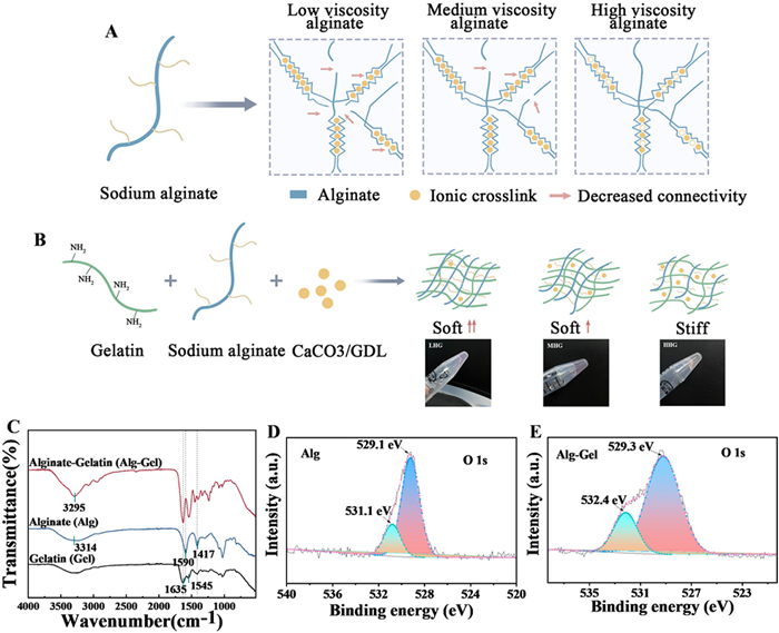
Here, we formulated calcium alginate-gelatin (Alg-Gel) hydrogels using three sodium alginates of varying viscosities, crosslinked via calcium ions to establish tunable network connectivity (Fig. 1A). The hydrogels were prepared by mixing sodium alginate with gelatin, with calcium ions gradually released from calcium carbonate and glucono-δ-lactone (GDL). Ionic crosslinking combined with hydrogen bonding between functional groups of alginate and gelatin yielded homogeneous Alg-Gel networks with modifiable mechanical properties [46,47]. The resulting hydrogels were designated as low- (LHG), medium- (MHG), and high-stiffness (HHG) hydrogels, corresponding to low-, medium-, and high-viscosity alginate respectively (Fig. 1B). The Fourier transform infrared (FT-IR) analysis confirmed Ca2+ coordination with alginate carboxyl groups (red-shifted vibrations) and enhanced hydrogen bonding (amide I shift from 1635 cm−1 to 1626 cm−1) (Fig. 1C). X-ray photoelectron spectroscopy (XPS) further validated Ca2+-carboxylate complexation, showing a 1.3 eV increase in O 1s binding energy (531.1–532.4 eV) (Figs. 1D and E). These spectroscopic results collectively establish the chemo-mechanical foundation of our hydrogel system.
Figure 1
Figure 1.
Schemes and characterizations of the Alg-Gel hydrogels showing interactions between alginate, gelatin and calcium ions. (A) Varying the viscosity of sodium alginate solutions to tune the mechanical properties of calcium-chelated alginate hydrogel. (B) Schematic illustration of the network structures of Alg-Gel hydrogels (LHG, MHG and HHG) of different mechanical properties. (C) The FT-IR spectra of Alg-Gel, Alg and Gel. (D) The O 1s peak in the XPS spectrum of Alg. (E) The O 1s peak in the XPS spectrum of Alg-Gel.
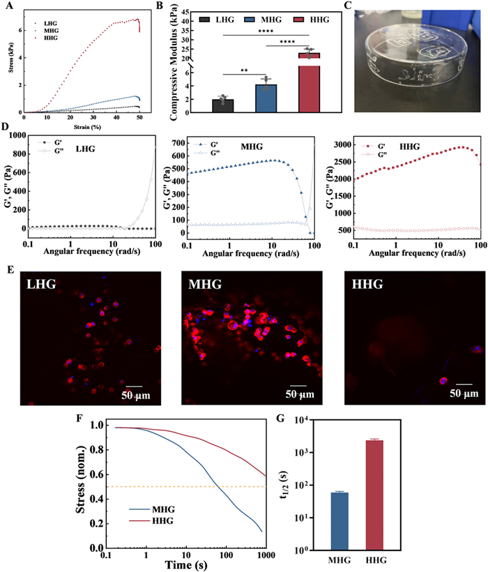
As shown in Fig. S1 (Supporting information), scanning electron microscopy (SEM) analysis revealed a positive correlation between the 3D network density of the composite hydrogels and sodium alginate viscosity (Fig. S1A). The average pore size decreased from 84.8 ± 17.6 µm (LHG) to 42 ± 12.1 µm (HHG) (Fig. S1B), while porosity dropped from 54.9% (LHG) to 17.2% (HHG) (Fig. S1C), attributable to enhanced molecular chain entanglement and denser calcium ion crosslinking in high-viscosity alginate [48,49]. Swelling experiments showed all hydrogels reached equilibrium within 5 h, with ratios of 241.2% (LHG), 207.6% (MGH), and 135.6% (HHG) (Fig. S1D). Degradation studies over 30 days revealed LHG and MHG degraded over 60%, faster than HHG (~40%) (Fig. S1E). To optimize islet cell culture, this study systematically evaluated the mechanical properties of Alg-Gel hydrogels. The results showed that with the increase in sodium alginate viscosity, the Young’s modulus of the hydrogels increased sequentially: LHG (1.98 kPa), MHG (4.21 kPa), and HHG (22.98 kPa) (Figs. 2A and B). Notably, the modulus values of LHG and MHG were consistent with the stiffness range (1–10 kPa) of the native pancreatic matrix [50,51]. Rheological analysis further revealed that MHG exhibited pronounced shear-thinning behavior (Fig. 2C and Fig. S2 in Supporting information), indicating good injectability [52,53]; in contrast, LHG showed weak gel properties due to limited molecular chain entanglement (Fig. 2D). In terms of cytocompatibility, INS-1 cells encapsulated in MHG demonstrated higher viability than those in LHG and HHG (Fig. 2E and Fig. S3 in Supporting information), suggesting that the MHG hydrogel was more suitable for the growth and functional maintenance of islet cells.
Figure 2
Figure 2.
Mechanical and rheological properties of LHG, MHG and HHG hydrogels. (A) Compressive stress of LHG, MHG and HHG hydrogels. (B) Young’s moduli of LHG, MHG and HHG hydrogels. The ordinary one-way ANOVA was performed (n = 5). (C) The photo showing continuously extruded MHG hydrogel through a double-syringe setup to write the word "slime" on the inner side of a glass petri dish. (D) Modulus analysis of LHG, MHG and HHG hydrogels under different frequency oscillations. (E) Representative images of the INS-1 cells cultured in LHG, MHG and HHG hydrogels for 48 h. The cells were stained for actin (red) and nuclei (blue). Scale bar: 50 µm. (F) Stress relaxation tests of hydrogels composed of sodium alginate with different viscosities and gelatin. (G) Quantification of the timescale for stress relaxation to half of its original value (n = 3), τ1/2 from the stress relaxation tests in (F). All data are shown as mean ± SD. **P < 0.01, ****P < 0.0001.
As established in previous studies, fast relaxation in hydrogels facilitates mechanical adaptation to cell expansion and migration [54–56]. We performed stress relaxation tests on composite hydrogels fabricated from sodium alginates of different viscosities. Owing to the excessive fluidity of the LHG hydrogel, reliable stress relaxation data could not be obtained; therefore, the comparison was focused on MHG and HHG hydrogels. Results indicated a significant increase in the stress relaxation rate with higher alginate viscosity (Fig. 2F), demonstrating that hydrogel viscoelasticity can be effectively modulated by varying alginate viscosity [24,57]. Calculation of the half-time for initial stress relaxation (τ1/2) (Fig. 2G) showed that the HHG hydrogel exhibited extremely slow stress relaxation, maintaining high stress under 1% strain (2354.70 ± 206.07 s), indicative of its static mechanical rigidity. In contrast, the MHG hydrogel rapidly dissipated internal stress under load (59.85 ± 2.34 s), promoting the rapid migration of islet cells (Fig. 2E) [57]. This rapid stress relaxation property also enables effective dissipation of injection-induced shear forces (Fig. S2), thereby preventing membrane rupture during the encapsulation process and ensuring high islet cell survival rates.
To elucidate how hydrogel mechanics govern islet cell behavior, we analyzed genomic responses of INS-1 cells to hydrogels of varying stiffness. As shown in Fig. S4 (Supporting information), transcriptomic analysis revealed that intermediate-stiffness hydrogel (MHG) specifically enriched phosphatidylinositol 3-kinase-protein kinase B (PI3K-Akt) and Wnt signaling pathways (Figs. S4A–C), promoting cellular proliferation and enhancing β-cell function through upregulated insulin secretion and modulated lipid metabolism. In contrast, both low and high stiffness conditions induced cellular stress or metabolic disruption (Figs. S4D and E). Quantitative real-time polymerase chain reaction (qPCR) confirmed that MHG significantly upregulated key genes (Akt1, Bcl2, Ctnnb1, Myc) in these pathways, with markedly attenuated effects in HHG (Fig. S4F), highlighting the critical role of stress-relaxation properties in activating pro-survival signaling. These findings establish that hydrogel mechanics orchestrate islet cell responses via mechanosensitive PI3K-Akt/Wnt signaling, with MHG optimally emulating physiological β-cell functionality. To evaluate biocompatibility for clinical translation, we co-cultured INS-1 cells with LHG, MHG, and HHG hydrogels. Cell counting kit-8 (CCK-8) assays revealed significant cell proliferation in all groups, with MHG showing the most prominent effect (Fig. S4G). Hemolysis tests confirmed the blood compatibility of all hydrogels, with hemolysis rates below the 5% threshold for biomedical materials (Fig. S4H).
PRP contains multiple growth factors (platelet-derived growth factor (PDGF), vascular endothelial growth factor (VEGF), insulin-like growth factor-1 (IGF-1), epidermal growth factor (EGF)) crucial for tissue repair, yet their clinical application is limited by burst release upon activation. To address this, we encapsulated PRP in Alg-Gel hydrogel where Ca2+ serves as both crosslinker and PRP stabilizer, enabling sustained growth factor release. Using the optimal MHG hydrogel, we demonstrated significantly prolonged release profiles of IGF-1 (Fig. 3A), VEGF (Fig. 3B), PDGF (Fig. S5A in Supporting information) and EGF (Fig. S5B in Supporting information) compared to PRP alone. This controlled release is attributed to synergistic 3D network encapsulation and Ca2+-mediated spatial activation of PRP. To evaluate the performance of MHG and MHG@PRP hydrogels in supporting β-cell growth, we assessed INS-1 cell viability and function. CCK-8 assays and live/dead staining showed no cytotoxicity in any group, with MHG@PRP maintaining over 90% cell viability after 7 days (Fig. 3C). Confocal imaging revealed improved 3D distribution and proliferation of INS-1 cells in MHG@PRP compared to MHG alone (Figs. 3D and E). Glucose-stimulated insulin secretion (GSIS) assays showed that while MHG/cells exhibited reduced insulin secretion vs. free INS-1 cells, MHG@PRP/cells demonstrated enhanced insulin production under both basal (2.2 mmol/L) and stimulatory (16.7 mmol/L) glucose conditions (Fig. 3F), indicating PRP supplementation creates a favorable microenvironment for β-cell function.
Figure 3
Figure 3.
The effect of PRP on the functions of islet cells. Release kinetic curves of IGF-1 (A) and VEGF (B) in PRP and PRP-loaded MHG hydrogel (MHG@PRP) (n = 5). (C) The viability of INS-1 cells was measured by CCK-8 assay after culture in the MHG and MHG@PRP hydrogel from day 1 to day 7. The ordinary two-way ANOVA was performed (n = 5). (D) The fluorescence microscopic images of live/dead staining of INS-1 cells cultured in MHG and MHG@PRP hydrogel for 48 h. Scale bar: 50 µm. (E) Representative images of the cultured INS-1 cells in MHG and MHG@PRP hydrogel stained for actin (red) and nuclei (blue) at 24 h. (F) The insulin secretion of the INS-1 cells in monolayer cultivation, encapsulated in the MHG or encapsulated in MHG@PRP hydrogel by GSIS assays, GSIS assays were performed under 2.2 mmol/L (basal) and 16.7 mmol/L (stimulatory) glucose to quantify both basal secretion and glucose responsiveness. The ordinary two-way ANOVA was performed (n = 5). All data are shown as mean ± SD. *P < 0.05, **P < 0.01. PI, propidium iodide; ns, not significant.
Transcriptome analysis revealed that PRP-released growth factors (e.g., PDGF, IGF-1) enhance islet cell function primarily through PI3K-Akt-mammalian target of rapamycin (mTOR) pathway activation, promoting proliferation and inhibiting apoptosis (Figs. S6A and B in Supporting information) [58,59]. PRP further improved metabolic homeostasis by downregulating lipotoxic pathways and reinforced cell-matrix interactions via extracellular matrix (ECM) component upregulation and focal adhesion activation (Figs. S6C and D in Supporting information). Notably, no inflammatory pathway activation was observed, indicating an immunologically silent phenotype. These multi–faceted mechanisms, including enhanced survival signaling, optimized metabolism, improved ECM integration, and suppressed inflammation, collectively explain the improved graft tolerance observed in vivo and underscore the capacity of MHG@PRP to foster an immune–tolerant microenvironment essential for long–term graft success.
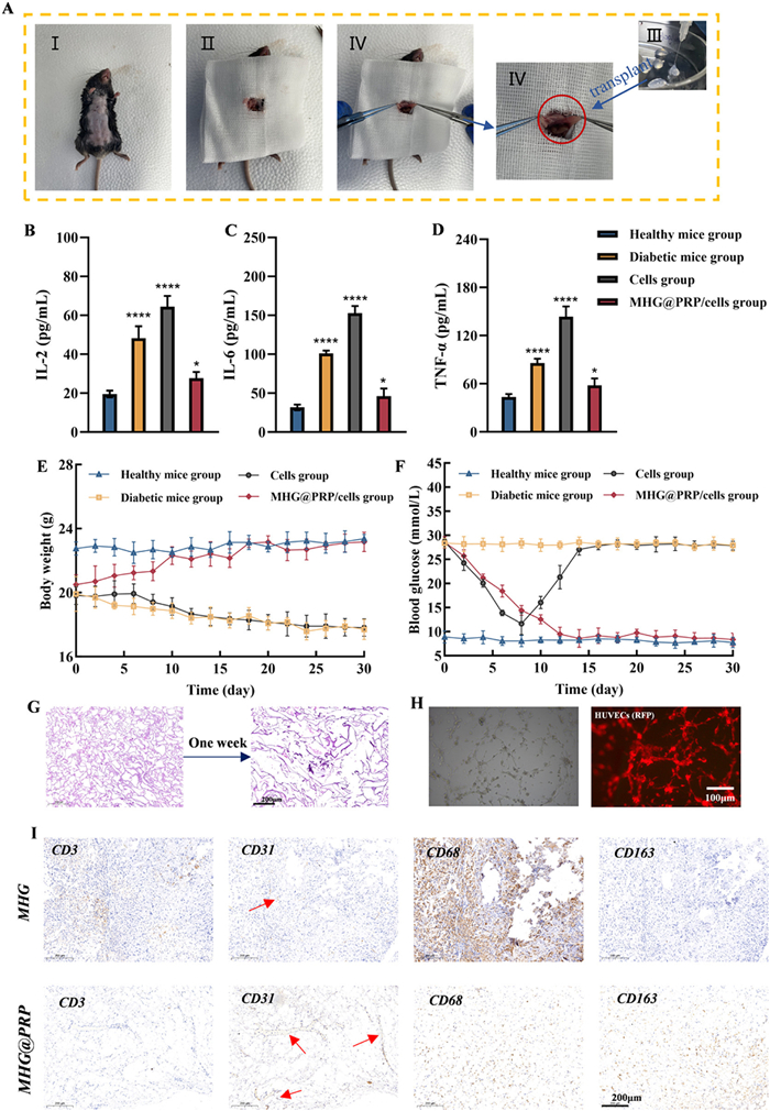
To evaluate the therapeutic potential of our MHG@PRP hydrogel for T1D, we transplanted either MHG@PRP-encapsulated INS-1 cells (MHG@PRP/cells group) or naked INS-1 cells (cells group) into the omental pouch of streptozotocin (STZ)-induced diabetic mice (Fig. 4A). All experimental protocols had been pre-approved by the Institutional Animal Care and Use Committee (IACUC) of Jinling Hospital, Medical School of Nanjing University (approval No. DZGZRDW2400240). While diabetic controls showed persistent hyperglycemia and weight loss (Figs. 4E and F), the naked cell group only exhibited transient glucose reduction followed by rejection, accompanied by elevated pro-inflammatory cytokines (interleukin-2 (IL-2), IL-6, and tumor necrosis factor-alpha (TNF-α)) (Figs. 4B–D). In contrast, the MHG@PRP/cells group maintained normoglycemia for 30 days with stable body weight and attenuated inflammation, demonstrating effective immune protection and sustained metabolic recovery. Metabolic function was further assessed in MHG@PRP/cells-transplanted mice via intraperitoneal glucose tolerance tests (IPGTT). Following a 16-h fast, these mice exhibited glucose clearance kinetics closely resembling healthy controls, with blood glucose peaking within 15 min and returning to baseline by 120 min post-injection (Fig. S7A in Supporting information). Quantitative analysis confirmed significantly reduced area under the curve (AUC) compared to pre-transplantation values (Fig. S7B in Supporting information), demonstrating restored glucose regulation.
Figure 4
Figure 4.
MHG@PRP hydrogel enables effective diabetes reversal through immunomodulation and graft protection in vivo. (A) The procedure of the MHG@PRP/cells graft transplanted into the omentum of diabetic mice modeled by STZ injection. Serum levels of IL-2 (B), IL-6 (C), and TNF-α (D) in healthy mice and diabetic mice were tested at 3 days post-transplantation. The ordinary one-way ANOVA was performed (n = 3). (E) The changes in body weight of healthy mice and diabetic mice within 30 days after the operation (n = 6). (F) The changes in non-fasting blood glucose levels of healthy mice and diabetic mice within 30 days after the operation (n = 6). (G) The H&E staining of the explanted subcutaneous implants after one week showed substantial structural collapse of the MHG@PRP hydrogel. Scale bar: 200 µm. (H) Tube formation assay of HUVECs cultured in vitro with MHG@PRP hydrogel. Scale bar: 100 µm. (I) IHC staining of subcutaneously transplanted MHG and MHG@PRP hydrogel. Scale bar: 200 µm. All data are shown as mean ± SD. *P < 0.05, ****P < 0.0001.
To comprehensively evaluate the biocompatibility, biodegradability, and immunomodulatory properties of the MHG@PRP hydrogel, we conducted both subcutaneous implantation studies and a diabetic mouse therapy model using hydrogel-encapsulated islets.
Subcutaneous implantation revealed significant pro-angiogenic effects of MHG@PRP, with CD31 staining demonstrating enhanced vascularization compared to MHG alone. This finding was further corroborated by an in vitro human umbilical vein endothelial cells (HUVECs) tube formation assay (Fig. 4H). Immunological analysis showed that MHG@PRP induced a higher ratio of CD163-positive M2 macrophages to CD68-positive total macrophages compared to the control, indicating a PRP-driven shift of the innate immune response toward an anti-inflammatory, pro-healing phenotype. Furthermore, a marked reduction in CD3-positive T-cell infiltration within and around the MHG@PRP hydrogel demonstrated its ability to effectively mitigate the activation of adaptive immunity (Fig. 4I). Hematoxylin and eosin (H&E) staining of the explanted subcutaneous implants after one week showed substantial structural collapse of the MHG@PRP hydrogel (Fig. 4G), confirming its biodegradable nature and suggesting a favorable clearance profile in vivo.
In the therapeutic model, H&E staining of the peri–graft omental tissue from mice transplanted with MHG@PRP-encapsulated islets revealed minimal lymphocyte infiltration and only a thin fibrous capsule, indicating a mild foreign body response (Fig. S8 in Supporting information). Notably, the transplanted islet clusters maintained well-defined boundaries and compact architecture, with a clear interface between the hydrogel and host tissue accompanied by only sporadic lymphocyte presence. In stark contrast, diabetic mice receiving unencapsulated islet transplants exhibited extensive lymphocyte infiltration, severe disruption of islet morphology, and substantial β-cell loss (indicated by red circles in Fig. S8) consistent with acute immune rejection that correlated with functional graft failure. Additionally, histopathological assessment of major organs (liver, kidney, and spleen) showed no apparent pathological abnormalities in the MHG@PRP/cells treatment group (Fig. S9 in Supporting information), affirming the biosafety of our approach.
In summary, this study demonstrates the feasibility of using a stress-relaxing hydrogel for the co-delivery of PRP and β-cells as a therapeutic approach for diabetes. MHG@PRP hydrogel regulates the density of reversible ionic cross-linking points and the mobility of polymer chains by adjusting the viscosity of sodium alginate, and incorporates gelatin to provide cell adhesion sites. The results show that the MHG hydrogel of rapid stress relaxation provides a three-dimensional microenvironment supporting the function of INS-1 cells by activating the PI3K-Akt/Wnt signaling pathway and promoting dynamic ECM remodeling, while the introduction of PRP further enhances the physiological performance of islet cells through the activation of the growth factor-PI3K/Akt/mTOR axis and the regulation of ECM homeostasis regulation. In vivo evaluation in diabetic mouse models confirmed the therapeutic potential, where transplantation of β-cell-laden MHG@PRP into omentum tissue restored glucose-responsive insulin secretion and maintained glycemic control for over 30 days. This study combines the regulation of the mechanical microenvironment with nutritional supply, establishing a novel paradigm for hydrogel design in diabetes cell therapy.
The authors declare that they have no known competing financial interests or personal relationships that could have appeared to influence the work reported in this paper.
Acknowledgment
The authors acknowledge financial support from the National Natural Science Foundation of China (Nos. 51873087, 81801839).
Supplementary materials
Supplementary material associated with this article can be found, in the online version, at doi:10.1016/j.cclet.2025.112128.
Figure 1
Schemes and characterizations of the Alg-Gel hydrogels showing interactions between alginate, gelatin and calcium ions. (A) Varying the viscosity of sodium alginate solutions to tune the mechanical properties of calcium-chelated alginate hydrogel. (B) Schematic illustration of the network structures of Alg-Gel hydrogels (LHG, MHG and HHG) of different mechanical properties. (C) The FT-IR spectra of Alg-Gel, Alg and Gel. (D) The O 1s peak in the XPS spectrum of Alg. (E) The O 1s peak in the XPS spectrum of Alg-Gel.
Figure 2
Mechanical and rheological properties of LHG, MHG and HHG hydrogels. (A) Compressive stress of LHG, MHG and HHG hydrogels. (B) Young’s moduli of LHG, MHG and HHG hydrogels. The ordinary one-way ANOVA was performed (n = 5). (C) The photo showing continuously extruded MHG hydrogel through a double-syringe setup to write the word "slime" on the inner side of a glass petri dish. (D) Modulus analysis of LHG, MHG and HHG hydrogels under different frequency oscillations. (E) Representative images of the INS-1 cells cultured in LHG, MHG and HHG hydrogels for 48 h. The cells were stained for actin (red) and nuclei (blue). Scale bar: 50 µm. (F) Stress relaxation tests of hydrogels composed of sodium alginate with different viscosities and gelatin. (G) Quantification of the timescale for stress relaxation to half of its original value (n = 3), τ1/2 from the stress relaxation tests in (F). All data are shown as mean ± SD. **P < 0.01, ****P < 0.0001.
Figure 3
The effect of PRP on the functions of islet cells. Release kinetic curves of IGF-1 (A) and VEGF (B) in PRP and PRP-loaded MHG hydrogel (MHG@PRP) (n = 5). (C) The viability of INS-1 cells was measured by CCK-8 assay after culture in the MHG and MHG@PRP hydrogel from day 1 to day 7. The ordinary two-way ANOVA was performed (n = 5). (D) The fluorescence microscopic images of live/dead staining of INS-1 cells cultured in MHG and MHG@PRP hydrogel for 48 h. Scale bar: 50 µm. (E) Representative images of the cultured INS-1 cells in MHG and MHG@PRP hydrogel stained for actin (red) and nuclei (blue) at 24 h. (F) The insulin secretion of the INS-1 cells in monolayer cultivation, encapsulated in the MHG or encapsulated in MHG@PRP hydrogel by GSIS assays, GSIS assays were performed under 2.2 mmol/L (basal) and 16.7 mmol/L (stimulatory) glucose to quantify both basal secretion and glucose responsiveness. The ordinary two-way ANOVA was performed (n = 5). All data are shown as mean ± SD. *P < 0.05, **P < 0.01. PI, propidium iodide; ns, not significant.
Figure 4
MHG@PRP hydrogel enables effective diabetes reversal through immunomodulation and graft protection in vivo. (A) The procedure of the MHG@PRP/cells graft transplanted into the omentum of diabetic mice modeled by STZ injection. Serum levels of IL-2 (B), IL-6 (C), and TNF-α (D) in healthy mice and diabetic mice were tested at 3 days post-transplantation. The ordinary one-way ANOVA was performed (n = 3). (E) The changes in body weight of healthy mice and diabetic mice within 30 days after the operation (n = 6). (F) The changes in non-fasting blood glucose levels of healthy mice and diabetic mice within 30 days after the operation (n = 6). (G) The H&E staining of the explanted subcutaneous implants after one week showed substantial structural collapse of the MHG@PRP hydrogel. Scale bar: 200 µm. (H) Tube formation assay of HUVECs cultured in vitro with MHG@PRP hydrogel. Scale bar: 100 µm. (I) IHC staining of subcutaneously transplanted MHG and MHG@PRP hydrogel. Scale bar: 200 µm. All data are shown as mean ± SD. *P < 0.05, ****P < 0.0001.


 DownLoad:


 下载:
下载:


 下载:
下载:

