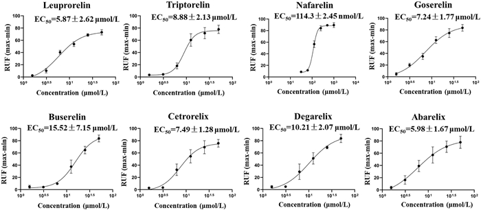
Figure 1.
The EC50 of GnRH analogs induce MRGPRX2-HEK293 cells calcium mobilization. n = 5 and experiments were repeated > 3 times. Data are presented as mean ± S.E.M.

Cationic modifications in gonadotropin-releasing hormone analogues drive mas-related G protein-coupled receptor X2-dependent histamine release through charge interactions
Yajing Hou , Qifan Yang , Nan Zhou , Li Li , Zhuo Li , Yaning Zhu , Zhiyuan Fang , Tongfei Yang , Peng Zhang , Dezhong Zhou , Delu Che


Gonadotropin-releasing hormone (GnRH) analogues are a class of synthetic peptide drugs derived from structural modifications of endogenous GnRH [1,2]. Clinically utilized in assisted reproductive technology and the treatment of various sex hormone-related disorders, including uterine fibroids, endometriosis, ovarian cancer, and prostate cancer, they have broad prospects for clinical application and development [3–5]. However, beyond well-documented adverse effects, such as increased cardiovascular risk, osteoporosis, and sexual dysfunction, allergic reactions-including urticaria, rash, pruritus, and anaphylactic shock-are among the most frequent complications [6,7]. For instance, a study on leuprorelin reported an allergy incidence of 0.49% in 1010 patients [8]. Local site reactions have been frequently reported in 10%–15% of patients and systemic hypersensitivity reactions in 3.8% of patients, using GnRH analogue preparations [9].
Early observations revealed that GnRH analogues induce histamine release at injection sites, leading to localized and systemic pseudo-allergic reactions that restrict their clinical use [10]. Researchers have attempted to mitigate this by modifying the N-terminal structure, optimizing flexibility, and adjusting basicity to reduce histamine-releasing activity [11]. Ex vivo human skin experiments have shown that degarelix induces minimal histamine release compared to cetrorelix, abarelix, and ganirelix [12]. However, histamine release remains the primary adverse reaction associated with these agents. As a key allergic mediator, histamine triggers vasodilation, smooth muscle contraction, and other hypersensitivity symptoms, prompting the hypothesis that GnRH analogue-induced histamine release causes anaphylactoid reactions. However, the molecular mechanisms underlying this process remain unclear.
Mas-related G protein-coupled receptor X2 (MRGPRX2), a recently identified mast cell membrane receptor [13], is a critical mediator of direct mast cell activation and drug-induced pseudo-allergic reactions [14]. Small-molecule drugs, such as fluoroquinolone antibiotics [15], phenothiazine antipsychotics [16], opioids [17], and antidepressants [18], trigger mast cell degranulation by directly activating MRGPRX2. These findings elucidate the specific mechanisms underlying the pseudo-allergic reactions caused by such drugs in clinical applications.
In addition to its activation by small-molecule drugs, MRGPRX2 has been identified as a critical receptor for the direct activation of mast cells by peptide molecules. Various endogenous peptides, including the antimicrobial peptide LL-37, substance P, neurokinin, and angiotensin, act as agonists of MRGPRX2 and participate in the development and progression of pathological processes, such as pruritus and inflammation, within the body [19,20]. Peptide-based drugs, such as icatibant, octreotide, and sermorelin, trigger the degranulation of human mast cells (LAD2) via MRGPRX2 activation. Furthermore, through their homologous receptor MRGPRB2, these drugs induce anaphylactoid symptoms in mice, including local tissue swelling, exudation, hypothermia, and release of inflammatory cytokines [14,21]. Moreover, GnRH analogues, such as cetrorelix and leuprorelin, have also been confirmed to act as MRGPRX2 agonists, inducing histamine release from mast cells [14].
Structural studies of MRGPRX2 have revealed that key amino acids, such as E164, D184, and W243, are indispensable for its activation by ligand binding [22–25]. Upon activation, MRGPRX2 mediates mast cell degranulation, leading to the release of granular substances containing various proinflammatory mediators [26]. Among these, histamine, one of the most prominent bioactive components, triggers pathophysiological effects, such as vasodilation and smooth muscle contraction, via the activation of histamine receptors [27]. Beyond histamine release, MRGPRX2 activation also promotes the synthesis and secretion of cytokines, interleukins, and chemokines by mast cells, thereby exacerbating inflammatory responses, pain, and pruritus [28,29]. In this study, we aimed to evaluate the risk of pseudo-allergic reactions induced by GnRH analogues through the activation of MRGPRX2 using both in vivo and in vitro experimental models. Additionally, molecular docking techniques and structural modification strategies were employed to elucidate the molecular mechanisms and structural determinants of MRGPRX2 activation by GnRH analogues.
First, an LAD2 cell model was established to investigate the activation effects of GnRH analogues on mast cells. After adding test drugs at different concentrations to LAD2 cells, the levels of Ca2+ mobilization, β-hexosaminidase, histamine, and tumor necrosis factor α (TNF-α) were measured. Leuprorelin was used as the positive control. As shown in Fig. S1 (Supporting information), although all tested compounds induced calcium mobilization in LAD2 cells, their activities exhibited significant differences. The degranulation activities of the GnRH analogues also varied (Fig. S2 in Supporting information). Abarelix and nafarelin significantly induced mast cell degranulation at 3.125 µmol/L, whereas triptorelin and buserelin required 12.5 µmol/L to activate mast cells. The other drugs triggered mast cell release at 6.25 µmol/L. These results indicate that GnRH analogues can be classified into three categories based on their potency to induce mast cell degranulation: Abarelix and nafarelin exhibit the strongest activity; cetrorelix, degarelix, and goserelin show moderate activity; and triptorelin and buserelin demonstrate the weakest activity. In vitro experiments suggest that triptorelin and buserelin may pose a lower risk of inducing anaphylactoid reactions.
To further evaluate the mechanism of mast cell activation induced by GnRH analogues, siRNA technology was employed to knockdown the expression of MRGPRX2 in LAD2 cells and examine the changes in the degranulation reaction caused by these drugs. The knockdown efficiency of MRGPRX2 was approximately 50%, as determined by quantitative real-time PCR (RT-qPCR) (Fig. S3 in Supporting information). As shown in Fig. S4 (Supporting information), all GnRH analogues reduced the release of β-hexosaminidase, histamine, and TNF-α in MRGPRX2-knockdown LAD2 cells. However, high doses of these drugs still activated the knockdown cells, likely because siRNA technology only reduces gene expression and cannot achieve complete knockout.
Calcium mobilization showed the same pattern, with an increase in intracellular calcium concentration in MRGPRX2-HEK293 cells but no effect on HEK293 cells (Fig. S5 in Supporting information). Calculation of median effective concentration (EC50) values for MRGPRX2 activation based on calcium mobilization revealed that nafarelin exhibited the strongest activity, whereas degarelix and buserelin showed the weakest activity (Fig. 1). Notably, previous literature on adverse reaction risks aligns with our observations. For instance, degarelix induced significantly less histamine release than cetrorelix, abarelix, or ganirelix [12]. Furthermore, World Health Organization data analyzing the immune-related adverse effects of GnRH analogues across 97 countries (1988–2010) indicated that leuprolide accounted for more than two-thirds of reported immune system disorders, whereas buserelin, goserelin, and triptorelin accounted for far fewer cases-even when analyses focused solely on anaphylactic shock and anaphylactoid reactions. Goserelin ranked second among reported cases, whereas buserelin and triptorelin had the lowest incidence [30]. Although the global market share of these agents remains unclear, real-world data partially reflect their relative pseudo-allergic risk profiles, which are consistent with our findings. Based on these results, triptorelin and buserelin may be safer choices for patients with a history of hypersensitivity or predisposition to allergic reactions, as they demonstrate the lowest risk of MRGPRX2-mediated pseudo-allergic responses.
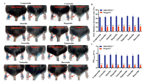
The Animal Ethics Committee at Xi’an Jiaotong University approved the mouse model experimental protocols (permit No. XJTUAE2023-1044). Leuprorelin, cetrorelix, degarelix, goserelin, triptorelin, and buserelin were injected subcutaneously, abarelix was administered intramuscularly, and nafarelin was delivered via nasal spray. They primarily cause immediate local allergic reactions in clinical settings. In our study, a murine passive cutaneous anaphylaxis model was used to validate the pseudo-allergic activity of GnRH analogues. Two groups of mice were utilized: Mrgprb2 knockout mice (Mrgprb2−/−) and a transgenic mouse model expressing human MRGPRX2 on a Mrgprb2−/− background (MRGPRX2+/+). GnRH analogues were subcutaneously injected into the paws of mice, and their sensitization potential was evaluated by measuring paw swelling and Evans blue extravasation in tissue fluid. Representative images are shown in Fig. 2A. GnRH analogues significantly increased both paw swelling (Fig. 2B) and Evans blue (Fig. 2C) leakage in MRGPRX2+/+ mice but showed no effects in Mrgprb2−/− mice. Furthermore, compared with Mrgprb2−/− mice, GnRH analogues induced local capillary dilation hematoxylin-eosin (H&E) staining) and irregular mast cell morphology with granular content leakage in MRGPRX2+/+ mice, as evidenced by avidin staining (Fig. S6 in Supporting information). These experimental results collectively demonstrate that GnRH analogues can induce mast cell degranulation through MRGPRX2 activation, posing the risk of localized pseudo-allergic reactions in mice.
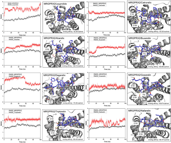
Molecular dynamics simulations and docking analyses confirmed the binding of GnRH analogues to MRGPRX2, revealing that the positively charged centers (Arg) of the GnRH analogues formed salt bridges with E164 and D184. However, in the case of abarelix and degarelix, whose positively charged centers are derived from N-isopropyl lysine, only one of these residues (either E164 or D184) participated in salt bridge interactions and the quantitative binding energy values was showed in Fig. 3.
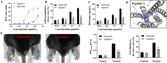
The initial virtual docking results suggested that the positively charged center of the GnRH analogues was a critical site for binding to MRGPRX2. To further validate this hypothesis, Peptide-1, a modified analogue in which the eighth-position Arg of triptorelin was substituted with Glu, was synthesized and compared with triptorelin for MRGPRX2 activation (Fig. S7 in Supporting information). To verify the core pharmacological efficacy of triptorelin and Peptide-1, a molecular docking analysis was performed. We supply the results of GnRHR binding with Peptide-1 and triptorelin. The molecular dynamics results show that R8E mutant of Triptorelin is slightly reduce the binding affinity (−91.68 vs. −81.19 kcal/mol by MM/GB-SA method). Although Arg8 is positioned to potentially form a cation-π interaction with His3067.36 and a salt bridge with Asp3027.32, the principal interactions of GnRH analogs with GnRHR are facilitated by pGlu1, His2, Trp3, Tyr5, Pro9, and Gly10. All of these key ligand residues are buried within the receptor’s binding pocket. From an evolutionary perspective, Arg8 is variable across species, whereas His3067.36 is highly conserved. Although Asp3027.32 is not a conserved residue, it does not appear to consistently form clear interactions (such as salt bridges, hydrogen bonds, or pi-interactions) (Fig. S8 in Supporting information). Additionally, we reviewed a large number of studies. Research indicates that two potential binding sites between GnRH and its receptor have been identified: Lys121 and Glu301. Lys121 is located in the ligand-binding pocket and is easily accessible by GnRH. It has been confirmed as a critical site for GnRH binding, and the agonistic binding of high-affinity GnRH analogues requires a positive charge at this position [31]. Based on this finding, since Arg8 in GnRH analogues inherently carries a positive charge, the likelihood of its binding to Lys121 of the GnRH receptor is extremely low. Therefore, we speculate that this structural feature explains why most modifications of GnRH analogues have focused on positions 6, 9 and 10 [1]. These modifications have been shown to delay peptide degradation, prolong half-life, and enhance receptor-binding affinity. Of course, this remains a theoretical hypothesis. Because our study primarily investigated the side effects of pseudo-allergic reactions, further research is required to validate this speculation.
Peptide-1 exhibited significantly reduced activity compared to triptorelin at equivalent concentrations in vitro, inducing lower calcium mobilization in MRGPRX2-HEK293 cells (Fig. 4A) and triggering substantially less β-hexosaminidase release (Fig. 4B) and histamine release (Fig. 4C) in LAD2 cells. Molecular docking analysis revealed that replacing positively charged Arg with negatively charged Glu at the eighth position abolished salt-bridge interactions with MRGPRX2, leading to significantly weakened receptor activation and the quantitative binding energy values of Peptide-1 showed in Fig. 4D. In vivo, Peptide-1 nearly failed to induce paw swelling, plasma extravasation (Fig. 4E), or mast cell (MC) activation in the paw skin of MRGPRX2+/+ mice (Fig. S9 in Supporting information). These results demonstrate the essential role of the positively charged center in GnRH analogues in activating MRGPRX2.
Flouret et al. also demonstrated that GnRH antagonists generated by relocating the cationic amino acid from position eight to position seven resulted in reduced histamine release, whereas combining lysine at position seven with a neutral amino acid at position eight led to further attenuation of histamine release [32]. Among proteogenic amino acids, lysine is extensively involved in posttranslational modifications and ligand-protein binding, making its selective modification attractive [33]. This conclusion aligns closely with our research findings, underscoring the critical role of the positively charged center of GnRH analogues in mediating histamine release. Based on these insights, structural modifications of GnRH analogues to minimize overall molecular positive charge while preserving therapeutic activity may effectively reduce histamine release and the associated adverse effects. These findings provide novel insights into the screening of peptide ligands targeting MRGPRX2. Peptide drugs hold great promise as therapeutic modalities [34], and our findings provide a foundation for research on their adverse effects.
In this study, we verified that GnRH analogues induce mast cell degranulation via MRGPRX2 in vivo and cause local anaphylactic reactions in MRGPRX2+/+ mice. Cationic modifications at position eight in GnRH analogues bind to MRGPRX2 through salt-bridge interactions, which are key factors in receptor activation. Overall, the evidence presented here comprehensively uncovers the mechanism of GnRH analogue-induced histamine release, which is significant for enhancing the safety of their clinical application and provides novel insights into the development of new GnRH-based therapeutics.
Yajing Hou: Writing – original draft, Supervision, Resources, Methodology, Investigation, Funding acquisition, Formal analysis, Data curation, Conceptualization. Qifan Yang: Supervision, Software, Resources. Nan Zhou: Methodology, Investigation, Formal analysis, Data curation. Li Li: Methodology, Investigation. Zhuo Li: Resources, Methodology. Yaning Zhu: Resources, Methodology. Zhiyuan Fang: Validation, Supervision, Software. Tongfei Yang: Validation, Supervision. Peng Zhang: Writing – review & editing, Supervision, Resources. Dezhong Zhou: Writing – review & editing, Methodology, Investigation, Data curation, Conceptualization. Delu Che: Writing – review & editing, Visualization, Validation, Supervision, Software, Resources, Project administration, Methodology, Investigation, Funding acquisition, Formal analysis, Data curation, Conceptualization.
The authors declare that they have no known competing financial interests or personal relationships that could have appeared to influence the work reported in this paper.
The authors gratefully acknowledge the financial support from the National Natural Science Foundation of China (Nos. 82304441, 82304689).
Supplementary material associated with this article can be found, in the online version, at doi:
O. Ortmann, J.M. Weiss, K. Diedrich, Reprod. Biomed. Online 5 (2002) 1–7.
D. Weckermann, R. Harzmann, Eur. Urol. 46 (2004) 279–284. doi: 10.1016/j.eururo.2004.05.006
X. Luo, Y. Wu, X. Zhang, et al., Chin. Chem. Lett. 36 (2025) 109724. doi: 10.1016/j.cclet.2024.109724
C. Gobello, Reprod. Domest. Anim. 47 (2012) 373–376. doi: 10.1111/rda.12025
S.J. Freedland, P.A. Abrahamsson, Asian J. Androl. 23 (2021) 3–10. doi: 10.4103/aja.aja_22_20
T. Raja, R. Sud, S. Addla, et al., Indian J. Cancer 59 (2022) S142–S159. doi: 10.4103/ijc.ijc_65_21
A.P. Metbulut, K.T. Adıgüzel, C. İslamoğlu, et al., Indian J. Endocrinol. Metab. 25 (2021) 527–531. doi: 10.4103/ijem.ijem_333_21
J.B. Engel, A. Schultze-Mosgau, K. Diedrich, Reprod. Biomed. Online 10 (2005) 61–65. doi: 10.1016/S1472-6483(11)60392-1
T. Kirkgoz, E. Karakoc-Aydiner, F. Bugrul, et al., Horm. Res. Paediatr. 93 (2020) 66–72. doi: 10.1159/000505329
J.E. Morgan, C.E. O’Neil, D.H. Coy, et al., Int. Arch. Allergy Appl. Immunol. 80 (1986) 70–75. doi: 10.1159/000234028
J.J. Nestor Jr, R. Tahilramani, T.L. Ho, et al., J. Med. Chem. 35 (1992) 3942–3948. doi: 10.1021/jm00099a023
W. Koechling, R. Hjortkjaer, L.B. Tankó, Br. J. Clin. Pharmacol. 70 (2010) 580–587. doi: 10.1111/j.1365-2125.2010.03730.x
H. Wu, M. Zeng, E.Y. Cho, et al., Prog. Histochem. Cyto. 50 (2015) 11–17. doi: 10.1016/j.proghi.2015.06.001
B.D. McNeil, P. Pundir, S. Meeker, et al., Nature 519 (2015) 237–241. doi: 10.1038/nature14022
T. Zhang, D.L. Che, R. Liu, et al., Eur. J. Immunol. 47 (2017) 1949–1958. doi: 10.1002/eji.201746951
Y.J. Hou, D.L. Che, D. Wei, et al., Mol. Immunol. 111 (2019) 118–127. doi: 10.1016/j.molimm.2019.04.008
K. Lansu, J. Karpiak, J. Liu, et al., Nat. Chem. Biol. 13 (2017) 529–536. doi: 10.1038/nchembio.2334
K. Wolf, H. Kühn, F. Boehm, et al., J. Allergy Clin. Immunol. 148 (2021) 506–522. doi: 10.1016/j.jaci.2020.12.655
H. Subramanian, K. Gupta, H. Ali, J. Allergy Clin. Immunol. 138 (2016) 700–710. doi: 10.1016/j.jaci.2016.04.051
H. Ali, J. Immunobiol. 1 (2016) 115. doi: 10.1109/IWCI.2016.7860350
M. Kumar, K. Duraisamy, B.K.C. Chow, Cells 10 (2021) 31. doi: 10.1515/openps-2021-0004
C. Chompunud Na Ayudhya, S. Roy, I. Alkanfari, et al., Int. J. Mol. Sci. 20 (2019) 5247. doi: 10.3390/ijms20215247
I. Alkanfari, K. Gupta, T. Jahan, H. Ali, J. Immunol. 201 (2018) 343–349. doi: 10.4049/jimmunol.1701793
C. Cao, H.J. Kang, I. Singh, et al., Nature 600 (2021) 170–175. doi: 10.1038/s41586-021-04126-6
F. Yang, L. Guo, Y. Li, et al., Nature 600 (2021) 164–169. doi: 10.1038/s41586-021-04077-y
S. Roy, A. Ganguly, M. Haque, H. Ali, J. Immunol. 202 (2019) 1229–1238. doi: 10.4049/jimmunol.1801227
R. Seifert, A. Strasser, E.H. Schneider, et al., Trends Pharmacol. Sci. 34 (2013) 33–58.
D.P. Green, N. Limjunyawong, N. Gour, et al., Neuron 101 (2019) 412–420. doi: 10.1016/j.neuron.2019.01.012
H. Kühn, P. Kolkhir, M. Babina, et al., J. Allergy Clin. Immunol. 147 (2020) 456–469.
A.B. Lüchinger, V. Mijatovic, T. Rustemeyer, P.G. Hompes, Am. J. Med. Sci. 341 (2011) 240–242. doi: 10.1097/MAJ.0b013e31820094da
W. Zhou, V. Rodic, S. Kitanovic, et al., J. Biol. Chem. 270 (1995) 18853–18857. doi: 10.1074/jbc.270.32.18853
G. Flouret, K. Mahan, T. Majewski, J. Med. Chem. 35 (1992) 636–640. doi: 10.1021/jm00082a004
J. Li, J. Chen, Q.L. Hu, et al., Chin. Chem. Lett. 36 (2025) 110126.
J. Deng, J. Shi, D. Li, J. Zheng, Chin. Chem. Lett. 31 (2020) 2365–2374.

Figure 1 The EC50 of GnRH analogs induce MRGPRX2-HEK293 cells calcium mobilization. n = 5 and experiments were repeated > 3 times. Data are presented as mean ± S.E.M.

Figure 2 GnRH analogs induce mice Evans blue extravasation or paw swell via MRGPRX2. (A) representative images showing Evans blue dye extravasation after intraplantar injection of 5 µL different concentrations of GnRH analogs (left) or saline (right). (B) Quantification of paw thickness increase. (C) Quantification of Evans blue leakage into the paw. n = 5 and experiments were repeated > 3 times. Data are presented as mean ± S.E.M. Two-tailed unpaired Student’s t-test was used to determine significance in statistical comparisons. ***P < 0.001.

Figure 3 Molecular dynamics simulations (left) and docking analysis (right) of GnRH analogs to MRGPRX2. The protein 3D structure was form the MRGPRX2-PAMP12 complex (PDB entry: 7VV0). Molecular dynamics simulation was carried out under Amber program, and simulation duration for 100 ns. Molecular docking analysis was conduct by HPEPDOCK 2.0.

Figure 4 Peptide-1 compared with triptorelin in activating MRGPRX2. (A) Intracellular Ca2+ mobilization of Peptide-1 and triptorelin on MRGPRX2-HEK293 cell. (B) β-hexosaminidase release of Peptide-1 and triptorelin on LAD2 cells. (C) Histamine release of Peptide-1 and triptorelin on LAD2 cells. (D) Molecular docking of Peptide-1 with MRGPRX2. (E) Evans blue dye extravasation and paw swelling after intraplantar injection of 5 µL Peptide-1 and triptorelin at concentration of 10 µg/mL. n = 5 and experiments were repeated > 3 times. Data are presented as mean ± S.E.M. Two-tailed unpaired Student’s t-test was used to determine significance in statistical comparisons. **P < 0.01, ***P < 0.001.

 扫一扫看文章


扫一扫关注我们
 DownLoad:
 下载:
下载:
 下载:
下载: