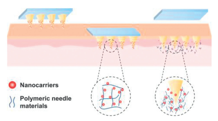
Figure 1.
Schematic diagram of PME.
Polymer modification effects in nanocarrier-loaded dissolving microneedles: Implications for transdermal drug delivery
Yuhuan Wu , Hoiian Ieong , Wenhao Wang , Chuanbin Wu , Anqi Lu , Xin Pan , Zhengwei Huang

Surface modification of nanocarriers with polymers represents a well-established strategy to optimize drug delivery performance. These intentional modifications significantly enhance nanocarrier stability, prolong systemic circulation, and improve targeting capability by precisely modulating their interactions with biological systems [1–3]. For instance, PEGylation effectively reduces opsonization [4], while hyaluronic acid (HA) modification enables CD44 receptor-mediated tumor targeting [5,6]. However, these intentional strategies face fundamental limitations in transdermal applications, as they cannot independently overcome the stratum corneum barrier, a critical obstacle preventing efficient percutaneous delivery of nanocarriers [7].
Dissolving microneedles (DMNs) have emerged as an innovative solution to this challenge. Unlike conventional transdermal patches, DMNs create transient microchannels that bypass the stratum corneum, enabling direct intradermal delivery of nanocarriers [8–10]. This unique approach synergistically combines the advantages of both systems: nanocarriers provide controlled drug release and targeting specificity [11], while DMNs address the crucial penetration barrier [12]. The surface modification of nanocarriers with HA markedly improves the tumor-targeting efficacy of paclitaxel-loaded nanocarriers by utilizing a transdermal drug delivery pathway facilitated by DMNs [13].
However, a previously overlooked phenomenon occurs when nanocarriers are incorporated into DMNs: the polymeric matrix may induce additional surface modifications, a process we term as the “polymer modification effect” (PME). Specifically, during DMNs fabrication or dissolution, polymeric components can alter nanocarrier properties (including size and surface charge) through non-covalent interactions such as hydrogen bonding and electrostatic adsorption. Unlike intentional surface modifications, PME arises as an uncontrolled, unintentional consequence of interactions between DMN materials and nanocarriers, which can significantly impact nanocarrier characteristics and their subsequent in vivo fate, including release kinetics, biodistribution, clearance, cellular uptake, and other interactions with the biological system [14]. For instance, while HA coating may be intentionally applied for tumor targeting, unintentional interactions between HA-nanocarriers within HA-based DMNs might impede release or alter targeting efficiency. In addition, PME is also different from the non-specific adsorption that occurs naturally in the biological environment. Unlike conventional “protein corona” and “non-specific adsorption” phenomena, PME constitutes a unique surface modification behavior specific to DMN systems. While protein coronas typically form through random adsorption of physiological proteins when nanocarriers enter biological fluids [15], PME originates from deliberate interactions between the DMN polymeric matrix and nanocarriers during fabrication and delivery processes.
While interactions between polymer and nanocarrier have been extensively studied in systemic delivery [16], the implications of PME in DMN systems remain poorly understood. A critical scientific question emerges: How might PME influence the in vivo fate of nanocarriers? Based on existing research, this perspective proposes a novel viewpoint: during transdermal delivery, the interaction between DMN polymers and nanocarriers may induce PME, consequently altering the in vivo fate of nanocarriers. We first systematically review cases where common DMN polymers independently modify nanocarriers, then elucidate potential PME mechanisms in DMNs system, and finally discuss strategies to manipulate PME effects, providing new insights for future development of nanocarrier-loaded DMNs.
Nanocarriers, due to their high surface energy, typically interact with and adsorb various biomolecules, such as proteins, phospholipids, sugars, and nucleic acids [17,18]. These interactions often significantly influence the behavior and fate of nanocarrier in vivo in most cases [19]. Building upon this fundamental phenomenon, researchers have developed surface modification strategies using polymers to precisely control nanocarrier-bioenvironment interactions, thereby endowing them with tailored functionalities such as prolonged circulation, enhanced targeting specificity, or improved drug release profiles [20–22].
In DMN delivery systems, polymeric materials serve as the primary matrix components for microneedle fabrication [23]. This creates a potential scenario where nanocarriers may interact with the surrounding polymeric materials of DMNs. We define this phenomenon as the PME, wherein DMN polymers may be modified on nanocarrier surfaces during preparation or release processes, consequently altering their intrinsic physicochemical properties (Fig. 1). Current evidence suggests that polymer-nanocarrier interactions in DMN systems are predominantly mediated by non-covalent forces, including electrostatic interactions, hydrogen bonding, and hydrophobic effects [16].
Therefore, this section will focus on the polymers commonly used in DMNs and their modification effects on nanocarriers. Such analysis is crucial for elucidating the delivery behavior of nanocarriers in DMN systems and optimizing their therapeutic performance.
DMNs are prepared of soluble or biodegradable polymers, including glucan, chitosan, HA, and alginate (Table S1 in Supporting information) [24–31]. Owing to their non-toxicity, biocompatibility, and biodegradability, these polymers demonstrate crucial functionalities in DMNs applications [32,33]. Notably, these polymers are also commonly employed for surface modification of nanocarriers to enhance their delivery performance.
Intriguingly, multiple experimental studies have observed that when these polymers served as the DMN matrix, they could induce alterations in the physicochemical properties of the loaded nanocarriers. For instance, Liu et al. reported that when methoxy poly(ethylene glycol) (mPEG)-poly(lactic acid) (PLA) nanomicelles were loaded into HA-based DMNs, HA could interact with mPEG-PLA, leading to an increase in particle size from 26.88 nm to 35.70 nm and a shift in zeta potential from −9.22 mV to –23.9 mV [34]. Similarly, Chen et al. developed C/I-Mil@MNs and found that after dissolution of HA-based DMNs, the particle size of C/I-Mil increased to approximately 90 nm, while the zeta potential changed from +6.30 mV to −8.34 mV [35]. Furthermore, Yang et al. fabricated ICG-NP MNs and demonstrated that the negatively charged HA in DMNs could adsorb onto the positively charged ICG-NPs via electrostatic interactions, resulting in increased nanoparticle size and enhanced accumulation of ICG-NPs at the microneedle tips [36].
Glucan is a neutral polysaccharide composed of glucose units associated with α−1,6 and α−1,3 bonds and is synthesized by bacteria from sucrose [37]. It offers several physicochemical advantages, including excellent water solubility, resistance to nonspecific protein adsorption on its surface, and ease of chemical derivatization, making it a suitable material for DMN drug delivery [38].
Owing to these favorable characteristics, glucan has been employed in surface modification of nanocarriers to confer specific functionalities. For instance, Ma et al. demonstrated that glucan-coated PLA nanoparticles exhibited significantly reduced interactions with bovine serum albumin (BSA), where the glucan surface coating provided steric hindrance protection that effectively prevented nonspecific protein interactions [39].
Based on the above experimental evidence, we hypothesize that when glucan serves as the matrix material in DMNs for nanocarrier loading, its hydrophilic nature and steric effects may exert complex influences on nanocarrier behavior. On one hand, the high-water solubility of glucan can accelerate microneedle dissolution, thereby promoting rapid nanocarrier release. On the other hand, physical entanglement or hydrogen bonding between glucan and nanocarriers might potentially hinder nanocarrier diffusion. It is further speculated that when nanocarriers are encapsulated into glucan-related DMNs, the interaction between glucan and nanocarriers promotes lymphatic drainage, facilitates dendritic cell uptake, enhances dendritic cell maturation, and supports antigen cross-presentation after nanoparticle administration, thereby inducing a robust cell-mediated immune response [40].
Chitosan is a partly deacetylated cationic polymer of N-acetyl glucosamine derived from chitin (2-acetamido-2-deoxy-1,4-D-glucan). It is a natural water-soluble derivative of chitin with unique characteristics [32]. Due to the abundant amounts of amino groups in its chain, chitosan carries a positive charge [41]. This cationic nature has strong electrostatic interactions with negatively charged components of the mucus, including sialic acids, as well as with the epithelial surface [42,43]. Consequently, chitosan can be electrostatically bound to negatively charged nanocarriers for surface modification.
Experimental evidence indicates that Malabanan et al. successfully coated chitosan onto HPIONP nanocarriers through electrostatic interactions, resulting in a significant increase in particle size and improved stability of the nanocarriers [44]. Similarly, Badran et al. modified 5-fluorouracil (5-FU)-loaded nanocarriers with chitosan, demonstrating that this modification substantially improved drug release and enhanced the anticancer efficacy of 5-FU [45].
Based on these findings, we hypothesize that when chitosan serves as the DMNs polymer matrix for nanocarrier loading, its electrostatic properties may introduce new challenges: the interactions between chitosan and nanocarriers could hinder the efficient release of nanocarriers from DMNs, potentially compromising drug delivery efficiency. It is further speculated that when nanocarriers are encapsulated within chitosan-related DMNs, the interaction between chitosan and nanocarriers can enhance both the stability of nanocarriers and their mucoadhesive properties, ultimately improving the oral bioavailability of drug-loaded nanocarriers [46].
HA is a linear macromolecular anionic polysaccharide composed of repeating pieces of D-glucuronic acid with N-acetylglucosamine, which are linked by β-(1–3) bonds [25,47]. HA is a natural component of the skin [48]. Its excellent biocompatibility, biodegradability, and high viscoelasticity make it a promising material for the development of DMNs. Under physiological conditions (pKa ≈ 3–4), the carboxyl groups of HA dissociate, rendering it negatively charged [49]. This property enables electrostatic interactions with positively charged nanocarriers. Researchers have utilized this property for surface modification of nanocarriers. For instance, Essam et al. utilized electrostatic interactions to coat HA onto simvastatin (SIM)-loaded zeolitic imidazolate framework-8 (ZIF-8) nanoparticles (SIM/ZIF-8).
It is noteworthy that the HA receptor CD44 has been confirmed to be overexpressed in various tumor cells [50]. Therefore, surface modification with HA may confer tumor-targeting capability to nanocarriers. Experimental studies have demonstrated this effect. For instance, Jiang et al. demonstrated that HA-coated hollow Prussian blue nanoparticles exhibited enhanced colloidal stability, prolonged circulation time, and specific targeting ability toward CD44-overexpressing HeLa cells [51].
Based on the above experimental evidence, we hypothesize that these properties of HA may introduce challenges in DMN delivery systems: HA could potentially adsorb onto nanocarriers via electrostatic interactions during the delivery process, thereby altering nanocarrier physical properties and in vivo behavior. It is further speculated that when nanocarriers are encapsulated into HA-related DMNs, the interaction between HA and nanocarriers enhances cellular uptake through HA receptor-mediated endocytosis, thereby improving tumor-targeting capability [52].
Alginate is composed of alternative blocks of 1–4 linked α-L-guluronic acid (GulA; G) and β-D-mannuronic acid (ManA; M) residues [53]. It is a water-soluble linear anionic polysaccharide, like HA, which contains abundant hydrophilic groups such as hydroxyl and carboxyl groups, endowing it with excellent water solubility [54,55]. Studies have shown that polymers with higher charge density exhibit superior adhesive, with polyanion polymers demonstrating greater effectiveness as bioadhesives compared to polycation or nonionic polymers [32]. Specifically, alginate, an anionic polymer containing carboxylic groups, possesses strong adhesive characteristics [56].
Researchers have exploited this property for nanocarrier surface modification. For example, Amin et al. developed alginate-coated chitosan nanoparticles, demonstrating that the alginate surface modification enhanced particle stability in simulated gastric and intestinal fluids while improving mucoadhesive properties, thereby establishing a potential nanocarrier platform for mucosal protein vaccine delivery [57].
Based on the above experimental evidence, it is hypothesized that when alginate is employed in DMNs fabrication, these adhesive properties may present a double-edged sword: while potentially beneficial for drug carrier applications, they may simultaneously hinder drug delivery, creating new challenges for therapeutic administration. It is further speculated that when nanocarriers are encapsulated into alginate-related DMNs, the interaction between alginate and nanocarriers can enhance stability, impede the interaction between drug-loaded nanocarriers and cell membranes, thereby attenuating cellular uptake while potentially decreasing drug-associated cytotoxicity [58].
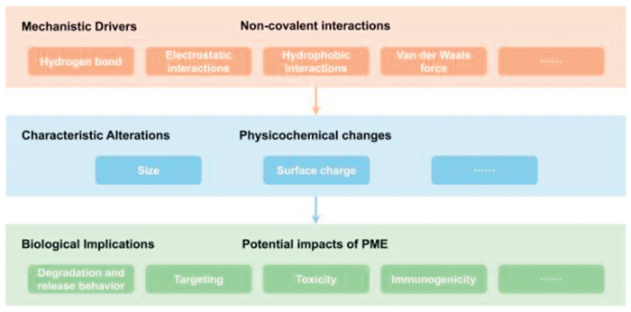
In nanocarrier research, significant overlap exists between the polymeric materials used for nanocarrier surface modification and those employed as DMNs matrix materials. During the preparation and release of nanocarrier-loaded DMNs, unintended interactions may occur between the nanocarriers and microneedle polymers. These modifications are primarily governed by non-covalent interactions, such as electrostatic forces, hydrogen bonding, hydrophobic effects, and van der Waals forces (Fig. 2) [59,60].

Current evidence demonstrates that polymer-nanocarrier non-covalent binding can confer specialized functionalities to drug delivery systems [59]. For example, alginate has been bound to the surface of liposomes containing 1,2-dipalmitoyl-sn-glycero-3-phosphocholine (DPPC), 1,2-distearoyl-sn-glycero-3-phosphocholine (DSPC), 1,2-distearoyl-sn-glycero-3-phosphoethanolamine-N-[amino(polyethylene glycol)-2000] (DSPE-PEG2000), and 1-stearoyl-2-hydroxy-sn-glycero-3-phosphocholine (MSPC) for protein delivery [61]. Similarly, HA has been bound to the surface of liposomes that containing egg phosphatidylcholine for sustained release and targeted delivery of drugs [62]. Furthermore, alginate and chitosan were bound to the surface of liposomes containing egg yolk for pH-responsive release of calcein [63]. Notably, these polymers used for liposome modification are identical to those commonly employed as DMN matrix materials. Structurally, DMN polymers typically contain both polar and nonpolar domains, allowing them to cooperatively modulate liposome surface properties through hydrophobic and electrostatic interactions, thereby modifying the DMN preparation and drug release process [59]. However, such interactions may also lead to a “material commonality-functional divergence” phenomenon: While enhanced nanocarrier stability and skin adhesion can be achieved, excessively strong binding may conversely impede effective drug release. Thus, a deeper understanding and precise control of these non-covalent interactions could open new avenues for developing next-generation nanocarrier-loaded DMN systems.
Current evidence demonstrates that polymer surface modifications can significantly alter a variety of physicochemical properties of the nanocarriers, including size and surface charge [64–66]. These physicochemical attributes are recognized as critical factors that influence the biological behavior of nanocarriers in vivo [67]. During transdermal delivery, PME influences the biological behavior of nanocarriers primarily in four aspects (Fig. 3).
When DMNs load nanocarriers, the interactions between polymeric microneedle matrices and encapsulated nanocarriers critically influence drug degradation and release profiles. Studies have shown that polymer biodegradability and drug release characteristics are essential parameters for effective drug delivery via nanocarriers [68]. Ahmad et al. have employed chitosan to prepare chitosan-DMNs for the sustained release of tacrolimus [69]. Chitosan has good adhesion properties by absorbing water and swelling in the weakly acidic environment of the skin [32]. Compared with other polymeric materials, chitosan has low water solubility [29]. When nanocarriers are encapsulated within chitosan-DMNs, polymer- nanocarrier interactions promote the formation of dense three-dimensional networks at nanocarrier surfaces, physically hindering nanocarriers degradation and consequently delaying drug release.
Furthermore, non-covalent interactions between polymers and nanocarriers significantly modulate release behavior [70]. In polymer-liposome systems, matrix swelling or surface erosion facilitates liposome liberation from the polymeric network [59]. Strategic selection of oppositely charged polymers enables precise tuning of interaction strength for pH-responsive release [59,71]. A representative example is Caddeo et al.'s hybrid system (TPP-chitosomes), where crosslinked chitosan-coated liposomes achieve intestinal pH-triggered release while preventing gastric drug loss [70]. When nanocarriers are encapsulated in chitosan-DMNs, the interaction between chitosan and nanocarriers will affect the release behavior of nanocarriers. These findings collectively underscore that chitosan-nanocarrier interactions in DMN systems exert profound effects on nanocarriers degradation and release behavior.
The unique physicochemical properties of nanocarriers grant them the capability for targeted therapeutic delivery [72], which shows promising potential in treating various diseases [73]. However, when nanocarriers are loaded into DMNs, interactions between the DMN polymeric matrix and nanocarriers can significantly alter nanocarriers’ characteristics, ultimately impacting their targeting efficiency.
This phenomenon is particularly prominent in cancer therapy [57]. Taking HA-related DMNs as an example, it is hypothesized that the PME influences targeting efficiency through four critical stages [74]:
(1) During in vivo circulation, negatively charged HA can adsorb onto the surface of positively charged nanocarriers via electrostatic interactions, thereby altering their surface charge. This modification promotes recognition and internalization by macrophages, ultimately leading to reduced circulation time [75,76].
(2) In the tumor site accumulation phase, polymer-nanocarrier interactions may increase particle size [77]. This alteration poses a dual challenge: on one hand, it complicates the extravasation of nanocarriers from blood vessels to the tumor site [78]. On the other hand, it renders the nanocarriers less prone to reflux after crossing the blood vessels, contributing to their accumulation at the tumor site [77].
(3) During tumor tissue penetration, polymer modifications influence nanocarrier surface potential, directly affecting tumor penetration capability (Fig. S1 in Supporting information) [79]. It has been reported that positively charged nanocarriers are particularly immobilized in tumor tissues and are least effective in tumor accumulation [80]. When loaded in HA-DMNs, the electrostatic interaction between negatively charged HA and positively charged nanocarriers minimizes adhesive interactions with tumor tissue components, enabling rapid tissue penetration.
(4) At the tumor cellular uptake stage, polymer-nanocarrier interactions increase the particle size of the nanocarrier, which has a steric effect that reduces interaction between the nanoparticles and the cell membrane, thus decreasing uptake [81,82]. Additionally, certain DMN polymeric materials (e.g., HA) can serve as natural targeting ligands, significantly enhancing tumor cell uptake through receptor-mediated internalization pathways [83]. Specifically, when nanocarriers are loaded in HA-related DMNs, the interaction between HA and nanocarriers can promote CD44 receptor-mediated targeting, substantially improving uptake by both macrophages and tumor cells [84].
Nanocarriers are generally prepared from lipids, polymeric and inorganic materials [85]. However, inorganic materials can accumulate in organs, provoke immune system stimulation and potentially cause long-term or chronic toxicity to the organism [86]. Currently, induction of inflammation and oxidative stress are common toxic responses reported for inorganic nanomaterials [87]. In contrast, microneedle polymeric materials are usually derived from polysaccharides [28,88], exhibit excellent biocompatibility, biodegradability and safety [24,26,28,89], generally causing no toxic side effects on the organism. Notably, when nanocarriers are encapsulated within polymeric MNs, polymer-nanoparticle interactions may significantly enhance their biocompatibility.
For instance, iron oxide nanoparticles (IONPs) have been reported to exhibit toxicity toward human dermal fibroblasts [90]. However, when coated with dextran, their toxicity is markedly reduced [91]. We hypothesize that within glucan-related DMNs, the interactions between the glucan and nanocarriers can form a protective coating, thereby alleviating cytotoxicity. Similarly, the introduction of hydrophilic polymers can neutralize the surface charge of cationic nanocarriers, mitigating their damaging effects on cell membranes. Lin et al. found that introducing hydrophilic polymers like (poly(isopropylacrylamide-co-2-(dimethylamino)ethyl methacrylate), P(IPAAm-co-DMAAm)) to the cationic nanoparticle surfaces can effectively mitigate cellular damage [92].
It is noteworthy that although DMN polymers possess excellent biosafety, their combined effects with nanoparticles warrant careful evaluation for potential cumulative toxicity. Singh et al. observed that PLA do not adversely affect cytokine activation, mitochondrial or lysosomal integrity in vitro at lower concentrations, but they can stimulate reactive oxygen species (ROS) production and inflammation at high concentrations [93]. These toxic effects are thought to be related to the accumulation of polymers in the cells [94]. Consequently, a thorough assessment of PME is essential, requiring optimization of DMN polymer composition based on nanocarrier properties to balance delivery efficiency and safety. These findings also suggest that DMNs not only enable transdermal nanocarriers delivery but also modulate nanocarriers biocompatibility through polymeric material interactions, offering novel strategies for safe and effective nanomedicine delivery.
When DMNs encapsulate nanocarriers, the interaction between the polymeric matrix material and the nanocarriers will significantly affect their immunogenicity.
Polymer components in DMNs can have a dual impact on the immunogenicity of nanoparticles. On one hand, polymers modified on the surface of nanoparticles can act as immune-blinding agents [95]. These polymers can partially or completely cover the molecules or antigens originally attached to the surface of the nanoparticle, thus inhibiting their interaction with specific receptors and potentially affecting the desired effect. On the other hand, certain DMN polymers (e.g., polyethylene glycol (PEG)-containing matrices) can trigger an anti-PEG antibody response via a classical T-cell-dependent pathway, enhancing the immunogenicity of the nanoparticles and thereby eliciting an immune response [96]. For instance, after HA-related DMNs dissolve, we hypothesize that their polymer chains may simultaneously provide surface shielding while activating CD44 receptor-mediated immune responses, leading to complex immunomodulatory effects.
Therefore, the rational design of nanocarrier-loaded DMNs requires careful material selection that balances two critical considerations: the capacity to modify nanocarrier surface properties and the potential to induce unintended immune activation. This dual optimization is essential for developing safe and effective transdermal immunotherapy platforms.
The PME frequently induces unintentional alterations in the in vivo behavior of loaded nanocarriers. Notably, these modifications typically manifest as undesirable negative effects. Consequently, deliberate modulation of these non-covalent interactions to mitigate PME occurrence could enhance the transdermal delivery performance of nanocarrier-loaded DMNs, thereby achieving closer alignment with predetermined therapeutic objectives.
The physicochemical properties of drug-loaded nanocarriers play a pivotal role in determining their behavior in vivo [97,98]. Polymer modification of the nanocarrier surface alters these properties, and these changes can be observed through in vitro monitoring techniques [19,99].
Currently, various in vitro detection methods are available for monitoring these properties. These include spectral analysis, zeta potentials, nuclear magnetic resonance (NMR), gas chromatography (GC), transmission electron microscopy (TEM), scanning electron microscopy (SEM), X-ray diffraction (XRD) and dynamic light scattering (DLS) [99–101]. These in vitro detection methods allow for the observation of the morphology, size, surface charge and other physicochemical properties of the nanocarriers in vitro. By employing these techniques, researchers can assess whether the nanocarriers interact with the polymer and determine the extent of any changes in their physicochemical properties [99].
Sadzuka et al. determined the thickness of the lipid layer on liposomes as a function of ionic strength by measuring their zeta potential (through electrophoretic mobility measurements) to ascertain whether polymers had adsorbed onto their surfaces [102,103]. Tirosh et al. characterized the state of PEG based on the amount of water bound using differential scanning calorimetry (DSC), densitometry and ultrasound velocity, enabling them to differentiate between PEG conjugated to the surface of nanoparticles and free PEG [104]. Similarly, Garcia-Fuentes et al. utilised NMR techniques to confirm the presence of PEG chains on the surface of the nanoparticles by measuring the nuclear Overhauser effect (NOE) of isolated nanoparticles suspended in a 90% H2O/10% D2O mixture [105].
PME significantly affects the nanocarrier and their behavior in vivo. Tracking the in vivo fate of drug-carrying nanocarriers is particularly crucial for understanding these effects [106].
Currently, in vivo fate studies of drug-loaded nanocarriers are mainly rely on fluorescent dyes or radio-labeling methods [66,107]. Microneedle polymers and nanocarriers can be labeled with suitable fluorescent dyes or radioisotopes, allowing for the tracking of PME in vivo by monitoring the labeled fluorescence or isotope [108,109].
Kreyling et al. labeled the polymer and gold nanoparticle (GNP) cores with 111In and 198Au, respectively, to investigate the behavior of polymer-coated nanoparticles in vivo [110]. The results showed that the polymer shells were more abundant in urine than GNP cores, and that polymer-coated GNPs were cleared by the immune system and transported to the liver for endocytosis. In another study, degradation was shown quantitatively using a double-labeling technique [111]. Bargheer et al. employed a double-labeling technique to quantitatively assess degradation, labeling the iron oxide cores with 59Fe and covalently binding a 14C-labeled peptide or a 125I-labeled protein to the surface [111]. The results demonstrated no separation between the polymer shell and the iron oxide core during blood circulation.
Antibiofouling polymers are synthetic polymers that are either hydrophilic or amphoteric in nature [112]. Their electrically neutral nature contributes to the reduction of electrostatic interactions with charged polymers [113]. Antibiofouling polymers are widely used to coat nanocarrier systems [114], endowing them with " invisible properties" to evade regular removal by the body.
PEG is the most used antibiofouling polymer for coating nanocarrier surfaces [114]. PEG is a polyether consisting of ethoxylated units derived from the ring-opening polymerization of ethylene oxide. It exists primarily in a linear form, with hydroxyl groups at both ends that can easily be conjugated to functional groups [115]. Owing to its electrically neutral, remarkably spatially repulsive and highly hydrophilic [116], PEG is frequently employed to modify the surface of nanocarriers.
Currently, two primary methods of PEGylated modifications are commonly used: (1) Physical adsorption, which involves interactions such as electrostatic, hydrophobic, and hydrogen bonding, to attach PEG to the nanocarrier surface [115]; (2) Chemical grafting, where stable chemical bonds are formed between PEG and functional groups on the nanocarrier surface, such as carboxyl groups [117]. These strategies can be effectively utilized in the preparation of nanocarrier-loaded DMNs to minimize potential PME.
In addition to PEG, several other polymers have been found to serve as effective antibiofouling agents for surface modification of nanocarriers, such as amphiphilic ionic polymers, poly(2-oxazoline), polyglycerol, and peptides [112]. Each of these polymers exhibits the promising potential to minimize PME and enhance the performance of nanocarriers.
Non-covalent forces, such as electrostatic adsorption, hydrophobic interactions, hydrogen bonding, play an essential role in PME [118–120]. In the practical preparation of nanocarrier-loaded DMNs, minimizing the occurrence of PME can be achieved by selecting appropriate polymers or nanocarrier materials to reduce the non-covalent forces between them [16,121,122]. For example, when preparing DMNs for loading cationic liposomes, it is advisable to avoid using anionic polymeric materials [123]. If the use of polymeric materials with opposite charges is unavoidable, they can be modified beforehand to minimize their interaction forces [16,124]. One such modification method involves altering the backbone of polymers, such as modifying the PVA with sulphobutyl or amine structures to produce negatively or positively charged polymer, respectively [124–126]. By adopting these strategies, the likelihood of PME can be significantly reduced during the preparation of nanocarrier-loaded DMNs.
Nanocarrier-loaded DMNs integrate the advantages of nanocarriers and DMNs [127–129]. Despite their therapeutic promises, their clinical translation remains constrained [130]. The PME represents a critical yet underappreciated barrier. In fact, unintentional modifications of nanocarriers by the DMN polymer matrix can lead to substantial changes in vivo performance, directly affecting reproducibility, dosing accuracy, and ultimately clinical efficacy. Therefore, to achieve successful clinical translation of nanocarrier-loaded DMNs, it is imperative to address the multiple challenges posed by PME.
First, PME can induce alterations in the physicochemical properties of nanocarriers, such as size and surface charge, which directly influence biodistribution, cellular uptake, and therapeutic outcomes [64–66]. The efficacy and safety of the formulation will thus become challengeable.
Second, there are currently no specific regulations globally for “nanocarrier-microneedle” combination products. Regulatory agencies like the Food and Drug Administration (FDA), National Medical Products Administration (NMPA) and European Medicines Agency (EMA) have yet to clearly classify these systems as drugs, devices, or combination products, resulting in a lack of defined critical quality attributes (CQAs) and standards. CQAs affected by PME, including sterility, stability, skin irritation, and potential immunogenicity assessments are required for additional validation [131].
More critically, PME often occurs unpredictably during scale-up manufacturing, leading to batch-to-batch inconsistencies in particle size, surface charge, and release kinetics [132]. The current absence of predictive models makes it particularly challenging to ensure product consistency, even when employing high-throughput molding or 3D printing continuous manufacturing platforms.
To overcome these challenge, future research should focus on in-depth understanding of PME. Advanced imaging techniques and bioanalytical methods should be employed to characterize nanocarrier surface properties and pharmacological behavior on both in vitro and in vivo levels (4.1 In vitro monitoring, 4.2 Tracking the behavior of nanocarrier-loaded DMNs in vivo), enabling correlation between PME and in vivo performance [99,106]. Under regulatory coordination, quality by design (QbD)-driven design strategies should be established [133]. This involves screening and optimizing preparation parameters through design of experiments (DoE) to ensure batch consistency. Concurrently, the development of process analytical technology (PAT) [134] for real-time monitoring of the production process involving DMN polymers and nanocarriers is essential, complemented by digital twin or AI models for scale-up risk prediction. Furthermore, multicenter clinical trials should be conducted to verify the safety and efficacy of nanocarriers, with particular attention to evaluating PME's impact on nanocarrier pharmacokinetics (PK) and pharmacodynamics (PD). Only through such comprehensive approaches can nanocarrier-loaded DMNs advance toward clinical application.
It is important to emphasize that PME posts not only challenges but also opportunities. With a deeper understanding of its mechanisms and improved precision in modulation, we may strategically leverage PME to optimize nanocarrier performance, including enhanced stability, improved targeting capability, and reduced toxicity. Continued advancements in materials science and nanofabrication technologies will provide more powerful tools to control and exploit PME. We anticipate that in the next generation of nanocarrier-loaded DMNs, PME can be rationally incorporated into system design, thereby harnessing its positive potential and facilitating more robust progress toward clinical translation.
Nanocarrier-loaded DMNs integrate the advantages of nanocarriers and DMNs with excellent biocompatibility, good targeting performance and controlled release profile. Although nanocarrier-loaded DMNs exhibit promising clinical applications, their clinical translation remains constrained, with the PME being a key contributing factor.
PME arises from non-covalent interactions between the nanocarriers and the DMN polymer matrix during preparation and drug release, which can significantly alter the physicochemical properties of the nanocarriers (e.g., size and surface charge). These alterations can significantly impact the nanocarrier's degradation, release profile, and targeting efficiency, thereby greatly hindering the clinical translation of nanocarrier-loaded DMNs. However, a growing body of evidence suggests that PME may also confer beneficial properties to the system, such as enhanced stability, reduced cytotoxicity, prolonged circulation time, or improved retention in specific tissues. Therefore, PME should not be viewed solely as a negative phenomenon, but rather as a design element that can be functionally optimized through material screening and process control.
Current research in this area is still in its infancy, and there is a long way to go before a comprehensive understanding of PME is achieved. We anticipate the development of more precise methods in the future, such as using techniques based on single nanoparticle imaging and bio-orthogonal imaging, to better understand and mitigate the effects of PME. We advocate investigators in this field to launch more in-depth studies on the PME, as this will be crucial for advancing the clinical translation of nanocarrier-loaded DMNs and realizing their full potential in medical applications.
PME represents both a major challenge in the clinical translation of nanocarrier-loaded DMNs and a source of new opportunities for optimizing formulation performance. Through interdisciplinary collaboration and technological innovation, transforming PME from an “uncontrolled variable” into a “designable parameter” will undoubtedly accelerate the development of next-generation nanocarrier-loaded DMN systems and broaden their prospects for medical application.
The authors declare that they have no known competing financial interests or personal relationships that could have appeared to influence the work reported in this paper.
Yuhuan Wu: Writing – review & editing, Writing – original draft, Conceptualization. Hoiian Ieong: Writing – review & editing. Wenhao Wang: Writing – review & editing. Chuanbin Wu: Resources. Anqi Lu: Writing – review & editing. Xin Pan: Resources. Zhengwei Huang: Writing – review & editing, Resources, Funding acquisition, Conceptualization.
We appreciate the financial support from Major Funding of National Natural Science Foundation of China (No. 82330112), National Natural Science Foundation of China (Nos. 82373800 and 82104070) and Guangdong Universities Keynote Regions Special Funded Project (No. 2022ZDZX2002).
Supplementary material associated with this article can be found, in the online version, at doi:
S. Priya, V.M. Desai, G. Singhvi, ACS Omega 8 (2023) 74–86. doi: 10.1021/acsomega.2c05976
T. Date, V. Nimbalkar, J. Kamat, et al., J. Control. Release 271 (2018) 60–73. doi: 10.1016/j.jconrel.2017.12.016
G.D. Mogosanu, A.M. Grumezescu, C. Bejenaru, L.E. Bejenaru, Int. J. Pharm. 510 (2016) 419–429. doi: 10.1016/j.ijpharm.2016.03.014
M. Papi, D. Caputo, V. Palmieri, et al., Nanoscale 9 (2017) 10327–10334. doi: 10.1039/C7NR03042H
C.S. Chen, H.N. Zhang, J.J. Han, et al., Carbohydr. Polym. 351 (2025) 123144. doi: 10.1016/j.carbpol.2024.123144
H. Hu, W. Zhang, L. Lei, et al., Chin. Chem. Lett. 35 (2024) 108765. doi: 10.1016/j.cclet.2023.108765
Z.X. Yu, X.X. Meng, S.N. Zhang, et al., Molecules 26 (2021) 3093. doi: 10.3390/molecules26113093
M. Gunawan, A.N. Bestari, D. Ramadon, et al., J. Drug Deliv. Sci. Technol. 107 (2025) 106807. doi: 10.1016/j.jddst.2025.106807
Salwa, N.T. Chevala, S.R. Jitta, et al., J. Drug Deliv. Sci. Technol. 65 (2021) 102711. doi: 10.1016/j.jddst.2021.102711
X. Jiang, S. Wang, L. Zhang, et al., Chin. Chem. Lett. 35 (2024) 108686. doi: 10.1016/j.cclet.2023.108686
M. Vázquez-González, I. Willner, ACS Appl. Mater. Interfaces 13 (2021) 9520–9541. doi: 10.1021/acsami.0c17121
A. Kheirieh, A. Abbasi, B. Malaekeh-Nikouei, et al., J. Drug Deliv. Sci. Technol. 107 (2025) 106836. doi: 10.1016/j.jddst.2025.106836
T.T. Peng, Y. Huang, X.Q. Feng, et al., Acta Pharm. Sin. B 11 (2021) 3297–3309. doi: 10.1016/j.apsb.2020.11.013
Y.F. Cai, J.P. Qi, Y. Lu, et al., Adv. Drug Deliv. Rev. 188 (2022) 114463. doi: 10.1016/j.addr.2022.114463
N. Onishchenko, D. Tretiakova, E. Vodovozova, Acta Biomater. 134 (2021) 57–78. doi: 10.1016/j.actbio.2021.07.074
H.B. He, X.X. Shen, Z.H. Nie, Prog. Polym. Sci. 143 (2023) 101710. doi: 10.1016/j.progpolymsci.2023.101710
S. Tenzer, D. Docter, J. Kuharev, et al., Nat. Nanotechnol. 8 (2013) 772–781. doi: 10.1038/nnano.2013.181
M.P. Monopoli, F.B. Bombelli, K.A. Dawson, Nat. Nanotechnol. 6 (2011) 11–12. doi: 10.1038/nnano.2010.267
D. Docter, S. Strieth, D. Westmeier, et al., Nanomedicine 10 (2015) 503–519. doi: 10.2217/nnm.14.184
S.M. D'Addio, S. Baldassano, L. Shi, et al., J. Control. Release 168 (2013) 41–49. doi: 10.1016/j.jconrel.2013.02.004
H.D. Li, S. Zha, H.L. Li, et al., Small 18 (2022) e2203629. doi: 10.1002/smll.202203629
J. Bugno, H.J. Hsu, S. Hong, J. Drug Target. 23 (2015) 642–650. doi: 10.3109/1061186X.2015.1052077
K.A.S. Al-Japairai, S. Mahmood, S.H. Almurisi, et al., Int. J. Pharm. 587 (2020) 119673. doi: 10.1016/j.ijpharm.2020.119673
M. Wang, L.Z. Hu, C.J. Xu, Lab Chip 17 (2017) 1373–1387. doi: 10.1039/C7LC00016B
D. Kulkarni, D. Gadade, N. Chapaitkar, et al., Sci. Pharm. 91 (2023) 27. doi: 10.3390/scipharm91020027
F.K. Aldawood, A. Andar, S. Desai, Polymers 13 (2021) 2815. doi: 10.3390/polym13162815
Z.J. Wang, Z.P. Yang, J.J. Jiang, et al., Adv. Mater. 34 (2022) e2106606. doi: 10.1002/adma.202106606
M. Ali, S. Namjoshi, H.A.E. Benson, et al., J. Control. Release 347 (2022) 561–589. doi: 10.1016/j.jconrel.2022.04.043
M.C. Chen, M.H. Ling, K.Y. Lai, E. Pramudityo, Biomacromolecules 13 (2012) 4022–4031. doi: 10.1021/bm301293d
H.H. Lu, J.L. Wang, J. Li, et al., Macromol. Biosci. 23 (2023) e2300141. doi: 10.1002/mabi.202300141
S. Dharadhar, A. Majumdar, S. Dhoble, V. Patravale, Drug Dev. Ind. Pharm. 45 (2019) 188–201. doi: 10.1080/03639045.2018.1539497
G.A. Martau, M. Mihai, D.C. Vodnar, Polymers 11 (2019) 1837. doi: 10.3390/polym11111837
Y. Liu, M. Zhu, M. Meng, et al., Chin. Chem. Lett. 34 (2023) 107583. doi: 10.1016/j.cclet.2022.06.006
T. Liu, J.T. Fu, M.L. Chen, et al., Eur. Polym. J. 210 (2024) 113008. doi: 10.1016/j.eurpolymj.2024.113008
M.L. Chen, D. Yang, Y. Sun, et al., ACS Nano 15 (2021) 3387–3401. doi: 10.1021/acsnano.0c10396
P.P. Yang, M.L. Chen, W.B. Qin, et al., ACS Appl. Nano Mater. 4 (2021) 5921–5931. doi: 10.1021/acsanm.1c00832
T.G. Barclay, C.M. Day, N. Petrovsky, S. Garg, Carbohydr. Polym. 221 (2019) 94–112. doi: 10.1016/j.carbpol.2019.05.067
T.X. Miao, J.Q. Wang, Y. Zeng, et al., Adv. Sci. 5 (2018) 1700513. doi: 10.1002/advs.201700513
W.J. Ma, X.B. Yuan, C.S. Kang, et al., Carbohydr. Polym. 72 (2008) 75–81. doi: 10.1016/j.carbpol.2007.07.033
W. Guo, X.Y. Zhang, L. Wan, et al., J. Pharm. Anal. 14 (2024) 100953. doi: 10.1016/j.jpha.2024.02.007
I. Fiebrig, S.E. Harding, A.J. Rowe, et al., Carbohydr. Polym. 28 (1995) 239– 244. doi: 10.1016/0144-8617(95)00105-0
T.M.M. Ways, W.M. Lau, V.V. Khutoryanskiy, Polymers 10 (2018) 267. doi: 10.3390/polym10030267
S. Cheng, M. Pan, D. Hu, et al., Chin. Chem. Lett. 34 (2023) 108276. doi: 10.1016/j.cclet.2023.108276
J.W.T. Malabanan, K.P. Alcantara, P. Jantaratana, et al., ACS Appl. Bio Mater. 6 (2023) 5426–5441. doi: 10.1021/acsabm.3c00657
M.M. Badran, M.M. Mady, M.M. Ghannam, F. Shakeel, Int. J. Biol. Macromol. 95 (2017) 643–649. doi: 10.1016/j.ijbiomac.2016.11.098
E. Antonio, O.D. Antunes, R. Marcano, et al., Int. J. Biol. Macromol. 172 (2021) 133–142. doi: 10.1016/j.ijbiomac.2021.01.041
G.F. Boafo, Y.J. Shi, Q.Q. Xiao, et al., Chin. Chem. Lett. 33 (2022) 4600–4604. doi: 10.1016/j.cclet.2022.04.033
P. Snetkov, K. Zakharova, S. Morozkina, et al., Polymers 12 (2020) 1800. doi: 10.3390/polym12081800
F. Dosio, S. Arpicco, B. Stella, E. Fattal, Adv. Drug Deliv. Rev. 97 (2016) 204–236. doi: 10.1016/j.addr.2015.11.011
G.L. Huang, H.L. Huang, Drug Deliv. 25 (2018) 766–772. doi: 10.1080/10717544.2018.1450910
L.J. Jing, S.M. Shao, Y. Wang, et al., Theranostics 6 (2016) 40–53. doi: 10.7150/thno.13250
T. Zhao, S.W. Qin, L.N. Peng, et al., Carbohydr. Polym. 214 (2019) 221–233. doi: 10.1016/j.carbpol.2019.03.043
K. Norajit, K.M. Kim, G.H. Ryu, J. Food Eng. 98 (2010) 377–384. doi: 10.1016/j.jfoodeng.2010.01.015
M. George, T.E. Abraham, J. Control. Release 114 (2006) 1–14.
N.N. Gao, Y. Zhang, Z.H. Yang, et al., Chin. Chem. Lett. 35 (2024) 108820. doi: 10.1016/j.cclet.2023.108820
J. Venkatesan, I. Bhatnagar, P. Manivasagan, et al., Int. J. Biol. Macromol. 72 (2015) 269–281. doi: 10.1016/j.ijbiomac.2014.07.008
M.K. Amin, J.S. Boateng, Mar. Drugs 20 (2022) 156. doi: 10.3390/md20030156
M. Azadi, A.E. David, ACS Biomater. Sci. Eng. 10 (2023) 429–441.
A.Umar Sriwidodo, N. Wathoni, et al., Heliyon 8 (2022) e08934. doi: 10.1016/j.heliyon.2022.e08934
S. Kumar, V.K. Aswal, J. Kohlbrecher, Langmuir 32 (2016) 1450–1459. doi: 10.1021/acs.langmuir.5b03998
M. van Elk, C. Lorenzato, B. Ozbakir, et al., Eur. Polym. J. 72 (2015) 620–631. doi: 10.1016/j.eurpolymj.2015.03.013
N. El Kechai, S. Geiger, A. Fallacara, et al., Int. J. Pharm. 523 (2017) 246–259. doi: 10.1016/j.ijpharm.2017.03.029
Y.J. Hong, J.C. Kim, J. Biomater. Sci. Polym. Ed. 26 (2015) 766–779. doi: 10.1080/09205063.2015.1058574
V.H. Nguyen, B.J. Lee, Int. J. Nanomed. 12 (2017) 3137–3151. doi: 10.2147/IJN.S129300
S.J. Park, Int. J. Nanomed. 15 (2020) 5783–5802. doi: 10.2147/ijn.s254808
N. Feliu, D. Docter, M. Heine, et al., Chem. Soc. Rev. 45 (2016) 2440–2457. doi: 10.1039/C5CS00699F
G. Sanità, B. Carrese, A. Lamberti, Front. Mol. Biosci. 7 (2020) 587012. doi: 10.3389/fmolb.2020.587012
J.W. Lee, M.R. Han, J.H. Park, J. Drug Target. 21 (2013) 211–223. doi: 10.3109/1061186X.2012.741136
Z. Ahmad, M.I. Khan, M.I. Siddique, et al., AAPS PharmSciTech 21 (2020) 68. doi: 10.1208/s12249-019-1611-9
C. Caddeo, O. Díez-Sales, R. Pons, et al., J. Colloid Interface Sci. 461 (2016) 69–78. doi: 10.1016/j.jcis.2015.09.013
M.G. Simoes, P. Alves, M. Carvalheiro, P.N. Simoes, Colloids Surf. B: Biointerfaces 152 (2017) 103–113. doi: 10.1016/j.colsurfb.2017.01.002
F. Qu, R. Geng, Y.J. Liu, J.T. Zhu, Theranostics 12 (2022) 3372–3406. doi: 10.7150/thno.69999
N. Habibi, D.F. Quevedo, J.V. Gregory, J. Lahann, Wiley Interdiscip. Rev. Nanomed. Nanobiotechnol. 12 (2020) e1625. doi: 10.1002/wnan.1625
Y.F. Pi, J.E. Zhou, J. Wang, et al., Curr. Pharm. Des. 21 (2015) 6236–6245. doi: 10.2174/1381612821666151027153611
G. Bozzuto, A. Molinari, Int. J. Nanomed. 10 (2015) 975–999.
Y.B. Liu, K.M.C. Bravo, J.W. Liu, Nanoscale Horiz 6 (2021) 78–94. doi: 10.1039/d0nh00605j
J. Lazarovits, Y.Y. Chen, E.A. Sykes, W.C.W. Chan, Chem. Commun. 51 (2015) 2756–2767. doi: 10.1039/C4CC07644C
M. Farshbaf, H. Valizadeh, Y. Panahi, et al., Int. J. Pharm. 614 (2022) 121458. doi: 10.1016/j.ijpharm.2022.121458
H.X. Wang, Z.Q. Zuo, J.Z. Du, et al., Nano Today 11 (2016) 133–144. doi: 10.1016/j.nantod.2016.04.008
K. Huang, R. Boerhan, C.M. Liu, G.Q. Jiang, Mol. Pharmaceutics 14 (2017) 4618–4627. doi: 10.1021/acs.molpharmaceut.7b00726
A. Tomak, S. Cesmeli, B.D. Hanoglu, et al., Nanotoxicology 15 (2021) 1331–1357. doi: 10.1080/17435390.2022.2025467
Y. Ma, J. Hong, Y. Ding, Adv. Healthc. Mater. 9 (2020) e1901448. doi: 10.1002/adhm.201901448
H. Lee, H. Fonge, B. Hoang, et al., Mol. Pharm. 7 (2010) 1195–1208. doi: 10.1021/mp100038h
Z.J. Luo, Y. Dai, H.L. Gao, Acta Pharm. Sin. B 9 (2019) 1099–1112. doi: 10.1016/j.apsb.2019.06.004
L.N. Borgheti-Cardoso, J.S.R. Viegas, A.V.P. Silvestrini, et al., Adv. Drug Deliv. Rev. 153 (2020) 109–136. doi: 10.1016/j.addr.2020.02.005
R. Mohammadpour, D.L. Cheney, J.W. Grunberger, et al., J. Control. Release 324 (2020) 471–481. doi: 10.1016/j.jconrel.2020.05.027
R. Mohammadpour, M.A. Dobrovolskaia, D.L. Cheney, et al., Adv. Drug Deliv. Rev. 144 (2019) 112–132. doi: 10.1016/j.addr.2019.07.006
Y.Q. Ye, J.C. Yu, D. Wen, et al., Adv. Drug Deliv. Rev. 127 (2018) 106–118. doi: 10.1016/j.addr.2018.01.015
J.T. Wang, J. Zeng, Z.D. Liu, et al., Pharmaceutics 14 (2022) 1736. doi: 10.3390/pharmaceutics14081736
S.J. Soenen, W.J. Parak, J. Rejman, B. Manshian, Chem. Rev. 115 (2015) 2109–2135. doi: 10.1021/cr400714j
A.K. Gupta, M. Gupta, Biomaterials 26 (2005) 1565–1573. doi: 10.1016/j.biomaterials.2004.05.022
H.P. Lin, J. Akimoto, Y.K. Li, Y. Ito, Macromol. Biosci. 20 (2020) e2000049. doi: 10.1002/mabi.202000049
R.P. Singh, P. Ramarao, Toxicol. Sci. 136 (2013) 131–143. doi: 10.1093/toxsci/kft179
T. Casalini, F. Rossi, A. Castrovinci, G. Perale, Front. Bioeng. Biotechnol. 7 (2019) 259. doi: 10.3389/fbioe.2019.00259
I. Mikelez-Alonso, A. Aires, A.L. Cortajarena, Int. J. Mol. Sci. 21 (2020) 519. doi: 10.3390/ijms21020519
B.M. Chen, T.L. Cheng, S.R. Roffler, ACS Nano 15 (2021) 14022–14048. doi: 10.1021/acsnano.1c05922
J.Q. Wang, W.W. Mao, L.L. Lock, et al., ACS Nano 9 (2015) 7195–7206. doi: 10.1021/acsnano.5b02017
Q.Q. Xiao, M. Zoulikha, M. Qiu, et al., Adv. Drug Deliv. Rev. 186 (2022) 114356. doi: 10.1016/j.addr.2022.114356
W.L. Jiang, R. Lionberger, L.X. Yu, Bioanalysis 3 (2011) 333–344. doi: 10.4155/bio.10.204
S. Mourdikoudis, R.M. Pallares, N.T.K. Thanh, Nanoscale 10 (2018) 12871–12934. doi: 10.1039/c8nr02278j
S. Hupfeld, A.M. Holsæter, M. Skar, et al., J. Nanosci. Nanotechnol. 6 (2006) 3025–3031. doi: 10.1166/jnn.2006.454
Y. Sadzuka, A. Nakade, R. Hirama, et al., Int. J. Pharm. 238 (2002) 171–180. doi: 10.1016/S0378-5173(02)00075-3
Y. Sadzuka, I. Sugiyama, T. Tsuruda, T. Sonobe, Int. J. Pharm. 312 (2006) 83–89. doi: 10.1016/j.ijpharm.2005.12.043
O. Tirosh, Y. Barenholz, J. Katzhendler, A. Priev, Biophys. J. 74 (1998) 1371–1379. doi: 10.1016/S0006-3495(98)77849-X
M. Garcia-Fuentes, D. Torres, M. Martín-Pastor, M.J. Alonso, Langmuir 20 (2004) 8839–8845. doi: 10.1021/la049505j
M.Z. Xu, Y.M. Qi, G.S. Liu, et al., ACS Nano 17 (2023) 20825–20849. doi: 10.1021/acsnano.3c05853
A. Sedlmeier, H.H. Gorris, Chem. Soc. Rev. 44 (2015) 1526–1560. doi: 10.1039/C4CS00186A
Q.Y. Lin, P. Fathi, X.Y. Chen, Ebiomedicine 59 (2020) 102958. doi: 10.1016/j.ebiom.2020.102958
M.Y.M. Corral, D.E. Alvarez, W. Poon, Curr. Opin. Biotechnol. 85 (2024) 103042. doi: 10.1016/j.copbio.2023.103042
W.G. Kreyling, A.M. Abdelmonem, Z. Ali, et al., Nat. Nanotechnol. 10 (2015) 619. doi: 10.1038/nnano.2015.111
D. Bargheer, A. Giemsa, B. Freund, et al., Beilstein J. Nanotechnol. 6 (2015) 111–123. doi: 10.3762/bjnano.6.11
S. Lowe, N.M. O'Brien-Simpson, L.A. Connal, Polym. Chem. 6 (2015) 198–212. doi: 10.1039/C4PY01356E
Y.J. Shih, Y. Chang, D. Quemener, et al., Langmuir 30 (2014) 6489–6496. doi: 10.1021/la5015779
T.T.H. Thi, E.H. Pilkington, D.H. Nguyen, et al., Polymers 12 (2020) 298. doi: 10.3390/polym12020298
L.W. Shi, J.Q. Zhang, M. Zhao, et al., Nanoscale 13 (2021) 10748–10764. doi: 10.1039/d1nr02065j
D. Shi, D. Beasock, A. Fessler, et al., Adv. Drug Deliv. Rev. 180 (2022) 114079. doi: 10.1016/j.addr.2021.114079
C.S. Ezquerro, J.M.G. Aznar, M. Laspalas, Soft Matter 18 (2022) 2800–2813. doi: 10.1039/d1sm01794b
P.N. Navya, A. Kaphle, S.P. Srinivas, et al., Nano Converg. 6 (2019) 23. doi: 10.1186/s40580-019-0193-2
A.E. Nel, L. Mädler, D. Velegol, et al., Nat. Mater. 8 (2009) 543–557. doi: 10.1038/nmat2442
D.J. McClements, Biotechnol. Adv. 24 (2006) 621–625. doi: 10.1016/j.biotechadv.2006.07.003
S.X. Ji, J.Y. Walz, Curr. Opin. Colloid Interface Sci. 20 (2015) 39–45. doi: 10.15837/ijccc.2016.1.617
M. Schulz, A. Olubummo, W.H. Binder, Soft Matter 8 (2012) 4849–4864. doi: 10.1039/c2sm06999g
J.Q. Lin, H.W. Zhang, Z. Chen, Y.G. Zheng, ACS Nano 4 (2010) 5421–5429. doi: 10.1021/nn1010792
L.A. Dailey, M. Wittmar, T. Kissel, J. Control. Release 101 (2005) 137–149. doi: 10.1016/j.jconrel.2004.09.003
T. Jung, A. Breitenbach, T. Kissel, J. Control. Release 67 (2000) 157–169. doi: 10.1016/S0168-3659(00)00201-7
C.G. Oster, M. Wittmar, F. Unger, et al., Pharm. Res. 21 (2004) 927–931. doi: 10.1023/B:PHAM.0000029279.50733.55
J.H. Cong, Z.Y. Zheng, Y.P. Fu, et al., Expert Opin. Drug Deliv. 21 (2024) 965–974. doi: 10.1080/17425247.2024.2375385
S.J. Oh, J.H. Jung, Chemistry 17 (2022) e202200333.
A.D. Permana, F. Nainu, K. Moffatt, et al., Wiley Interdiscip. Rev. Nanomed. Nanobiotechnol. 13 (2021) e1690. doi: 10.1002/wnan.1690
P. Khairnar, V. Phatale, S. Shukla, et al., Mol. Pharmaceutics 21 (2024) 2118–2147. doi: 10.1021/acs.molpharmaceut.4c00144
M.K. Alshammari, J.A. Ghazwani, F.O. Alsharari, et al., J. Drug Deliv. Sci. Technol. 75 (2022) 103668. doi: 10.1016/j.jddst.2022.103668
H.P. Chen, B.Y. Wu, M.M. Zhang, et al., Drug Deliv. Trans. Res. 9 (2019) 240–248. doi: 10.1007/s13346-018-00593-z
P.K. Srivastava, H.P. Thakkar, Eur. J. Pharm. Sci. 164 (2021) 105909. doi: 10.1016/j.ejps.2021.105909
M.K. Maruthamuthu, S.R. Rudge, A.M. Ardekani, et al., Trends Biotechnol. 38 (2020) 1169–1186. doi: 10.1016/j.tibtech.2020.07.004

Figure 2 Schematic diagram of the types of interactions between polymers and nanocarriers.

 扫一扫看文章

扫一扫关注我们
 DownLoad:

 下载:
下载:
