Department of Medicinal Chemistry, Shandong Key Laboratory of Druggability Optimization and Evaluation for Lead Compounds, School of Pharmaceutical Sciences, Shandong University, Ji'nan 250012, China
*Corresponding authors. E-mail addresses: shh_gao@163.com (S. Gao)
Nipah virus (NiV) is characterised as a highly pathogenic enveloped RNA virus belonging to the genus NiV within the family Paramyxoviridae, and classified as a non-segmented negative-strand RNA virus. Following an initial outbreak which occurred in Malaysia in 1998, the virus has caused multiple zoonotic epidemics in Southeast Asia, resulting in a high fatality rate (40%–70%) [1]. However, vaccines and antiviral drugs targeting the NiV are not yet available for clinical use [2]. Against this backdrop, four studies on the NiV have been published recently, all employing high-resolution cryo-electron microscopy (cryo-EM) to analyze the structure and function of the NiV large polymerase (L)-phosphoprotein (P) complex, with each study having its distinct focus. Specifically, the study published in Cell by Abraham's group analyzed the structure and functional motifs of the L-P complex of NiV [3], while the study published in Protein & Cell by Xiong's group explored in depth the zinc-finger site and L-P interactions [4], and Hauke's group published in Nature Communications focuses on capturing the conformational changes of the L-P complex [5]. Building on these, the study published in Cell by Kohnberg's group used an inhibitor-bound complex as the entry point to demonstrate that conformational changes in conserved motifs can suppress polymerase activity [6]. Collectively, given the critical role of the NiV L-P complex in mediating viral genome replication and mRNA transcription, processes indispensable for viral survival and pathogenesis, the structural insights from these four cryo-EM studies establish a foundational framework for dissecting the complex’s molecular architecture and functional mechanisms, which is prerequisite to rational antiviral drug design.
The NiV L-P complex is constituted primarily by the L protein and P protein, which are responsible for viral genome replication and mRNA transcription [7,8]. The complex formed by the L and P proteins exhibits a highly conserved architecture. Among them, the L protein functions as the catalytic core and contains multiple functional domains, including the RNA-dependent RNA polymerase domain (RdRp), the polyribonucleotidyltransferase domain (PRNTase), the connecting domain (CD), the methyltransferase domain (MTase), and the C-terminal domain (CTD) [9,10]. Among these, Jonathan Abraham's group reported that the RdRp domain adopts a classic "right-handed" conformation, with the Gly-Asp-Asn-Glu (GDNE) motif in its palm subdomain serving as the core active site for RNA synthesis [3]. The PRNTase domain is responsible for the capping modification of viral mRNA and contains two conserved zinc finger structures (ZF1 and ZF2), which are crucial for maintaining enzyme activity [4]. The RdRp and PRNTase domains form a spherical core. As visualized in the cryo-EM structures reported by Abraham's group [3] and Hauke's group [5], the P protein (a cofactor of the L polymerase) forms a tetramer via its N-terminal domain (NTD), central oligomerization domain (OD), and C-terminal X domain (XD); the OD domain mediates P-L binding, while XD binds to the CTD and RdRp domains of L to form a mushroom-like complex that stabilizes the L-P conformation and regulates enzymatic activity [3-5].
While the domain composition of the L protein (RdRp, PRNTase, MTase, CTD) and the oligomeric state of the P protein (tetramer via NTD/OD/XD) define the basic scaffold of the L-P complex, the dynamic conformational transitions of the complex across distinct functional states (apo vs. early-elongation) are key to deciphering how the complex executes its enzymatic activities in RNA synthesis and mRNA capping. Recent four studies have provided a comprehensive architectural overview of the NiV L-P complex, along with elucidating several key structural features and dynamic mechanisms. Researchers have successfully determined the high-resolution structures of the NiV L-P complex in two distinct states: the apo state (absence of nucleic acid binding) and the early elongation state (bound to template RNA, product RNA, and incoming nucleotide triphosphate NTP) [3,5]. The study demonstrated that in the apo state, the NiV L-P complex manifests an overall compact structure, with the L protein firmly bound to the four P protein subunits, the RdRp domain located at the core [5], and the PRNTase domain positioned above it, forming a cap-like structure [3-5]. Within the RNA-bound state, the insertion loop and priming loop within the PRNTase domain undergo dynamic conformational changes [5]. Roger Kornberg’s group identified the catalytic Gly-Asp-Asn (GDN) loop (residues 830–833) within the larger GDNE motif in the RdRp palm subdomain as a structurally conserved motif within the NiV L-P complex. In the apo state, the loop resides in a GDN-in conformation permissive for chain elongation. Upon inhibitor binding, an 2.3 Å outward transition to the GDN-out conformation lengthens the distance between the Mg2+-NTP complex and the nascent RNA 3′ end, thereby arresting translocation and abolishing polymerase activity [6]. In contrast, Abraham's group has delved deeper into the subject by conducting molecular dynamics simulations [3]. These simulations show that it forms functional pockets during the catalytic cycle through folding, which mediate substrate binding and product release. These domains are essential for RNA synthesis and offer new opportunities for drug development. Structural and dynamic analysis of the NiV L-P complex, including conserved active sites (e.g., RdRp GDN/GDNE motifs), PRNTase zinc-binding domains, L-P interaction interfaces, and flexible loops (insertion/priming loops), reveals the molecular basis of NiV replication and highlights multiple druggable sites, providing a foundation for targeted antiviral strategies. By targeting these key functional domains and active sites of the L-P complex, combined with novel drug discovery technologies and strategies, researchers can design highly specific and potent antiviral drugs.
In consideration of the NiV L-P complex's pivotal function in viral replication and transcription, and given that targeting novel sites represents an effective strategy for combating drug resistance [11], the development of pharmaceuticals targeting this complex has emerged as a pivotal direction in NiV antiviral research. We posit that antiviral drug design against NiV could be directed toward the following aspects (Fig. 1):
Figure 1
Figure 1.
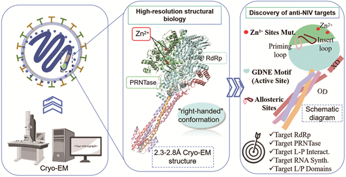
Schematic of the Cryo-EM-Derived NiV L-P complex structure and key druggable targets (PDB code: 9BDQ). The mushroom-shaped L-P complex is depicted, with annotations of the L protein’s catalytic domains (RdRp containing GDN/GDNE motifs, PRNTase with ZF1/ZF2 zinc fingers, MTase, CTD) and the tetrameric P protein’s domains (NTD, OD, XD). Critical regulatory elements (insertion loop, priming loop) and antiviral target sites (RdRp active site, PRNTase zinc-binding pockets, L-P interface [OD-CTD/XD]) are also labeled to illustrate their spatial distribution and functional relevance to viral replication.
(1) Targeting of the RdRp domain: The RdRp domain has been identified as the catalytic centre of the L-P complex, and is responsible for RNA chain elongation. The RdRp domain contains multiple conserved catalytic motifs (such as the GDNE motif, GDN loop), which play a key role in viral RNA synthesis [3]. Competitive inhibitors, such as nucleoside analogues, can be designed to target the GDNE motif at the RdRp active site, leading to chain termination by incorporation into the nascent RNA chain. For the GDN loop, non-nucleoside inhibitors can be used to design allosteric antagonists. These antagonists regulate the conformational transition of the "GDN loop" from "GDN-in" to "GDN-out", thereby inhibiting polymerase activity [6].
(2) Targeting of the PRNTase domain: The PRNTase domain is involved in the capping process of mRNA, which is crucial for viral replication and transcription. The two zinc-binding sites in this domain have been demonstrated to play a pivotal role in viral RNA capping [4]. The design of small-molecule compounds capable of chelating zinc ions has been shown to disrupt the stability of the PRNTase domain, thereby inhibiting the mRNA capping process, hindering proper mRNA processing, and impeding viral proliferation.
(3) Targeting L-P protein interactions: Disrupting L-P protein interactions, mediated primarily by the P protein’s OD domain [5], interferes with the assembly and function of the L-P complex. The 3D structure of the L-P interaction interface provides a basis for designing peptide or non-peptide small-molecule inhibitors, which can block L-P binding by occupying binding sites or inducing conformational changes.
(4) Targeting dynamic changes during viral RNA synthesis: During the processes of RNA binding and early elongation, the insertion loop and priming loop undergo dynamic conformational changes to stabilize the RNA template and nascent RNA chain [5]. It is therefore hypothesized that the insertion and priming loops function as allosteric regulatory sites, inhibiting the activity of the enzyme by stabilizing the inactive conformation and thereby preventing viral RNA synthesis.
In addition to the design of inhibitors targeting the active sites of enzymes, the structural biology information have also given rise to the possibility of multi-target drug design [12]. It is evident that, in consideration of the multi-component characteristics of the NiV L-P complex, the design of bifunctional molecules that simultaneously bind multiple domains of the L and P proteins is a potential avenue for enhancement of inhibitory effects through synergistic action, whilst concomitantly reducing the risk of resistance. For instance, an inhibitor that binds to both the RdRp active site of the L protein and the XD domain of the P protein may simultaneously impede RNA synthesis and complex stability [4], thereby achieving more efficient antiviral effects; this approach leverages the close proximity of the RdRp and XD-binding interfaces observed in the cryo-EM structures [3,4].
By targeting key functional domains and active sites of the L-P complex, combined with novel drug discovery technologies and strategies [13], researchers can design highly specific and potent antiviral drugs. Outstanding challenges include validating loop-stabilising compounds in NiV-infected cells and determining whether conformational restriction of the GDN loop can be achieved without off-target effects. Future cryo-EM studies under more native or inhibitor-bound conditions, along with the continuous advancement of structural biology technologies, will be critical to refine these pharmacological strategies, deepen our understanding of NiV molecular mechanisms, and ultimately accelerate the development of novel antiviral drugs against NiV.
Declaration of competing interest
The authors declare that they have no known competing financial interests or personal relationships that could have appeared to influence the work reported in this paper.
The authors are supported by the Key Research and Development Program, Ministry of Science and Technology of the People’s Republic of China (No. 2023YFC2606500 to P. Zhan), Shandong Provincial Natural Science Foundation for Young Scholars Project (No. ZR2022QH036 to S. Gao), Young Innovation Team of Colleges and Universities in Shandong Province (No. 2024KJJ063 to S. Gao)
[1]
J.F. Cotino, G. Reina, J. Pueyo, Viruses 16 (2024) 179. doi: 10.3390/v16020179
Figure 1
Schematic of the Cryo-EM-Derived NiV L-P complex structure and key druggable targets (PDB code: 9BDQ). The mushroom-shaped L-P complex is depicted, with annotations of the L protein’s catalytic domains (RdRp containing GDN/GDNE motifs, PRNTase with ZF1/ZF2 zinc fingers, MTase, CTD) and the tetrameric P protein’s domains (NTD, OD, XD). Critical regulatory elements (insertion loop, priming loop) and antiviral target sites (RdRp active site, PRNTase zinc-binding pockets, L-P interface [OD-CTD/XD]) are also labeled to illustrate their spatial distribution and functional relevance to viral replication.

 DownLoad:

 下载:
下载:


 下载:
下载:
