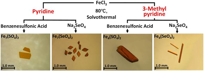
Scheme 1.
The synthetic routes and optical micrographs of crystals for Fe7(SO4)2, Fe7(SeO4)2, Fe4(SO4)2, and Fe4(SeO4)2.

β-Anderson-type antiferromagnetic iron oxo clusters for catalysis in Aldehyde Cyanosilylation
Chunhui Wang , Xinyi Ji , Puyong He , Chaoyue Fan , Yuan Gao , Zhe Han , Jie Qiu


Metal oxo clusters (MOCs) have garnered significant interest due to their rich structures and properties, making them highly promising for diverse applications such as catalysis [1], magnetism [2], biomedicine [3], and materials science [4,5]. Polyoxometalates (POMs) are traditionally defined as anionic metal oxo clusters comprising early transition metals (W, Mo, V, Nb, or Ta) in their high oxidation states [6]. They exhibit potential applications in diverse fields, including biomedicine [7], energy [8,9], and catalysis [10-15]. So far, POMs with classic structural archetypes, including Keggin, Anderson, Dawson, Waugh, Silverton, and Lindqvist types, have been progressively discovered [7]. Among them, the Anderson-type POM was first reported in 1937 and features a central XO6 octahedron surrounded by six peripheral MO6 octahedra [16], where X represents a heteroatom (typically a transition metal, such as V, Cr, or Fe) and M represents an addenda atom (mostly Mo or W) [17]. Anderson-type POMs are commonly heterometallic oxo clusters and can exist as either α or β isomers, depending on their conformations [18]. While α-Anderson-type POMs have been extensively synthesized and well-documented, β-Anderson-type POMs are relatively rare, with only a few clusters reported in the literature [17].
Iron oxo clusters are one important subset of metal oxo clusters and have attracted considerable interest. Research has yielded diverse iron clusters, such as Keggin-type Fe13 [19,20], Lindqvist-type Fe6 [21], and clusters without classical POM-type structures like Fe17 [22], Fe30 [23], and Fe34 [24]. These discoveries have substantially expanded the family of iron oxo clusters. However, despite these advances, iron oxo clusters with classical POM-type structures remain notably under-explored compared to those of other transition metals.
Intense studies on iron oxo clusters are driven by their pivotal role in understanding the biomineralization process [25] and their applications in numerous fields, such as lithium storage [26], single-molecule magnets [27], cationic dye adsorption and separation [28], and catalysis [29]. As catalysts, iron-oxo clusters have been applied in water oxidation [29,30] and oxidative coupling reactions [31], whereas their potential for C-C bond formation remains unexplored. The cyanosilylation of aldehydes with nucleophilic trimethylsilyl cyanide (TMSCN) is a pivotal reaction for C-C bond formation to produce cyanohydrin derivatives [32,33]. This reaction has been facilitated by diverse catalysts, including solid acids and bases [34], small molecules [35,36], organometallic complexes [37], and crystalline materials [38-41]. Metal-oxo clusters represent a class of crystalline materials with well-defined atomic structures and compositions, enabling atomic-level analysis of catalytic mechanisms [38-41]. While previous studies have reported catalytic performances of POM-based compounds [42,43] and Al-Ln hybrid clusters [38] for this reaction, research employing iron oxo clusters remains unexplored.
Herein, we report the synthesis of two novel heptanuclear iron clusters Fe7(SO4)2 and Fe7(SeO4)2, which exhibit unexpected β-Anderson-type structures when pyridine is used as the base. By switching to 3-methylpyridine, we successfully isolated two unique tetranuclear clusters Fe4(SO4)2 and Fe4(SeO4)2, which possess half β-Anderson structures. The catalytic experiments revealed excellent catalytic activities and stabilities of these clusters in the cyanosilylation of aldehydes. Details on the syntheses, structural features, spectral properties, magnetic behaviors, and catalytic performances of the four clusters are provided below.
Brown crystals of the cluster Fe7(SO4)2 were synthesized by mixing solutions of FeCl3, benzenesulfonic acid, and pyridine, followed by allowing the mixture to react under solvothermal conditions at 80 ℃ (Scheme 1). To investigate the effect of ligands on the formation of iron oxo clusters, we replaced benzenesulfonic acid with sodium selenate, resulting in the formation of crystals of the cluster Fe7(SeO4)2. Next, we substituted pyridine with 3-methylpyridine in the two reactions, resulting in the formation of crystals containing clusters Fe4(SO4)2 and Fe4(SeO4)2. This result indicates the critical role of organic amines in directing the formation and structures of iron oxo clusters. Previous studies suggested amines act as templates for the formation of clusters [44,45].
Single crystal X-ray diffraction revealed that the two heptanuclear clusters, Fe7(SO4)2 and Fe7(SeO4)2, crystallize in space groups of monoclinic C2/c and triclinic P
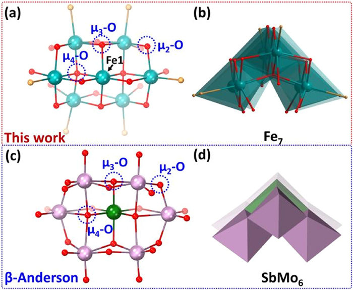
The structural difference of clusters Fe7(SO4)2 and Fe7(SeO4)2 lies in the substitution of the two sulfate groups in the former with two selenate groups in the latter (Figs. 2a-c). Specifically, their structures comprise a six-membered ring of iron ions, with an additional iron ion (Fe1) located at the center. The central Fe1 ion connects to iron ions in the ring via two μ4-O atoms, along with two μ3-O and two μ2-O atoms from CH3O− groups (Fig. 1a). Meanwhile, the adjacent iron ions in the ring are bridged by μ2-O atoms from CH3O− groups. Notably, the Fe1 ion aligns co-planarly with Fe3 and Fe6 ions, while the remaining four iron ions occupy the other plane (Fig. S1 in Supporting information). Apart from bridging O atoms, the remaining coordination sites of irons were occupied by six monodentate coordinated Cl− ions and six O atoms from two tridentate coordinated sulfate or selenate groups, resulting in a distorted octahedral geometry of [FeO6] or [FeO5Cl] for each iron ion (Fig. S2 in Supporting information). In addition, the crystallographic analysis also revealed the presence of co-crystallized pyridine and water molecules distributed among clusters (Figs. S3 and S4 in Supporting information). Since the two clusters are formed in an acidic reaction solution, their negative charges may be balanced by protonated pyridine molecules [23]. The corresponding Fe-O and Fe-Cl bond lengths, as well as Fe-O-Fe bond angles in the two clusters, exhibit common values among iron complexes (Tables S2-S5 in Supporting information) [24].
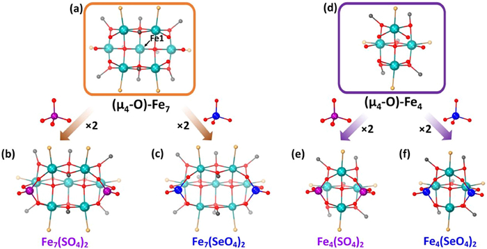
The tetranuclear clusters Fe4(SO4)2 and Fe4(SeO4)2 crystallizes in the monoclinic space group of C2/c (Table S1) and exhibit similar compositions of [Fe4O(SO4/SeO4)2(OCH3)5Cl4]3−. Their negative charges may be balanced by protonated 3-methylpyridine molecules distributed among clusters (Figs. S5 and S6 in Supporting information). These two clusters display similar structures with half β-Anderson configurations (Figs. 2d-f), which is extremely rare in previous studies (Table S15 and Fig. S8 in Supporting information). Specifically, the four Fe ions in their structures are linked by a μ4-O atom, while most of the previously reported tetranuclear clusters are mainly connected by μ3-O atoms [48,49]. Besides the μ4-O atom, the remaining coordination sites of the iron ions are occupied by two tridentate coordinated SO42− (SeO42−) groups, five bidentate coordinated CH3O− groups and four monodentate coordinated Cl− ions, thus resulting in a distorted octahedral geometry of [FeO5Cl] with common bond lengths and angles for each iron ion (Figs. S5 and S6, Tables S6-S9 in Supporting information). The iron ions in the four clusters are all in the +3 oxidation states as indicated by the bond valence sum (BVS) analysis (Tables S10-S13 in Supporting information).
Crystals of clusters Fe7(SO4)2, Fe7(SeO4)2, and Fe4(SeO4)2 were further characterized using powder X-ray diffraction (PXRD), scanning electron microscopy-energy dispersive spectroscopy (SEM-EDS), inductively coupled plasma mass spectrometry (ICP-MS), thermogravimetric analysis (TGA), and infrared (IR) spectroscopy to investigate their purity and composition. Specifically, the experimental PXRD patterns closely matched the calculated ones using single crystal X-ray diffraction data (Figs. S9-S11 in Supporting information), indicating these crystals have high crystalline purity. SEM-EDS analysis confirmed the existence of Fe, S (Se), Cl, O, C, and N elements in these crystals (Figs. S12-S15 in Supporting information), and further quantitative analysis using ICP-MS revealed that the Fe:Se ratios in the structures of Fe7(SeO4)2 and Fe4(SeO4)2 are 6.22:2 and 3.80:2 (Table S16 in Supporting information), respectively, which were consistent with the ratios obtained via crystallographic analysis. The TGA results confirmed the number of H2O molecules in three clusters (Figs. S16-S18 in Supporting information), and the PXRD patterns revealed that TGA residues contain hematite-Fe2O3 (Fig. S19 in Supporting information). The IR spectra demonstrated the existence of the SO42− group, SeO42- group, CH3OH, pyridine, and/or 3-methylpyridine in respective crystals (Fig. S20 in Supporting information).
UV-vis spectroscopy revealed that the three clusters exhibit similar absorption spectra in the range of 200-800 nm (Fig. 3a), which should be attributed to the ligand-to-metal charge transfer (LMCT) transition [50]. By applying the Kubelka–Munk function, their calculated bandgap values are 2.22, 2.50, and 2.39 eV, respectively (Figs. S21-S23 in Supporting information), indicating the semiconducting properties of the crystals [51].
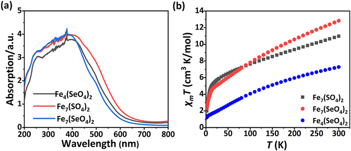
The direct-current molar magnetic susceptibility of the crystals of clusters Fe7(SO4)2, Fe7(SeO4)2, and Fe4(SeO4)2 was measured under a magnetic field of 0.1 T. As shown in Fig. 3b, the heptanuclear clusters Fe7(SO4)2 and Fe7(SeO4)2 exhibit similar magnetic behaviors, characterized by decreasing χmT values as the temperature decreases. At 300 K, their χmT values are 10.96 and 12.82 cm3 K/mol, respectively (Fig. 3b). These values are significantly lower than the Curie constant (C = 30.63 cm3 K/mol, with S = 5/2 and g = 2) expected for seven noncoupled high-spin Fe3+ ions. As the temperature drops to 2 K, the χmT values decrease to 1.99 and 2.31 cm3 K/mol, respectively. In addition, the slopes of 1/χm vs. T plots within the temperature range of 100-300 K are negative (Figs. S24 and S25 in Supporting information). Moreover, their molar magnetizations (Mm) were measured at 2 K. At a magnetic field of 7.0 T, the Mm values are 3.75 NA μB and 3.61 NA μB, respectively (Fig. S26 in Supporting information). These values are lower than the saturation value (Mm = 30 NA μB) of seven Fe3+ ions. The above results indicate that antiferromagnetic exchange interactions are dominant among Fe3+ ions in Fe7(SO4)2 and Fe7(SeO4)2 clusters.
Similarly, the tetranuclear cluster Fe4(SeO4)2 also exhibits antiferromagnetic behavior. Specifically, its χmT value is 7.23 cm3 K/mol at 300 K (Fig. 3b), falling below the Curie constant (C = 17.50 cm3 K/mol) anticipated for four Fe3+ ions. As the temperature decreases to 2 K, the χmT value decreases to 1.20 cm3 K/mol (Fig. 3b). The slope of the corresponding 1/χm vs. T plot ions is also negative (Fig. S27 in Supporting information). At a magnetic field of 7.0 T, its molar magnetization is 1.35 NA μB (Fig. S26), below the saturation value of four Fe3+ ions.
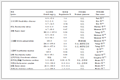
We selected the cyanosilylation of benzaldehyde as a model reaction to evaluate the catalytic activity of Fe7(SO4)2, Fe7(SeO4)2, and Fe4(SeO4)2 clusters. The reaction of benzaldehyde with TMSCN yields 2-phenyl-2-((trimethylsilyl)oxy)acetonitrile as the target product (Table 1). First, the stability of these clusters in organic solvents was assessed. Specifically, crystals of Fe7(SO4)2, Fe7(SeO4)2, and Fe4(SeO4)2 were individually soaked in the same organic solvents for 24 h at room temperature. PXRD patterns confirmed that all clusters remained structurally intact in n-propanol, diethyl ether, toluene, n-hexane, and cyclohexane (Figs. S28-S30 in Supporting information). Next, the effect of solvents on the cyanosilylation of aldehydes was investigated. Using Fe7(SeO4)2 as the catalyst, reactions between benzaldehyde and TMSCN were conducted in each solvent. The experiment shows that n-hexane afforded the highest yield of the target product (89.2%) after 12 h (Table S17 in Supporting information). Consequently, n-hexane was selected as the solvent for subsequent experiments. Then, the effect of reaction time on product yield was investigated using Fe7(SeO4)2 as the catalyst. The yield increases with the increasing reaction time and reaches 89.2% at 12 h (Table 1). Further extension of the reaction time resulted in no significant change in yield. To confirm the catalytic activity of Fe7(SO4)2, Fe7(SeO4)2, and Fe4(SeO4)2 for the cyanosilylation of aldehydes, control experiments were performed (Table 1). The blank reaction without a catalyst yielded only trace amounts of the target product. Reactions catalyzed by benzenesulfonic acid or Na2SeO4 produced almost no detectable product. These comparative results demonstrate the essential catalytic role of these iron clusters in this reaction.
 DownLoad:
CSV
 |
|||||||||||||||||||||||||||||||||||
| Catalyst | Time (h) | Yieldb(%) | |||||||||||||||||||||||||||||||||
| Fe7(SeO4)2 | 4 | 23.8 | |||||||||||||||||||||||||||||||||
| Fe7(SeO4)2 | 6 | 30.7 | |||||||||||||||||||||||||||||||||
| Fe7(SeO4)2 | 8 | 38.4 | |||||||||||||||||||||||||||||||||
| Fe7(SeO4)2 | 10 | 85.6 | |||||||||||||||||||||||||||||||||
| Fe7(SeO4)2 | 12 | 89.2 | |||||||||||||||||||||||||||||||||
| Fe7(SeO4)2 | 16 | 89.5 | |||||||||||||||||||||||||||||||||
| Fe7(SO4)2 | 12 | 86.0 | |||||||||||||||||||||||||||||||||
| Fe4(SeO4)2 | 12 | 80.0 | |||||||||||||||||||||||||||||||||
| None | 12 | Trace | |||||||||||||||||||||||||||||||||
| FeCl3 | 12 | 3.3 | |||||||||||||||||||||||||||||||||
| Benzenesulfonic acid | 12 | Trace | |||||||||||||||||||||||||||||||||
| Na2SeO4 | 12 | Trace | |||||||||||||||||||||||||||||||||
| a Reaction condition: Aldehyde (0.1 mmol), TMSCN (0.22 mmol), Cat. (0.01 mmol), and 2 mL of hexane at room temperature. b Determined by GC-MS. |
|||||||||||||||||||||||||||||||||||
Pyridine adsorption IR spectroscopy was employed to characterize acid site types in these iron clusters. As observed in the IR spectra of Fe7(SeO4)2 powder after exposure to pyridine vapor and subsequent evacuation at different temperatures (Fig. 4a), the strong band at 1450 cm−1 is attributed to the pyridine molecules adsorbed on the Lewis (L) acid sites [52]. The relatively weaker band at 1540 cm−1 is assigned to the pyridine molecules adsorbed on the Brønsted (B) acid sites [52]. The band at 1490 cm−1 is ascribed to the pyridine molecules co-adsorbed on both Lewis and Brønsted acid sites [52]. These findings demonstrate that Fe7(SeO4)2 possesses dominant Lewis sites, with the B/L of 0.24, 0.29, and 0.36 at 273, 423, and 473 K, respectively.
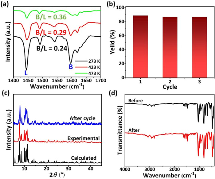
To evaluate catalyst stability, Fe7(SO4)2, Fe7(SeO4)2, and Fe4(SeO4)2 crystals were recovered post-reaction via filtration and reused for three cycles. As shown in Fig. 4b and Fig. S31 (Supporting information), the recycled crystals maintained excellent activity with no significant decline. The structural stability of these crystals was further examined using PXRD and FT-IR analyses of pre- and post-reaction samples. Nearly identical PXRD patterns and IR spectra (Figs. 4c and d, Figs. S32-S35 in Supporting information) verify the preservation of cluster structures. These experimental results not only demonstrated the exceptional stability of these clusters as catalysts but also confirmed their heterogeneous nature in cyanosilylation reactions.
Furthermore, the effect of organic substrates on the cyanosilylation reaction was investigated using Fe7(SeO4)2 as the catalyst. Specifically, various aldehydes were allowed to individually react with TMSCN, yielding corresponding cyanosilylation products. As shown in Table S18 (Supporting information), bromobenzaldehyde, especially 2-bromobenzaldehyde and 3-bromobenzaldehyde, afford the highest yield (98%), 4-chlorobenzaldehyde and 4-cyanobenzaldehyde afford relatively lower yields (73%, 86%), and whereas 9-anthraldehyde and vanillin perform the lowest yields (4.2% and 2.8%). This indicates that the type and position of substituent on the aromatic ring and spatial site resistance of the substrates do affect the yield of cyanosilylation reaction
In the cyanosilylation of aldehydes, Lewis acid catalysts activate carbonyl compounds through electrophilic interactions, while Lewis base catalysts activate TMSCN via nucleophilic interactions [53]. For example, the cluster Al4Ln4, containing unsaturated sites, acts as a Lewis acid catalyst and exhibits enhanced catalytic activity [38]. Similarly, terminal oxygen atoms on the POMs surface may function as Lewis base catalytic sites [53]. In this study, the active sites in Fe7(SO4)2, Fe7(SeO4)2, and Fe4(SeO4)2 likely originate from labile methanol ligands bound to Fe centers. Transient dissociation of these ligands may generate coordinatively unsaturated sites responsible for catalytic activity in aldehyde cyanosilylation [54].
In summary, four novel iron oxo clusters were successfully synthesized by varying ligands and organic amines and characterized using various techniques. Among them, the two heptanuclear clusters, featuring an unprecedented β-Anderson-type structure, were synthesized using pyridine. Replacing pyridine with 3-methylpyridine in the reactions resulted in the formation of the two tetranuclear clusters with half β-Anderson configurations, highlighting the crucial role of organic amines in the formation and types of iron oxo clusters. These clusters exhibit similar magnetic and optical properties. Moreover, they exhibit excellent Lewis acid catalytic performance and stability in the cyanosilylation of aldehydes. This study expands the family of iron oxo clusters and demonstrates the potential applications of iron clusters in the catalytic field.
Chunhui Wang: Writing – original draft, Methodology, Formal analysis, Data curation, Conceptualization. Xinyi Ji: Investigation, Data curation. Puyong He: Investigation, Data curation. Chaoyue Fan: Investigation. Yuan Gao: Data curation. Zhe Han: Software. Jie Qiu: Writing – review & editing, Validation, Supervision, Software, Resources, Project administration, Methodology, Investigation, Funding acquisition, Formal analysis, Data curation, Conceptualization.
The authors declare that they have no known competing financial interests or personal relationships that could have appeared to influence the work reported in this paper.
This research was funded by the National Natural Science Foundation of China (Nos. 22076152, 22276147, 22476158, 21806127). Single-crystal X-ray diffraction data were collected using a Bruker D8 Quest diffractometer in the State Key Laboratory of Multiphase Flow in Power Engineering. We would like to thank Dr. Nan Deng at the Instrument Analysis Center of Xi’an Jiao Tong University for her expert assistance with the GC-MS analysis.
CCDC (239601-239604) contains the supplementary crystallographic data for this paper. These data can be obtained free of
charge via
Supplementary material associated with this article can be
found, in the online version, at doi:
N. Li, J. Liu, B.X. Dong, Y.Q. Lan, Angew. Chem. Int. Ed. 59 (2020) 20779–20793. doi: 10.1002/anie.202008054
S. Bertaina, S. Gambarelli, T. Mitra, et al., Nature 453 (2008) 203–206. doi: 10.1038/nature06962
G.X. Wang, W.M. Guan, B. Li, L.X. Wu, Curr. Opin. Colloid. In. 35 (2018) 91–103. doi: 10.1016/j.cocis.2018.01.016
L. Chen, W.L. Chen, X.L. Wang, et al., Chem. Soc. Rev. 48 (2019) 260–284. doi: 10.1039/c8cs00559a
G.H. Zhang, B.Y. Li, Y. Zhou, et al., Nat. Commun. 11 (2020) 425. doi: 10.1038/s41467-019-14227-6
Z.M. Wang, X. Xin, M. Zhang, et al., Sci. China Chem. 65 (2022) 1515–1525. doi: 10.1007/s11426-022-1276-4
M.J.W. Budych, K. Staszak, A. Bajek, et al., Coord. Chem. Rev. 493 (2023) 215306. doi: 10.1016/j.ccr.2023.215306
M.R. Horn, A. Singh, S. Alomari, et al., Energy Environ. Sci. 14 (2021) 1652–1700. doi: 10.1039/d0ee03407j
L. Zhang, Z.Q. Chen, Int. J. Energy Res. 44 (2020) 3316–3346. doi: 10.1002/er.5124
Y. Jiang, H. Liu, J. Zhang, et al., Chin. J. Struct. Chem. 44 (2025) 100603. doi: 10.1016/j.cjsc.2025.100603
X. Zhang, H. Li, N. Xu, X. Liu, X.L. Wang, Inorg. Chem. Front. 12 (2025) 3823–3832. doi: 10.1039/D5QI00460H
Y. Liu, J. Cao, J. Wu, et al., Inorg. Chem. 64 (2025) 12619–12626. doi: 10.1021/acs.inorgchem.5c01101
K. Hasnain, Y. Feng, B. Song, et al., Polyoxometalates 4 (2025) 9140094. doi: 10.26599/pom.2025.9140094
Q.L. Hu, K. Li, X.J. Chen, Y.F. Liu, G.P. Yang, Polyoxometalates 3 (2024) 9140048. doi: 10.26599/pom.2023.9140048
X.Q. Huang, S. Liu, G. Liu, et al., Appl. Catal. B 323 (2023) 122134. doi: 10.1016/j.apcatb.2022.122134
J.S. Anderson, Nature 140 (1937) 850-850. doi: 10.1038/140850a0
P. Wu, Y. Wang, B. Huang, Z. Xiao, Nanoscale 13 (2021) 7119–7133. doi: 10.1039/d1nr00397f
Q.H. Zhuang, Z.Q. Sun, C.G. Lin, B. Qi, Y.F. Song, Inorg. Chem. Front. 10 (2023) 1695–1711. doi: 10.1039/d2qi02690b
O. Sadeghi, L.N. Zakharov, M. Nyman, Science 347 (2015) 1359–1362. doi: 10.1126/science.aaa4620
A. Bino, M. Ardon, D. Lee, B. Spingler, S.J. Lippard, J. Am. Chem. Soc. 124 (2002) 4578–4579. doi: 10.1021/ja025590a
M. Ashafaq, M. Khalid, M. Raizada, et al., Polyhedron 163 (2019) 131–143. doi: 10.1016/j.poly.2019.02.019
G.W. Powell, H.N. Lancashire, E.K. Brechin, et al., Angew. Chem. Int. Ed. 43 (2004) 5772–5775. doi: 10.1002/anie.200460636
A.E. Dearle, D.J. Cutler, M. Coletta, et al., Chem. Commun. 58 (2021) 52–55. doi: 10.1039/D1CC06224G
A.E. Dearle, D.J. Cutler, H.W.L. Fraser, et al., Angew. Chem. Int. Ed. 58 (2019) 16903–16906. doi: 10.1002/anie.201911003
J.C. Mendez, T. Hiemstra, G.F. Koopmans, Environ. Sci. Technol. 54 (2020) 11990–12000. doi: 10.1021/acs.est.0c02163
J.Q. Lv, B. Li, Q. Liu, et al., Angew. Chem. Int. Ed. 62 (2023) e202306193. doi: 10.1002/anie.202306193
L. Poggini, E. Tancini, C. Danieli, et al., Adv. Mater. Interfaces 8 (2021) 1–9. doi: 10.1002/admi.202101182
M.N. Akhtar, M.I.M. Shahid, M.A. AlDamen, M. Khalid, M. Akram, New J. Chem. 45 (2021) 1415–1422. doi: 10.1039/d0nj05242f
J.Q. Lv, Z.L. Lang, J.Q. Fu, et al., Angew. Chem. Int. Ed. 61 (2022) e202202650. doi: 10.1002/anie.202202650
J. Deutscher, T. Corona, K. Warm, et al., Eur. J. Inorg. Chem. 2018 (2018) 4925–4929. doi: 10.1002/ejic.201800269
L.L. He, S.N. Sun, J.W. Shi, J. Liu, N. Li, Chin. Chem. Lett. 37 (2026) 111111. doi: 10.1016/j.cclet.2025.111111
R.J. Gregory, Chem. Rev. 99 (1999) 3649–3682. doi: 10.1021/cr9902906
M. North, Tetrahed.: Asym. 14 (2003) 147–176. doi: 10.1016/S0957-4166(02)00825-X
K. Higuchi, M. Onaka, Y. Izumi, Bull. Chem. Soc. Jpn. 66 (1993) 2016–2032. doi: 10.1246/bcsj.66.2016
V. Swamy, M.K. Bisai, T. Das, S.S. Sen, Chem. Commun. 53 (2017) 6910–6913. doi: 10.1039/C7CC03948D
G.K. Prakash, H. Vaghoo, C. Panja, et al., Proc. Natl. Acad. Sci. U. S. A. 104 (2007) 3026–3030. doi: 10.1073/pnas.0611309104
G. Vinoth, S. Indira, M. Bharathi, et al., Inorg. Chim. Acta 514 (2021) 120006. doi: 10.1016/j.ica.2020.120006
D. Luo, C.H. Liu, Y.B. Chen, S.T. Wang, W.H. Fang, J. Zhang. Adv. Sci. 11 (2024) e2305833. doi: 10.1002/advs.202305833
Q. Han, X. Sun, J. Li, P. Ma, J. Niu, Inorg. Chem. 53 (2014) 6107–6112. doi: 10.1021/ic5004879
S. Horike, M. Dinca, K. Tamaki, J.R. Long, J. Am. Chem. Soc. 130 (2008) 5854–5855. doi: 10.1021/ja800669j
T. Kundu, J. Wang, Y. Cheng, et al., Dalt. Trans. 47 (2018) 13824–13829. doi: 10.1039/c8dt03005g
H. An, L. Wang, Y. Hu, F. Fei, CrystEngComm 17 (2015) 1531–1540. doi: 10.1039/C4CE01802H
K. Suzuki, M. Sugawa, Y. Kikukawa, et al., Inorg. Chem. 51 (2012) 6953– 6961. doi: 10.1021/ic3008365
J.N. Behera, C.N. Rao, Inorg. Chem. 45 (2006) 9475–9479. doi: 10.1021/ic061457y
J.N. Behera, C.N. Rao, Chem. Asian J. 1 (2006) 742–750. doi: 10.1002/asia.200600083
A. Ogawa, H. Yamato, U. Lee, et al., Acta Crystallogr. C 44 (1988) 1879–1881. doi: 10.1107/S0108270188007073
A. Blazevic, A. Rompel, Coord. Chem. Rev. 307 (2016) 42–64. doi: 10.1016/j.ccr.2015.07.001
K.H.K. Lee, L. Aebersold, J.E. Peralta, K.A. Abboud, G. Christou, Inorg. Chem. 61 (2022) 17256–17267. doi: 10.1021/acs.inorgchem.2c02841
A.R. Hale, M.E. Lott, J.E. Peralta, et al., Inorg. Chem. 61 (2022) 11261–11276. doi: 10.1021/acs.inorgchem.2c01371
Z. Shi, J. Jin, L. Zhao, P. Chen, S. Niu, Chin. Sci. Bull. 55 (2010) 124–130. doi: 10.1007/s11434-009-0483-x
Y. Xue, S. Zhang, D. Song, et al., J. Semicond. 42 (2021) 112101. doi: 10.1088/1674-4926/42/11/112101
B. Liu, Y. Chai, Y. Li, et al., Appl. Catal. A 471 (2014) 70–79. doi: 10.1016/j.apcata.2013.11.017
Y. Kikukawa, K. Suzuki, M. Sugawa, et al., Angew. Chem. Int. Ed. 51 (2012) 3686–3690. doi: 10.1002/anie.201200486
J.B. Xie, J.J. Bao, H.X. Li, et al., RSC Adv. 4 (2014) 54007–54017. doi: 10.1039/C4RA11743C

Scheme 1 The synthetic routes and optical micrographs of crystals for Fe7(SO4)2, Fe7(SeO4)2, Fe4(SO4)2, and Fe4(SeO4)2.

Figure 1 Comparison between the Fe7(S/SeO4)2 cluster in this work and the reported β-Anderson-type cluster [SbMo6O24]7− [46]. Color code: Fe, lime green; Mo, pink; Sb, blue; O, red; C, grey; Cl, pale yellow.

Figure 2 Ball-stick representations of (a) cores and (b, c) crystal structures of clusters Fe7(SO4)2 and Fe7(SeO4)2, as well as (d) cores and (e, f) crystal structures of clusters Fe4(SO4)2 and Fe4(SeO4)2. Color code: Fe, lime green; S, purple; Se, blue; O, red; C, grey; Cl, pale yellow.

Figure 3 (a) UV–vis spectra and (b) χmT vs. T plots for Fe7(SO4)2, Fe7(SeO4)2 and Fe4(SeO4)2.

Figure 4 (a) IR spectra of Fe7(SeO4)2 powder after exposure to pyridine vapor and subsequent evacuation at 373, 423, and 473 K, respectively. (b) Change of the yield during the recycling catalytic experiments. (c) PXRD patterns and (d) IR spectra of Fe7(SeO4)2 powder before and after the recycling catalytic experiments.
Table 1. Optimizing reaction conditions for cyanosilylation of benzaldehyde.a
|
|||||||||||||||||||||||||||||||||||
| Catalyst | Time (h) | Yieldb(%) | |||||||||||||||||||||||||||||||||
| Fe7(SeO4)2 | 4 | 23.8 | |||||||||||||||||||||||||||||||||
| Fe7(SeO4)2 | 6 | 30.7 | |||||||||||||||||||||||||||||||||
| Fe7(SeO4)2 | 8 | 38.4 | |||||||||||||||||||||||||||||||||
| Fe7(SeO4)2 | 10 | 85.6 | |||||||||||||||||||||||||||||||||
| Fe7(SeO4)2 | 12 | 89.2 | |||||||||||||||||||||||||||||||||
| Fe7(SeO4)2 | 16 | 89.5 | |||||||||||||||||||||||||||||||||
| Fe7(SO4)2 | 12 | 86.0 | |||||||||||||||||||||||||||||||||
| Fe4(SeO4)2 | 12 | 80.0 | |||||||||||||||||||||||||||||||||
| None | 12 | Trace | |||||||||||||||||||||||||||||||||
| FeCl3 | 12 | 3.3 | |||||||||||||||||||||||||||||||||
| Benzenesulfonic acid | 12 | Trace | |||||||||||||||||||||||||||||||||
| Na2SeO4 | 12 | Trace | |||||||||||||||||||||||||||||||||
| a Reaction condition: Aldehyde (0.1 mmol), TMSCN (0.22 mmol), Cat. (0.01 mmol), and 2 mL of hexane at room temperature. b Determined by GC-MS. |
|||||||||||||||||||||||||||||||||||
 下载: 导出CSV

 扫一扫看文章


扫一扫关注我们
 下载: