Citation:
Junpu Yang, Yaoyao Bai, Huangjie Lu, Jingqi Ma, Jian Xie, Jie Qiu, Xiaofeng Guo, Yaxing Wang, Shuao Wang, Jian Lin. Emergence of acidity modulation as a new strategy for eliciting stable radicals in multi-stimuli-responsive metal-oxo clusters[J]. Chinese Chemical Letters,
2026, 37(11): 111702.
doi:
10.1016/j.cclet.2025.111702
Emergence of acidity modulation as a new strategy for eliciting stable radicals in multi-stimuli-responsive metal-oxo clusters
English
Emergence of acidity modulation as a new strategy for eliciting stable radicals in multi-stimuli-responsive metal-oxo clusters
School of Nuclear Science and Technology, Xi’an Jiaotong University, Xi’an 710049, China
b.
Shanghai Institute of Applied Physics, Chinese Academy of Sciences, Shanghai 201800, China
c.
School of Life and Environmental Sciences, Shaoxing University, Shaoxing 312000, China
d.
Department of Chemistry and Alexandra Navrotsky Institute for Experimental Thermodynamics, Washington State University Pullman, WA 99164-4630, United States
e.
State Key Laboratory of Radiation Medicine and Protection, School of Radiation Medicine and Protection, Collaborative Innovation Centre of Radiological Medicine of Jiangsu Higher Education Institutions, Soochow University, Suzhou 215123, China
Received Date:
06 July 2025 Accepted Date:
09 August 2025 Revised Date:
03 August 2025 Available Online:
15 November 2026
Abstract:
Stable radicals have applications in numerous fields, yet their generation and stabilization remain methodologically challenging, with current achievements primarily limited to metal-organic frameworks (MOFs). In this work, we report the stabilization of radicals within metal-oxo clusters (MOCs) through an acidity modulation method, resulting in two thorium-oxo nanoclusters with identical structures but distinct photophysical properties. Under high-acidity conditions, yellow single crystals of Th-aca-Y, containing highly stable radical species, are obtained in situ, whereas lower acidity leads to the formation of Th-aca-C, a colorless and radical-free counterpart. Remarkably, the radical formed through acid modulation in Th-aca-Y exhibits unique UV-vis absorbance properties and considerably enhanced stability compared to the Xe light-induced radicals in Th-aca-C-Xe. Fourier-transform infrared (FTIR) spectroscopy, nuclear magnetic resonance (NMR) spectroscopy, X-ray photoelectron spectroscopy (XPS), and photoluminescence characterization further confirmed the fundamentally different nature of the in situ generated radicals in Th-aca-Y compared to the light-induced radicals in Th-aca-C-Xe. In addition, these clusters display multi-stimuli-responsive properties, including photochromism, photothermal conversion, and notably, mechanochromic luminescence, which represents the first such instance in actinide-based materials.
Organic radicals are highly reactive species characterized by an unpaired electron in an organic molecule, often contributing to significant chemical reactivity and stability challenges [1,2]. Beyond their well-known roles in organic synthesis and polymerization, radicals have potential in many advanced physicochemical properties, such as photoswitching, capacitors, photovoltaics, chemosensing, magnetism, and catalysis [3-8]. However, organic radicals are typically thermodynamically and kinetically unstable owning to their subvalent nature, which facilitates the formation of covalent bonds [9]. To address these challenges, bulky substituents are commonly utilized to provide steric protection, effectively shielding unstable radicals from external reactive species and preventing dimerization of the radicals themselves [10,11]. Moreover, stabilizing radicals can be achieved by enhancing the delocalization of the unpaired electron through constructing π-conjugated systems, thereby mitigating radical reactivity and enhancing their resilience against external perturbations [12,13].
Metal-organic frameworks (MOFs), a class of crystalline materials consisting of metal ions or clusters and organic linkers to form one-, two-, or three-dimensional structures, have emerged as a novel platform for stabilizing organic radicals [14-16]. The ordered and spatial distribution of organic ligands in MOFs effectively inhibits collisional quenching of radicals [17-19]. Moreover, their porous architectures enable the incorporation of electron-donating guest species, which facilitate electron transfer to radical formation in response to external stimuli [20-23]. Persistent or stable radicals of naphthalenediimides, viologens, tetrathiafulvalene (TTF), 2,2,6,6-tetramethyl-1-piperidinyloxy (TEMPO), triazine, perylenediimides, have been embedded in MOFs [24-29]. In stark contrast, molecular metal-oxo clusters (MOCs), which can also be composed of metal nodes and organic ligands similar to MOFs, have lagged behind in this regard. Although MOCs possess great potential in catalysis, sensing, magnetism, medicine, and biotechnology, studies of stable radicals in MOCs remain extremely limited [30-34]. To date, only a few examples have demonstrated radical generation in MOCs upon light irradiation [35-37].
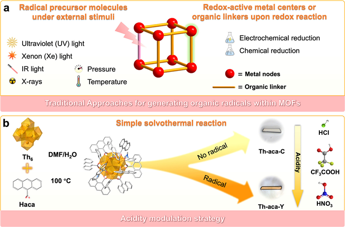
Generating organic radicals within MOFs involves intricate methodologies that leverage the unique structural and chemical properties of these porous materials. One approach is the incorporation of radical precursor molecules into the framework during synthesis, which can then be activated through external stimuli to generate radicals. For instance, exposure to ultraviolet (UV) light, xenon (Xe) light, IR light, X-rays, or variations in pressure and temperature can trigger electron transfer processes that lead to radical formation (Fig. 1a) [21-23,38]. Alternatively, in situ generation of radicals can be achieved through redox-active metal centers or organic linkers within MOFs and covalent-organic frameworks (COFs). For example, TTF•+ and BPPTzTz•– (2,5-bis(4-(pyridine-4-yl)phenyl)thiazolo[5,4-d]thiazole) radicals can be generated by oxidizing neutral redox-active TTF-based or reducing BPPTzTz–based MOFs via electrochemical or chemical methods [39-45].
Figure 1
Figure 1.
(a) Schematic representation of traditional approaches for generating organic radicals within MOFs. (b) A reaction scheme illustrating the acidity modulation strategy that leads to the formation of non-radical containing Th-aca-C under low acidity conditions and radical-bearing Th-aca-Y under high acidity conditions. Color code: Th = orange, O = red, N = blue, C = grey, and H atoms are omitted for clarity.
In this study, we have designed an acidity modulation strategy to stabilize radicals within MOCs. Two thorium-oxo nanoclusters featuring distinct colors, yellowish-brown Th-aca-Y (Y stands for yellow) and colorless Th-aca-C (C stands for colorless), with the identical molecular formula [Th6(OH)4(O)4(H2O)4(DMF)2](aca)12 (aca = anthracene-9-carboxylate) and structure, have been isolated. The Th(Ⅳ) cation offers several advantages that make it an ideal building unit for crystalline materials, including its large ionic radius, which facilitates the growth of high-quality single crystals and promotes open coordination environments conducive to structural diversity [35]. By simply increasing the reaction acidity, Th-aca-Y is obtained in situ, which incorporates highly stable radical species, whereas lower acidity produces Th-aca-C, which lacks such radicals. Notably, this radical generated through acid modulation displays distinct UV-vis absorbance characteristics and markedly superior stability compared to the Xe light-induced radicals in Th-aca-C-Xe. The incorporation of radical species into Th-aca-Y and Th-aca-C-Xe effectively activates photothermal properties of these thorium-oxo nanoclusters [46-50]. Additionally, Th-aca-C displays a reversible fluorochromic response to mechanical stress, making it as the first case of mechanochromic luminescence in actinide-based materials.
The solvothermal reactions between Th(NO3)4·6H2O and anthracene-9-carboxylic acid (Haca) in a DMF/H2O (v/v = 4:6), with modulator of trifluoroacetic acid, hydrochloric acid, or nitric acid, yielded two types of single crystals exhibiting identical crystal habits but differing in color (Fig. 1b). Under conditions of higher acidity, yellowish-brown prismatic crystals of Th-aca-Y were obtained, whereas lowering the acidity led to the formation of colorless crystals of Th-aca-C. Single crystal X-ray diffraction (SCXRD) analysis unveiled that Th-aca-Y and Th-aca-C exhibit an identical zero-dimensional (0D) structure, characterized by the monoclinic C2/c space group (Table S1 in Supporting information). The asymmetric units of Th-aca-Y and Th-aca-C consist of 1/2 [Th6(μ3-OH)4(μ3-O)4(H2O)4(DMF)2]12+ inorganic secondary building unit (SBU) and six aca– ligands. These hexanuclear Th6 SBUs, observed in both compounds, arise from the connection of six Th4+ cations through four oxo and four hydroxo groups. Notably, the resulting [Th6(μ3-OH)4(μ3-O)4(H2O)4(DMF)2]12+ moiety is capped by four H2O and two DMF molecules, a departure from the conventional arrangement typically featuring six H2O molecules in hexanuclear Th6-based MOCs or MOFs (Fig. S1 in Supporting information). The affording [Th6(μ3-OH)4(μ3-O)4(H2O)4(DMF)2]12+ SBUs are further stabilized by 12 monocarboxylate aca– ligands, resulting in the formation of a discrete nanocluster. Powder X-ray diffraction (PXRD) analysis demonstrates that the diffraction peaks of Th-aca-Y and Th-aca-C match well with the simulated patterns, confirming phase purities of the bulk samples (Figs. S2 and S3 in Supporting information).
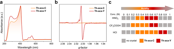
To unravel the origin of the differing colors between Th-aca-Y and Th-aca-C, we conducted solid-state UV-vis absorption spectroscopy on single crystals of both complexes. As shown in Fig. 2a, Th-aca-C exhibits a broad absorption band spanning from 200 nm to 450 nm. The lack of absorbance within the visible range of the spectrum is consistent with the colorless appearance of Th-aca-C. Alongside a comparable UV absorption band, Th-aca-Y displays a bimodal absorption band within the visible range of 490–590 nm, imparting the yellow hue observed in its crystals. There is precedence in literature indicating that the coloration in MOFs and MOCs can be attributed to the generation of radical species upon external stimuli, which can influence the electronic structure within the crystal lattice [51,52]. Indeed, electron paramagnetic resonance (EPR) spectroscopy unveiled a sharp and intense resonance signal in Th-aca-Y, whereas Th-aca-C remained EPR silent under the identical measurement conditions (Fig. 2b). The g value of 2.0003 corresponds well to the tensor value of a free electron, confirming the formation of organic radicals [53].
Figure 2
Figure 2.
(a) UV-vis absorption spectra of Th-aca-C and Th-aca-Y. (b) EPR spectra of Th-aca-C and Th-aca-Y. (c) Concentrations of HNO3, CF3COOH, and HCl used in the synthesis of Th-aca-C and Th-aca-Y.
While the stabilization of stable radicals within MOFs through heating and solvothermal methods has been previously documented, the generation of radicals via acidity adjustment has remained rare in the literature [7,13,21,39,54,55]. To further investigate the impact of acidity on radical generation, we fine-tuned the concentrations of hydrochloric acid, trifluoroacetic acid, and nitric acid to study the effects of acid type and concentration on the resulting products (Figs. S4 and S5 in Supporting information). After combining Th(NO3)4·6H2O, Haca, and the modulator, the resulting solutions exhibit light yellow prior to initiation of the reaction. Reaction conditions with low concentrations of HNO3 (0.3-0.4 mol/L) CF3COOH (0.2-0.5 mol/L), and HCl (0.3-0.7 mol/L) for 72 h yielded Th-aca-C, whereas higher concentrations of these acids (HNO3: 0.5-0.7 mol/L, CF3COOH: 0.6-0.7 mol/L, and HCl: 0.8-0.9 mol/L) led to the crystallization of Th-aca-Y (Fig. 2c and Fig. S5 in Supporting information). It is particularly noteworthy that the efficacy in eliciting radicals in Th-aca-Y follows the tendency of HNO3 > CF3COOH > HCl. This trend can be attributed their differing oxidizing strengths and abilities to accept electrons, with HNO3 being the strongest oxidizing agent, followed by CF3COOH and HCl being primarily a strong acid with limited radical-inducing capability. Moreover, HNO3 can generate unstable nitrogen dioxide (•NO2) and hydroxyl (•OH) radicals, which can react with the conjugated aca– ligands, inducing the formation of stable radicals within Th-aca-Y through crystallization [56]. Additionally, the trifluoromethyl group (–CF3) of CF3COOH provides addition electron-withdrawing capability, aiding in radical generation [57].
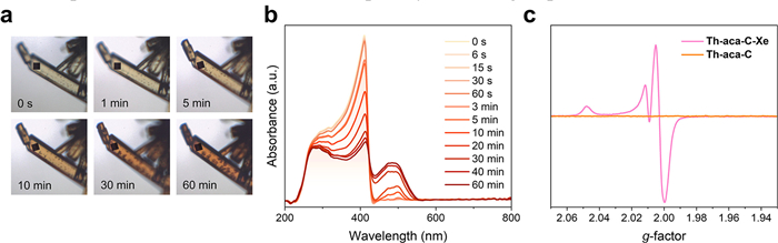
Markedly, pristine colorless Th-aca-C exhibits photochromism and its color gradually transitions to dark orange when irradiated with a Xe lamp (420-780 nm) (Fig. 3a). This photochromic response is irreversible, as the material retains its dark orange color even after heating at 100 ℃ for three days or storing under ambient conditions for three months. Correspondingly, the UV-vis absorption band of irradiated Th-aca-C (named as Th-aca-C-Xe) diminishes between 200 nm and 440 nm, while a new absorption band gradually emerges in the range of 440-560 nm (Fig. 3b). Previous studies have elucidated the generation of radical species in MOFs incorporating anthracene moieties upon UV irradiation, attributing these radicals to the observed alterations in optical properties [58-60]. In a similar vein, EPR spectroscopy confirmed the presence of radical species in Th-aca-C-Xe, revealing a strong EPR signal upon Xe lamp irradiation (Fig. 3c). This anisotropic EPR spectrum closely resembles the signal reported by Farha and co-workers for UV-induced superoxide radicals on Zr6 nodes [17]. Therefore, we attribute the observed species to superoxide radicals coordinated to the Th6 nodes, rather than to organic radicals found in Th-aca-Y. Furthermore, Th-aca-C exhibits photoluminescence quenching upon irradiation with a Xe lamp, further supporting the generation of radical species (Fig. S6 in Supporting information). Moreover, the PXRD patterns of Th-aca-C and Th-aca-C-Xe are almost identical, suggesting the structure Th-aca-C remains unchanged upon Xe lamp irradiation (Fig. S7 in Supporting information). These results indicate that the photochromic behavior of Th-aca-C can be ascribed to the generation of radical species, a mechanistic feature frequently occurring in photochromic materials [51,52].
Figure 3
Figure 3.
(a) Crystal images of Th-aca-C upon continuous Xe-lamp irradiation. (b) UV-vis absorption spectra of Th-aca-C upon continuous Xe-lamp irradiation. (c) EPR spectra of Th-aca-C before and after Xe-lamp irradiation.
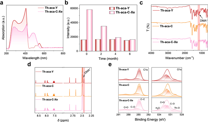
Intriguingly, a wavelength discrepancy arises in the radical-induced UV-vis absorption peaks between Th-aca-Y (490–590 nm) and Th-aca-C-Xe (440–560 nm), notable for a shift of approximately 50 nm (Fig. 4a). Other photogenerated anthracene radical species Ni(TEA)2·2(ACA) (TEA = triethanolamine), Cd2(ADC)2(DMF)2(H2O) (ADC = anthracene-9,10-dicarboxylate), and Zn(ADC)(H2O)·DMA·H2O, M(ADC)(H2O)2(DMF)2 (M = Mn and Zn) exclusively display absorbance bands, with intensities peaking below 500 nm [58-60]. Furthermore, Th-aca-Y displays a bimodal absorption band, starkly contrasting with the monomodal absorption band observed in Th-aca-C-Xe. Moreover, photoluminescence spectroscopy study reveals that Th-aca-Y, which contains stable radicals, maintains its fluorescence with an emission maximum at 450 nm. In contrast, Th-aca-C-Xe exhibits a fluorescence quenching upon Xe light irradiation (Fig. S8 in Supporting information). In addition, the radicals in Th-aca-Y demonstrate exceptional stability, as evidenced by the retention of EPR signal intensity over a period of 6 months under ambient conditions (Fig. 4b and Fig. S9 in Supporting information). To our best knowledge, Th-aca-Y represents a rare instance of radicals reported to maintain remarkable stability for such a long period under ambient conditions. In comparison, the radical signal in Th-aca-C-Xe showed a significant 72% decay within 6 months (Fig. 4b and Fig. S10 in Supporting information). These discrepancies prompt a compelling inquiry into the potential identity of these in-situ synthesized and photogenerated radicals.
Figure 4
Figure 4.
(a) UV-vis absorption spectra of Th-aca-Y and Th-aca-C-Xe. (b) EPR intensities of Th-aca-Y and Th-aca-C-Xe over time under ambient conditions. (c) FTIR spectra of Th-aca-Y, Th-aca-C, and Th-aca-C-Xe. (d) NMR spectra of Th-aca-Y, Th-aca-C, and Th-aca-C-Xe. (e) O 1s and C 1s XPS spectra of Th-aca-Y, Th-aca-C, and Th-aca-C-Xe.
Despite the identical structures of Th-aca-Y and Th-aca-C-Xe, their FTIR spectra are different (Fig. 4c). Specifically, Th-aca-Y exhibits two prominent vibrational peaks at 1202 and 1132 cm–1, which are difficult to discern in Th-aca-C and Th-aca-C-Xe. These two peaks are assignable to the CN stretching vibrations of guest dimethylammonium (DMA+) cations, presumably derived from the decomposition of DMF solvent [61,62]. Particularly, the acidic environment promotes the protonation of DMF, facilitating the hydrolysis of the DMF molecules [12,13,62]. Modeling the guest species in Th-aca-Y and Th-aca-Cvia SCXRD was attempted but was unsuccessful due to the disorder of the guest species within the void space. Alternatively, the presence of DMA+ cations was further confirmed by proton nuclear magnetic resonance (1H NMR) spectrum of Th-aca-Y, showing a significantly higher peak (δ = 2.45 ppm) derived from DMA+ cations than those of Th-aca-C and Th-aca-C-Xe (Fig. 4d) [12,13]. To further confirm the presence of DMA+, elemental analysis was performed, showing that the C:N mass ratios are 28.3 and 24.1 in Th-aca-C and Th-aca-Y, respectively (Table S2 in Supporting information). These values are close to the C:N mass ratios calculated from the integration of proton signals corresponding to DMA+ and DMF in the 1H NMR spectra. These electron-rich organic amines facilitate electron transfer and the formation of radical species in Th-aca-Y, a key factor contributing to the long-term stability of radicals in Th-aca-Y. In Th-aca-C-Xe, radical formation is induced by photo-induced intramolecular electron transfer, where the carboxyl oxygen acts as the electron donor and anthracene serves as the electron acceptor [58-60] ,63]. This assumption is strongly corroborated by X-ray photoelectron spectroscopy (Fig. 4e) results, revealing that the binding energy of O 1s follows the trend of Th-aca-C-Xe (531.98 eV) > Th-aca-Y (531.88 eV) > Th-aca-C (531.68 eV) [64,65]. Owning to the delocalization of electrons across the carbon-carbon bonds of anthryl groups, the radicals of Th-aca-Y and Th-aca-C-Xe exhibit excellent stability [58]. In particular, the electron-donating effect of DMA+ in Th-aca-Y further enhances the long-term stability of the radicals. In addition, the XPS spectra of Th exhibit characteristic Th 4f peaks, with the Th 4f7/2 binding energies measured at 335.08 eV for both Th-aca-C and Th-aca-Y, and 334.79 eV for Th-aca-C-Xe (Fig. S11 in Supporting information). These values are consistent with Th4+ and confirm that thorium remains in the tetravalent oxidation state in all three samples [66].
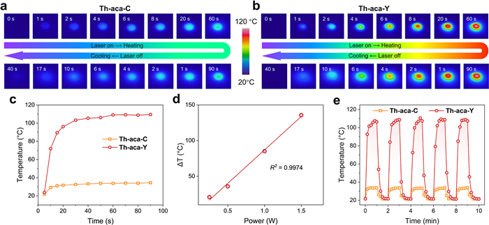
Th-aca-Y exhibits a noticeable darkening and its extensive absorption band extends into the near-infrared (NIR) region with higher absorption efficiency than Th-aca-C, making it advantageous for efficient photothermal conversion (Fig. S12 in Supporting information) [67]. Thorium clusters offer distinct advantages for photothermal conversion. The exclusive +4 oxidation state of Th(Ⅳ) prevents undesired redox reactions with radicals, ensuring excellent chemical stability. Strong coordination bonds between Th(Ⅳ) and carboxylate ligands further enhance thermal and chemical robustness. Additionally, the versatile coordination chemistry of Th(Ⅳ) enables diverse and highly tunable cluster architectures, providing greater structural flexibility to optimize photothermal properties. To verify this assumption, we initially conducted thermogravimetric analysis (TGA), indicating that Th-aca-Y and Th-aca-C exhibit excellent thermal stability up to 200 ℃ (Figs. S13 and S14 in Supporting information). To evaluate the photothermal conversion efficiency, we recorded temperature changes in real-time using an IR camera (Figs. 5a and b). Pallets of Th-aca-Y and Th-aca-C, each with a diameter of 5 mm, were subjected to irradiation with an 808 nm laser at a constant power of 1 W. The temperature of Th-aca-Y escalated rapidly from 23 ℃ to 109 ℃ within 40 s, whereas that of Th-aca-C only rose to 33 ℃ under identical irradiation conditions (Fig. 5c). This observation underscores the critical role of radicals in the photothermal conversion process. Indeed, Th-aca-C-Xe, which features photo-induced radicals upon Xe light irradiation, exhibits excellent photothermal conversion efficiency as well, achieving a temperature of 118 ℃ (Figs. S15 and S16 in Supporting information). Notably, the photothermal conversion performances of Th(NO3)4·6H2O and purely organic Haca ligand are far inferior to Th-aca-Y (Figs. S17 and S18 in Supporting information). This outcome implies that the strategy of metal-ligand assembly to construct rigid organic-inorganic hybrids via modulated synthesis functions effectively to promote the formation of radicals and improve photothermal conversion performance [68-70]. Furthermore, the temperature evolution profiles of Th-aca-Y were investigated under different laser power densities (0.25, 0.5, 1, and 1.5 W), demonstrating a linear relationship between its photothermal effect and the NIR laser power (Fig. 5d). In addition, the cyclic test highlights the exceptional photostability of Th-aca-Y, as evidenced by the average temperature rapidly reaching 110 ℃ after five consecutive illumination cycles, showcasing their robust performance and excellent reusability (Fig. 5e).
Figure 5
Figure 5.
(a) IR thermal images of Th-aca-C under 808 nm laser irradiation (1 W) and after the laser was turned off. (b) The IR thermal images of Th-aca-Y under 808 nm laser irradiation (1 W) and after the laser was turned off. (c) Photothermal conversion curves for Th-aca-C and Th-aca-Y under 808 nm laser irradiation (1 W). (d) Correlation between photothermal effect and NIR laser power for Th-aca-Y. (e) Photothermal conversion cycling of Th-aca-C and Th-aca-Y.
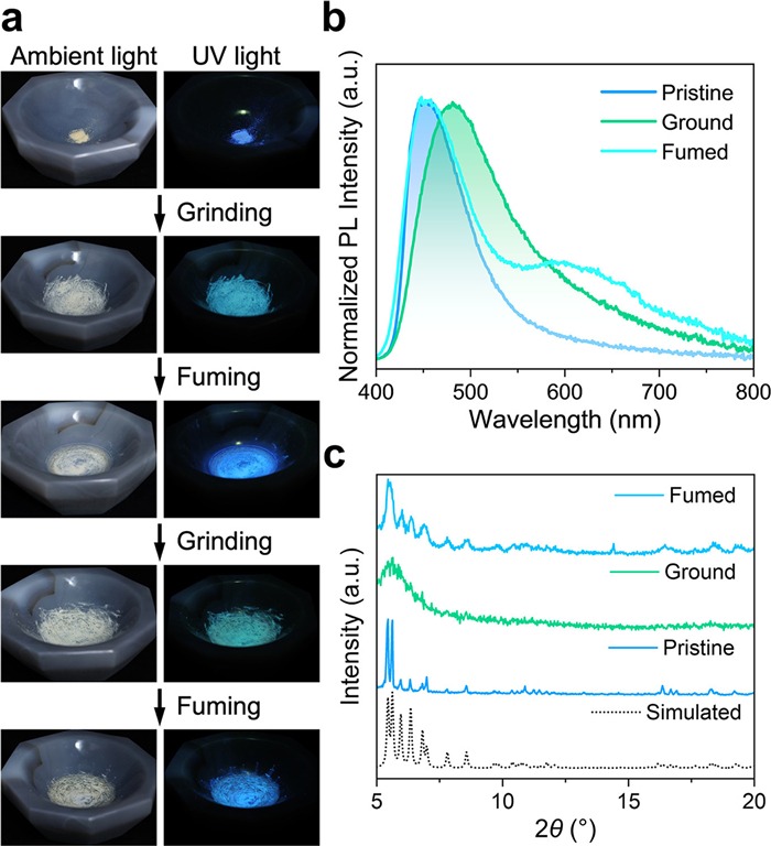
Beyond the photothermal conversion property feature of actinide-based material, both Th-aca-C and Th-aca-Y exhibit (Fig. S19 in Supporting information) luminescence color change upon mechanical grinding, rendering it as the first reported mechanochromic luminescence of its kind (Fig. 6a) [71]. Specifically, the pristine crystals of Th-aca-C display the typical blue ligand-based emission, with a peak wavelength at 450 nm (λex = 365 nm), which can be attributed to the intermolecular π-π* transition of the aca– ligand (Fig. 6b) [72]. Time-resolved luminescence spectroscopy revealed that the photoluminescence lifetime of Th-aca-C is 1.42 ns based on bi-exponential decay fitting, shorter than the 11.08 ns of Haca (Figs. S20-S22 in Supporting information). Intriguingly, grinding Th-aca-C with a mortar and pestle results in a visible alteration in emission color. The emission maximum shifts from λemmax of 450 nm to 500 nm, changing the emission color from blue to green as illustrated in the photos of Th-aca-C powder and the CIE chromaticity diagram (Fig. 6a and Fig. S23 in Supporting information). Upon mechanical grinding, the photoluminescence lifetime of Th-aca-C increases from 1.42 ns to 4.3 ns, while the photoluminescence quantum yield decreases from 2.82% to 0.75% (Figs. S24 and S25 in Supporting information). PXRD and FTIR analyses revealed that the change in emission color of Th-aca-C is related to morphological transitions from crystalline to amorphous phases, with its functional groups remaining unaffected by grinding (Fig. 6c and Fig. S22). Explicitly, the preliminary blue-emissive crystals of Th-aca-C displays sharp diffraction peaks, while the green-emissive powder of Th-aca-C after grinding shows no diffraction bands, indicating its amorphous state. Indeed, literatures have revealed that the crystalline-to-amorphous phase transition, which can be induced by mechanical grinding or external pressure, plays a pivotal role in the initiation of mechanochromic luminescence in organic-inorganic hybrid and organic-co-crystal materials [71,73-75]. This transition induces alterations in molecular packing and intermolecular interaction, such as π–π stacking, thereby resulting in a luminescence color transition [76,77]. It is noteworthy that our finding represents the first successful demonstration of mechanochromic luminescence in actinide materials. When the ground powder of Th-aca-C is exposed to dichloromethane vapor, its crystallinity and blue-emission properties can be restored, as evidenced by the emergence of the original diffraction peaks and blue shift of the emission band, respectively (Fig. 6c). The reversible nature of this transition can be attributed to the solvent vapor-induced reestablishment of short-range ordering of the crystal lattice. The emission spectrum of fumed Th-aca-C is slightly different from that of pristine Th-aca-C and exhibits a weak shoulder peak at 600 nm, which is likely attributed to the formation of an excimer between the organic ligands (Fig. 6b) [72,78]. In addition, Th-aca-C retains its mechanochromic luminescence with no significant degradation after three consecutive grinding and fuming cycles, which underscores its excellent reusability and stability [51].
Figure 6
Figure 6.
(a) Photographs of Th-aca-C under ambient light and UV (365 nm) irradiation before and after grinding, and after fuming with dichloromethane. (b) Solid-state photoluminescence spectra of Th-aca-C before and after grinding and fuming with dichloromethane. (c) PXRD patterns of Th-aca-C before and after grinding or fuming, compared with the simulated pattern.
In conclusion, this study introduces an approach for stabilizing radicals within MOCs by leveraging acidity modulation, a breakthrough that expands the scope of radical stabilization beyond MOFs. By tuning the reaction acidity, we successfully synthesized two thorium-oxo nanoclusters, yellow Th-aca-Y and colorless Th-aca-C, with identical structures but markedly different photophysical properties. Th-aca-Y, formed under higher acidity, incorporates highly stable radicals, which exhibit unique UV-vis absorbance and superior stability compared to the Xe light-induced radicals in Th-aca-C-Xe. These findings highlight the potential of acidity modulation as a versatile tool for tuning radical behavior in MOCs and potentially other crystalline materials such as MOFs and COFs. Furthermore, unprecedented mechanochromic luminescence has been unraveled in actinide materials by Th-aca-C. These findings open new directions for exploring multi-stimuli-responsive behaviors and other photophysical peculiarities in actinide complexes. Future work may explore the broader applicability of this acidity modulation strategy to other radical systems and materials, potentially unlocking new applications in sensing, imaging, and catalysis.
The authors declare that they have no known competing financial interests or personal relationships that could have appeared to influence the work reported in this paper.
Supplementary materials
Supplementary material associated with this article can be found, in the online version, at doi:10.1016/j.cclet.2025.111702.
L. Zeng, L. Huang, Z. Wang, et al., Angew. Chem. Int. Ed. 60 (2021) 23569-23573. doi: 10.1002/anie.202108076
[70]
Q. Zheng, X. Liu, Y. Zheng, et al., Chem. Soc. Rev. 50 (2021) 5086-5125. doi: 10.1039/d1cs00056j
[71]
Z. Chi, X. Zhang, B. Xu, et al., Chem. Soc. Rev. 41 (2012) 3878-3896. doi: 10.1039/c2cs35016e
[72]
K. Yuhara, K. Tanaka, Angew. Chem. Int. Ed. 63 (2024) e202319712. doi: 10.1002/anie.202319712
[73]
Y. Yang, X. Fang, S.S. Zhao, et al., Chem. Commun. 56 (2020) 5267-5270. doi: 10.1039/c9cc09806b
[74]
B. Dutta, K. Debsharma, S. Dey, et al., Adv. Mater. Interfaces 9 (2022) 2201120 doi: 10.1002/admi.202201120
[75]
S.N. Lei, H. Xiao, Y. Zeng, et al., Angew. Chem. Int. Ed. 59 (2020) 10059-10065. doi: 10.1002/anie.201913340
[76]
L. Wilbraham, M. Louis, D. Alberga, et al., Adv. Mater. 30 (2018) 1800817. doi: 10.1002/adma.201800817
[77]
C. Liu, G. Xiao, M. Yang, et al., Angew. Chem. Int. Ed. 57 (2018) 1893-1897. doi: 10.1002/anie.201711409
[78]
S. Yamane, Y. Sagara, T. Kato, Chem. Commun. 49 (2013) 3839–3841. doi: 10.1039/c3cc00072a
Figure 1
(a) Schematic representation of traditional approaches for generating organic radicals within MOFs. (b) A reaction scheme illustrating the acidity modulation strategy that leads to the formation of non-radical containing Th-aca-C under low acidity conditions and radical-bearing Th-aca-Y under high acidity conditions. Color code: Th = orange, O = red, N = blue, C = grey, and H atoms are omitted for clarity.
Figure 2
(a) UV-vis absorption spectra of Th-aca-C and Th-aca-Y. (b) EPR spectra of Th-aca-C and Th-aca-Y. (c) Concentrations of HNO3, CF3COOH, and HCl used in the synthesis of Th-aca-C and Th-aca-Y.
Figure 3
(a) Crystal images of Th-aca-C upon continuous Xe-lamp irradiation. (b) UV-vis absorption spectra of Th-aca-C upon continuous Xe-lamp irradiation. (c) EPR spectra of Th-aca-C before and after Xe-lamp irradiation.
Figure 4
(a) UV-vis absorption spectra of Th-aca-Y and Th-aca-C-Xe. (b) EPR intensities of Th-aca-Y and Th-aca-C-Xe over time under ambient conditions. (c) FTIR spectra of Th-aca-Y, Th-aca-C, and Th-aca-C-Xe. (d) NMR spectra of Th-aca-Y, Th-aca-C, and Th-aca-C-Xe. (e) O 1s and C 1s XPS spectra of Th-aca-Y, Th-aca-C, and Th-aca-C-Xe.
Figure 5
(a) IR thermal images of Th-aca-C under 808 nm laser irradiation (1 W) and after the laser was turned off. (b) The IR thermal images of Th-aca-Y under 808 nm laser irradiation (1 W) and after the laser was turned off. (c) Photothermal conversion curves for Th-aca-C and Th-aca-Y under 808 nm laser irradiation (1 W). (d) Correlation between photothermal effect and NIR laser power for Th-aca-Y. (e) Photothermal conversion cycling of Th-aca-C and Th-aca-Y.
Figure 6
(a) Photographs of Th-aca-C under ambient light and UV (365 nm) irradiation before and after grinding, and after fuming with dichloromethane. (b) Solid-state photoluminescence spectra of Th-aca-C before and after grinding and fuming with dichloromethane. (c) PXRD patterns of Th-aca-C before and after grinding or fuming, compared with the simulated pattern.


 DownLoad:

 下载:


 下载:

