Figure 1.
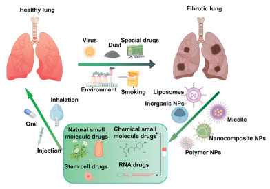
Schematic illustration of healthy lung and fibrotic lung experienced influencing factors, drug selection, drug delivery, and administration methods. This figure was created using Figdraw.
Recent advances in drug delivery systems for pulmonary fibrosis therapy
Yan Yu , Cailing Gan , Kun Shi , Zhongwu Bei , Yang Yu , Meng Pan , Hanzhi Deng , Zhiyong Qian

Fibrosis is an outcome of the tissue repair process during chronic inflammatory disorders and various forms of tissue injury [1-3]. There are some common diseases associated with fibrosis, including systemic sclerosis, liver cirrhosis, pulmonary fibrosis (PF), myocardial infarction, renal interstitial fibrosis and arteriosclerosis [3,4]. In the worldwide, fibrosis is a significant contributor to morbidity and mortality on a global scale and increasingly recognized as a major health and longevity challenge [5]. Among these fibrotic conditions, PF especially is a devastating disease with a lethal progression to respiratory failure and main outcome of most chronic respiratory diseases [1,2,6]. Numerous studies have identified several risk factors associated with PF occurrence [4]. These factors include the cumulative effects of tissue repair responses triggered by smoking, viral infections, genetic predispositions, and the inhalation of environmental and occupational pollutants, as well as exposure to certain drugs (Fig. 1). PF has a dysregulated repair stage, when these specific factors result in damage to pulmonary tissue. The main characteristics of PF are the formation of scars in the lungs over time, deposition of extracellular matrix (ECM), and thickening of the alveolar walls [7,8]. Notably, ECM components accumulated continuously will lead to tissue architecture disruption, organ dysfunction and organ failure [9]. As research on PF advances, the pathogenesis of the disease remains incompletely understood, for example, abnormal angiogenesis and inflammation may be related to the formation of fibrosis [10]. Ding et al. identified that targeting and inhibiting endothelial Rhoj can mitigate collagen deposition, decrease nested angiogenesis, and alleviate fibrosis [10]. So that, accelerating the penetration of the collagen barrier is crucial, as it requires not only effective treatment but also early detection.
In recent years, the development of PF therapeutic drugs has also made relatively slow progress, thereby impeding the targeted pharmacological therapies [11]. Most of small molecules or compounds are currently in clinical trials for PF [12,13]. There are only two drugs approved by the Food and Drug Administration (FDA), pirfenidone and nintedanib for PF therapy. Due to patients need to oral pirfenidone daily 2403 mg. The most commonly reported adverse events included gastrointestinal disorders, fatigue, skin rash, nausea, and weight loss. Some cases of abnormal liver function have been reported and it is imperative for patients to undergo regular reevaluation of their liver function [14]. Similarly, patients receive nintedanib at a dose of 150 mg twice daily. Diarrhea was the most common side effect for 66.9% of patients [15]. To some up, drug research of PF will face major challenges for reducing their side effects and improving the therapeutic effect. Several important drugs are discovered from natural products [16,17]. For examples, baicalin is a flavonoid component derived from Radix Scutellariae. It is very effective in treating lung diseases. But baicalin has poor water and lipid solubility resulting in low bioavailability. Nanodrugs delivery technology can increase the solubility of drugs [18]. Besides, other technological drugs are also worth paying more attention. Macromolecular drug are therapeutic drugs produced using innovative biotechnology, such as stem cell technology, cytokines, recombinant protein drugs, and antibodies [19,20]. Based on extensive literature research, more RNA drugs are developed for treatment various illness [21]. They have advantages, such as strong targeting, lasting efficacy, but still in the preclinical research stage due to immunogenicity and degradation. In response to this situation, researchers have implemented a range of strategies in the design of medication regimens and delivery systems to address the challenges presented.
With the development of nanotechnology application, the emergence of nano-drug delivery systems enables to target organs or tissues for clinical treatment [22]. Some nano-drugs have been used for clinical patients and have achieved better therapeutic effects. These drugs are packaged in nanoscale carriers made of lipids or polymers, which can deliver different drugs to lung tissues in order to reduce side effects [23]. In other words, changing the way of drug treatment and improving drug efficacy are urgent needed through nano-delivery system. Among the related delivery systems, nanodrug delivery systems are a popular research trend for enhancing efficacy, enabling sustained, and controlled drug release [24]. Nano delivery systems effectively address the shortcomings of current PF treatments obviously. Currently, the drugs for PF are mainly systemic therapies such as oral and injection. On the contrary, nanodrug can meet multiple delivery methods [25]. In this review, we provide recent research on the drugs delivery technology of PF. There are classified drugs for treating PF based on their sources and properties (Fig. 2). Then, delivery systems of PF treatment drugs are reviewed respectively. Finally, the future outlook of PF treatments was discussed.
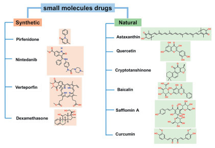
Most of new drugs and their products are small molecules in the pharmaceutical industry [26]. However, the majority of small molecule drugs exhibit a lack of specificity and are susceptible to interacting with multiple targets due to their relatively small structure. That's why small molecule drugs can easily induce more toxic side effects [27]. Small molecule drugs exhibit relatively short half-life in vivo and require one or three large doses of medication per day. Therefore, drug delivery systems for small molecule drugs have become increasingly urgent in order to meet the treatment needs of patients [28]. Pirfenidone and nintedanib have approved small molecule drugs by FDA for the treatment of PF. Different methods for delivering small molecule drugs are analyzed and discussed in this part (Fig. 2).
Pirfenidone is a small molecule drug derived from pyridine. The molecular weight of pirfenidone is 185.22 g/mol, which is easy to distribute good crystal stability. Due to pirfenidone has pyridone ring as key functional groups, it has poor aqueous solubility and higher solubility in organic solvents, which make it oral capsule formulations necessarily [29]. But phenyl and pyridone of pirfenidone molecular structure make it have photosensitivity side-effect and may have the potential to cause skin cancer [30]. More and more research on formulation of pirfenidone encounters challenges in minimizing dosage and side effects. As shown Fig. 2, pirfenidone's molecular structure regulates multiple targets synergistically because of its pyridone ring, methyl group, and phenyl group [29]. Thereby, pirfenidone exhibits anti-inflammatory, antioxidant, and anti-fibrotic properties [14]. Liposomes are nanoscale spherical structures characterized by phospholipid bilayers that encapsulate an aqueous core. They can encapsulate diverse small molecules drugs coupled with their biocompatibility [31]. Several studies have confirmed that liposomes are more suitable for pulmonary drug delivery systems, based on the extremely similar performances between liposomes and pulmonary surfactants [32]. A prominent characteristic of PF is the persistent accumulation of aberrant collagen, creating a formidable obstacle to drug permeation. Liposome targeting modification and inhalation delivery are crucial to improve the therapeutic efficacy [33]. Nuclear factor erythroid 2 related factor 2 (Nrf2) is a key molecule regulating reactive oxygen species (ROS) and transforming growth factor-β (TGF-β) expression. A ROS-responsive liposome loaded dimethyl fumarate (Nrf2 agonist) were synthesized by Wang's team. The study found that these ROS-responsive liposomes exhibited prolonged accumulation in the lung, indicating that the nano-delivery system has very good biocompatibility and improves the efficacy in the treatment of PF [34].
Albumin as protein drug carrier with binding sites for metabolic substrates and therapeutic agents, is biodegradable and non-immunogenic [35]. It is widely studied in anti-tumor drug delivery systems but has seen less research in PF drug delivery [36]. Albumin is considered a more suitable carrier for drug delivery due to its cost-effectiveness, biodegradable, nontoxic, low immunogenicity, and uptake in inflamed tissues [37]. Gong et al. established a nanoplatforms for multi-targeting delivery [38]. They used FnBAP5 peptide for facilitating targeted interaction with fibroblastic foci, thereby enhancing the ability of bovine serum albumin (BSA) nanoparticles to recognize and accumulate at fibrotic sites. This method represents an effective strategy for enhancing the lung microenvironment and its functional capacity. Youn et al. fabricated inhalable albumin nanoparticles and drugs were released gradually for 24 h. The inhaled nanoparticles demonstrated superior anti-fibrotic efficacy compared to that achieved through intraperitoneal administration [39]. In the future, increased attention could be paid on chemical modifications, which may potentially expanding the applications of albumin nanoparticles [37]. Albumin nanoparticles will be served as a promising drug delivery system in the clinical development of therapeutic PF.
With DNA nanotechnology developing, it has been utilized in the treatment of a range of clinical conditions [40]. Tetrahedral framework nucleic acid (tFNA) has emerged as a central focus in the research area of DNA nanotechnology [41]. tFNA were initially introduced by Turberfield and his team in 2004 [42]. tFNAs show some excellent advantages, including easy design, structural stability, biodegradability, and functional diversity [43]. It is very suitable for drug carrier research in the biomedical field due to mechanisms such as embedding, electrostatic interactions and chemical cross-linking [44]. Our team synthesized a loading pirfenidone complex using tFNA as the delivery vehicle and achieved quantitative nebulized drug delivery to the lungs through a micronebulizer for treatment PF [45]. tFNA has been demonstrated as a drug delivery system with exceptional delivery capabilities [46]. It is anticipated as a prominent nucleic acid nanocarrier for respiratory diseases [47]. In short, there are many studies on drug delivery for pirfenidone, and we look forward to them entering the clinical market as soon as possible.
Nintedanib is a triple vascular kinase inhibitor that inhibits growth factor receptors in the pathological mechanism of PF, thereby slowing down the progression of idiopathic PF [15]. The molecular weight of nintedanib is 539.64 g/mol, classifying it as a small molecule drug. Due to its limited water solubility and significant protein binding properties, high-dose administration as oral medication is necessary but nintedanib is known for its severe dose-limiting side effects [48]. There is a need to develop sustained-release formulations to improve its bioavailability. These factors prompted Brenner et al. to devise solid lipid nanoparticles containing nintedanib. The presented data indicates that nintedanib-loaded liposomes maintain their structural integrity and do not aggregate following extended exposure to the alveolar cavity in vivo. In comparison to oral medications, inhaled nintedanib-loaded liposomes exhibit order of magnitude increase in lung area under the curve (AUC) showing better bioavailability [24]. This phenomenon can be attributed to the effective drug release capabilities of liposome delivery platforms. The study findings also indicate that the inhaled liposomes may be harmonious with pulmonary surfactant, thereby indicating the potential suitability of the liposome delivery system for clinical use in PF. At the same time, Sistla et al. also designed a liposome formulation loaded with nintedanib because of the low bioavailability and adverse therapeutic side effects of nintedanib. They found that the relative oral bioavailability in liposome formulation was 2.87 times that of free nintedanib. Furthermore, respiratory mechanics analysis showed good anti PF effects [49]. It is evident that drug delivery system has the potential to substantially enhance bioavailability and extend the duration of action.
The distinct mechanisms of pirfenidone and nintedanib offer a physiological basis for their combined use in efforts to further mitigate the decline lung function in PF. Clinical trials have assessed the safety profile of the combined therapy of pirfenidone/nintedanib and other small molecule drugs for the treatment of PF [50].
Jiang et al. has constructed nanoparticles loaded with dual drugs (verteporfin and pirfenidone, Lip@VP) [51]. They administered Lip@VP to the lungs via nebulized inhalation. Verteporfin is widely accumulated in lung tissue and released to inhibit airway epithelial fluidization and the formation of honeycomb cysts. Pirfenidone inhibits excessive activation of fibroblasts, reduces cytokine secretion, and promotes airway epithelial fluidization. The liposome loaded dual-drug demonstrates greater efficacy compared to single drug delivery. The collagen barrier in PF has always been of great concern. Jiang et al. employed self-assembly and Michael addition reactions in the construction of pathological collagen targeting and penetrating liposomes. Through the utilization of collagen binding peptides with a high affinity for collagen, the identification of the pathological collagen environment can be achieved, followed by the application of collagenase to penetrate the collagen barrier. The antifibrotic drugs of pirfenidone and dexamethasone were injected into the injured lungs [52]. The release of two-drug demonstrated a synergistic anti-fibrotic impact by facilitating the repair of impaired epithelial cells and inhibiting the activation of myofibroblasts. Drawing inspiration from the physiological characteristics of pulmonary surfactant, Zhang et al. have developed a nano biomimetic liposome drug delivery system, which is loaded with astaxanthin (AST) and pirfenidone administered through nebulized lung inhalation. This nano-system is administered through nebulized lung inhalation, allowing AST and pirfenidone to be further transported to penetrate lung tissue and exerting therapeutic effects. Liposomes delivery system has the advantage of prolonging retention time in the lungs, with nebulization every other day, which is more convenient than injection and multiple oral medications, and has a better therapeutic effect on PF [53].
Inorganic nanoparticles are frequently investigated for their potential applications in theranostics [54,55]. Qian et al. has investigated and developed a greater number of inorganic nanoparticles for the diagnosis and treatment of diseases, including tumors and fibrosis [54-58]. The inorganic nanoparticles most frequently used in biomedical applications include the following, noble metals nanoparticles such as Au, Pt, and Ag [59,60], metal oxides/ metal sulfide (e.g., solid or mesoporous SiO2, Ag2S) [61,62], semiconductor materials (quantum dots) [63]. Inorganic nanoparticles are attracting substantial attention due to their high biocompatibility, chemical stability, high electrical conductivity and potential for bioconjugation [57,63]. Zhang et al. demonstrated the successful synthesis of Janus Au core-mesoporous silica shell nanoparticles. Mesoporous silica loaded with pirfenidone not only increases the drug loading capacity, but also achieves responsive release of pirfenidone due to the modification of ROS responsive groups and 1,2-distearoyl-sn‑glycero‑3-phosphoethanolamine (DSPE) by mesoporous silica, while reducing the number of drug administration. This study demonstrates the collaborative effect of pirfenidone and stem cells utilizing nano drug delivery systems and creates a favorable microenvironment and enhance the treatment of PF [64]. However, the utilization of inorganic nanoparticles has also raised concerns regarding their potential long-term impact due to immunogenicity and cytotoxicity [65]. Although inorganic nanoparticles have the capability to deliver drugs, due to the non-degradability, the safety issues of inorganic nanoparticles remain a significant obstacle [66]. In this field of drug delivery, inorganic nanoparticles could possess some difficulty and potential to be incorporated as an effective nanoscale platform for PF.
These studies suggest outstanding potential of employing nanocarriers to improve the delivery and efficacy of chemically synthesized small molecules drugs. Most studies use clinical conventional drugs pirfenidone, nintedanib and other candidate drugs used to treat PF. From the perspectives of animal models, cell models, and mechanisms of PF, it has been confirmed that they greatly reduce their toxic side effects, avoid high-dose and frequent medication. This review is beneficial for promoting the adjustment of clinical drug dosage forms of nintedanib and pirfenidone, and promoting the application research of nanocarrier systems in translation and clinical practice (Fig. 3). In short, there is a huge demand for nanocarrier platforms during the process of clinical application.
As previously discussed, chemical synthetic small molecules drugs have low bioavailability, insufficient targeting, and easy drug resistance or side effects when used for a long time. Emerging more increasing evidence have revealed the efficacy of natural products in PF treatment [71,72]. We focus on to widen potential clinical applications of natural products in PF treatment. In fact, there are many natural small molecule drugs derived from natural products have used in clinical practice, such as artemisinin, taxol, curcumin and silymarin. Natural small-molecule drugs have emerged as a crucial resource in multiple diseases drug development, attributed to their structural diversity, multi-target effects, and low toxicity [73,74]. Natural products, originating from plants, animals, and microorganisms, play a crucial role in maintaining human health, such as tanshinone ⅡA [67,75], flavonoids [76], asiatic acid [77,78], triptolide [79] and others more small molecules drugs [80,81]. An increasing number of approved pharmaceuticals are derived from natural compounds or their derivatives, a trend attributable to their diverse chemical components and high biocompatibility. For instance, the anti-fibrotic properties of artemisinin compounds have been investigated using both in vivo and in vitro models of PF [82].
Quercetin is the main representative of flavonoids, widely present in fruits, vegetables, and grains. The molecular weight of quercetin is 302.23 g/mol, and its molecular structure comprises hydroxyl groups, benzene rings, and pyran ring (Fig. 2). The hydroxyl group and pyran ring structure are the key to its antioxidant activity [83]. Nevertheless, the limited water solubility, low bioavailability, and rapid metabolism of quercetin constrain its efficacy in clinical applications. Yao et al. developed chitosan-coated quercetin nanoparticles (Qu/CS-NPs) for the treatment of silica induced occupational PF. Following administration via the pulmonary route, Qu/CS-NPs exhibit an enhanced therapeutic effect, leading to a significant reduction in oxidative stress, mitigation of lung structural damage, suppression of α-smooth muscle actin (α-SAM) expression, and decreased ECM deposition. The delivery of Qu/CS-NPs through CS NPs not only enhances its hydrophilicity, antioxidant, and anti-inflammatory properties, but also improves bioavailability and anti-fibrotic characteristics of quercetin [84]. In the case of quercetin, the bioavailability of the substance in the body after oral administration is as low as 1%, due to its poor solubility in water (2.84 mg/mL). Furthermore, the limited oral absorption (< 50%) of the insoluble flavonoid silybin restricts its characteristic, resulting in diminished clinical efficacy [85]. We were pleasantly surprised to find PhytosomeⓇ technology as a delivery system to enhance absorption of certain compounds [86]. This study aimed to assess and compare the solubility of a novel quercetin formulation (Quercetin PhytosomeⓇ) with that of unformulated quercetin in simulated gastrointestinal fluids, as well as its oral absorption. These pharmacokinetic findings indicate that the new formulation significantly increased plasma levels of quercetin, achieving concentrations up to 20 times greater than those typically observed following the administration of standard quercetin doses.
CTS is the predominant fat-soluble compound extracted from Salvia miltiorrhiza, characterized by its diterpene quinone structure featuring a distinctive ternary or quaternary ring o-quinone or p-quinone configuration (Fig. 2). These structures make it with multiple biological activities, such as antitumor, antioxidant, anti-inflammatory, and antibacterial properties. More and more studies have demonstrated promising potential in the treatment of cardiovascular diseases, antibacterial applications, anti-tumor effects, anti-inflammatory properties, metabolic disorders, and neurodegenerative diseases. Liu et al. developed and evaluated two drug delivery systems about CTS-loaded modified liposomes [68]. CTS-loaded modified liposomes exhibit targeted delivery to pulmonary myofibroblasts through the specific interaction of the CREKA peptide with fibronectin and the inherent homing capability of exosomes to progenitor cells, respectively. This mechanism enhances the efficient transport of CTS to pulmonary fibrotic lesions. Notably, the inhaled administration of CTS-loaded liposomes every two days achieved comparable efficacy to the control drug pirfenidone and conventional microspheres administered daily.
AST is a keto-carotenoid characterized by its distinctive conjugated double bond and ketone structure (Fig. 2), which confer potent antioxidant and anti-inflammatory properties. It is abundantly found in marine organisms such as fish, shrimp, and crabs, and is called the super antioxidant in nature [87]. AST exhibits limited solubility in aqueous environments, necessitating the development of advanced drug delivery systems to enhance its bioavailability and broaden its potential applications. Huang's team has designed a nano-pharmaceuticals that co-delivers AST and siTGF-β1 through ionizable liposome nanoparticles for improving PF treatment [69]. This study offers a unique and pioneering approach in co-delivering siRNA and AST for the treatment of PF. Liposomes are fabricated through the utilization of microfluidic technology in order to facilitate efficient drug delivery. The inclusion of AST within these liposomes serves to mitigate excessive levels of ROS within cells, thereby diminishing aberrant damage to alveolar epithelial type 2 cells. Additionally, the incorporation of siTGF-β1 within the liposomes functions to suppress the expression of the TGF-β1 gene, consequently impeding the accumulation of ECM by inhibiting the differentiation and migration of fibroblasts. Compared with monotherapy, this combination therapy significantly suppressed the worsening of PF. This study combines natural products with gene therapy, with a therapeutic effect of 1 + 1 > 2, providing a promising strategy for the clinical treatment of PF.
Baicalin, as a flavonoid derived from Scutellaria baicalensis, possesses several phenolic hydroxyl groups in molecular structure (Fig. 2), enabling it to interact with various free radicals. This interaction underlies its antioxidant and anti-inflammatory properties [88]. More research has been shown in prior research to possess various pharmacological properties that are beneficial in the treatment of respiratory illnesses, demonstrating favorable therapeutic outcomes [89,90]. Wang et al. reviewed the protective effects of baicalin on respiratory diseases in recent years and summarized the therapeutic effects of baicalin nanocarrier systems in respiratory diseases [91]. Baicalin has demonstrated protective effects on the lungs across various animal models, including lung injury, PF, pulmonary embolism, and lung remodeling. Baicalin exhibits poor water solubility, necessitating enhancement via nanomedicine delivery systems to improve its therapeutic efficacy.
HSYA is a compound characterized by a monochalcone glycoside structure. As a bioactive constituent of the natural product safflower. HSYA distinguishes it from other natural small molecule drugs due to its disaccharide and polyhydroxy groups, which confer high water solubility and low membrane permeability. However, it exhibits low oral bioavailability [92]. The chalcone structure imparts antioxidant properties through the scavenging of free radicals. So that HSYA exhibits pharmacological effects including anti-inflammatory, antioxidant, and anti-apoptotic effects [93]. Zhang et al. designed a phospholipid complex based on the van der Waals forces/hydrogen bonding between phospholipids and HSYA molecules, which not only improved the encapsulation efficiency of water-soluble HSYA, but also enhanced the lipid solubility of free HSYA and increased its bioavailability. They further evaluated the administration of HSYA via intervaginal space injection (ISI) as an anti-PF treatment [94]. Findings have demonstrated that HSYA effectively decreases the secretion of inflammatory factors and inhibits collagen deposition in the lungs of mice, suggesting a promising anti-fibrotic potential for HSYA. A notable and innovative aspect of this study is the utilization of an alternative route of administration-ISI [95]. It is demonstrating favorable biosafety and efficacy and elicits a targeted response in the lungs. ISI could be a promising lung-targeted delivery system.
Curcumin, a compound frequently utilized in culinary applications, possesses both medicinal and nutritional properties. It is characterized by a diketone structure and phenolic hydroxyl groups, which enable it to effectively scavenge free radicals and downregulate the expression of inflammatory mediators. Consequently, curcumin exhibits significant antioxidant, anti-tumor, and anti-inflammatory activities [96]. Curcumin is a well-documented natural compound with anti-inflammatory properties and has been demonstrated to exert beneficial effects in PF [97-99]. Capsule LongvidaⓇ technology (solid lipid curcumin particles) have optimized role of curcumin and alleviated pain in knee osteoarthritis [100]. A highly bioavailable curcumin preparation (LongvidaⓇ) have improved working memory and reduced fatigue and stress reactivity [101]. After literature research, we are gratified that some companies are developing nanoliposomes for natural products preparations to improve the stability and bioavailability of drugs, such as Yunnan Baiyao Group Company. While the current domestic market features a limited number of natural small molecules drugs preparations and health products utilizing nano-delivery technology, with the development of technology, this field is expected to foster in more innovations and products. Jiang et al. have developed a Lipomicelles loaded curcumin for enhancing ECM penetration and exhibiting excellent therapeutic effect in PF [70]. Jiang et al. not only made breakthroughs in basic research, but also actively promoted the transformation of these research results into practical applications, providing new strategies and methods for the treatment of related fibrosis diseases.
With the development of biotherapy, macromolecular drugs have been large-scale produced, including DNA, RNA, peptides and proteins [102]. They have gained more attention in lung disease, especially in PF field. Due to their sophisticated functions, several drug delivery strategies have been developed for overcoming difficulties, such as the instability and easily restricted by physiologic barriers [103].
RNA is a multifunctional macromolecule characterized by its ability to adopt a diverse array of structural conformations [104]. Since the approval of the first RNA-based drug named fomivirsen for the market in 1998, there has been significant progress in the field of RNA therapeutics [105]. In the field of pharmaceutical research, RNA drug therapy can accurately regulate the expression of target genes compared to small molecule drugs [106]. Currently, more and more clinical data supports the potential of overcoming the major limitation of existing medicines and treating variety of diseases. Meanwhile, RNA-based therapies demonstrate high specificity and efficacy for the treatment of lung diseases [107-109]. However, RNA drug delivery is a valuable medical approach known for its specificity and facile production, but it suffers from poor stability and rapid degradation [110,111]. In the treatment of PF, siRNAs are usually designed to target genes that play a key role in the fibrosis process, such as cytokines, growth factors and key molecules in signaling pathways, to inhibit the development of fibrosis [112,113]. Currently, the mainstream gene delivery carriers are non-viral vectors, such as liposomes [114,115], polymer nanoparticles [116,117], and inorganic nanoparticles [118,119]. At the international conference on novel formulations and process technologies, K. Raemdonck of Ghent University presented his research accomplishments and shared several studies on gene therapy for lung disorders. In order to maximize the efficacy of RNA drugs, it is essential to utilize safe and efficient nano-formulations for targeted delivery into target cells [120].
Surfactant protein B (SP-B) plays a crucial role in lung surfactants, facilitating the potential for inhalation delivery of siRNA and mRNA through the use of cationic amphipathic drugs (CAD) and SP-B [121,122]. Oupický et al. developed perfluorocarbon nano-emulsions can deliver siRNA to the lungs, achieving gene silencing treatment of PF safely and effectively [123]. Considering the strong correlation between the expression of protein and the epithelial-mesenchymal transition process, Lee et al. hypothesized that the silencing of G2 and S phase-expressed protein 1 (GTSE1) might represent a promising therapeutic strategy for addressing PF. To this end, they developed a mannose-incorporated engineered lipid nanoparticle for efficient delivery of GTSE1 siRNA to the lungs. These findings support the notion that targeting GTSE1 may hold potential for the treatment of PF by modulating pulmonary epithelial-mesenchymal transition [124].
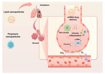
In addition, researchers are making many attempts in other non-viral vector directions presently. Ahn et al. has developed a complex as a lung-targeted gene delivery system. The underlying mechanism is formed a self-assembled nanoparticles (pASO/DhBD23) through charge interactions. These nanoparticles facilitate systemic delivery through tail-vein administration, achieving lung-specific targeting of TGF-β mRNA to effectively treat PF [125]. Mannosylated albumin nanoparticles encapsulating TGFβ1-siRNA selectively targeted the profibrotic subpopulation of CD206+ macrophages, effectively inhibiting the progression of PF [126]. Zhang et al. developed a mucus-penetrative nanoparticles, which were formulated through self-assembly of a biodegradable copolymer and cationic lipid for delivery of siIL11. The highlight of their work is cationic lipid-like molecule G0-C14 for that penetrates mucus [127]. Yang et al. also constructed stable carrier polymer nanoparticles and it may effectively encapsulate siIL-11 through hydrophobic, hydrogen bonding, and electrostatic interactions. Fig. 4 demonstrates a marked mitigation of disease progression in PF model [128].
In addition to single gene delivery, some studies have also discovered multi gene targeted delivery systems, which have better therapeutic effects due to their synergistic therapeutic effects. The protein tyrosine phosphatase-N13 (PTPN13) is a key protein that inhibits apoptosis of myofibroblasts, and NADPH oxidase-4 (NOX4) is a key transcription factor that promotes activation of myofibroblasts. Han et al. first innovatively synthesized siRNA delivery nanomaterials with myofibroblast targeting effects. After self-assembly, they synthesized targeted polymer micelles to jointly deliver siPTPN13 and siNOX4 for the treatment of PF. In comparison to single-gene targeted therapies, the concurrent targeting of PTPN13 and NOX4 demonstrated substantial antifibrotic effects in the treatment of PF [129]. The progress in nano-delivery technology has substantially enhanced the efficacy of mRNA-based therapeutics and has partially mitigated the challenges related to their clinical implementation. Gene therapies have shown considerable potential and application value in the treatment of PF [130].
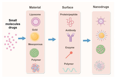
As a result, in the development of RNA drugs, lipid nanoparticles is the main carrier for RNA drugs widely. Nevertheless, lipid nanoparticles as carriers face poor targeting to the lungs, as well as some toxicity and immunogenicity issues [131,132]. At present, the scientific community is making many attempts to improve these shortcomings. Especially, the methods of modifying liposomes become necessary, which mainly include inserting targeted ligands or peptide and adding modified substances or surfactants as shown in Fig. 3. These modified vectors can achieve enhanced capability for site-specific targeting [133,134]. Moreover, the delivery strategy of virus particles and exosomes undoubtedly adds choices [135]. It has broad application prospects for treatment PF in the future.
Protein therapy involves the modulation of cellular biological functions and the treatment of diseases through the application of biological molecules, such as exogenous proteins or polypeptides [136,137]. Compared with gene therapy and chemotherapy, protein therapy has unique advantages. Protein therapy does not induce permanent genetic alterations or result in significant adverse reactions [136,138]. It can be employed in the treatment of various diseases, including diabetes. At present, the primary method for the clinical administration of protein and peptide pharmaceuticals is via injection [139]. Oral delivery of peptide and protein drugs faces great challenges [140]. Cellular communication network factor 2 (CCN2) gene as connective tissue growth factor, encodes a protein that plays a role in cell growth, differentiation, adhesion, and ECM remodeling. Pavlidou et al. presented the preclinical profile of the inhaled anti-CCN2 AnticalinⓇ protein PRS-220 [141]. A biotherapeutic protein drug targeting CCN2 is administered via inhalation directly to the lungs for the treatment of PF. Owing to its generally advantageous biophysical characteristics, it is highly suitable for inhalation delivery and has superior anti-fibrotic activity. Their work also has highlighted the advantage of inhaled protein drug delivery for treatment of respiratory disease in the future. The delivery of proteins and peptides represents an emerging frontier in delivery research. Several protein drugs are currently in used to treat cancer, inflammatory bowel disease and brain disease [137,140,142]. We hope protein drugs with the potential to significantly expand the range of PF drug therapy.
Cell therapies represent an innovative therapeutic strategy, attributed to their intrinsic anti-inflammatory and anti-fibrotic characteristics [143,144]. Many preclinical studies have demonstrated that stem cells therapy exhibits significant therapeutic efficacy across a variety of pulmonary diseases [145]. Stem cells therapy is among the most extensively researched cellular products in the field of regenerative medicine. Currently, mesenchymal stem cells (MSCs) as new biotech drugs have received much attention due to their substantial efficacy in regulating epithelial permeability, reducing inflammation, and promoting tissue regeneration [146,147]. More researchers have found that it can be beneficial for the repair and reconstruction of fibrotic lung. Stem cell drugs are typically labeled and delivered to the target site using specific carriers including nanoparticles and exosomes [148]. Zhang et al. have used the pH-sensitive Au nanotracers transplanted MSCs for the first time. This innovative approach enables monitoring through computed tomography (CT) imaging for 35 days and contributes to the mitigation of PF [149]. Chen et al. have developed a novel self-assembly multifunctional nanoagent labeling MSCs. They found that labeled MSCs enhanced pulmonary function and mitigated symptoms of fibrosis [150]. Li et al. have fabricated AuPtCoPS nanozyme as carrier for tracking MSCs and eradicating ROS [151]. Based the harmful interaction between myofibroblasts and vascular endothelial cells leads to a significant loss of pulmonary capillaries in worsening fibrosis. Jiang et al. developed a modifying the surface of MSC with ROS-responsive lipid polymeric hybrid nanoparticles encapsulating the metformin and macitentan. Owing to the intrinsic homing capability of MSCs and the influence of growth factors secreted by MSCs, this approach not only efficiently targets pulmonary tissue but also facilitates the reconstruction of normal vascular architecture within the fibroblast region, thereby reversing PF [146]. The treatment of PF with MSCs demonstrates significant efficacy. In short, Fig. 5 shows that cell drug combined with small molecule drugs for realizing better [148-150], such as 1 + 1 ≥ 2. Nevertheless, further research is necessary to validate these preliminary and promising findings. Jiang et al. constructed a MSC nanoengineered platform through the bioconjugation of MSCs and type Ⅰ collagenase-modified liposomes encapsulating with nintedanib for the treatment of advanced fibrosis. This MSC nanoengineered platform not only facilitates the repair of alveolar structures but also significantly enhances the regeneration of damaged lung in aged mice [148]. MCS-derived exosomes has capability of targeting and penetrating efficiently [152]. Researchers think it is a good strategy for integration of treatment methods for PF. For instance, macrophage-based delivery of anti-fibrotic proteins and achieving successful application of chimeric antigen receptor T cells (CAR-T) therapy in the reversal of PF according to the latest study [153,154].

It is worth noting that clinical medication for PF has indeed encountered certain difficulties, including the development of nanomedicine [8[160-162]. Nanomedicines have demonstrated substantial benefits in the realms of healthcare, disease diagnosis, and therapeutic interventions. The integration of PF treatment with nanomedicine is anticipated to foster a broader spectrum of possibilities and innovations [163,164]. Nanomedicine has the potential to significantly enhance the bioavailability, minimize adverse effects, and optimize the delivery mechanisms of small molecule pharmaceuticals, active constituents of natural products, and gene therapies (Table 1). Currently, the discussion is limited to FDA-approved small molecule drugs, specifically pirfenidone and nintedanib, with no mention of those still under development. Numerous studies have demonstrated that the formulation of nanomedicine significantly enhances drug bioavailability and therapeutic efficacy. Consequently, the promotion of nanomedicine represents a critical and urgent strategy in the realms of drug development and clinical validation. The treatment of PF using natural products involves complex components and diverse pharmacological effects. Consequently, it is essential to precisely select active ingredients from natural products for formulation as natural small molecule drugs nanomedicines [72,165,166]. Comprehensive and systematic in-depth research on these nanomedicines is required to further elucidate their regulatory mechanisms and safety profiles in vivo. Chinese medicine nanomedicines have the potential to offer new impetus and direction for the prevention and treatment of PF. Gene delivery constitutes a fundamental aspect of gene therapy, particularly for the treatment of PF. The utilization of delivery vectors, such as viral vectors or liposomes, can elicit robust immune responses in the host, potentially diminishing therapeutic efficacy and inducing adverse effects. Achieving sustained and stable gene expression in pulmonary tissues presents a significant challenge, with particular emphasis on mitigating side effects associated with gene overexpression. Concurrently, ensuring the long-term safety of the therapeutic intervention remains paramount. Therefore, it is imperative for researchers in the field of gene therapy to closely integrate technological innovations across multiple disciplines, including biology, clinical medicine, materials science, and drug delivery, to enhance the efficacy of treatments for PF. As technological advancements continue and research deepens, gene therapy holds the potential to offer new hope for the treatment of diseases such as PF.
 DownLoad:
CSV
DownLoad:
CSV
| Type | Drug | Delivery system | Design targeting | Delivery route | Ref. |
| Small molecule drugs | Pirfenidone | BSA nanoparticles | FnBAP5 peptide modified PEGylated BSA | Vein injection | [38] |
| HSYA | Phytosomes | HSYA phytosome | ISI | [94] | |
| Nintedanib/colchicine | Lipid NPs | liposomes modified E5 peptide | Vein injection | [155] | |
| Grape seed extract | N-Succinyl chitosan-coated liposomes | Liposomes loaded GSE and N-succinyl chitosan was used as a coating layer to protect the formulation | Oral administration | [156] | |
| Astragaloside Ⅳ (AS-Ⅳ) and ligustrazine (LIG) | Polylactic-co-glycolic acid (PLGA) particles | The fundamental theory of traditional Chinese medicine (TCM) of “tonifying qi and activating blood” | Intratracheally injected | [157] | |
| Salvianolic acid B | Dry power inhaler | Sal B-DPI was successfully prepared by spray drying method with L-leucine as excipient | Oral administration | [158] | |
| Macromolecular drugs | siRNA (Drp1) | Exosome | Delivery of ExoMMP19 for degraded excessive ECM and then delivery of ExoTx for siRNA | Intratracheally inhaled | [135] |
| siRNA (STAT3) | Perfluorocarbon nanoemulsions | Core-shell structure of nanoemulsions providing the positive surface charge necessary for siRNA binding | Intratracheal instillation | [123] | |
| IL-10 | Hydrogel | hydrogels loaded with IL-10 (heparin-immobilized protein drug IL-10) | Intranasal instillation | [159] | |
| Cell drugs | IL-10, sTGFR-Fc and CD147 | Macrophage-based delivery | engineered macrophages to continuously secrete interleukin-10, TGFRcFc, and CD147 | Intranasal infusions | [153] |
| MSC and nintedanib | Liposomes | MSCs and type Ⅰ collagenase–modified liposomes loaded with nintedanib | Vein injection | [148] |
Protein delivery is also a direct and simple drug delivery method for treating PF, such as macrophage-based delivery of anti-fibrotic proteins can also alleviate bleomycin-induced PF in mice [153]. Stem cell therapy is being explored as a potential treatment for idiopathic PF (IPF). However, several limitations must be addressed, including immunological compatibility, cell survival and high costs [167]. Future strategies for drug delivery in the treatment of PF are likely to advance towards the use of multi-drug combination therapies. These may encompass small molecule drugs, components of natural products, nucleic acids, peptide and stem cells, while also addressing challenges related to stability and safety. Additionally, with advancements in artificial intelligence, its role in drug delivery is expected to gain increasing attention. Artificial intelligence (AI) technology may be utilized in the design of drugs, optimization of delivery systems, and the personalized development of treatment plan.
In summary, PF drug delivery systems encounter numerous challenges. These challenges primarily arise from the unclear pathogenesis of PF and the intricate pathological environment associated with the condition. The most notable and distinctive features is pathological environment with excessive accumulation of ECM components, including collagen, plastids, and fibronectin, which can significantly increase the rigidity of lung tissue [168]. This hard lung parenchyma is called collagen barrier, forming a "barrier" that hinders the normal exchange of oxygen and carbon dioxide, thereby affecting respiration. Furthermore, it is imperative to select suitable drug delivery systems for small molecule drugs, antibodies, proteins, and gene. The future challenge is to improve drug delivery performance while enabling more convenient administration with fewer doses. Many studies are mostly based on results from mouse, rat and cell culture experiment. Animal models may be a significant limitation of research [23]. There may also be more suitable research methods to accelerate the overcoming of collagen barriers and improve drug delivery performance with development of organoid structures, lung chips, 3D printing technology and immunotherapies. Despite notable advancements in the development of sophisticated drug delivery systems for the treatment of PF, further research is imperative to elucidate the underlying mechanisms, assess drug safety and efficacy, and explore combination therapies to enhance treatment specificity [25]. Future PF treatments will rely on the deep intersection of materials science, gene, cell engineering, and immunology, could be customized to each patient and their disease stage, necessitating early diagnosis and effective drug delivery. Through these ongoing investigative efforts, we remain optimistic that the field will develop effective treatment responses for patients with PF.
Drug delivery technologies have shown significant potential in the treatment of PF. The pulmonary administration of drug therapies presents significant challenges due to the intricate anatomy and pathophysiological characteristics of lung. This review provides a detailed overview and framework for delivery of small-molecule drugs, macromolecular therapeutics, and cellular drugs treatment PF. Drugs need to overcome the specific barriers through designed delivery carriers. So safe and efficient pulmonary delivery system are constructed of critical importance for PF. Drug delivery application shown great effectiveness in PF treatment through numerous preclinical studies. Multidrug delivery therapy with a range of capabilities may be a promising treatment of PF in the near future.
The authors declare that they have no known competing financial interests or personal relationships that could have appeared to influence the work reported in this paper.
Yan Yu: Writing – review & editing, Writing – original draft, Funding acquisition, Data curation, Conceptualization. Cailing Gan: Visualization, Methodology, Investigation, Formal analysis. Kun Shi: Software, Methodology, Investigation, Conceptualization. Zhongwu Bei: Visualization, Software, Conceptualization. Yang Yu: Visualization, Resources, Investigation. Meng Pan: Visualization, Resources, Methodology. Hanzhi Deng: Visualization, Methodology, Formal analysis. Zhiyong Qian: Methodology, Conceptualization.
This work was funded by the National Natural Science Foundation of China (No. NSFC82400096), Science and Technology Department of Sichuan Province (No. 2025ZNSFSC1538), and Xihua University Internal Talent Introduction Project with Scientific Research Funding (No. ZX20250087). We thank for their financial support.
J.P. Anna, N. Imre, R. Ganesh, Eur. Respir. J. 57 (2021) 2003551. doi: 10.1183/13993003.03551-2020
Y. Toyoshi, G. Christophe, R.J.K. Martin, Eur. Respir. J. 61 (2023) 2300407. doi: 10.1183/13993003.00407-2023
M. Zhao, L. Wang, M. Wang, et al., STTT 7 (2022) 206.
N.C. Henderson, F. Rieder, T.A. Wynn, Nature 587 (2020) 555–566. doi: 10.1038/s41586-020-2938-9
Y. Chen, Z. Li, G. Ji, et al., MedComm 5 (2024) e494. doi: 10.1002/mco2.494
L. Zhao, Y. Zhu, H. Tao, et al., Acta Pharm. Sin. B 14 (2024) 3543–3560. doi: 10.1016/j.apsb.2024.04.013
C. Zhang, J. Ma, X. Zhang, et al., Int. Immunopharmacol. 134 (2024) 112176. doi: 10.1016/j.intimp.2024.112176
A.J. Podolanczuk, C.C. Thomson, M. Remy-Jardin, et al., Eur. Respir. J. 61 (2023) 2200957. doi: 10.1183/13993003.00957-2022
S. Wu, X. Zhou, Z. Jin, et al., Collagen Leather 5 (2023) 19. doi: 10.1186/s42825-023-00126-6
J. Ma, L. Zhang, X. Zhang, et al., Sci. Transl. Med. 16 (2024) eado5266. doi: 10.1126/scitranslmed.ado5266
X. Chen, H. Wang, C. Wu, et al., Redox Biol. 70 (2024) 103038. doi: 10.1016/j.redox.2024.103038
I.P.F.C.R. Network., New Engl. J. Med. 366 (2014) 1968–1977.
I.P.F.C.R. Network, New Engl. J. Med. 370 (2014) 2093–2101. doi: 10.1056/NEJMoa1401739
T.M. Maher, T.J. Corte, A. Fischer, et al., Lancet Respir. Med. 8 (2020) 147–157. doi: 10.1016/S2213-2600(19)30341-8
R. Flaherty Kevin, U. Wells Athol, V. Cottin, et al., New Engl. J. Med. 381 (2019) 1718–1727. doi: 10.1056/NEJMoa1908681
A. Najmi, S.A. Javed, M. Al Bratty, et al., Molecules 27 (2022) 349. doi: 10.3390/molecules27020349
L. Meng, C. Zhang, P. Yu, Pharmacol. Res. 203 (2024) 107179. doi: 10.1016/j.phrs.2024.107179
S.N. Adin, G. Isha, A. Mohd, et al., J. Drug Target 32 (2024) 707–723. doi: 10.1080/1061186x.2024.2347371
Q. Zeng, Z. Liu, T. Niu, et al., Chin. Chem. Lett. 34 (2023) 107747. doi: 10.1016/j.cclet.2022.107747
Y. Tian, M. Li, Y. Yang, et al., Chin. Chem. Lett. 35 (2024) 109270. doi: 10.1016/j.cclet.2023.109270
B. Hu, S. Kong, Y. Weng, et al., Chin. Chem. Lett. 34 (2023) 108210. doi: 10.1016/j.cclet.2023.108210
Y. Chen, Q. Zeng, B. Chu, et al., Chin. Chem. Lett. 34 (2023) 108133. doi: 10.1016/j.cclet.2023.108133
R. Li, Y. Jia, X. Kong, et al., J. Control. Release 348 (2022) 95–114. doi: 10.1016/j.jconrel.2022.05.039
L.T. Ferguson, X. Ma, J.W. Myerson, et al., Adv. NanoBiomed. Res. 3 (2023) 2200106. doi: 10.1002/anbr.202200106
S. Singh, S. Wairkar, AAPS PharmSciTech. 25 (2024) 78. doi: 10.1208/s12249-024-02793-y
A. Kidane, P.P. Bhatt, Curr. Opin. Chem. Biol. 9 (2005) 347–351. doi: 10.1016/j.cbpa.2005.06.006
X. Wang, Y. Wang, Z. Xue, et al., J. Colloid Interface Sci. 636 (2023) 388–400. doi: 10.1016/j.jcis.2023.01.007
X. Yang, G. Lin, Y. Chen, et al., ACS Cent. Sci. 10 (2024) 1789–1802. doi: 10.1021/acscentsci.4c00798
Q. Yin, Y. Chen, M. Zhou, et al., Spectrochim. Acta A 204 (2018) 88–98. doi: 10.1016/j.saa.2018.06.016
H. Taniguchi, M. Ebina, Y. Kondoh, et al., Eur. Respir. J. 35 (2009) 821–829.
L. Tomasini, M. Ferrere, J. Nicolas, Nat. Rev. Bioeng. 2 (2024) 501–520. doi: 10.1038/s44222-024-00161-w
C. Liu, L. Xi, Y. Liu, et al., ACS Nano 17 (2023) 11626–11644. doi: 10.1021/acsnano.3c02075
L. Rao, P. Zhu, M. Guo, et al., Nano Today 56 (2024) 102298. doi: 10.1016/j.nantod.2024.102298
J. Liu, Z. Wu, Y. Liu, et al., J. Nanobiotechnol. 20 (2022) 213. doi: 10.1186/s12951-022-01435-4
Q. Wan, X. Zhang, D. Zhou, et al., J. Nanobiotechnol. 21 (2023) 215. doi: 10.1186/s12951-023-01971-7
X. Wang, L. Tian, Y. Li, et al., Angew. Chem. Int. Ed. 64 (2025) e202421949. doi: 10.1002/anie.202421949
N. Qu, S. Ke, J. Yating, et al., Int. J. Nanomed. 19 (2024) 6945–6980. doi: 10.2147/ijn.s467876
T. Zhao, B. Gong, S. Luo, et al., Acta Biomater. 167 (2023) 574–582. doi: 10.1016/j.actbio.2023.06.024
J. Seo, C. Lee, H.S. Hwang, et al., Pulm. Pharmacol. Ther. 36 (2016) 53–61. doi: 10.1016/j.pupt.2016.01.001
T. Zhang, W. Cui, T. Tian, et al., ACS Appl. Mater. Interfaces 12 (2020) 47115–47126. doi: 10.1021/acsami.0c13806
Z. Yang, L. Shi, Y. Wang, et al., Small 21 (2025) 2410162. doi: 10.1002/smll.202410162
R.P. Goodman, R.M. Berry, A.J. Turberfield, Chem. Commun. (2004) 1372–1373.
T. Zhang, T. Tian, Y. Lin, Adv. Mater. 34 (2022) 2107820. doi: 10.1002/adma.202107820
Y. Chen, M. Lin, D. Ye, et al., Nat. Protoc. 19 (2024) 985–1014. doi: 10.1038/s41596-023-00943-3
Y. Xie, S. Shi, W. Lv, et al., ACS Nano 18 (2024) 26704–26721. doi: 10.1021/acsnano.4c06598
Y. Liu, S. Li, S. Lin, et al., Chin. Chem. Lett. 34 (2023) 107987. doi: 10.1016/j.cclet.2022.107987
M.Y. Yang, M.M. Han, L. Tang, et al., Adv. Funct. Mater. 34 (2024) 2315128. doi: 10.1002/adfm.202315128
G.J. Roth, R. Binder, F. Colbatzky, et al., J. Med. Chem. 58 (2015) 1053–1063. doi: 10.1021/jm501562a
R. Kaur, T.B. Shaikh, H.P. Sripadi, et al., Int. J. Pharm. 649 (2024) 123644. doi: 10.1016/j.ijpharm.2023.123644
K.R. Flaherty, C.D. Fell, J.T. Huggins, et al., Eur. Respir. J. 52 (2018) 1800230. doi: 10.1183/13993003.00230-2018
M.M. Han, L. Tang, B. Huang, et al., J. Control. Release 366 (2024) 732–745. doi: 10.1016/j.jconrel.2024.01.032
M.Y. Yang, Y.J. Lin, M.M. Han, et al., J. Control. Release 351 (2022) 623–637. doi: 10.1016/j.jconrel.2022.09.054
B. Wang, Y. Gao, L. sun, et al., Biomaterials 303 (2023) 122404. doi: 10.1016/j.biomaterials.2023.122404
Y. Yu, J. Peng, M. Pan, et al., Small Methods 5 (2021) 2001212. doi: 10.1002/smtd.202001212
H. Liu, Y. Yu, T. Xue, et al., Chin. Chem. Lett. 35 (2024) 108574. doi: 10.1016/j.cclet.2023.108574
W. Chen, K. Shi, Y. Yu, et al., Chin. Chem. Lett. 35 (2024) 109159. doi: 10.1016/j.cclet.2023.109159
M. Pan, D. Hu, L. Yuan, et al., Acta Pharm. Sin. B 13 (2023) 2926–2954. doi: 10.1016/j.apsb.2022.12.021
Y. Yang, Q. Liu, M. Wang, et al., STTT 9 (2024) 158.
H. Deng, Y. Qu, B. Chu, et al., Biomaterials 317 (2025) 123072. doi: 10.1016/j.biomaterials.2024.123072
Y. Jia, K. Shi, L. Dai, et al., Small Methods 7 (2023) 2201087. doi: 10.1002/smtd.202201087
X. He, Y. Hao, B. Chu, et al., Nano Today 39 (2021) 101174. doi: 10.1016/j.nantod.2021.101174
R. Han, Y. Xiao, Q. Yang, et al., Biomaterials 264 (2021) 120451. doi: 10.1016/j.biomaterials.2020.120451
Y. Yan, P. Meng, P. Jinrong, et al., Chin. Chem. Lett. 33 (2022) 4133–4145. doi: 10.1016/j.cclet.2022.02.045
X. Li, Y. Li, C. Yu, et al., ACS Nano 17 (2023) 6387–6399. doi: 10.1021/acsnano.2c11112
J.K. Tee, F. Peng, H.K. Ho, Biochem. Pharmacol. 160 (2019) 24–33.
B. Tu, Y. Gao, X. An, et al., Acta Pharm. Sin. B. 13 (2023) 1828–1846. doi: 10.1016/j.apsb.2022.09.011
W. Chen, Y. Gao, Y. Liu, et al., ACS Biomater. Sci. Eng. 10 (2024) 6250–6262. doi: 10.1021/acsbiomaterials.4c00532
X. Wang, W. Wan, J. Lu, et al., Int. J. Pharm. 656 (2024) 124096. doi: 10.1016/j.ijpharm.2024.124096
X. Cao, C. Yu, S. Cheng, et al., ACS Appl. Mater. Interfaces 16 (2024) 20260–20272.
X. Chang, Y.M. Han, Q.L. Li, et al., J. Control. Release 376 (2024) 861–879. doi: 10.1016/j.jconrel.2024.10.061
F. Chen, Q. Liu, Adv. Drug Deliv. Rev. 186 (2022) 114317. doi: 10.1016/j.addr.2022.114317
H. Xu, T. Wu, L. Huang, Adv. Drug Deliv. Rev. 177 (2021) 113911. doi: 10.1016/j.addr.2021.113911
Y. Hao, J. Li, L. Dan, et al., J. Ethnopharmacol. 318 (2024) 116836. doi: 10.1016/j.jep.2023.116836
Y. Zhang, L. Zhou, G. Cheng, et al., MedComm Future Med. 3 (2024) e91. doi: 10.1002/mef2.91
Y. Xu, Y. Wang, W. Peng, et al., Phytother. Res. 39 (2025) 282–297. doi: 10.1002/ptr.8372
A. Sharma, S. Wairkar, Phytother. Res. 38 (2024) 4406–4423. doi: 10.1002/ptr.8285
S.H. Dong, Y.W. Liu, F. Wei, et al., Biomed. Pharmacother. 89 (2017) 1297–1309. doi: 10.1016/j.biopha.2017.03.005
L. Marinaccio, G. Gentile, G. Zengin, et al., Food Chem. X 26 (2025) 102273. doi: 10.1016/j.fochx.2025.102273
W. Lin, Y. Song, T. Li, et al., Biomed. Pharmacother. 166 (2023) 115394. doi: 10.1016/j.biopha.2023.115394
J. Yong, H. Shu, X. Zhang, et al., Int. J. Nanomed. 19 (2024) 1723–1748. doi: 10.2147/ijn.s451206
Q. Wang, W. Li, H. Hu, et al., Biomed. Pharmacother. 159 (2023) 114226. doi: 10.1016/j.biopha.2023.114226
D. Dolivo, P. Weathers, T. Dominko, Acta Pharm. Sin. B 11 (2021) 322–339. doi: 10.1016/j.apsb.2020.09.001
Y. Su, Q. Zhou, H. Xu, et al., Food Hydrocolloid. 149 (2024) 109502. doi: 10.1016/j.foodhyd.2023.109502
J. Yao, Y. Li, F. Meng, et al., Environ. Toxicol. 38 (2023) 1494–1508. doi: 10.1002/tox.23781
Y.B. Chen, Y.B. Zhang, Y.L. Wang, et al., J. Nanobiotechnol. 20 (2022) 272. doi: 10.1145/3537693.3537734
A. Riva, M. Ronchi, G. Petrangolini, et al., Eur. J. Drug Metab. Pharmacokinet. 44 (2019) 169–177. doi: 10.1007/s13318-018-0517-3
K. Li, W. Wang, W. Xiao, Pharmacol. Res. 188 (2023) 106657. doi: 10.1016/j.phrs.2023.106657
Z.W. Sharawi, I.M. Ibrahim, E.K. Abd-alhameed, et al., Naunyn Schmiedebergs Arch. Pharmacol. 397 (2024) 1405–1419. doi: 10.1007/s00210-023-02704-1
D. Qi, B. Jia, H. Peng, et al., Eur. J. Pharm. Biopharm. 188 (2023) 243–253. doi: 10.1016/j.ejpb.2023.05.017
J. Li, Y. Xiao, Y. Zhang, et al., ACS Nano 17 (2023) 15354–15370. doi: 10.1021/acsnano.2c10388
S. Song, L. Ding, G. Liu, et al., Front. Pharmacol. 14 (2023) 1129817. doi: 10.3389/fphar.2023.1129817
Z. -L. Wang, H. -T. Wang, G. Chang, et al., Nat. Commun. 16 (2025) 4489. doi: 10.1038/s41467-025-59774-3
W. Wang, M. Liu, X. Fu, et al., Phytomedicine 132 (2024) 155814. doi: 10.1016/j.phymed.2024.155814
T. Li, D. Han, Z. Li, et al., Pharmaceuticals 15 (2022) 1394. doi: 10.3390/ph15111394
Z. Li, Q. Zhang, J. Xiang, et al., Mater. Today Bio 20 (2023) 100653. doi: 10.1016/j.mtbio.2023.100653
E. Ghoushi, M. Poudineh, N. Parsamanesh, et al., Food Chem.: Mol. Sci. 8 (2024) 100198. doi: 10.1016/j.fochms.2024.100198
M. Fathimath Muneesa, R.R. Barki, S.B. Shaikh, et al., Toxicol. Appl. Pharmacol. 449 (2022) 116116. doi: 10.1016/j.taap.2022.116116
Y. Miao, Y. Zhang, S. Qiao, et al., Acta Pharmacol. Sin. 42 (2021) 422–435. doi: 10.1038/s41401-020-0469-4
F. Hanyu, H. Zheng, W. Jiaqi, et al., Front. Pharmacol. 14 (2023) 1258885. doi: 10.3389/fphar.2023.1258885
P.A. Gupte, S.A. Giramkar, S.M. Harke, et al., J. Inflamm. Res. 12 (2019) 145–152. doi: 10.2147/jir.s205390
K.H.M. Cox, D.J. White, A. Pipingas, et al., Nutrients 12 (2020) 1678. doi: 10.3390/nu12061678
Q. Guo, C. Jiang, Acta Pharm. Sin. B 10 (2020) 979–986. doi: 10.1016/j.apsb.2020.01.009
A. Kakkar, G. Traverso, O.C. Farokhzad, et al., Nat. Rev. Chem. 1 (2017) 0063. doi: 10.1038/s41570-017-0063
Y. Zhu, S.S. Cai, J. Ma, et al., Biomaterials 308 (2024) 122559. doi: 10.1016/j.biomaterials.2024.122559
I. Melnikova, Nat. Rev. Drug Discov. 6 (2007) 863–864. doi: 10.1038/nrd2443
N. Yoshinaga, J.K. Zhou, C. Xu, et al., Nano Lett. 23 (2023) 757–764. doi: 10.1021/acs.nanolett.2c02306
M. Zhang, H. Lu, L. Xie, et al., Adv. Drug Deliv. Rev. 203 (2023) 115144. doi: 10.1016/j.addr.2023.115144
A.Y. Jiang, S. Lathwal, S. Meng, et al., J .Am. Chem. Soc. 146 (2024) 32567–32574. doi: 10.1021/jacs.4c11347
S. Zhang, X. Tong, S. Liu, et al., Cell Death. Dis. 14 (2023) 389. doi: 10.18208/ksdc.2023.29.4.389
O.M. Merkel, J. Control. Release 345 (2022) 549–556. doi: 10.1016/j.jconrel.2022.03.039
W. Li, C. Wang, Y. Zhang, et al., Small 20 (2024) 2310531. doi: 10.1002/smll.202310531
Q. Ji, J. Hou, X. Yong, et al., Adv. Mater. 33 (2021) 2007798. doi: 10.1002/adma.202007798
R.J. Allen, J.M. Oldham, D.A. Jenkins, et al., Lancet Respir. Med. 11 (2023) 65–73. doi: 10.1016/S2213-2600(22)00251-X
K. Huang, Y. Liu, H. Miao, et al., ACS Nano 19 (2025) 1128–1139. doi: 10.1021/acsnano.4c13053
E.L. Han, S. Tang, D. Kim, et al., Nano Lett. 25 (2025) 800–810. doi: 10.1021/acs.nanolett.4c05186
A.K. Blakney, P.F. McKay, K. Hu, et al., J. Control. Release 338 (2021) 201–210. doi: 10.1016/j.jconrel.2021.08.029
T. Kim, H.S. Han, K. Yang, et al., ACS Nano 18 (2024) 7972–7988. doi: 10.1021/acsnano.3c10732
B.E. Ferdows, D.N. Patel, W. Chen, et al., Nanoscale 14 (2022) 4448–4455. doi: 10.1039/d1nr06991h
G. Kandasamy, D. Maity, Biomed. Mater. 19 (2024) 022001. doi: 10.1088/1748-605x/ad1baf
H. Zhang, J. Vandesompele, K. Braeckmans, et al., Chem. Soc. Rev. 53 (2024) 317–360. doi: 10.1039/d3cs00194f
R. Guagliardo, L. Herman, J. Penders, et al., ACS Nano 15 (2021) 8095–8109. doi: 10.1021/acsnano.0c04489
P. Merckx, G. Conickx, E. Blomme, et al., Eur. J. Pharm. Biopharm. 197 (2024) 114223. doi: 10.1016/j.ejpb.2024.114223
L. Ding, S. Tang, W. Tang, et al., Adv. Sci. 9 (2022) 2103676. doi: 10.1002/advs.202103676
H. Jin, M. Jeong, G. Lee, et al., Adv. Funct. Mater. 33 (2023) 2209432. doi: 10.1002/adfm.202209432
J. Kim, S. Jeon, S.J. Kang, et al., J. Control. Release 322 (2020) 108–121. doi: 10.1016/j.jconrel.2020.03.016
A. Singh, S. Chakraborty, S.W. Wong, et al., Proc. Natl. Acad. Sci. U. S. A. 119 (2022) e2121098119. doi: 10.1073/pnas.2121098119
X. Bai, G. Zhao, Q. Chen, et al., Sci. Adv. 8 (2022) eabn7162. doi: 10.1126/sciadv.abn7162
S. Dong, H. Fang, J. Zhu, et al., ACS Nano 19 (2025) 2742–2758. doi: 10.1021/acsnano.4c15130
J. Hou, Q. Ji, J. Ji, et al., Theranostics 11 (2021) 3244–3261. doi: 10.7150/thno.54217
X. Bai, Q. Chen, F. Li, et al., Nat. Commun. 15 (2024) 6844. doi: 10.1038/s41467-024-51056-8
L. Yan, Y. Su, I. Hsia, et al., Mol. Ther. Nucleic Acids 32 (2023) 36–47. doi: 10.1016/j.omtn.2023.02.031
R. Li, M. Zhu, X. Hu, et al., Chin. Chem. Lett. 36 (2025) 110736. doi: 10.1016/j.cclet.2024.110736
Y. Huang, J. Zhang, X. Wang, et al., Biomolecules 14 (2024) 904. doi: 10.3390/biom14080904
J. Guo, M. Gu, Y. Chen, et al., Chin. Chem. Lett. 36 (2025) 110849. doi: 10.1016/j.cclet.2025.110849
W. Zhang, Z. Wan, D. Qu, et al., Bioact. Mater. 32 (2024) 488–501.
Y. Li, Z. Ye, H. Yang, et al., Acta Pharm. Sin. B 12 (2022) 2624–2639. doi: 10.1016/j.apsb.2022.04.013
H. Lu, S. Xu, Z. Guo, et al., ACS Nano 15 (2021) 18214–18225. doi: 10.1021/acsnano.1c07166
T. Liu, M. Chen, J. Fu, et al., Acta Pharm. Sin. B 11 (2021) 2326–2343. doi: 10.1016/j.apsb.2021.03.003
S. Mitragotri, P.A. Burke, R. Langer, Nat. Rev. Drug Discov. 13 (2014) 655–672. doi: 10.1038/nrd4363
S. Arora, T. Bajaj, J. Kumar, et al., J. Pharmacol. Exp. Ther. 388 (2024) 54–66. doi: 10.1124/jpet.123.001690
V. Neiens, E.M. Hansbauer, T.J. Jaquin, et al., Nat. Commun. 16 (2025) 3251. doi: 10.1038/s41467-025-58568-x
B. Huang, T. Yin, S. Fu, et al., Proc. Natl. Acad. Sci. U. S. A. 121 (2024) e2320482121. doi: 10.1073/pnas.2320482121
S. Geiger, D. Hirsch, F.G. Hermann, Eur. Respir. Rev. 26 (2017) 170044. doi: 10.1183/16000617.0044-2017
H. Duan, Y. Liu, Z. Gao, et al., Acta Pharm. Sin. B 11 (2021) 55–70. doi: 10.1016/j.apsb.2020.09.016
A. Serrano-Mollar, Med. Sci. 6 (2018) 64. doi: 10.3390/medsci6030064
Y.F. Fang, C. Zhang, M.M. Han, et al., Adv. Mater. 37 (2025) 2414601. doi: 10.1002/adma.202414601
J. Xu, Y. Sun, Y. You, et al., Acta Pharm. Sin. B 14 (2024) 1412–1427. doi: 10.1016/j.apsb.2023.11.009
M.M. Han, X.Y. He, L. Tang, et al., Sci. Adv. 9 (2023) eadg5358. doi: 10.1126/sciadv.adg5358
C. Yu, Z. Chen, X. Li, et al., Small 17 (2021) 2101861. doi: 10.1002/smll.202101861
Y. Shu, M. Ma, X. Pan, et al., Bioact. Mater. 21 (2023) 129–141.
H. Bao, M. Wu, J. Xing, et al., Sci. Adv. 10 (2024) eadq0703. doi: 10.1126/sciadv.adq0703
Y. Zhou, W. Zhou, X. Chen, et al., Acta Pharm. Sin. B 10 (2020) 1563–1575. doi: 10.1016/j.apsb.2019.11.013
H. Liu, C. Yang, Y. Gao, et al., Bioeng. Transl. Med. 8 (2023) e10555. doi: 10.1002/btm2.10555
J. Yan, S.Y. Wang, Q. Su, et al., Nat. Commun. 16 (2025) 3748. doi: 10.1038/s41467-025-59093-7
X. Chang, L. Xing, Y. Wang, et al., Nanoscale 12 (2020) 8664–8678. doi: 10.1039/d0nr00750a
N. Bavarsad, A. Hemmati, F. Jafarian, et al., J. Basic Med. Sci. 26 (2023) 1237–1244.
M. Zheng, K. Liu, L. Li, et al., J. Nanobiotechnol. 22 (2024) 14. doi: 10.1186/s12951-023-02251-0
P. Lu, J. Li, C. Liu, et al., Asian J. Pharm. Sci. 17 (2022) 447–461.
E.A. Shamskhou, M.J. Kratochvil, M.E. Orcholski, et al., Biomaterials 203 (2019) 52–62. doi: 10.1016/j.biomaterials.2019.02.017
L. Chibaya, K.D. DeMarco, C.F. Lusi, et al., Sci. Transl. Med. 16 (2024) eadj9366. doi: 10.1126/scitranslmed.adj9366
P. Xue, H. Zhuang, S. Shao, et al., ACS Nano 18 (2024) 25795–25812. doi: 10.1021/acsnano.4c08574
Q. Yao, B. Wang, J. Yu, et al., J. Control. Release 374 (2024) 112–126. doi: 10.1016/j.jconrel.2024.08.006
M.M. Rahman, J. Wang, G. Wang, et al., Nat. Nanotechnol. 19 (2024) 818–824. doi: 10.1038/s41565-024-01620-6
K. Jiang, K. Tian, Y. Yu, et al., Nat. Commun. 15 (2024) 6136. doi: 10.1038/s41467-024-50568-7
Y. Xie, X. Shen, F. Xu, et al., Phytochem. Anal. (2024), doi: 10.1002/pca.3381.
J. Guan, W. Chen, M. Yang, et al., Adv. Drug Deliv. Rev. 174 (2021) 210–228. doi: 10.1016/j.addr.2021.04.015
M. Ikrama, M. Usama, S. Israr, et al., J. Taibah Univ. Med. Sci. 19 (2024) 82–89.
M. Kamiya, H. Carter, M.S. Espindola, et al., Cell 187 (2024) 3506–3530. doi: 10.1016/j.cell.2024.05.015

Figure 1 Schematic illustration of healthy lung and fibrotic lung experienced influencing factors, drug selection, drug delivery, and administration methods. This figure was created using Figdraw.

Figure 2 Example of various small molecule drugs used in drug delivery for PF therapy.

Figure 3 Schematic representation of various modified nanoparticles delivery systems utilizing for the therapeutic of PF. This figure was created using Figdraw.

Figure 4 Inhalable siRNA targeting lipid nanoparticles or polymeric nanoparticles for PF treatment. This figure was created using Figdraw.

Figure 5 Illustration of lung-targeting nanoengineered MSCs for reversing PF in mice. This figure was created using Figdraw.
Table 1. Different drug delivery systems in PF.
| Type | Drug | Delivery system | Design targeting | Delivery route | Ref. |
| Small molecule drugs | Pirfenidone | BSA nanoparticles | FnBAP5 peptide modified PEGylated BSA | Vein injection | [38] |
| HSYA | Phytosomes | HSYA phytosome | ISI | [94] | |
| Nintedanib/colchicine | Lipid NPs | liposomes modified E5 peptide | Vein injection | [155] | |
| Grape seed extract | N-Succinyl chitosan-coated liposomes | Liposomes loaded GSE and N-succinyl chitosan was used as a coating layer to protect the formulation | Oral administration | [156] | |
| Astragaloside Ⅳ (AS-Ⅳ) and ligustrazine (LIG) | Polylactic-co-glycolic acid (PLGA) particles | The fundamental theory of traditional Chinese medicine (TCM) of “tonifying qi and activating blood” | Intratracheally injected | [157] | |
| Salvianolic acid B | Dry power inhaler | Sal B-DPI was successfully prepared by spray drying method with L-leucine as excipient | Oral administration | [158] | |
| Macromolecular drugs | siRNA (Drp1) | Exosome | Delivery of ExoMMP19 for degraded excessive ECM and then delivery of ExoTx for siRNA | Intratracheally inhaled | [135] |
| siRNA (STAT3) | Perfluorocarbon nanoemulsions | Core-shell structure of nanoemulsions providing the positive surface charge necessary for siRNA binding | Intratracheal instillation | [123] | |
| IL-10 | Hydrogel | hydrogels loaded with IL-10 (heparin-immobilized protein drug IL-10) | Intranasal instillation | [159] | |
| Cell drugs | IL-10, sTGFR-Fc and CD147 | Macrophage-based delivery | engineered macrophages to continuously secrete interleukin-10, TGFRcFc, and CD147 | Intranasal infusions | [153] |
| MSC and nintedanib | Liposomes | MSCs and type Ⅰ collagenase–modified liposomes loaded with nintedanib | Vein injection | [148] |
 下载: 导出CSV

 扫一扫看文章

扫一扫关注我们
 下载:
下载: