
Figure 1.
Schematic illustration of nanomaterial-mediated drug delivery strategies to enhance BBB penetration for CNS diseases treatments.
Advances in CNS drug delivery strategies to cross the blood-brain barrier
Guanlong Li , Zhuoyan Li , Yan Sun , Tiange Bu , Shaochuan Chen , Leixin Yang , Zhi Li , Wenyue Mao , Yanpeng Jia

Central nervous system (CNS) diseases are among the leading causes of mortality and disability worldwide, posing a severe threat to global public health and quality of life [1,2]. Despite significant efforts to improve clinical outcomes for patients with CNS disorders, several formidable challenges remain unresolved [3-5]. One of the most critical barriers is the blood-brain barrier (BBB), which significantly restricts the transport of therapeutic agents into the brain, thereby limiting the efficacy of many treatment strategies [6-8]. Indeed, the complexity of the BBB’s pathophysiological characteristics has been a major contributing factor to the failure of numerous CNS-targeted therapies. In response to these challenges, nanomaterials have emerged as a promising class of platforms for CNS disease diagnosis and treatment. Their unique properties, such as high stability, biodegradability, and capacity for targeted delivery, have spurred extensive research into their potential for crossing the BBB and enhancing drug accumulation in brain tissue [9-16].
In this review, we discuss the structural and functional features of the BBB, followed by an overview of the pathogenesis of representative CNS diseases. We then focus on recent advances in nanomaterial-based BBB penetration strategies. As illustrated in Fig. 1, various nanocarriers, including liposomes, micelles, nanoemulsions, and polymeric nanoparticles, have been engineered to increase drug delivery efficiency to the brain and thereby improve therapeutic outcomes in CNS disorders [1,17,18]. Special attention is given to brain-targeting approaches such as receptor-mediated transport (RMT), reversible BBB disruption, biomimetic membrane coating, and intranasal delivery.
BBB is a significant physiological and metabolic barrier that forms a tight structural and functional block between the peripheral circulation and CNS. It precisely regulates the cellular and molecular exchange between the systematic circulation and brain parenchyma by controlling the permeation of materials entering and exiting the nervous system [19]. BBB plays a vital role in maintaining brain homeostasis and protecting the CNS from harmful substances in the peripheral circulation. As a regulatory interface, BBB is often involved in the progression of various CNS disorders with increased permeability, including Alzheimer’s disease (AD), stroke, and brain tumors, leading to irreversible damage to the CNS [20]. As shown in Fig. S1 (Supporting information), BBB is typically composed of five integral components: endothelial cells, pericytes, astrocytes, basement membrane, and junctional complexes. Endothelial cells lining cerebral capillaries, along with tight junctions, form the inner layer of the BBB, preventing the cargo exchange between adjacent endothelial cells through the paracellular passage. The middle layer (basement membrane) contains extracellular matrix proteins and pericytes, while the outer layer consists of astrocytes and additional extracellular matrix [21].
Two primary pathways mediate transport across the BBB: the paracellular route, located between adjacent endothelial cells, facilitates the movement of ions, solutes, and some small drugs; and the transcellular route, which enables the passage of cargo through endothelial cells. The latter includes passive diffusion of small lipophilic molecules, gas exchange via specific receptors, and transporter-mediated trafficking of hydrophilic or polar compounds [2]. Leveraging these pathways, multiple strategies have been developed to enhance drug delivery across the BBB. This review focuses on several promising approaches, including adsorptive-mediated transcytosis, RMT, membrane-coated nanocarriers, and strategies for transient BBB disruption.
Representative CNS disorders, including gliomas, stroke, AD, Parkinson’s disease (PD), depression, and epilepsy, pose distinct challenges for effective drug delivery across the BBB. In gliomas, BBB disruption is heterogeneous, with pronounced permeability in tumor cores but relatively intact barriers at the invasive margins. This variability hampers the efficacy of chemotherapeutic agents and contributes to tumor recurrence [22-24]. In stroke, the integrity of the BBB is compromised by neuroinflammatory responses and a cascade of pathological events. The current standard treatment, recombinant tissue plasminogen activator (rt-PA), is constrained by a narrow therapeutic window and an increased risk of hemorrhagic complications [25-29]. In AD, BBB dysfunction impairs the clearance of amyloid-β and tau proteins, exacerbating disease progression. Although nanoparticle-based delivery systems have shown promise in improving therapeutic outcomes, further optimization is required [30-32]. PD is characterized by the degeneration of dopaminergic neurons, often associated with neuroinflammation and BBB disruption. While levodopa remains the gold standard treatment, its clinical utility is restricted by inefficient CNS delivery and peripheral side effects [33,34]. Emerging evidence also links depression to BBB dysfunction, wherein stress-induced loss of tight junction proteins permits the entry of peripheral inflammatory cytokines into the brain, potentially aggravating depressive symptoms [35,36]. In epilepsy, BBB disruption is both a cause and a consequence of recurrent seizures, further diminishing the effectiveness of antiepileptic drugs [37]. Overall, these pathologies highlight the dual role of BBB impairment as both a pathological hallmark and a major therapeutic barrier in CNS diseases. Addressing these challenges necessitates the development of advanced, targeted drug delivery systems capable of crossing or modulating the BBB to enhance treatment efficacy and clinical outcomes.
A range of innovative drug delivery strategies have emerged, taking the unique advantages of nanomaterials to facilitate the efficient transportation of therapeutic agents across the BBB. As summarized in Table S1 (Supporting information), we briefly introduce representative nanoplatforms that have been developed to facilitate BBB penetration, including lipid-based, polymeric, magnetic, carbon-based and inorganic nanoparticles (NPs). Lipid nanoparticles (LNPs) are highly biocompatible and can encapsulate both hydrophilic and lipophilic drugs. They can prolong drug half-life and regulate drug release, but they may have issues with drug leakage and poor stability [38,39]. Polymeric NPs, including polymeric micelles, can effectively deliver hydrophilic drugs and improve drug solubility and bioavailability. However, their clinical translation is limited by concerns regarding physical stability and potential immunogenicity [40-42]. Magnetic NPs provide external magnetic responsiveness for targeted delivery. However, concerns remain regarding their long-term toxicity and in vivo aggregation [43-45]. Carbon-based NPs, such as carbon nanotubes and graphene oxides, have high drug-loading capacity and favorable biocompatibility, but their potential toxicity and difficulty in biodegradation are still under debate [46]. Inorganic NPs, like gold NPs (AuNPs) and mesoporous silica NPs, have excellent stability and can be easily surface-modified for various drug delivery applications. However, potential long-term toxicity and immunogenicity remain important concerns [47,48]. The following sections outline a series of BBB-penetrating strategies that leverage advances in nanotechnology, highlighting their feasibility and therapeutic potential for brain-targeted drug delivery.
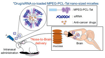
CPPs are a distinct class of short peptide sequences, typically composed of fewer than 30 amino acids and rich in cationic residues such as arginine and/or lysine [49]. Their cationic nature enables efficient intracellular accumulation [49]. A defining feature of CPPs is their ability to traverse cellular membranes without causing significant membrane damage, allowing them to deliver hydrophilic macromolecules into cells and subcellular compartments both in vitro and in vivo [50,51]. Owing to these properties, CPPs have been widely employed to deliver diverse therapeutic cargos including LNPs, plasmid DNA, oligonucleotides, siRNA, proteins, and peptides [52]. As illustrated in Fig. 2, one notable example involves the use of Tat, a CPP derived from the human immunodeficiency virus (HIV) transactivator protein, to construct methoxy polyethylene glycol-polycaprolactone (MPEG-PCL)-Tat micelles for siRNA and drug delivery, significantly extending survival in a rat model of malignant glioma [53]. Despite encouraging findings, the lack of systematic, quantitative comparisons among different CPPs poses a challenge. This gap limits our ability to accurately assess subtle differences in their BBB penetration capabilities. To address this issue, Stalmans et al. conducted a comparative study evaluating the in vivo behavior of five structurally distinct CPPs: pVEC, SynB3, Tat47–57, transportan 10 (TP10), and TP10–2. Using multiple-time regression analysis, the study revealed significant differences in BBB permeability among the peptides. Notably, Tat47–57, SynB3, and pVEC exhibited high unidirectional influx rates of 4.73, 5.63, and 6.02 µL g−1 min−1, respectively. In contrast, TP10 and TP10–2 demonstrated considerably lower permeability. Importantly, the study also found that BBB influx rates did not consistently correlate with overall cellular uptake, suggesting that efficient BBB penetration is not necessarily indicative of general cell-penetrating efficiency [54].
CPPs mainly utilize their cationic properties to efficiently accumulate within cells. They have the potential to directly interact with the siRNA anionic phosphate backbone when applied for siRNA transportation [55]. For example, the work of Hu and colleagues, as illustrated in Fig. S2 (Supporting information), developed a lipoplex with a core-shell structure which was specifically designed for intranasal delivery of siRNA to treat glioma via gene therapy [56]. And then a CPP was chosen to modify lipoplex, enhancing its permeation across various absorptive barriers [56]. However, the intrinsic positive charges of the CPP were partially neutralized upon complexation with negatively charged siRNA, resulting in a reduction of free cationic residues. This, in turn, impaired the internalization efficiency of the CPP and ultimately limited the effectiveness of siRNA delivery. Mo et al. designed a novel polyplex architecture specially designed to address the CPP neutralization issue [49]. This system was composed of siRNA and a specially designed CPP delivery unit. A 21-mer oligolysine (K21) was modified chemically to introduce conjugation sites for CPP, forming K21-PDP. A dual CPP strategy was employed in which two distinct CPP moieties served complementary functions: one facilitated siRNA transport, while the other mitigated the effects of charge neutralization, thus preserving delivery efficiency [49].
Peptide shuttles, owing to their unique properties, can exploit BBB transport mechanisms to facilitate the delivery of compounds that otherwise cannot cross the barrier [57]. These peptides exhibit advantages such as ease of synthesis, high specificity and potency, and cost-effective production. Notably, compared to conventional chemotherapeutics, peptides demonstrate minimal accumulation in tissues and organs, reducing systemic toxicity and adverse effects [58]. Consequently, the application of peptide-based strategies, particularly for glioblastoma (GBM), has expanded significantly in recent years. Currently, the realm of glioma therapy still stands as a difficult frontier, plagued by two main issues: increasing therapeutic effectiveness and minimizing the severe side effects cause by anticancer drugs. Using an AS1411 aptamer, a ligand specific to cancer cells, and a phage-displayed TGN peptide targeted at the BBB, Gao and colleagues have developed a novel technique [59]. By ingeniously combining these substances with NPs, researchers developed the AsTNP, a cascade delivery system specifically designed to treat brain gliomas. Extensive experimental investigation has highlighted AsTNP’s superior tumor localization abilities and advantageous tumor-to-normal brain concentration ratio. To further improve the anti-glioma efficacy, Yu et al. proposed D-T7 peptide-modified NPs capable of crossing the BBB for glioma targeting. Experimental results demonstrated that D-T7-modified NPs exhibited superior BBB penetration and tumor targeting. Cediranib (anti-angiogenic) and paclitaxel (chemotherapeutic) were co-loaded into PEGylated bilirubin NPs (BRNPs), forming the dual-delivery system CD & PTX@TBRBPs. Subsequent experiments confirmed both the safety and therapeutic efficacy of the delivery system [12].
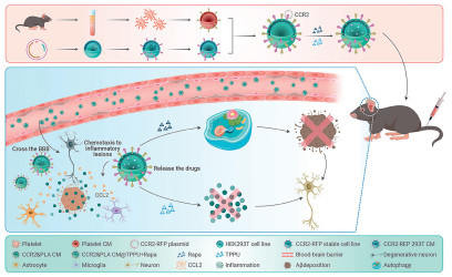
The intricate mechanism of RMT paves the way for a wide range of endogenous macromolecules to traverse the barrier [60]. Specific ligands selectively bind to receptors expressed on the surface of brain endothelial cells. This interaction initiates a cascade that triggers the formation of vesicles via endocytosis. These vesicles then cross BBB and release their cargoes into CNS via exocytosis. This process exemplifies the precision and complexity of endogenous transport mechanisms [61]. Moreover, it provides a promising avenue for therapeutic intervention in CNS diseases. Nanotechnology enables targeted delivery to CNS by functionalizing nanomaterials with receptor-specific ligands. For example, Fig. 3 illustrates a hybrid nanoplatform that integrates platelet membranes with CCR2-overexpressing membranes. This hybrid membrane is used to deliver two small-molecule drugs, rapamycin and TPPU, targeting pathological regions in an AD model [62]. Here, three typical instances are provided below.
Low-density lipoprotein receptor (LDLR), a prominent member of its own family, is highly expressed at the BBB, making it an outstanding candidate for BBB-transversing nanomaterials via its corresponding ligands. Notably, LDLR is also overexpressed in various tumor types, underscoring its potential as a therapeutic target in glioma [63,64].
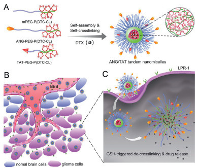
Angiopep-2, a peptide derived from the Kunitz domain (sequence: TFFYGGSRGKRNNFKTEEY), specifically targets the LDLR-related protein-1 (LRP1), a receptor closely related to LDLR. Due to its high affinity for LRP1, angiopep-2 has attracted considerable attention for BBB-targeted drug delivery [65]. Angiopep-2 exhibits superior transcytosis efficiency and parenchymal accumulation compared to other ligands such as lactoferrin (Lf) and transferrin (Tf), making it a leading candidate for BBB-targeted delivery [66]. Recent studies have demonstrated the efficacy of angiopep-2 in facilitating nanomaterial-mediated BBB transport [67,68]. One example involves 2-methoxyestradiol (2-ME2), a potent hypoxia inducible factor-1 (HIF-1) inhibitor and microtubule disruptor used to treat cerebral ischemia-reperfusion injury [69]. However, its poor aqueous solubility has limited its clinical application [70]. To overcome this limitation, Hu et al. employed an emulsion-solvent evaporation method to formulate PEGylated 2-ME2 and encapsulate it into angiopep-2-functionalized PEG micelles (ANG-PEG-2ME2/2ME2) [71]. This formulation improved both solubility and brain-targeted delivery. In oxygen-glucose deprivation/reoxygenation models using PC12 cells, ANG-PEG-2ME2/2ME2 significantly reduced cellular injury. This finding highlighted the promise of this micellar formulation as a therapeutic approach for cerebral ischemia-reperfusion injury [71]. Fig. 4 illustrates the construction of angiopep-2-modified micelles, which demonstrated enhanced selectivity and efficacy in glioma-targeted chemotherapy [72]. Zong et al. developed lipid-poly(hypoxic radiosensitized polyprodrug) NPs decorated with angiopep-2 for temozolomide (TMZ) delivery, termed A2-p(MIs)25/TMZ, aiming to reduce the increased toxicity arising from the combined therapy of TMZ and radiotherapy. This system exhibited excellent drug encapsulation and a synergistic antitumor effect in glioma models [73]. In pursuit of effective brain glioma treatment, Ren et al. also explored the field of dual-targeting drug delivery systems. Their system was based on PEGylated oxidized multi-walled carbon nanotubes modified with angiopep-2. Histopathological analysis confirmed the system’s high biocompatibility, low toxicity, and favorable safety profile [74]. Furthermore, Yang et al. designed a dual-peptide-modified liposomal system. The system combined ligands for neuropilin-1 receptor and LRP1 to enhance BBB permeability and tumor targeting [75]. Kim et al. further advanced this strategy by developing Dual-LP-TMZ, an immunoliposome co-targeting LRP1 and CD133, and encapsulating TMZ. This system used angiopep-2 to mediate BBB transcytosis and an anti-CD133 monoclonal antibody for targeting glioma stem cells, offering a promising approach for GBM therapy [76].
Although the BBB presents a major obstacle to brain drug delivery, its complex network of receptors and transport proteins facilitates the entry of essential nutrients into the brain and offers promising opportunities for therapeutic targeting. A notable example is the Tf receptor, which has been widely explored for receptor-mediated drug delivery across the BBB.
Tf is a 78 kDa monomeric glycoprotein and a member of a closely related family of iron-binding glycoproteins, which also includes Lf, ovotransferrin, and melanotransferrin, each with distinct biological roles [77]. Among plasma proteins, Tf is one of the most abundant (2–4 µg/mL), and it plays a critical role in iron metabolism by binding nearly all circulating iron under normal physiological conditions [78]. While Tf can be synthesized locally in tissues such as the testes, oligodendrocytes, and choroid plexus epithelial cells of the CNS, the liver remains its primary site of production [79-81]. The primary function of Tf is to act as an iron transporter, facilitating the transfer of iron between cells. Due to its hydrophilic nature and strong binding affinity for the transferrin receptor (TfR), Tf-bound iron can be efficiently taken up by cells with high TfR expression, enabling transport across the BBB and into the brain parenchyma [82-84]. Notably, iron binding induces a conformational change in the Tf molecule, which is essential for its specific recognition by TfR and subsequent receptor-mediated endocytosis [85]. Johnsen et al. demonstrated that TfR-targeted AuNPs exhibit remarkable accumulation within brain capillaries and effectively cross the BBB to reach the brain parenchyma. This conclusion was supported by both in vitro BBB models and in vivo quantification of gold accumulation [86]. Their study highlights the substantial enhancement in uptake efficiency enabled by the specific affinity and valency of antibody-conjugated AuNPs. Cabezón et al. examined the behavior of AuNPs coated with the 8D3 anti-TfR antibody in the mouse BBB [87]. Their results revealed a clathrin-dependent, TfR-mediated endocytosis process, in which 8D3-AuNPs were encapsulated into intracellular vesicles. These vesicles then followed two distinct trafficking pathways, indicating complex intracellular routing dynamics following TfR engagement. Another noteworthy BBB-targeting approach involves small peptides such as T7, which are favored for their low cost, stability, and ease of synthesis. The D-enantiomeric form of T7 (D-T7), a heptapeptide with high affinity for TfR, has been shown to facilitate efficient brain-targeted drug delivery. As illustrated in Fig. 5, the functionalization of nanoparticles with D-T7 peptides underscores their potential utility in designing targeted nanocarriers for CNS therapeutics [12].

Lf, an 80 kDa cationic glycoprotein with excellent iron-binding capabilities, is a notable member of the Tf superfamily. It plays a crucial role in facilitating the efficient transportation of iron within blood serum [88]. Lf is distinguished by possessing a superior iron-binding affinity, greater resistance to proteolysis, and its capability of retaining iron over a wide pH range [89,90]. Various physiological functions of Lf have been indicated, such as anti-inflammatory, antioxidant, anticarcinogenic, and immunomodulatory activities [91,92]. The receptors of Lf are essential to the coordination of its diverse biological functions. Numerous evidence from reverse transcription-polymerase chain reaction (RT-PCR) analyses and immunohistochemistry analyses has confirmed the presence of Lf receptors and highlighted their significance in neurological processes [93]. Given its classification within the Tf family, RMT serves as the primary mechanism for its uptake into CNS, as supported by both in vitro and in vivo studies [94,95]. The application of Lf as a BBB targeting ligand has been studied extensively. Using high-precision confocal microscopy, Huang et al. investigated the intricate binding sites of Lf within brain endothelial capillary cells (BCECs). Their findings revealed the striking abundance of Lf receptors on the BCEC surface, which is an essential feature for RMT-mediated cellular entry [96]. Based on this finding, the same group conducted a follow-up study demonstrating that Lf-modified NPs exhibit efficient brain-targeting capabilities. Both in vitro and in vivo data consistently showed that these NPs successfully cross the BBB, highlighting their promise as a platform for exogenous gene delivery, particularly for genes encoding secretory proteins [96]. Further evidence comes from the work of Qiao et al., who developed a brain-delivery probe making use of the capacity of Lf, covalently attached to PEG-coated Fe3O4 NPs, to coordinate RMT across the BBB [97]. To address the formidable challenges faced by glioma patient after chemotherapy, Pang et al. developed a revolutionary antitumor drug delivery system, Lf-conjugated biodegradable polymersomes that contain doxorubicin and tetrandrine (Lf-PO-Dox/Tet). This innovative design demonstrates its potential as a novel therapeutic vehicle for glioma-targeted therapy [98].
Table 1 [12,71-73,76,98] is listed to help readers have a clear understanding.
 DownLoad:
CSV
DownLoad:
CSV
| Receptor | Material | Nanoparticle/nanoplatform | Cargo/core particle | Application | Ref. |
| LDLR | Angiopep-2 | ANG-PEG-2ME2/2ME2 | 2-ME2 | Stroke | [71] |
| LDLR | Angiopep-2 & TAT | ANG/TAT-Ms micelles | Micelles | Glioma | [72] |
| LDLR | Angiopep-2 | A2-p(MIs)25/TMZ | TMZ | Glioma | [73] |
| LDLR | Angiopep-2 & tLyP-1 | Dual-LP-TMZ | TMZ | Glioma | [76] |
| Tf receptor | D-T7 peptide | Dual-loaded PEGylated nanocarriers | Cediranib & paclitaxel | Glioma | [12] |
| Lf receptor | Lf | Lf-PO-Dox/Tet | Doxorubicin & tetrandrine | Glioma | [98] |
Cell membrane coating nanotechnology involves the integration of specific cell membranes onto synthetic substrates, encompassing AuNPs, polymeric NPs and liposomes. Due to their distinct characteristics, membranes generated from various parent cells such as erythrocytes, neutrophils, and macrophages have recently garnered increasing attention. These cell membranes can serve as the outer shell of drug delivery vehicles, playing a critical role in facilitating targeted delivery to the brain. Chimeric antigen receptor T-cell immunotherapy (CAR-T) cell membrane coating is an emerging delivery strategy in the field of cancer immunotherapy [99,100]. Researchers have explored coating NPs with CAR-T cell membranes to enhance the targeting and therapeutic effects of CAR-T therapy. For instance, a study exemplifies the innovative use of CAR-T therapy in a highly targeted manner, demonstrating the potential of cell-based engineering to overcome traditional barriers in CNS treatment. It involves the use of synNotch receptors to direct therapeutic agents to specific tissues, thereby improving the precision and effectiveness of interventions [101]. The CAR-T cell membrane coating strategy leverages the specific targeting ability of CAR-T cells to deliver therapeutic agents directly to tumor sites, potentially reducing side effects and improving therapeutic outcomes. Moreover, it offers a promising solution to limitations of traditional CAR-T therapy, such as poor tumor infiltration and limited persistence in solid tumors. However, this field remains in its early stages, and further research is required to optimize the coating process, enhance the stability and biocompatibility of membrane-coated NPs, and explore their clinical applications in various cancers types.
Due to their unique advantages, including the ability to greatly increase the circulatory lifespan of therapeutic drugs and avoid immune system destruction, erythrocytes have been employed in drug delivery systems. In particular, red blood cell (RBC) membrane-coated NPs have been extensively explored to address issues such as enzyme instability, poor biocompatibility or toxicity of various NPs [102]. However, despite these explorations, a major limitation persists: RBC—NPs inherently lack targeting specificity, making it extremely difficult for them to cross the BBB. This limits their utility in targeted brain delivery for both research and therapeutic applications. To overcome this challenge, Gu et al. conducted a pioneering study to develop a brain-targeted drug delivery system utilizing the TGN peptide as a BBB-penetrating ligand [31]. Their approach involved the preparation and characterization of curcumin-encapsulated NPs coated with erythrocyte membranes to enhance bioactivity. To improve delivery specificity, the TGN peptide was incorporated into the RBC—NPs using an innovative lipid-insertion technique. This design demonstrated superior BBB penetration and remarkable brain targeting capabilities, as supported by in vitro and in vivo evidence. Additionally, behavioral assessments showed the encouraging potential of TGN-RBC—NPs-Cur in reversing cognitive impairments in AD animal models, emphasizing the therapeutic potential of this strategy. Similar studies have also investigated the application of RBC membranes in AD treatments, particularly focusing on the elimination of Aβ. As illustrated in Fig. 6, Ma et al. developed CuxO@EM-K by incubating the erythrocyte membrane with Aβ-targeting molecules to produce the EM-K, which was then fused with CuxO. CuxO@EM-K complex effectively absorbed Aβ from the bloodstream, after which the liver facilitated its clearance, thereby reducing Aβ burden in the brain [103].

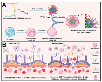
Neutrophils, the initial and predominant subset of leukocytes responsible for defending against pathogens, possess the unique ability to traverse the BBB, enabling them to swiftly migrate from the circulatory system to sites of brain injury [104]. Upon recruitment, endothelial cells upregulate membrane adhesion proteins such as intercellular adhesion molecule-1, while neutrophils concurrently overexpress membrane proteins including integrin β2, macrophage-1 antigen, and lymphocyte function-associated antigen 1. These coordinated alterations facilitate the transmigration of neutrophils [105,106]. Consequently, neutrophils represent promising donors for cell membrane coating in the treatment of inflammatory brain disorders [107-109]. For example, leonurine, an alkaloid from Herba Leonuri, has been extensively studied for ischemic stroke treatment [110,111]. However, its limited BBB penetration, poor lesion-targeting capability and restricted interaction with the complex pathological microenvironment have hindered its therapeutic efficacy. To address these challenges, Tang et al. developed a neutrophil-inspired nanoplatform for non-invasive administration of leonurine [108]. This system utilizes membranes derived from neutrophil-mimetic human promyelocytic leukemia cells to construct proteolipid structures termed Leo@NM-Lipo. These biomimetic carriers leverage the abundance of membrane proteins to facilitate targeted delivery to inflamed brain regions. Importantly, neutrophil membrane camouflage exhibits a high affinity to inflammatory cytokines, enabling precise lesion targeting while simultaneously enhancing leonurine’s anti-inflammatory effects through synergistic action. Similarly, Zhao et al. reported the development of a neutrophil membrane-coated NP system for reactive oxygen species (ROS)-responsive delivery of FTY720 to ischemic brain tissue [109]. As illustrated in Fig. 7, ROS-responsive FTY720 monomers were polymerized into amphiphilic block copolymers, which self-assembled into polyprodrug NPs loaded with FTY720 in an aqueous solution. Subsequently, these NPs are coated with neutrophil membrane to impart inflammation-oriented chemotaxis. The resulting system demonstrated enhanced delivery efficiency and selective drug release in inflamed brain tissue, thereby improving therapeutic outcomes while reducing drug retention in the heart and circulation, key factors in minimizing the adverse effects associated with high systemic doses of FTY720 in ischemic stroke treatment.
Macrophages play an important role in the complex physiological microenvironment with their polarization phenotypes significantly influencing tumor progression and metastasis. In the context of neuroinflammation, these adaptable cells can adopt an anti-inflammatory phenotype, functioning as protectors that eliminate pathogens and cellular debris while releasing anti-inflammatory mediators and initiating the activation cascade of complementary immune sentinels [112,113]. Consequently, macrophage-based drug delivery systems leverage their inherent capabilities-including prolonged circulatory lifespan, abundant surface receptors, and phenotype-specific targeting abilities-offering a promising therapeutic strategy [114]. In an innovative study, Han et al. developed macrophage membrane-coated solid lipid nanoparticles (MASLNs) to evade recognition by the reticuloendothelial system and prolong systemic circulation [115]. To address challenges in AD therapy, they engineered a biomimetic delivery system, termed RVG/TPP-MASLNs, designed to deliver Genistein specifically to neuronal mitochondria. The macrophage membranes exhibited the macrophage-like immunological characteristics, such as the high expression of F4/80 and CD11b, thus facilitating targeted accumulation of the biomimetic nanoplatform in the AD-affected brain. Recently, hybrid membrane-based systems have gained increasing attention. Yin et al. introduced a nanoplatform combining membranes from both neutrophils and macrophages, termed NMm-PLGA/RAPA, for efficient rapamycin delivery across the BBB [116]. This hybrid system integrates neutrophil-mediated inflammatory chemotaxis with macrophage-driven stimulus responsiveness, enabling spontaneous BBB penetration. Rapamycin was encapsulated in PLGA nanoparticles and then coated with the dual membranes to form NMm-PLGA/RAPA. As illustrated in Fig. S3 (Supporting information), alternative membrane sources have also been explored, including platelet membranes and tumor cell membranes [117]. Inspired by inherent ability of platelets to target injured vasculatures and the rapid BBB penetrability of 4T1 tumor cells, Tang et al. developed a biomimetic nanoplatform (PP@PCL) for ischemic stroke therapy. This system was constructed by fusing membranes from 4T1 tumor cells and platelets and applying them to liposomes co-loaded with paeonol and polymetformin. By efficiently targeting the ischemic lesion, preventing neuroinflammation, eliminating excess ROS, reprogramming microglia phenotypes, and promoting angiogenesis, PP@PCL could significantly reduce ischemia-reperfusion injury [117]. Promising results in both injured PC12 neuronal cells and rat models of ischemic stroke underscore the therapeutic potential of membrane-coating strategies for cerebral ischemic injury.
Table 2 [31,103,108,109,115-117] summarizes various membrane coating applications.
DownLoad:
CSV
| Material | Nanoplatform | Cargo | Application | Ref. |
| Erythrocyte membrane | TGN-RBC—NPs-Cur | Curcumin | AD | [31] |
| Erythrocyte membrane | CuxO@EM-K | CuxO | AD | [103] |
| Neutrophil membrane | Leo@NM-Lipo | Leonurine (Leo) | Stroke | [108] |
| Neutrophil membrane | (Untitled) | FTY720 | Stroke | [109] |
| Macrophage membrane | RVG/TPP-MASLNs | Genistein (GS) | AD | [115] |
| Neutrophil membrane & macrophage membrane | NMm-PLGA/RAPA | Rapamycin (RAPA) | Glioma | [116] |
| 4T1 tumor cell membrane & platelet membrane | PP@PCL | Paeonol & polymetformin (Glucophage) | Stroke | [117] |
In contrast to the intrusive methods of directly injecting therapeutic compounds into brain regions via intraventricular or intraparenchymal routes, intranasal delivery is a non-invasive method that facilitates the rapid transportation of therapeutic agents from the nasal cavity to the brain [118]. As illustrated in Fig. S4 (Supporting information), such process can occur via either intracellular or extracellular pathways [118]. The intracellular route efficiently bypasses the BBB by leveraging the trigeminal and olfactory nerve pathways [119]. Owing to the highly vascularized nasal epithelium, intranasal administration, known for its non-invasiveness and safety profile, improves absorption efficiency and may achieve higher drug concentrations in the brain [120]. Nonetheless, certain limitations exist in the nasal-to-brain delivery route, including reduced drug permeability and challenges associated with nasal mucosa and mucociliary clearance mechanisms [121]. NPs offer a promising strategy to address these drawbacks by facilitating greater cargo loading, precise targeting, and enhanced retention of therapeutic agents on the nasal mucosa [122]. Various nanocarrier systems, such as dendrimers, micelles, and various polymeric NPs, have been employed to improve nose-to-brain drug delivery efficiency [123,124]. Wang et al. developed a thermosensitive gel-based intranasal delivery system comprising rotigotine-loaded polymer micelles. This formulation was designed to increase drug solubility, prolong residence time, and increase the accumulation of rotigotine within brain tissue [125]. These micelles enabled sustained drug release and, according to permeability studies, facilitated direct brain delivery of rotigotine via the nasal route, resulting in improved cerebral distribution. Furthermore, this system exhibited no notable adverse effects on the cilia or nasal mucosa of rats, underscoring its potential for clinical application. Jia et al. established circSCMH1@LNP1 to deliver the therapeutic circular RNA circSCMH1 (Fig. 8) [25]. Intranasal administration of circSCMH1@LNP1 significantly increased its distribution in the peri‑infarct region and reduced off-target accumulation compared to intravenous delivery. Therapeutic results demonstrated that circSCMH1@LNP1 promoted synaptic plasticity, vascular repair, neuroinflammation relief, and myelin sheath formation, leading to enhanced sensorimotor and cognitive function recovery in post-stroke mice. These findings highlight the potential of LNP-based intranasal delivery system for brain injury management.

Among the diverse strategies employed for drug delivery to the CNS, the temporary modulation of the BBB remains a central area of research [126,127]. One promising approach involves intra-arterial injection after osmotically induced BBB disruption. This allows for access to extensive brain regions in a single treatment, outperforming the direct and localized administration approaches [128]. This procedure utilizes osmotic agents such as mannitol, urea and glycerol to transiently increase osmotic pressure, thereby causing temporary BBB permeability. This technique is especially valuable in the field of brain tumor treatment [129,130]. Nevertheless, considering the non-selective nature of BBB opening, this increased permeability could lead to the enhanced entry of unintended macromolecules, which may result in irreversible CNS damage, or even adverse effects such as seizures and chronic neuropathological changes [131,132]. To mitigate these risks, an innovative nanoplatform, TPP@(CeO2+ROF), has been developed, as illustrated in Fig. S5 (Supporting information). This system incorporates ceria nano-enzymes with mitochondria-targeting and potent antioxidant capabilities. This design, as demonstrated by the authors, successfully mitigates mitochondrial damage, alleviates oxidative stress and apoptosis, minimizes BBB trauma, and has an excellent biosafety profile. This customized approach offers a promising way to avoid the problems associated with temporary disruption of the BBB, paving the way for safer and more effective CNS drug delivery strategies [133].
In recent years, ultrasound-based BBB opening techniques has been widely explored [134,135]. In particular, focused ultrasound (FUS), which has emerged as an innovative tool for enhancing BBB permeability to facilitate targeted brain therapy delivery, is gaining more and more attention in treating brain tumors and neurodegenerative diseases [136,137]. Numerous studies have demonstrated the safety and practicality of non-invasive and low-density FUS for transient BBB disruption [138-140]. Notably, Rezai et al. provided a groundbreaking demonstration of safely, non-invasively, and precisely induced transient and repeatable BBB permeabilization of FUS within the hippocampus/entorhinal cortex of humans, thus paving the way for innovative therapeutic delivery strategies in AD and beyond [138]. In a pioneering phase Ⅰ, single-arm, open-label clinical investigation, Mainprize et al. confirmed the safety and practicality of magnetic resonance-guided focused ultrasound (MRgFUS) facilitating BBB opening in glioma patients receiving systemic chemotherapy. Their analysis of chemotherapy concentrations in both targeted (sonicated) and non-targeted (non-sonicated) peritumor tissues underscored the transformative potential of MRgFUS to enhance treatment efficacy [139]. Based on these, Park et al. demonstrated the accuracy, reliability, and safety of MRgFUS-mediated BBB disruption, offering a translational pathway for therapies previously hindered by limited BBB permeability [140]. While further follow-up is necessary to fully assess long-term effects, these findings open new avenues for previously inaccessible CNS treatments.
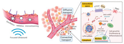
Among its emerging applications, MRgFUS has shown considerable promise in advancing liquid biopsy technologies for early cancer detection, disease monitoring, and recurrence prevention. BBB presents a significant obstacle to the release and detection of brain-derived biomarkers using conventional techniques [141]. Having recognized the unique ability of transcranial MRgFUS to safely and temporarily disrupt the BBB, Meng et al. hypothesized that this technology could enhance the detectability of circulating brain-derived biomarkers. In a first-in-human proof-of-concept study involving GBM patients, MRgFUS significantly increased the presence of neuron-derived extracellular vesicles, brain-specific proteins, and cell-free DNA in circulation [142]. These findings not only confirmed the safety and feasibility of this technology, but also demonstrated its ability to enhance the signal of circulating biomarkers generated from the brain, signaling a breakthrough for liquid biopsy applications in neuro-oncology. To address the gap between preclinical success and limited drug delivery in human subjects, Meng et al. also conducted a pivotal study demonstrating enhanced delivery of trastuzumab in HER2-positive breast cancer patients with brain metastases via MRgFUS. The observed elevation in the standardized uptake value ratio in MRgFUS-treated regions, alongside the absence of severe adverse effects, marked a milestone in non-invasive and spatially precise monoclonal antibody delivery across the BBB [143]. In addition to these landmark studies, other investigations have similarly highlighted the efficacy of MRgFUS in improving therapeutic delivery to the brain. As illustrated in Fig. 9, the synergistic combination of FUS and LPHs@siRNA facilitates enhanced siRNA delivery to brain tumors. This combination improves delivery efficiency and significantly promotes tumor cell apoptosis in experimental models, indicating a substantial improvement in therapeutic outcomes [144]. Collectively, these comprehensive studies underscore the transformative potential of MRgFUS in treating brain tumors and neurodegenerative diseases, offering renewed hope for patients afflicted by these conditions.
Light disruption has emerged as a promising candidate approach for the temporary opening of BBB because of its unique characteristics including minimal scattering propensity, the ability to induce nonlinear absorption, and spatiotemporal accuracy [145]. Kiesslin et al. were the first to present this novel technique, envisioning the use of a neodymium-doped yttrium aluminum garnet laser pulse for precise focal disruptions of the BBB [146]. This technique works by slowly and steadily delivering laser energy at low levels over extended periods of time, ingeniously utilizing near-infrared light’s deep tissue penetration capabilities, with its wavelengths positioned between 700 nanometers and 1600 nanometers. The use of near-infrared light for BBB modulation has garnered significant attention, positioning it at the forefront of research aimed at delicately tuning BBB permeability [147-149]. Li and colleagues developed AuNPs with an unparallelled affinity for tight junction, demonstrating that intravenous injection of these engineered AuNPs, followed by transcranial stimulation with picosecond laser pulses, effectively enhances BBB permeability [150]. Laser intensity is carefully adjusted to determine the degree of BBB permeability augmentation, guaranteeing a completely reversible procedure that promotes improved paracellular diffusion.
Despite the crucial function of the BBB in preserving the normal physiological operations of the CNS, the delivery of certain conventional medications for CNS disorders remains significantly hindered. Drawing upon a concise overview of the basic structure of BBB and several major types of CNS diseases, different types of drug administration strategies across the BBB were discussed in this review. Undoubtedly, the utilization of nanomaterials for traversing the BBB presents a promising avenue for the future treatment of CNS diseases. Nevertheless, further in-depth fundamental investigations into nanomaterial-based strategies for traversing BBB and their mechanisms of drug release within the brain are imperative. On the premise of ensuring safety and feasibility, how to further improve the loading capacity of therapeutic drugs and the targeting of delivery to the brain tissues is still a challenge that should to be focused on in the future. To achieve this goal, several unresolved issues are expected be addressed. Biological barriers such as the formation of a “protein corona” can alter nanomaterial properties, hindering drug delivery. Immune system interactions may lead to clearance or inflammation. The tumor microenvironment and the heterogeneity of the enhanced permeability and retention (EPR) effect can impede nanomaterial penetration. Safety concerns include acute and chronic toxicity, as well as immunogenicity should also been considered. Manufacturing issues involve difficulties in achieving consistent quality and scalability. Regulatory hurdles and economic considerations, such as high development costs, further complicate clinical translation. Addressing these challenges requires a multidisciplinary approach to optimize nanomaterial design, production, and safety evaluation, ensuring effective and safe treatments for brain-related diseases.
The authors declare that they have no known competing financial interests or personal relationships that could have appeared to influence the work reported in this paper.
Guanlong Li: Writing – original draft. Zhuoyan Li: Writing – original draft, Conceptualization. Yan Sun: Software. Tiange Bu: Software. Shaochuan Chen: Software. Leixin Yang: Validation. Zhi Li: Methodology. Wenyue Mao: Validation. Yanpeng Jia: Writing – review & editing, Supervision.
This research was funded by the Fundamental Research Funds for the Central Universities (No. 2242022R42012).
Supplementary material associated with this article can be found, in the online version, at doi:
M. Srikanth, J.A. Kessler, Nat. Rev. Neurol. 8 (2012) 307–318. doi: 10.1038/nrneurol.2012.76
J. Xie, Z. Shen, Y. Anraku, K. Kataoka, X. Chen, Biomaterials 224 (2019) 119491. doi: 10.1016/j.biomaterials.2019.119491
D.J. Begley, Pharmacol. Ther. 104 (2004) 29–45. doi: 10.1016/j.pharmthera.2004.08.001
S. Wohlfart, S. Gelperina, J. Kreuter, J. Control. Release 161 (2012) 264–273. doi: 10.1016/j.jconrel.2011.08.017
H. Gao, Z. Pang, X. Jiang, Pharm. Res. 30 (2013) 2485–2498. doi: 10.1007/s11095-013-1122-4
E. Bender, Nature 561 (2018) S46–S47. doi: 10.1038/d41586-018-06707-4
M.G. Baratta, Nat. Nanotechnol. 13 (2018) 536. doi: 10.1038/s41565-018-0182-3
W.M. Pardridge, J. Neurochem. 70 (1998) 1781–1792. doi: 10.1046/j.1471-4159.1998.70051781.x
W. Chen, K. Shi, Y. Yu, et al., Chin. Chem. Lett. 35 (2024) 109159. doi: 10.1016/j.cclet.2023.109159
S. Cheng, M. Pan, D. Hu, et al., Chin. Chem. Lett. 34 (2023) 108276. doi: 10.1016/j.cclet.2023.108276
Z. Cao, X. Zuo, X. Liu, G. Xu, K.T. Yong, Adv. Colloid Interface Sci. 330 (2024) 103206. doi: 10.1016/j.cis.2024.103206
M. Yu, D. Su, Y. Yang, et al., ACS Appl. Mater. Interfaces 11 (2019) 176–186. doi: 10.1021/acsami.8b16219
Z. Wang, Z. Feng, F. Du, et al., Chin. Chem. Lett. 34 (2023) 108137. doi: 10.1016/j.cclet.2023.108137
A. Ivask, E.H. Pilkington, T. Blin, et al., Biomater. Sci. 6 (2018) 314–323. doi: 10.1039/c7bm01012e
H. Liu, Y. Yu, T. Xue, et al., Chin. Chem. Lett. 35 (2024) 108574. doi: 10.1016/j.cclet.2023.108574
Y. Liu, T. Huang, Z. Qian, W. Chen, Chin. Chem. Lett. 34 (2023) 108103. doi: 10.1016/j.cclet.2022.108103
Q. Feng, Y. Shen, Y. Fu, et al., Theranostics 7 (2017) 1875–1889. doi: 10.7150/thno.18985
L. Ribovski, N.M. Hamelmann, J.M.J. Paulusse, Pharmaceutics 13 (2021) 2045. doi: 10.3390/pharmaceutics13122045
R. Yang, B. Xu, B. Yang, et al., RNA Biol. 18 (2021) 108–116. doi: 10.1080/15476286.2021.1950465
R. Yang, W. Liu, L. Miao, et al., Oncotarget 7 (2016) 63839–63855. doi: 10.18632/oncotarget.11696
E. Candelario-Jalil, R.M. Dijkhuizen, T. Magnus, Stroke 53 (2022) 1473–1486. doi: 10.1161/strokeaha.122.036946
L. Tang, R. Zhang, Y. Wang, et al., J. Control. Release 369 (2024) 642–657. doi: 10.3390/s24020642
F. Mo, A. Pellerino, R. Soffietti, R. Rudà, Int. J. Mol. Sci. 22 (2021) 12654. doi: 10.3390/ijms222312654
S. Quader, K. Kataoka, H. Cabral, Adv. Drug Deliv. Rev. 182 (2022) 114115. doi: 10.1016/j.addr.2022.114115
Y. Jia, L. Xu, S. Leng, et al., Adv. Mater. 37 (2025) 2500598. doi: 10.1002/adma.202500598
C. Qin, S. Yang, Y. -H. Chu, et al., Signal Transduct. Target. Ther. 7 (2022) 215. doi: 10.1038/s41392-022-01064-1
C. Xie, J. Liao, N. Zhang, et al., Chin. Chem. Lett. 35 (2024) 109149. doi: 10.1016/j.cclet.2023.109149
S. Sasannia, R. Leigh, P.B. Bastani, et al., Neurotherapeutics 22 (2025) e00516. doi: 10.1016/j.neurot.2024.e00516
Y. Wang, W. Jia, J. Zhu, R. Xu, Y. Lin, Chin. Chem. Lett. 34 (2023) 107746. doi: 10.1016/j.cclet.2022.107746
J. Han, X. Zhang, L. Kang, J. Guan, J. Neuroinflamm. 22 (2025) 120. doi: 10.20855/ijav.2025.30.2e115
J. Gu, C. Yan, S. Yin, et al., J. Control. Release 366 (2024) 448–459. doi: 10.1016/j.jconrel.2023.12.030
H. Zeng, Y. Qi, Z. Zhang, et al., Chin. Chem. Lett. 32 (2021) 1857–1868. doi: 10.1016/j.cclet.2021.01.014
N. Vijiaratnam, T. Simuni, O. Bandmann, H.R. Morris, T. Foltynie, Lancet Neurol. 20 (2021) 559–572. doi: 10.1016/S1474-4422(21)00061-2
E.F. van Vliet, M.J. Knol, R.M. Schiffelers, M. Caiazzo, M. Fens, J. Control. Release 360 (2023) 212–224. doi: 10.1016/j.jconrel.2023.06.026
L. Cui, S. Li, S. Wang, et al., Signal Transduct. Target. Ther. 9 (2024) 30. doi: 10.1038/s41392-024-01738-y
K.A. Dudek, L. Dion-Albert, M. Lebel, et al., Proc. Natl. Acad. Sci. U. S. A. 117 (2020) 3326–3336. doi: 10.1073/pnas.1914655117
C. Greene, N. Hanley, C.R. Reschke, et al., Nat. Commun. 13 (2022) 2003. doi: 10.1038/s41467-022-29657-y
I. Hamad, A.A. Harb, Y. Bustanji, Pharmaceutics 16 (2024) 400. doi: 10.3390/pharmaceutics16030400
Y. Zheng, L. Cui, H. Lu, et al., Int. J. Nanomedicine 19 (2024) 12343–12368. doi: 10.2147/ijn.s497480
M.A. Beach, U. Nayanathara, Y. Gao, et al., Chem. Rev. 124 (2024) 5505–5616. doi: 10.1021/acs.chemrev.3c00705
Y. Chen, Q. Zeng, B. Chu, et al., Chin. Chem. Lett. 34 (2023) 108133. doi: 10.1016/j.cclet.2023.108133
W. Jia, Y. Wu, Y. Xie, M. Yu, Y. Chen, Adv. Mater. 37 (2025) e2413603. doi: 10.1002/adma.202413603
Y. Yu, C. Zhang, X. Yang, L. Sun, F. Bian, Small Methods 9 (2025) e2401220. doi: 10.1002/smtd.202401220
W. Sun, X. Chai, Y. Zhang, et al., Chem. Rec. 24 (2024) e202400179. doi: 10.1002/tcr.202400179
T. Muthukumaran, J. Philip, Adv. Colloid Interface Sci. 334 (2024) 103314. doi: 10.1016/j.cis.2024.103314
M. Bai, X. Shao, C. Wang, et al., Mater. Horiz. 12 (2025) 673–693. doi: 10.1039/d4mh01256a
J. Wang, X. Fan, X. Han, et al., Adv. Mater. 36 (2024) e2312374. doi: 10.1002/adma.202312374
Y. Meng, J. Zhang, Y. Liu, et al., J. Adv. Res. 71 (2025) 551–570. doi: 10.1016/j.jare.2024.05.023
R.H. Mo, J.L. Zaro, W.C. Shen, Mol. Pharm. 9 (2012) 299–309. doi: 10.1021/mp200481g
F. Madani, S. Lindberg, U. Langel, S. Futaki, A. Graslund, Biophys. J. 2011 (2011) 414729.
S. Stalmans, E. Wynendaele, N. Bracke, et al., PLoS One 8 (2013) e71752. doi: 10.1371/journal.pone.0071752
M. Lindgren, U. Langel, Methods Mol. Biol. 683 (2011) 3–19. doi: 10.1007/978-1-60761-919-2_1
T. Kanazawa, K. Morisaki, S. Suzuki, Y. Takashima, Mol. Pharm. 11 (2014) 1471–1478. doi: 10.1021/mp400644e
S. Stalmans, N. Bracke, E. Wynendaele, et al., PLoS One 10 (2015) e0139652. doi: 10.1371/journal.pone.0139652
P. Lundberg, S. El-Andaloussi, T. Sutlu, H. Johansson, U. Langel, FASEB J. 21 (2007) 2664–2671. doi: 10.1096/fj.06-6502com
Y. Hu, K. Jiang, D. Wang, et al., Acta Biomater. 138 (2022) 478–490. doi: 10.1016/j.actbio.2021.10.042
M. Sanchez-Navarro, E. Giralt, Pharmaceutics 14 (2022) 1874. doi: 10.3390/pharmaceutics14091874
G. Guidotti, L. Brambilla, D. Rossi, Curr. Opin. Pharmacol. 47 (2019) 102–109. doi: 10.1016/j.coph.2019.02.007
H. Gao, J. Qian, S. Cao, et al., Biomaterials 33 (2012) 5115–5123. doi: 10.1016/j.biomaterials.2012.03.058
A.R. Jones, E.V. Shusta, Pharm. Res. 24 (2007) 1759–1771. doi: 10.1007/s11095-007-9379-0
V.I. Brown, M.I. Greene, DNA Cell Biol. 10 (1991) 399–409. doi: 10.1089/dna.1991.10.399
R.R. Lin, L.L. Jin, Y.Y. Xue, et al., Adv. Sci. 11 (2024) 2306675. doi: 10.1002/advs.202306675
M. Yamamoto, K. Ikeda, K. Ohshima, et al., Cancer Res. 57 (1997) 2799–2805.
L. Maletinska, E.A. Blakely, K.A. Bjornstad, et al., Cancer Res. 60 (2000) 2300–2303.
X. Tian, S. Nyberg, S.S. P., et al., Sci. Rep. 5 (2015) 11990. doi: 10.1038/srep11990
M. Demeule, J.C. Currie, Y. Bertrand, et al., J. Neurochem. 106 (2008) 1534–1544. doi: 10.1111/j.1471-4159.2008.05492.x
P. Figueiredo, V. Balasubramanian, M.A. Shahbazi, et al., Int. J. Pharm. 511 (2016) 794–803. doi: 10.1016/j.ijpharm.2016.07.066
F. Lu, Z. Pang, J. Zhao, et al., Int. J. Nanomedicine 12 (2017) 2117–2127. doi: 10.2147/IJN.S123422
H.I. Cho, M.J. Seo, S.M. Lee, Biochem. Pharmacol. 131 (2017) 40–51. doi: 10.1016/j.bcp.2017.02.008
B. Du, Y. Li, X. Li, et al., Int. J. Pharm. 384 (2010) 140–147. doi: 10.1016/j.ijpharm.2009.09.045
L. Hu, Y. Wang, Y. Zhang, et al., Colloids Surf. B 171 (2018) 638–646. doi: 10.1016/j.colsurfb.2018.08.009
Y. Zhu, Y. Jiang, F. Meng, et al., J. Control. Release 278 (2018) 1–8. doi: 10.1016/j.jconrel.2018.03.025
Z. Zong, L. Hua, Z. Wang, et al., Drug Deliv. 26 (2019) 34–44. doi: 10.1080/10717544.2018.1534897
J. Ren, S. Shen, D. Wang, et al., Biomaterials 33 (2012) 3324–3333. doi: 10.1016/j.biomaterials.2012.01.025
Z.Z. Yang, J.Q. Li, Z.Z. Wang, D.W. Dong, X.R. Qi, Biomaterials 35 (2014) 5226–5239. doi: 10.1016/j.biomaterials.2014.03.017
J.S. Kim, D.H. Shin, J.S. Kim, J. Control. Release 269 (2018) 245–257. doi: 10.1016/j.jconrel.2017.11.026
H.A. Huebers, C.A. Finch, Physiol. Rev. 67 (1987) 520–582. doi: 10.1152/physrev.1987.67.2.520
G.J. Anderson, C.D. Vulpe, Cell. Mol. Life Sci. 66 (2009) 3241–3261. doi: 10.1007/s00018-009-0051-1
T. Moos, E.H. Morgan, Cell. Mol. Neurobiol. 20 (2000) 77–95. doi: 10.1023/A:1006948027674
A.R. Aldred, P.W. Dickson, P.D. Marley, G. Schreiber, J. Biol. Chem. 262 (1987) 5293–5297. doi: 10.1016/S0021-9258(18)61187-1
P.W. Dickson, A.R. Aldred, P.D. Marley, et al., Biochem. Biophys. Res. Commun. 127 (1985) 890–895. doi: 10.1016/S0006-291X(85)80027-9
T. Moos, T. Rosengren Nielsen, T. Skjorringe, E.H. Morgan, J. Neurochem. 103 (2007) 1730–1740. doi: 10.1111/j.1471-4159.2007.04976.x
T. Skjorringe, A. Burkhart, K.B. Johnsen, T. Moos, Front. Mol. Neurosci. 8 (2015) 19.
S. Sato, S. Liu, A. Goto, et al., J. Control. Release 357 (2023) 379–393. doi: 10.1016/j.jconrel.2023.04.012
H. Sun, H. Li, P.J. Sadler, Chem. Rev. 99 (1999) 2817–2842. doi: 10.1021/cr980430w
K.B. Johnsen, M. Bak, P.J. Kempen, et al., Theranostics 8 (2018) 3416–3436. doi: 10.7150/thno.25228
I. Cabezon, G. Manich, R. Martin-Venegas, et al., Mol. Pharm. 12 (2015) 4137–4145. doi: 10.1021/acs.molpharmaceut.5b00597
S.A. Gonzalez-Chavez, S. Arevalo-Gallegos, Q. Rascon-Cruz, Int. J. Antimicrob. Agents 33 (2009) e301–e308.
R.M. Bennett, T. Kokocinski, Br. J. Haematol. 39 (1978) 509–521. doi: 10.1111/j.1365-2141.1978.tb03620.x
R.Q. Huang, W.L. Ke, Y.H. Qu, et al., J. Biomed. Sci. 14 (2007) 121–128. doi: 10.1007/s11373-006-9121-7
D. Legrand, A. Pierce, E. Elass, et al., Adv. Exp. Med. Biol. 606 (2008) 163–194. doi: 10.1007/978-0-387-74087-4_6
O.M. Conneely, J. Am. Coll. Nutr. 20 (2001) 389S–395S. doi: 10.1080/07315724.2001.10719173
B.A. Faucheux, N. Nillesse, P. Damier, et al., Proc. Natl. Acad. Sci. U. S. A. 92 (1995) 9603–9607. doi: 10.1073/pnas.92.21.9603
C. Fillebeen, L. Descamps, M.P. Dehouck, et al., J. Biol. Chem. 274 (1999) 7011–7017. doi: 10.1074/jbc.274.11.7011
B. Ji, J. Maeda, M. Higuchi, et al., Life Sci. 78 (2006) 851–855. doi: 10.1016/j.lfs.2005.05.085
R. Huang, W. Ke, L. Han, et al., Brain Res. Bull. 81 (2010) 600–604. doi: 10.1016/j.brainresbull.2009.12.008
R. Qiao, Q. Jia, S. Huwel, et al., ACS Nano 6 (2012) 3304–3310. doi: 10.1021/nn300240p
Z. Pang, L. Feng, R. Hua, et al., Mol. Pharm. 7 (2010) 1995–2005. doi: 10.1021/mp100277h
Q. Zeng, Z. Liu, T. Niu, et al., Chin. Chem. Lett. 34 (2023) 107747. doi: 10.1016/j.cclet.2022.107747
Y. Xie, X. Li, J. Wu, et al., Chin. Chem. Lett. 34 (2023) 108202. doi: 10.1016/j.cclet.2023.108202
Y. Wu, Z. Zhang, Y. Wei, Z. Qian, X. Wei, Chin. Chem. Lett. 34 (2023) 108098. doi: 10.1016/j.cclet.2022.108098
Z. Chai, X. Hu, X. Wei, et al., J. Control. Release 264 (2017) 102–111. doi: 10.1016/j.jconrel.2017.08.027
M. Ma, Z. Liu, N. Gao, et al., J. Am. Chem. Soc. 142 (2020) 21702–21711. doi: 10.1021/jacs.0c08395
D. Chu, X. Dong, X. Shi, C. Zhang, Z. Wang, Adv. Mater. 30 (2018) e1706245. doi: 10.1002/adma.201706245
G.C. Jickling, D. Liu, B.P. Ander, et al., J. Cerebr. Blood Flow Met. 35 (2015) 888–901. doi: 10.1038/jcbfm.2015.45
S. Liu, J. Xu, Y. Liu, et al., ACS Appl. Mater. Interfaces 14 (2022) 27743–27761. doi: 10.1021/acsami.2c09020
X. Dong, J. Gao, C.Y. Zhang, et al., ACS Nano 13 (2019) 1272–1283.
Z. Tang, S. Meng, Z. Song, et al., Mater. Today Bio 20 (2023) 100674. doi: 10.1016/j.mtbio.2023.100674
Y. Zhao, Q. Li, J. Niu, et al., Adv. Mater. 36 (2024) e2311803. doi: 10.1002/adma.202311803
Y.Z. Zhu, W. Wu, Q. Zhu, X. Liu, Pharmacol. Ther. 188 (2018) 26–35. doi: 10.1016/j.pharmthera.2018.01.006
Q.Y. Zhang, Z.J. Wang, D.M. Sun, et al., Oxid. Med. Cell. Longevity 2017 (2017) 7150376. doi: 10.1155/2017/7150376
Y. Zhao, M.J. Haney, N.L. Klyachko, et al., Nanomedicine 6 (2011) 25–42. doi: 10.2217/nnm.10.129
A.M. Brynskikh, Y. Zhao, R.L. Mosley, et al., Nanomedicine 5 (2010) 379–396. doi: 10.2217/nnm.10.7
Y. Zhang, K. Cai, C. Li, et al., Nano Lett. 18 (2018) 1908–1915. doi: 10.1021/acs.nanolett.7b05263
Y. Han, C. Gao, H. Wang, et al., Bioact. Mater. 6 (2021) 529–542.
Y. Yin, W. Tang, X. Ma, et al., Chem. Eng. J. 433 (2022) 133848. doi: 10.1016/j.cej.2021.133848
L. Tang, Y. Yin, H. Liu, et al., Adv. Mater. 36 (2024) e2312897. doi: 10.1002/adma.202312897
Q. Huang, Y. Chen, W. Zhang, et al., J. Control. Release 366 (2024) 519–534. doi: 10.1016/j.jconrel.2023.12.054
C.V. Pardeshi, V.S. Belgamwar, Expert Opin. Drug Deliv. 10 (2013) 957–972. doi: 10.1517/17425247.2013.790887
Y. Chen, C. Zhang, Y. Huang, et al., Adv. Drug Deliv. Rev. 207 (2024) 115196. doi: 10.1016/j.addr.2024.115196
S.H. Jeong, J.H. Jang, Y.B. Lee, J. Pharm. Investig. 53 (2023) 119–152. doi: 10.1007/s40005-022-00589-5
L. Biddlestone-Thorpe, N. Marchi, K. Guo, et al., Adv. Drug Deliv. Rev. 64 (2012) 605–613. doi: 10.1016/j.addr.2011.11.014
R. Awad, A. Avital, A. Sosnik, Acta Pharm. Sin. B 13 (2023) 1866–1886. doi: 10.1016/j.apsb.2022.07.003
S. Zha, K.L. Wong, A.H. All, Adv. Healthc. Mater. 11 (2022) e2102610. doi: 10.1002/adhm.202102610
F. Wang, Z. Yang, M. Liu, et al., Int. J. Pharm. 577 (2020) 119046. doi: 10.1016/j.ijpharm.2020.119046
C.T. Curley, N.D. Sheybani, T.N. Bullock, R.J. Price, Theranostics 7 (2017) 3608–3623. doi: 10.7150/thno.21225
.M. Kemper, W. Boogerd, I. Thuis, J.H. Beijnen, O. van Tellingen, Cancer Treat. Rev. 30 (2004) 415–423. doi: 10.1016/j.ctrv.2004.04.001
C.P. Foley, D.G. Rubin, A. Santillan, et al., J. Control. Release 196 (2014) 71–78. doi: 10.1016/j.jconrel.2014.09.018
T.F. Cloughesy, Y.P. Gobin, K.L. Black, et al., J. Neurooncol. 35 (1997) 121–131. doi: 10.1023/A:1005856002264
N.G. Rainov, K. Ikeda, N.H. Qureshi, et al., Hum. Gene Ther. 10 (1999) 311–318. doi: 10.1089/10430349950019093
D. Fortin, C. Gendron, M. Boudrias, M.P. Garant, Cancer 109 (2007) 751–760. doi: 10.1002/cncr.22450
T. Siegal, R. Rubinstein, F. Bokstein, et al., J. Neurosurg. 92 (2000) 599–605. doi: 10.3171/jns.2000.92.4.0599
J. Liao, Y. Li, L. Fan, et al., ACS Nano 18 (2024) 5510–5529.
G. Toccaceli, G. Barbagallo, S. Peschillo, Theranostics 9 (2019) 537–539. doi: 10.7150/thno.31765
F.M. Kashkooli, A. Jakhmola, T.K. Hornsby, J.J. Tavakkoli, M.C. Kolios, J. Control. Release 355 (2023) 552–578. doi: 10.1016/j.jconrel.2023.02.009
C. Gasca-Salas, B. Fernandez-Rodriguez, J.A. Pineda-Pardo, et al., Nat. Commun. 12 (2021) 779. doi: 10.1038/s41467-021-21022-9
P.J. Martinez, A.L. Green, M.A. Borden, J. Control. Release 365 (2024) 412–421. doi: 10.1016/j.jconrel.2023.11.037
A.R. Rezai, M. Ranjan, P.F. D’Haese, et al., Proc. Natl. Acad. Sci. U. S. A. 117 (2020) 9180–9182. doi: 10.1073/pnas.2002571117
T. Mainprize, N. Lipsman, Y. Huang, et al., Sci. Rep. 9 (2019) 321. doi: 10.1038/s41598-018-36340-0
S.H. Park, M.J. Kim, H.H. Jung, et al., J. Neurosurg. 134 (2020) 475–483.
D.W. Cescon, S.V. Bratman, S.M. Chan, L.L. Siu, Nat. Cancer 1 (2020) 276–290. doi: 10.1038/s43018-020-0043-5
Y. Meng, C.B. Pople, S. Suppiah, et al., Neuro Oncol. 23 (2021) 1789–1797. doi: 10.1093/neuonc/noab057
Y. Meng, R.M. Reilly, R.C. Pezo, et al., Sci. Transl. Med. 13 (2021) eabj4011. doi: 10.1126/scitranslmed.abj4011
Y. Guo, H. Lee, Z. Fang, et al., Sci. Adv. 7 (2021) eabf7390. doi: 10.1126/sciadv.abf7390
D.H. Upton, C. Ung, S.M. George, et al., Theranostics 12 (2022) 4734–4752. doi: 10.7150/thno.69682
M. Kiessling, E. Herchenhan, H.R. Eggert, J. Neurosurg. 73 (1990) 909–917. doi: 10.3171/jns.1990.73.6.0909
C. Xu, K. Pu, Chem. Soc. Rev. 50 (2021) 1111–1137. doi: 10.1039/d0cs00664e
W. Tao, O.C. Farokhzad, Chem. Rev. 122 (2022) 5405–5407. doi: 10.1021/acs.chemrev.2c00089
X. Wu, Y. Jiang, N.J. Rommelfanger, et al., Nat. Biomed. Eng. 6 (2022) 754–770. doi: 10.1038/s41551-022-00862-w
X. Li, V. Vemireddy, Q. Cai, et al., Nano Lett. 21 (2021) 9805–9815. doi: 10.1021/acs.nanolett.1c02996

Figure 1 Schematic illustration of nanomaterial-mediated drug delivery strategies to enhance BBB penetration for CNS diseases treatments.

Figure 2 Prolongation of life in rats with malignant glioma by intranasal siRNA/drug codelivery to the brain with CPP-modified micelles. Copied with permission [53]. Copyright 2014, American Chemical Society.

Figure 3 Scheme of innovative strategy for crafting drug-loaded hybrid cell membrane liposomes: harnessing CCR2-RFP overexpression in HEK293Tcells for targeted AD therapy. Copied with permission [62]. Copyright 2022, Wiley Publishing Group.

Figure 4 Tandem micelles co-functionalized with brain tumor-targeting and cell penetrating peptides, angiopep-2 and TAT, for highly efficacious and specific anti-glioma chemotherapy. Copied with permission [72]. Copyright 2018, Elsevier B.V.

Figure 5 Enhanced anti-glioma efficacy of CD and PTX loaded nanocarriers with D-T7 peptide modification. Copied with permission [12]. Copyright 2019, American Chemical Society.

Figure 6 CuxO@EM-K synthesis and peripheral Aβ clearance by CuxO@EM-K. Copied with permission [103]. Copyright 2020, American Chemical Society.

Figure 7 The formation and anti-inflammation effects of NRNs. Reproduced with permission [109]. Copyright 2024, Wiley Publishing Group.

Figure 8 Intranasal delivery of circSCMH1@LNP improved post-stroke neurological function repair in an ischemic stroke mouse model. Copied with permission [25]. Copyright 2025, Wiley Publishing Group.

Figure 9 Focused ultrasound enhanced delivery of siRNA. Copied with permission [144]. Copyright 2021, The American Association for the Advancement of Science.
Table 1. Receptor mediated BBB crossing strategies.
| Receptor | Material | Nanoparticle/nanoplatform | Cargo/core particle | Application | Ref. |
| LDLR | Angiopep-2 | ANG-PEG-2ME2/2ME2 | 2-ME2 | Stroke | [71] |
| LDLR | Angiopep-2 & TAT | ANG/TAT-Ms micelles | Micelles | Glioma | [72] |
| LDLR | Angiopep-2 | A2-p(MIs)25/TMZ | TMZ | Glioma | [73] |
| LDLR | Angiopep-2 & tLyP-1 | Dual-LP-TMZ | TMZ | Glioma | [76] |
| Tf receptor | D-T7 peptide | Dual-loaded PEGylated nanocarriers | Cediranib & paclitaxel | Glioma | [12] |
| Lf receptor | Lf | Lf-PO-Dox/Tet | Doxorubicin & tetrandrine | Glioma | [98] |
 下载: 导出CSV
Table 2. Representative membrane coating strategies for delivering drugs to the brain.
| Material | Nanoplatform | Cargo | Application | Ref. |
| Erythrocyte membrane | TGN-RBC—NPs-Cur | Curcumin | AD | [31] |
| Erythrocyte membrane | CuxO@EM-K | CuxO | AD | [103] |
| Neutrophil membrane | Leo@NM-Lipo | Leonurine (Leo) | Stroke | [108] |
| Neutrophil membrane | (Untitled) | FTY720 | Stroke | [109] |
| Macrophage membrane | RVG/TPP-MASLNs | Genistein (GS) | AD | [115] |
| Neutrophil membrane & macrophage membrane | NMm-PLGA/RAPA | Rapamycin (RAPA) | Glioma | [116] |
| 4T1 tumor cell membrane & platelet membrane | PP@PCL | Paeonol & polymetformin (Glucophage) | Stroke | [117] |
下载: 导出CSV

 扫一扫看文章

扫一扫关注我们
 下载:
下载: