School of Perfume and Aroma Technology, Shanghai Institute of Technology, Shanghai 201418, China
b.
School of Chemical Engineering, East China University of Science and Technology, Shanghai 200237, China
c.
Key Laboratory for Advanced Materials and Joint International Research Laboratory of Precision Chemistry and Molecular Engineering, Feringa, Nobel Prize Scientist Joint Research Center, Institute of Fine Chemicals, School of Chemistry and Molecular Engineering, East China University of Science and Technology, Shanghai 200237, China
Received Date:
13 May 2025 Accepted Date:
25 June 2025 Revised Date:
24 June 2025 Available Online:
15 January 2026
Abstract:
The diagnostic efficacy of contemporary bioimaging technologies remains constrained by inherent limitations of conventional imaging agents, including suboptimal sensitivity, off-target biodistribution, and inherent cytotoxicity. These limitations have catalyzed the development of intelligent stimuli-responsive block copolymers-based bioimaging agents, which was engineered to dynamically respond to endogenous biochemical cues (e.g., pH gradients, redox potential, enzyme activity, hypoxia environment) or exogenous physical triggers (e.g., photoirradiation, thermal gradients, ultrasound (US)/magnetic stimuli). Through spatiotemporally controlled structural transformations, stimuli-responsive block copolymers enable precise contrast targeting, activatable signal amplification, and theranostic integration, thereby substantially enhancing signal-to-noise ratios of bioimaging and diagnostic specificity. Hence, this mini-review systematically examines molecular engineering principles for designing pH-, redox-, enzyme-, light-, thermo-, and US/magnetic-responsive polymers, with emphasis on structure-property relationships governing imaging performance modulation. Furthermore, we critically analyze emerging strategies for optical imaging, US synergies, and magnetic resonance imaging (MRI). Multimodal bioimaging has also been elaborated, which could overcome the inherent trade-offs between resolution, penetration depth, and functional specificity in single-modal approaches. By elucidating mechanistic insights and translational challenges, this mini-review aims to establish a design framework of stimuli-responsive block copolymers-based for high fidelity bioimaging agents and accelerate their clinical translation in precise diagnosis and therapy.
The World Health Organization estimates that delayed diagnosis contributes to approximately 70% of cancer-related mortality worldwide, underscoring the urgent need for advanced diagnostic solutions [1]. Therefore, as the "visual gateway" of modern precision medicine, bioimaging serves as a cornerstone for early disease detection, therapeutic intervention monitoring, and prognostic evaluation. High-fidelity bioimaging modalities have demonstrated the potential to elevate early diseases detection rates beyond 90% through enhanced lesion characterization [2]. Clinically established technique, including optical imaging, ultrasonography, and magnetic resonance imaging (MRI), fundamentally depend on bioimaging agents to amplify pathophysiological signals. However, conventional bioimaging agents face three critical limitations: (1) intrinsic sensitivity constraints (e.g., relaxation rate limitations in MRI contrast media), (2) nonspecific biodistribution relying on passive targeting via the enhanced permeability and retention (EPR) effect, and (3) systemic toxicity risks from long-term bioaccumulation (e.g., gadolinium-induced nephrogenic systemic fibrosis) [3]. These deficiencies severely compromise imaging signal fidelity, with tumor-to-background ratios often insufficient for definitive clinical interpretation [4,5].
Stimuli-responsive block copolymers have emerged as a transformative class of "smart" materials poised to overcome the fundamental limitations of conventional bioimaging agents. These macromolecular systems exhibit programmable physicochemical transitions such as conformational swelling/deswelling, reversible hydrophilic-hydrophobic phase shifts, or stimulus-cleavable bond dissociation, in response to endogenous biochemical cues (e.g., pH gradients, redox potential, enzyme activity, hypoxia environment) or exogenous triggers (e.g., photoirradiation, thermal gradients, ultrasound (US)/magnetic stimuli) [6,7]. This dynamic responsiveness enables precise spatiotemporal control over bio-interfacial behaviors, positioning stimuli-responsive block copolymers with three paradigm-shifting advantages: (1) Signal dynamic modulation: stimuli-responsive block copolymers amplify imaging signals through stimulus-triggered structural reconfiguration. (2) Precision Targeting: stimuli-responsive block copolymers achieve selective enrichment of imaging agents at the lesion site by responding to the local microenvironment of the lesion or exogenous stimuli. (3) Biosafety: stimuli-responsive block copolymers can use degradable polymers as the backbone, ensuring that the material gradually breaks down into non-toxic small molecules in the body. Meanwhile, the on-demand release mechanism of the stimulus response further avoids the cytotoxicity caused by the high dose of traditional imaging agents. Therefore, stimuli-responsive polymers show great potential in the field of bioimaging due to their dynamic response properties and precise modulation capabilities.
Herein, in this mini-review, we present a comprehensive analysis of overview and challenges in stimuli-responsive block copolymers for bioimaging, with particular emphasis on structure-activity relationships. First, we systematically examine the principles of design strategies underlying pH-, redox-, enzyme-, light-, thermo-, and US/magnetic-responsive materials (Section 2). Building upon this foundation, we subsequently investigate how stimuli-responsive block copolymers can be engineered to enable optical, MRI, and US modalities for high-fidelity bioimaging. In addition, combining multimodal imaging integration through unified material platforms, multimodal bioimaging has also been elaborated to overcome the inherent sensitivity and resolution limitations of single-modality approaches. This discussion is complemented by critical evaluations of how specific structural modifications directly influence imaging performance parameters (Section 3). While acknowledging foundational historical developments, our analysis primarily emphasizes cutting-edge research published within the past five years. Through this focused examination, we aim to provide strategic guidance for the rational design of stimuli-responsive block copolymers-based bioimaging agents, ultimately seeking to accelerate the development of next-generation intelligent bioimaging technologies.
2.
Stimuli-responsive block copolymers
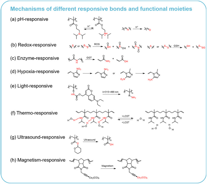
The phenomenon of stimuli-responsiveness, alternatively described as environmental responsiveness or adaptive functionality, derives its "intelligent" characteristics from the capacity to detect external stimulus signals and execute programmed response behaviors through structural or conformational adaptations [8]. Based on stimulus origin, stimuli-responsive block copolymers are categorized into two distinct classes: (1) endogenous-responsive systems targeting intrinsic physiological variations (e.g., pH gradients, redox potential, enzymatic activity) and (2) exogenous-responsive systems activated by external physical interventions (e.g., photonic energy, thermal fluctuations, magnetic/US fields). In this section, we methodically analyze the dynamic covalent chemistries underpinning the functionality of stimuli-responsive block copolymers while elucidating critical structure-property correlations governing their stimulus-actuated response mechanisms. Figs. 1 and 2 summarize the response bonds and representative molecular structures of endogenous and exogenous stimuli-responsive types, respectively. Fig. 3 illustrates the response mechanisms of several typical responsive bonds or functional units. Table 1 provides a comprehensive overview of the advantages and disadvantages of different responsive polymers. This systematic categorization bridges molecular design principles with macroscopic functional outputs, emphasizing contemporary advances in the past five years.
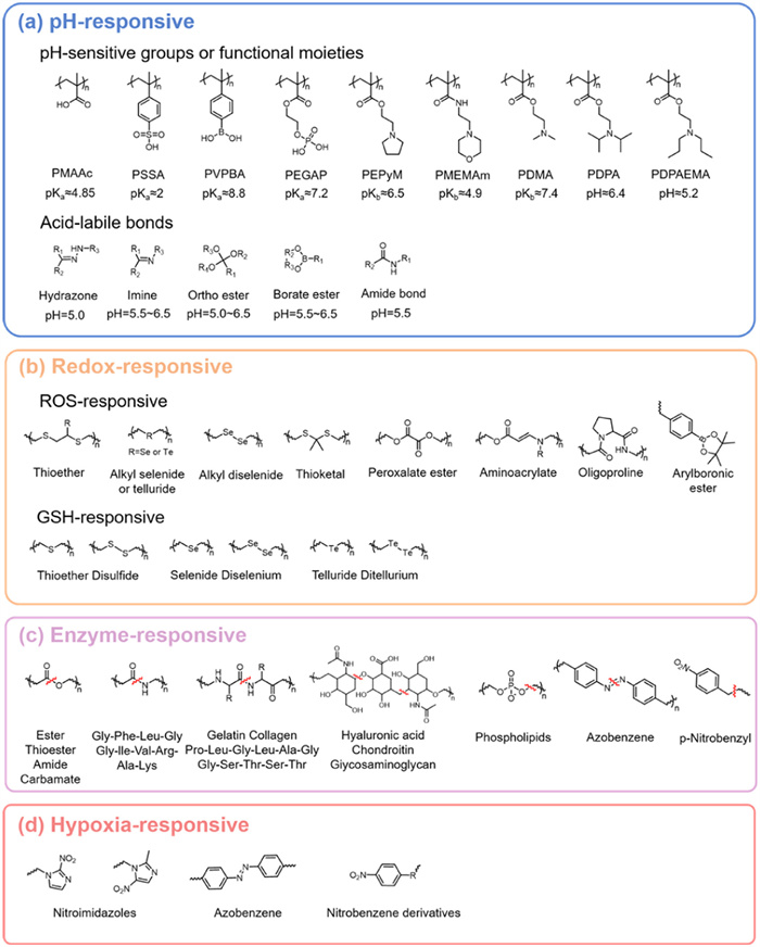
Figure 1
Figure 1.
Endogenous stimuli-responsive chemical bonds and responsive conditions. (a) pH-responsive moieties or chemical bonds. (b) Redox-responsive chemical bonds. (c) Enzyme-responsive chemical bonds. (d) Hypoxia-responsive chemical bonds.
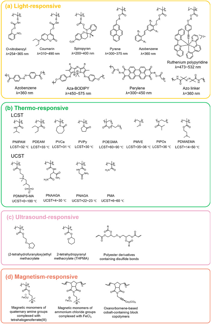
Figure 2.
Exogenous stimuli-responsive chemical bonds and responsive conditions. (a) Light-responsive chemical bonds. (b) Thermo-responsive moieties. (c) US-responsive moieties and chemical bonds. (d) Magnetism-responsive moieties.
(1) Narrow physiological pH range; (2) Diminished response at high ionic strength.
Redox-responsive
Polymers containing disulfide bonds
(1) Specific response to tumor/inflammatory high GSH environment; (2) Efficient intracellular drug release;
(1) Susceptible to nonspecific reduction; (2) Complex synthesis and potentially toxic by-products.
Enzyme-responsive
Peptide-polymer conjugates
(1) Ultra-high biological specificity; (2) Generation of active molecules in response.
(1) Slow response rate (dependent on enzyme diffusion and catalysis); (2) Heterogeneity of enzyme expression leading to imaging fluctuations;
Hypoxia-responsive
Nitroaromatics/quinone polymers
(1) Precise targeting of tumour hypoxic areas; (2) Lack of oxygen to activate the release of prodrugs.
(1) Slow rate of reduction (requires high expression of reductase); (2) Off-target hypoxic activation in healthy tissues; (3) Nitroreduction products may be cytotoxic.
Light-responsive
Azobenzene/spiropyran-containing polymers
(1) Non-contact precise control; (2) Fast response time (milliseconds); (3) Reversible deformation (optical switching).
(1) Poor tissue penetration; (2) Possible generation of biotoxicity.
(1) Non-invasive deep penetration; (2) Controlled cavitation/thermal effects.
(1) Low precision of energy focusing; (2) Dependence of the cavitation effect on the density of the medium.
Magnetism-responsive
SPIONs-polymer complexes (e.g., Fe3O4@PEG)
(1) Remotely controllable; (2) Multifunctional (thermal therapy/MRI imaging/targeted delivery); (3) Extremely fast response time (milliseconds).
(1) Potential oxidative stress at high concentrations of iron ions; (2) Decay of magnetic field gradients with distance; (3) Potential organ fibrosis from prolonged retention.
2.1
Endogenous stimuli-responsive
Endogenous stimuli response means that the material will respond to stimuli in the body such as pH, redox, enzymes. Changes in the environment at the lesion site can cause chemical or physical changes in the delivery system, allowing for effective and accurate bioimaging.
2.1.1
pH-responsive
pH-responsive polymers are a class of special functional polymer nanoparticles with a broad range of applications in bioimaging [9-11]. Currently, these polymers are primarily categorized into two types. The first kind of pH-responsive block copolymers contain pH-sensitive groups or functional moieties, which enable the polymer nanoparticles to respond to pH changes by altering their charge density. Examples of such pH-sensitive groups or functional moieties include polymethacrylic acid (PMAAc), poly(styrene sulfonic acid) (PSSA), poly(N-ethylpyrrolidine methacrylate) (PEPyM) and poly(N,N-dimethylacrylamide) (PDMA). The key mechanisms for these copolymer behaviors involves protonation or deprotonation processes, which results in changes to their physical properties or structures such as molecular chain conformation, morphology, conductivity, stability, and hydrophobicity [12]. Preparation of pH-responsive block copolymeric nanoparticles using reversible ionization of weakly acidic or weakly basic groups that become polyanions, polycations, or polyzwitterions in response to pH changes. The second category of pH-responsive block copolymers are those with acid-labile bonds. The primary mechanism here is that the acid-labile bonds can be cleaved under acidic or basic conditions, leading to a charge-induced transformation. Examples of such acid-labile bonds include hydrazone bonds, acetal bonds, amide bond, benzoic acid imine bonds, and adjacent ester bonds (Fig. 1a). These acid-labile copolymers are particularly useful for controlling the release of encapsulated substances in acidic environments by breaking the bonds upon exposure to acidic conditions. For example, the amide bond is an acid-unstable chemical bond formed by the reaction of a carboxyl group and an amino or hydrocarbon amino group in a carboxylic acid molecule [13]. This bond is unstable under acidic conditions and is capable of being hydrolyzed in weak acidic environments to form positively charged primary amines, thus causing a change in charge.
2.1.2
Redox-responsive
To meet the demand for specific responses in complex physiological environments, the researchers introduced a reactive oxygen species/glutathione (ROS/GSH)-responsive mechanism, and the core of molecular design is to take advantage of the controllable breaking or functional switching mechanism of ROS/GSH-sensitive bonds. The ROS-responsive units that have been developed so far include: thioether, selenium/tellurium compounds, thioketals, polysaccharides, aminoacrylates, boronic esters, peroxalate esters, and polyproline derivatives (Fig. 1b) [14]. These functionalized structures provide diverse molecular tools for the construction of smart delivery systems by specifically responding to different ROS types. Thioketal linkers have gained significant attention in recent years due to their ability to undergo facile cleavage by ROS, resulting in the generation of ketones and thiols. These linkers are particularly useful for targeted delivery to sites of inflammation or cancer cells where ROS levels are elevated for bioimaging [15]. Polymers incorporating thioether groups also are among the most widely studied ROS-responsive materials in biomedical research [14]. Upon exposure to oxidative environments, these polymers undergo a phase transition from hydrophobic sulfides to more hydrophilic states such as sulfoxides or sulfones [16]. Selenium (Se) and tellurium (Te), sharing similar chemical properties with sulfur due to their position in the same periodic group, exhibit comparable oxidation sensitivity. Like thioether groups, the oxidation of Se or Te-containing polymers leads to a transition from hydrophobic to hydrophilic states [16,17]. Moreover, targeted bioimaging using the high GSH levels of tumor tissue is also one of the common modalities in therapeutic diagnostics. As a result, GSH-responsive polymers have emerged as particularly promising carriers for tumor-specific drug delivery and cancer theranostics. Over the past decades, a variety of functional chemical bonds including disulfide bonds, diselenium, ditellurium, and thioether linkages have been utilized to conjugate drugs to polymers, facilitating the synthesis of GSH-responsive prodrugs [18]. In contrast to ROS-responsive mechanisms, GSH-responsive polymers focus on intracellular release. For example, high concentrations of GSH in tumor cells can rapidly break disulfide bonds in polymers, leading to targeted release.
2.1.3
Enzyme-responsive
In order to achieve highly selective and catalytically efficient responses in living organisms, scientists have explored enzyme-responsive block copolymers. These polymers undergo catalytic reactions facilitated by enzymes through processes such as the reduction/oxidation of substrates and the formation/cleavage of chemical bonds, leading to significant changes in the chemical properties of the polymers [19]. For example, enzymes can trigger specific responses by cleaving ester and amide bonds in enzyme-responsive block copolymers, which in turn results in substantial alterations in polymer morphology, solubility, and permeability. Fig. 1c outlines the various regions that binding enzymes can target for cleavage, including biomolecular fragments, traditional biodegradable polymer units, and degradable chemical bonds [20]. Peptide chains contain multiple cleavage sites and are therefore susceptible to hydrolysis by proteases. Polyester polymers, such as polylactic acid, polycaprolactone, and polybutylene terephthalate (PBT), are common biodegradable materials that are efficiently degraded by lipases [21,22]. In addition, fatty acid esters and amide bonds are often used as enzyme cleavage linkages in delivery systems. Azobenzene bonds are another strategy employed in the modification of enzymatically reactive polymers, which are likewise capable of being cleaved by specific enzymes. While most azo reductase-responsive probes are covalently bonded for enzyme-driven reactions, Li et al. introduced a novel method for selectively emitting fluorescence signals in oxygen-depleted cells by incorporating azobenzene into nanoparticles. The presence of azobenzene in the assembly effectively quenches the fluorescence signal. Upon reaction with reductases, the azobenzene undergoes cleavage, restoring the fluorescence signal as it is converted into amines [23].
2.1.4
Hypoxia-responsive
Hypoxia serves as a typical microenvironmental feature of pathological tissues. Polymers can achieve intelligent target modulation through specific response to hypoxia markers (e.g., nitroreductase, hypoxia-inducible factor-1α (HIF-1α)), leading to precise drug release or bioimaging signal enhancement in hypoxic regions. Currently, several hypoxia-responsive moieties, such as nitroimidazoles, nitrobenzoyl alcohols, and azo derivatives, are commonly used in delivery and bioimaging systems [24]. These moieties generally function by accepting electrons under hypoxic conditions, which generates hydrophilic functional groups that alter the physicochemical properties of the polymer, such as particle size and hydrophilicity [18]. For example, when exposed to hypoxia, nitroimidazoles can be bio-reduced into hydrophilic 2-aminoimidazoles, which modulate the hydrophobicity/hydrophilicity balance of the polymer [25]. This transformation enables the control of polymer nanoparticle assembly/disassembly, thereby facilitating more precise and controlled bioimaging. Although endogenous stimuli-responsive polymers are biocompatible, enabling controlled degradation of the material and localized therapy, the response is often inconsistent due to the complexity of the human physiological environment, and newly introduced groups may trigger adverse reactions.
2.2
Exogenous stimuli-responsive
Endogenous stimuli response means that the material will respond to external stimuli such as light, temperature, US. Exogenous stimuli-response block copolymers offer greater flexibility and control than endogenous stimuli-response block copolymers.
2.2.1
Light-responsive
Light-responsive polymers are a class of smart polymers that trigger dynamic changes in molecular structure or physicochemical properties by absorbing light at specific wavelengths. Their core feature is the use of light as a non-invasive, high spatial and temporal resolution means of regulation for diagnosis and therapy. The response of light-responsive materials can be readily modulated by adjusting the excitation source, such as by varying the intensity or wavelength of the light source [26]. The spectrum of light typically spans from ultraviolet (UV) to near-infrared (NIR, 780–840 nm), with certain light-responsive block copolymers also being activated by exposure to lower wavelengths, such as X-rays [19,26]. From the perspective of molecular design, light-responsive block copolymers can be broadly categorized into two types: one utilizes photothermal agents combined with temperature-responsive block copolymers [27], where light exposure induces a temperature change that triggers the desired release [28]. The second type involves the incorporation of light-reactive chromophores into polymers that undergo a photochemical reaction (light-chemical bond breaking and light-isomerization), activating the polymer's response (Fig. 2a) [29]. These processes drive macroscopic phase transitions, deformation, or release of functional molecules by changing the hydrophilicity, chain conformation, or cross-linking density of the polymer. Coumarin, a typical photochemically active molecule, not only acts as a cleavable group but also acts as a cross-linking agent. Coumarin exhibits faster release rates due to its higher molar absorption coefficient and stronger absorption characteristics. This facilitates the efficient release of the leaving group, which leads to the dissociation of the polymer [30,31]. Unlike coumarin polymers, spiropyran derivatives undergo an efficient transition from closed to open rings under UV light (200–400 nm) without causing polymer decomposition [32]. Spiropyran derivatives as a typical example of photoisomerism can be incorporated into copolymer matrices, either as side chain or as a part of the main copolymer chain [33]. Upon light switching, these derivatives cause a significant change in the hydrophobicity of the polymer. UV light triggers the cleavage of the C—O bond in the closed-ring structure of spiropyran, converting it into the zwitterionic form, merocyanine. Upon exposure to blue light (420 nm), merocyanine can reversibly revert to its hydrophobic ring-closed form [34].
2.2.2
Thermo-responsive
In addition to light-responsive block copolymers, thermo-responsive block copolymers have also received a lot of attention due to the superior imaging capabilities they exhibit when combined with photothermal agents. Thermo-responsive block copolymers are a class of smart polymeric materials that respond specifically to temperature changes in the body’s internal or external environments through reversible or irreversible changes in molecular chain conformation or aggregation states [35]. In general, thermo-responsive block copolymers demonstrate either a lower critical solution temperature (LCST) or an upper critical solution temperature (UCST), characterized by a transition between monophasic and biphasic states as the solution temperature changes (Fig. 2b). Poly(N-isopropylacrylamide) (PNIPAM) is one of the most widely studied thermos-responsive polymers, with an LCST that lies between body temperature and room temperature. The phase transition in PNIPAM is driven by an entropic advantage as the temperature increases. Below the LCST of PNIPAM, intermolecular hydrogen bonds dominate, leading to extended hydrophilic polymer chains. Above the LCST of PNIPAM, intramolecular hydrogen bonds become more prominent, causing the polymer chains to collapse and exhibit hydrophobicity [36]. In contrast to the LCST transition, which is driven primarily by cooperative entropy, the UCST transition is primarily governed by enthalpy. When the solution temperature exceeds the UCST, thermo-responsive block copolymers exhibiting UCST behavior undergo a shift from a biphasic to a monophasic state, which is the opposite of the behavior seen in thermo-responsive block copolymers with an LCST. The UCST phenomenon in aqueous solutions is attributed to strong interactions between polymer segments [19]. At lower temperatures, the interactions between polymer segments are more important than the polymer-solvent interactions. However, as the temperature exceeds the UCST, the polymer-solvent interactions become dominant, leading to the dissolution of the polymer. The strength of the interactions between polymer segments, and thus the UCST behavior can be enhanced by incorporating more hydrophobic side chains into the copolymer structure [37].
2.2.3
US/magnetism-responsive
US-responsive block copolymers exhibit the advantages of fast response time and high tissue penetration depth compared responsive polymers are precisely manipulated by exogenous mechanical wave energy. They absorb US energy, which triggers a physical or chemical change. The mechanism is mainly based on the ultrasonic cavitation-induced heating ability (local high pressure/high temperature microbubble rupture), which modifies the carrier structure or breaks the US-sensitive bonds to achieve drug delivery systems that can be remotely activated [38,39]. In addition to US stimuli, magnetic stimuli have also been explored as non-invasive techniques for bioimaging. Magnetism-responsive block copolymers are defined as composites that produce functional responses through the interaction of magnetic components with an external magnetic field. The mechanisms can be divided into two categories: the magneto-thermal effect, in which magnetic particles generate heat under an alternating magnetic field, triggering a phase transition in a temperature-sensitive block copolymer. The magneto-mechanical force actuation, in which a magnetic field gradient induces deformation or displacement of the material to enable remotely manipulated drug release or material reconfiguration [40]. Exogenous stimuli-responsive block copolymers have demonstrated its unique advantages in the field of bioimaging. However, despite the advantages of these polymers in bioimaging, such as ease of handling and on-demand control, they also face a number of challenges, including a higher risk of side effects, limited mechanism of action studies, and dependence on external stimulation devices.
3.
Stimuli-responsive block copolymers for multimodal bioimaging
Stimuli-responsive block copolymers provide highly sensitive and specific smart probes for bioimaging by virtue of their dynamic response to endogenous stimuli or exogenous stimuli. By precisely modulating the optical signal, acoustic impedance, or magnetic relaxation rate, such materials enable multi-scale imaging from the molecular to the tissue level and facilitate the integration of diagnosis and therapy. In this chapter, we have discussed the progress of their innovative design and application in optical imaging, US imaging, and magnetic resonance imaging, respectively. Furthermore, multimodal bioimaging has also been elaborated, which could overcome the inherent trade-offs between resolution, penetration depth, and functional specificity in single-modal bioimaging.
3.1
Optical imaging
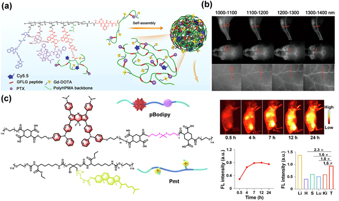
Optical imaging is a widely employed technique in the field of medical bioimaging, with NIR light (700–1000 nm) being particularly favored due to its low tissue absorption. Small organic molecules with unique optical properties, such as cyanine dyes, are commonly used as NIR fluorescent probes. However, these molecules tend to degrade rapidly in aqueous environments and exhibit short circulation times in physiological settings. To address these shortcomings, cyanine derivatives have been integrated into various polymers to enhance their bioavailability and longevity. Through integrating NIR fluorescent probes into polymeric micelle structures, it becomes possible not only to monitor the real-time release of targeted drug but also to utilize the probes for bioimaging purposes. For example, Cai et al. designed an innovative, biodegradable, multifunctional theranostic nanoplatform using branched copolymers for the bioimaging, diagnosis, and treatment of breast cancer (Fig. 4a). The nanoplatform was designed by incorporating a Gly-Phe-Leu-Gly tetrapeptide, an anticancer drug (paclitaxel (PTX)), a fluorescence dye cyanine5.5 (Cy5.5), and an MRI contrast agent (Gd-DOTA) into a branched copolymer (pHPMA) through RAFT polymerization, click chemistry, and chemical complexation. Once internalized by tumor cells, this nanomedicine is expected to provide both MRI and fluorescent bioimaging, while also enabling the controlled release of the anticancer drug for chemotherapeutic purposes [41].
Figure 4
Figure 4.
(a) Schematic illustration of cathepsin B-responsive biodegradable theranostic nanomedicine derived from branched pHPMA. Reprinted with permission [41]. Copyright 2020, Wiley. (b) High-quality NIR-Ⅱ fluorescence images obtained using the telechelic glycopolymer TTQ-TC-PFru. Reprinted with permission [42]. Copyright 2020, American Chemical Society. (c) Schematic illustration of cationic anticancer nanoparticles; NIR-Ⅱ fluorescence bioimaging/fluorescence analysis in mice injected with mt-NPBodipy. Reprinted with permission [43]. Copyright 2024, Nature Publishing Group.
In recent years, NIR-Ⅱ (1000–1700 nm) fluorescence bioimaging has received much attention from researchers due to its deeper tissue penetration and higher resolution. Chen et al. developed a glycosylated NIR-Ⅱ fluorescent probe polymer (TTQ-TC-PFru) and successfully constructed a multifunctional nano-diagnostic system with microenvironmental responsive properties (PFru-BTZ-PBOB) (Fig. 4b). BTZ-PBOB exhibits dual functional advantages: on the one hand, it achieves precise tumor localization imaging through high-intensity fluorescence signals in the NIR-Ⅱ window (1300–1400 nm); on the other hand, it relies on the highly efficient photothermal conversion capability to generate localized hyperthermia under NIR laser excitation. It is worthy noting that the smart carrier can specifically respond to the acidic microenvironment of the tumor, triggering the targeted release of bortezomib (BTZ) drugs and ultimately realizing chemotherapy-photothermal synergistic treatment under NIR-Ⅱ fluorescence. This integrated design of diagnosis and treatment not only breaks through the limitations of traditional single therapy but also provides an innovative solution for the precise visualization of malignant tumors [42]. Tang's team has developed a new type of cationic anticancer nanoparticles with dual functions of targeted therapy and imaging. These nanoparticles use their cationic surface properties to specifically anchor cancer cell membranes (Fig. 4c). Under NIR laser irradiation, they act through a triple synergistic mechanism. First, the NIR-Ⅱ fluorescent BODIPY units (1000–1700 nm) in the core of the particles generate highly penetrating imaging signals. Secondly, photoexcitation leads to the breakage of the sulfur-based aldehyde bond, releasing large amounts of ROS. Finally, ROS-induced lipid peroxidation and photothermal effect together destroy the tumor cell membrane and trigger apoptosis. This intelligent diagnostic platform not only achieves real-time tumor visualization and tracking in the NIR-Ⅱ window but also significantly improves anti-tumor efficiency through light-controlled dualmode therapy [43].
3.2
US imaging
US waves, characterized by high frequencies (≥2 MHz), are capable of both focusing and propagating through specific media. These waves have become widely utilized in numerous clinical settings due to their cost-effectiveness, non-invasive property, real-time capabilities, and powerful visualization potential, particularly for in vivo bioimaging [44]. The waves are reflected by various body tissues and are subsequently transformed into visual images by a converter. However, biosafety and lesion-targeted imaging have been the two core challenges that need to be overcome for the clinical translation of US imaging diagnostic systems. In oncology, a prevalent therapeutic and diagnostic approach combines targeted, biodegradable polymers with US imaging for cancer treatment [45]. Gao and co-workers developed an innovative US-activated delivery system, PFP/C9F17-PAsp (DET)/CAD/PGA-g-mPEG nanodroplets (Fig. 5a). This system integrates an acid-cleavable doxorubicin prodrug, an US-responsive phase-change contrast agent, and a cationic amphiphilic fluorinated polymer carrier to enhance both imaging contrast and the therapeutic efficacy of US-activation. The polymers used in this system serve as efficient theranostic agents, offering low cytotoxicity, excellent serum compatibility, and sustainable performance. When the nanodroplets were activated by 3.5 MHz US irradiation at a physiological temperature of 37 ℃, significant US contrast enhancement was observed and the high-intensity imaging signals could be stably maintained for >40 min. This result not only verifies the efficient response of the nanodroplets in the US field but also reveals their unique advantage over the traditional microbubble imaging agents, which is the long-lasting signal output through the kinetic modulation of the phase transition to provide a novel solution [46]. In addition, Jung et al. designed a curcumin-loaded nanoparticle (CUR-PVAX) by using as a contrast agent for enhanced US imaging and as a targeted therapeutic agent for ischemic injuries (Fig. 5b). CUR-PVAX activates the synergistic therapeutic effect of curcumin and PVAX by triggering the oxidation of peroxalate via H2O2 to generate CO2 bubbles. Notably, CUR-PVAX was able to amplify the overexpressed H2O2 signal at the ischemic site by generating CO2 bubbles, enhancing the echo intensity on US imaging for >1 h [47].
Figure 5
Figure 5.
(a) Fabrication of PFP-TNDs and delivery of CAD. Reprinted with permission [46]. Copyright 2019, Elsevier Ltd. (b) Schematic diagram and US imaging of the synthetic process of CUR-PVAX nanoparticles. Reprinted with permission [47]. Copyright 2018, Elsevier Ltd. (c) Acoustic imaging performance and blasting performance of Mag-Tar-DL NBs. Reprinted with permission [48]. Copyright 2024, Elsevier Ltd.
However, single targeting cannot address the problem of attenuated efficacy due to insufficient focal targeting, which is driving breakthroughs in dual-targeted precise delivery technologies. Lin et al. designed a dual-targeting drug delivery and imaging system using a combination of poly(lactic-co-glycolic acid) (PLGA), Biotin-DSPE-PEG2000, R406, Fe3O4, and Biotin-VCAM-1, fabricated via the double emulsification method and the biotin-avidin bridging approach (Fig. 5c). The Fe3O4 component imparts magnetic properties to the nanobubbles (NBs), while the VCAM-1 antibody enables specific antibody targeting. This dual-targeting strategy, integrating both magnetic and antibody-based targeting, facilitates imaging-guided antiatherosclerosis therapy. The drug-loaded NBs preferentially accumulate at sites of vascular inflammation due to the combined effects of an external magnetic field and antibody targeting. When exposed to US irradiation, the NBs undergo disruption to release R406, thereby alleviating the inflammatory response and preventing the progression of atherosclerosis. This research presents a controlled drug delivery system guided by US imaging, enhancing the efficiency of antiatherosclerotic treatments and providing a promising approach for drug delivery within large and medium arteries [48].
3.3
MRI
In addition to the diagnostic imaging tools previously discussed, magnetic resonance imaging has been widely used as a non-invasive medical technology for disease diagnosis, testing, and treatment. MRI offers several advantages over other diagnostic modalities, including high-resolution imaging and the absence of ionizing radiation risk [49,50]. However, small molecule MRI imaging agents have a short half-life in vivo and are not suitable for long-term repeat imaging. Conventional polymers, although able to circulate in the body for long periods of time, tend to accumulate in organs such as the liver and kidneys. Therefore, there is an urgent need for a contrast agent that can be activated and localized at the lesion for repeated, long-term, and selective imaging of the lesion. In response to this problem, stimuli-responsive MRI imaging agents have been developed. Stimuli-responsive magnetic resonance imaging diagnostics can be achieved by modulating the chemical environment or magnetic relaxation rate of specific block copolymer chain segments.
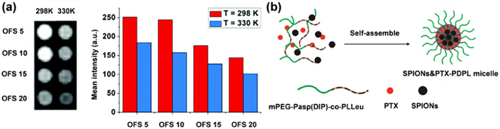
Usman et al. designed styrene-containing fluorinated PEG-methacrylate thermo-responsive terpolymers through free radical polymerization. The incorporation of the hydrophobic comonomer styrene was used to fine-tune the phase transition temperature of these terpolymers. Thermo-responsive block copolymers with controlled LCST were coupled to the hydrophobic drug fluorine-2-carboxaldehyde via acid-cleavable hydrazone bonds. Upon drug release, the polymer’s hydrophobicity decreased, and the LCST increased. When a certain temperature threshold was reached, the signal of 19F MRI was activated (Fig. 6a). The study provides a foundation for developing a switchable 19F MRI-based drug delivery and theranostic system [51]. At the same time, the synergy between the responsive polymer and the focal site targeting function further enhances its capability in dynamic monitoring. For example, Xiao et al. designed a pH-responsive micellar system derived from the biodegradable block copolymer mPEG-PAsp (DIP)-co-PLLeu to encapsulate the chemotherapeutic agent PTX and the MRI contrast agent superparamagnetic iron oxide nanoparticles (SPIONs) (Fig. 6b). This theranostic system (SPIONs/PTX-PDPL) not only facilitated the effective delivery of PTX and SPIONs into Bel-7402 liver cancer cells but also promoted the rapid release of PTX in acidic environment of lysosomes to induce cell apoptosis. In vitro MRI enabled precise localization of liver cancer cells following co-incubation with the SPIONs/PTX-PDPL micelles [52].
Figure 6
Figure 6.
(a) 19F MR images, and 19F MR signal intensities of OFS polymers at 298 and 330 K. Reprinted with permission [51]. Copyright 2021, Royal Society of Chemistry. (b) Preparation of SPIONs/PTX PDPL micelle in Bel-7402 cancer cells. Reprinted with permission [52]. Copyright 2020, Springer.
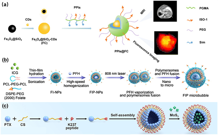
However, complex pathological microenvironments still require multi-mechanism synergistic responses with multimodal imaging validation. To this end, Shen et al. developed a ROS-responsive nano-micelle system (PPIS) for the diagnosis and treatment of atherosclerosis. This system was constructed by linking polyglycidyl methacrylate (PGMA) with polyethylene glycol (PEG) using oxaloyl chloride (Fig. 7a). To enhance the monitoring of atherosclerotic progression during therapy, the researchers integrated carbon dots (CDs) with Fe3O4@SiO2, a type of superparamagnetic iron oxide nanoparticles, to form a dual-modal probe, Fe3O4@SiO2CDs. These probes were subsequently encapsulated within nano-micelles to create PPIS. In the oxidative microenvironment of atherosclerotic plaques, the dicarbonyl group in the PGMA–PEG component was cleaved due to the elevated ROS levels, resulting in the release of the dual-modal probe along with Simvastatin [53]. The strategy enables simultaneous imaging and therapeutic intervention at the plaque site, offering a promising approach for both the early diagnosis and treatment of atherosclerosis. At the same time, the synergistic use of multimodal imaging modalities exploits the complementary strengths of different mechanisms to cross-validate and visualize information, thereby significantly reducing diagnostic uncertainty. Huang's team designed a folate-targeted laser-responsive perfluorohexane polymer nanoparticles (FIP-NPs) to achieve US imaging-phototherapy synergy through an innovative phase transition mechanism. The system is based on the self-assembly of the triblock copolymer PCL-PEG-PCL to form a dense membrane structure, loading with the NIR dye ICG and liquid perfluorohexane (PFH) (Fig. 7b). Under laser triggering, the photothermal conversion effect of ICG melted the polymer membrane and drove the gas-liquid phase transition of PFH, which prompted the dynamic reorganization of nanoparticles into micron-sized bubbles. The "nano-micron" trans-scale transition was captured by imaging for the first time, revealing that the membrane fusion and swelling properties can significantly enhance US contrast. This phase transition strategy solves the technical bottleneck between conventional microbubbles (poor tumor targeting) and nanoparticles (weak acoustic response) through the "nano-micron" dynamic transformation mechanism, and builds a multifunctional diagnostic platform with high sensitivity imaging and precise therapeutic feedback [54].
Figure 7
Figure 7.
(a) Illustration of the PPIS@FC nanomicelles for atherosclerosis diagnosis and therapy. Reprinted with permission [53]. Copyright 2023, Royal Society of Chemistry. (b) Schematic diagram of the synthetic process of FIP-NPs. Reprinted with permission [54]. Copyright 2024, American Chemical Society. (c) Schematic illustration of the construction of MoS2@PTX-CS-K237 NPs for CT/PA imaging and synergistic therapy. Reprinted with permission [55]. Copyright 2024, Elsevier Ltd.
In multimodal imaging materials, molybdenum disulfide (MoS2) and its derivatives have received significant attention for biomedical applications, particularly due to their optimal photothermal conversion efficiency and excellent imaging properties. Wang and co-workers synthesized a novel MoS2-loaded and K237-functionalized composite NPs to serve as multifunctional nanoplatforms with thermal/photoacoustic/computed tomography multimodal imaging-guided photothermal therapy (PTT) for the precise diagnosis and treatment of tumors (Fig. 7c). The fabricated nanoparticles possess a uniform spherical nanostructure and demonstrate favorable biocompatibility in the bloodstream. The incorporation of disulfide and amide bonds into the nanocomposites ensures that they remain inactive until they reach the tumor site. Upon endocytosis by tumor cells, the NPs are gradually triggered within the tumor microenvironment, leading to the degradation of the nanomaterial's core structure. This dual-functionality, combining both imaging and therapeutic capabilities, offers substantial potential for clinical applications in tumor treatment [55]. The development of these polymer systems has great potential to advance the early detection, prevention, and therapeutic management of diseases, as well as to help improve the effectiveness of diagnosis and treatment.
4.
Summary and outlook
Bioimaging techniques are instructive for monitoring drug delivery efficiency as well as tumor treatment efficacy. Stimuli-responsive block copolymers significantly enhance the targeting and signal-to-noise ratio of bioimaging by dynamically responding to pathological microenvironments or exogenous stimuli while reducing the non-specific distribution and toxicity of conventional imaging agents. However, several challenges remain, and there are emerging trends that will likely shape future developments in this area. The following outlines key challenges and potential future directions.
Challenges:
(1) Insufficient response specificity: In complex biological environments, such as inflammatory and tumor microenvironments, stimuli-responsive block copolymers often lack sufficient response specificity due to similar pH and redox properties, leading to off-target activation. This not only reduces the accuracy of imaging, but may also trigger unwanted side effects. Therefore, further advancing the research on stimuli-responsive block copolymers to broaden their applications is an important direction at present. Meanwhile, continuous exploration of new synthetic strategies and functional groups is crucial to improve the stimuli-responsive properties of copolymers.
(2) Development of multi-stimuli synergistic responsive systems: the development of multi-stimuli synergistic responsive systems can significantly improve the targeting accuracy through logic gate design. In recent years, the deep integration of diagnostic and therapeutic integration has become an important trend in the biomedical field. Through the construction of stimuli-responsive drug delivery and real-time bioimaging platform to achieve a complete closed loop of "monitoring–treatment–feedback", not only can improve the therapeutic effect, but also provide real-time data support for clinical decision-making.
Future development direction:
(1) Improving response specificity: Future research should focus on developing stimuli-responsive block copolymers with higher response specificity. This can be achieved by designing more complex molecular structures, introducing new responsive moieties or developing new synthetic methods.
(2) Integration of multimodal imaging techniques: As imaging technologies continue to evolve, the integration of multimodal imaging techniques (e.g., optical imaging, magnetic resonance imaging, and US imaging) will become an important trend in the future. By combining the advantages of different imaging modalities, more comprehensive biological information can be provided, thus improving the accuracy and reliability of diagnosis.
(3) Intelligent diagnostic and therapeutic platform: Building an intelligent platform integrating diagnosis, treatment and feedback is an important development direction for future bioimaging technology. Through the development of intelligent systems capable of real-time monitoring of drug release and therapeutic effects, personalized therapeutic plans can be realized to improve therapeutic effects and reduce side effects.
In summary, stimuli-responsive block copolymers show great potential in the field of multimodal bioimaging, but some key challenges still need to be overcome. The field can be further advanced by improving response specificity, developing multi-stimulus synergistic response systems, integrating multimodal imaging technologies, and building intelligent diagnostic and therapeutic platforms. Future research should focus on clinical translation to ensure that these innovations provide real benefits to patients.
Declaration of competing interest
The authors declare that they have no known competing financial interests or personal relationships that could have appeared to influence the work reported in this paper.
CRediT authorship contribution statement
Lizhuang Zhong: Writing – original draft, Investigation, Data curation, Conceptualization. Ming Liu: Writing – review & editing, Writing – original draft, Supervision, Resources, Investigation, Funding acquisition, Formal analysis, Data curation, Conceptualization. Shilong Su: Investigation, Conceptualization. Dongxin Zeng: Investigation, Data curation. Jing Hu: Writing – review & editing, Supervision, Resources, Funding acquisition. Zhiqian Guo: Writing – review & editing, Supervision, Resources, Funding acquisition.
Acknowledgments
This work was supported by the National Natural Science Foundation of China (Nos. 22208218, 22078196, and 22278268), the Natural Science Foundation of Shanghai (No. 22ZR1460400), Collaborative Innovation Center of Fragrance Flavour and Cosmetics, and Collaborative Innovation Project of Shanghai Institute of Technology (No. XTCX2023–07).
P. Wang, Z. Peng, Y.Y. Zhang, et al., Carbohydr. Polym. 335 (2024) 122073.
Figure 1
Endogenous stimuli-responsive chemical bonds and responsive conditions. (a) pH-responsive moieties or chemical bonds. (b) Redox-responsive chemical bonds. (c) Enzyme-responsive chemical bonds. (d) Hypoxia-responsive chemical bonds.
Figure 2
Exogenous stimuli-responsive chemical bonds and responsive conditions. (a) Light-responsive chemical bonds. (b) Thermo-responsive moieties. (c) US-responsive moieties and chemical bonds. (d) Magnetism-responsive moieties.
Figure 5
(a) Fabrication of PFP-TNDs and delivery of CAD. Reprinted with permission [46]. Copyright 2019, Elsevier Ltd. (b) Schematic diagram and US imaging of the synthetic process of CUR-PVAX nanoparticles. Reprinted with permission [47]. Copyright 2018, Elsevier Ltd. (c) Acoustic imaging performance and blasting performance of Mag-Tar-DL NBs. Reprinted with permission [48]. Copyright 2024, Elsevier Ltd.
Figure 6
(a) 19F MR images, and 19F MR signal intensities of OFS polymers at 298 and 330 K. Reprinted with permission [51]. Copyright 2021, Royal Society of Chemistry. (b) Preparation of SPIONs/PTX PDPL micelle in Bel-7402 cancer cells. Reprinted with permission [52]. Copyright 2020, Springer.
Figure 7
(a) Illustration of the PPIS@FC nanomicelles for atherosclerosis diagnosis and therapy. Reprinted with permission [53]. Copyright 2023, Royal Society of Chemistry. (b) Schematic diagram of the synthetic process of FIP-NPs. Reprinted with permission [54]. Copyright 2024, American Chemical Society. (c) Schematic illustration of the construction of MoS2@PTX-CS-K237 NPs for CT/PA imaging and synergistic therapy. Reprinted with permission [55]. Copyright 2024, Elsevier Ltd.
(1) Narrow physiological pH range; (2) Diminished response at high ionic strength.
Redox-responsive
Polymers containing disulfide bonds
(1) Specific response to tumor/inflammatory high GSH environment; (2) Efficient intracellular drug release;
(1) Susceptible to nonspecific reduction; (2) Complex synthesis and potentially toxic by-products.
Enzyme-responsive
Peptide-polymer conjugates
(1) Ultra-high biological specificity; (2) Generation of active molecules in response.
(1) Slow response rate (dependent on enzyme diffusion and catalysis); (2) Heterogeneity of enzyme expression leading to imaging fluctuations;
Hypoxia-responsive
Nitroaromatics/quinone polymers
(1) Precise targeting of tumour hypoxic areas; (2) Lack of oxygen to activate the release of prodrugs.
(1) Slow rate of reduction (requires high expression of reductase); (2) Off-target hypoxic activation in healthy tissues; (3) Nitroreduction products may be cytotoxic.
Light-responsive
Azobenzene/spiropyran-containing polymers
(1) Non-contact precise control; (2) Fast response time (milliseconds); (3) Reversible deformation (optical switching).
(1) Poor tissue penetration; (2) Possible generation of biotoxicity.
(1) Non-invasive deep penetration; (2) Controlled cavitation/thermal effects.
(1) Low precision of energy focusing; (2) Dependence of the cavitation effect on the density of the medium.
Magnetism-responsive
SPIONs-polymer complexes (e.g., Fe3O4@PEG)
(1) Remotely controllable; (2) Multifunctional (thermal therapy/MRI imaging/targeted delivery); (3) Extremely fast response time (milliseconds).
(1) Potential oxidative stress at high concentrations of iron ions; (2) Decay of magnetic field gradients with distance; (3) Potential organ fibrosis from prolonged retention.

 DownLoad:


 下载:
下载:


 下载:
下载:
