Received Date:
18 March 2025 Accepted Date:
29 May 2025 Revised Date:
27 May 2025 Available Online:
15 September 2025
Abstract:
Wound dressings with tissue adhesion, good mechanical, antioxidant and anti-inflammatory performance are urgently needed. In this work, we present a multifunctional selenium nanoparticles (SeNPs)/citric acid/gelatin/hydroxysuccinimide-grafted polyacrylic acid nanocomposite hydrogel adhesive (SCA) specifically designed for wound healing applications. The SCA was prepared via a one-pot processing, where SeNPs synthesized via chemical reduction were incorporated. These SeNPs not only endowed SCA with robust wet adhesion ability, excellent stretchability, and skin-matched elasticity modulus by serving as a physical crosslinker to modulate swelling equilibrium and molecular slippage, but also enhanced the biocompatibility and free radical scavenging capacity of SCA. Furthermore, in vivo evaluation of full-thickness cutaneous defects of rats revealed that SCA effectively reduced inflammation, promoted wound closure, and increased collagen deposition. All these results demonstrated that the developed SCA offers a promising therapeutic strategy for wound healing applications.
Skin is the largest organ of the body and serves as a physical barrier to resist dehydration, chemical or radiation damage, and microbial invasion [1,2]. However, the skin is also the organ most prone to damage owing to trauma, surgery, accidents, etc. [3-5]. It has been estimated that millions of people worldwide experience significant inconvenience due to wounds annually [6,7]. Although human skin has self-regeneration ability, skin defects beyond a certain diameter cannot heal spontaneously and may further deteriorate into nonhealing wounds, imposing a huge economic burden on healthcare systems [8-10]. Thus, effective treatments for promoting wound healing are urgently needed.
The wound dressing acts as a temporary shield against external infections and also functions as a guiding template to facilitate the reorganization of skin cells, as well as the subsequent infiltration and integration of host tissues, thereby significantly enhances the wound healing process [11-13]. An ideal skin wound dressing should present good tissue compatibility, excellent moisture retention, sufficient physical mechanical strength, and promotion of cell adhesion, proliferation, and differentiation [14-16]. Recent studies have witnessed the fast progress in various wound dressings, such as membranes, ointments, films, fibers, and hydrogels [17,18]. Among these, hydrogels are particularly promising because of their intrinsic feature to create a hydrated environment [19,20]. The hydrogels exhibit a high degree of design flexibility in functionalization to mimic the extracellular matrix (ECM) [21]. Recently, wet adhesion to wound tissue is attracting great attention for its role in preventing bleeding and promoting wound healing, especially in emergency situations [22,23]. Hydrogel dressings with wet adhesion can also simplify surgical procedures, shorten recovery times, and improve the quality of patient care [24-26]. However, achieving wet adhesion to tissue surfaces faces a great challenge because the presence of a hydration layer can impede direct contact between the adhesive and the tissue to reduce the first-line interfacial interactions [27,28].
Wound healing is a complex and dynamically ordered process that involves hemostasis, immune response and inflammation, proliferation, and remodeling [29-31]. Reactive oxygen species (ROS) are oxygen-derived small molecules generated by the respiratory chain in mitochondria and are indispensable cellular byproducts [32]. However, during the inflammatory stage of wound healing, excessive release of ROS significantly hinders wound healing, which in turn strengthens the inflammatory phase [30,33]. A hydrogel dressing with the ability to scavenge ROS can alleviate inflammation at the wound site [34-36]. Selenium (Se) is an essential dietary nutrient and a crucial trace element for human health and is primarily known for its antioxidant role in the form of glutathione peroxidase (GSH-Px) [37]. This enzyme takes a critical part in safeguarding cells against oxidative stress-induced damage [38]. Recent studies have highlighted the remarkable bioactivity, minimal toxicity, and enhanced bioavailability of Se nanoparticles (SeNPs). Xu et al. developed a polydopamine/alginate/nanoselenium composite hydrogel, where SeNPs mitigated infection-induced inflammation by scavenging ROS [39]. Mao et al. in situ synthesized SeNPs in a bacterial cellulose/gelatin hydrogel for full-thickness skin defect repair [40]. Zhao et al. prepared chondroitin sulfate-modified SeNPs for diabetic wound healing, playing important roles in downregulating inflammatory mediators [41]. Nevertheless, there are few reports on the hydrogel adhesive with the loading of SeNPs.
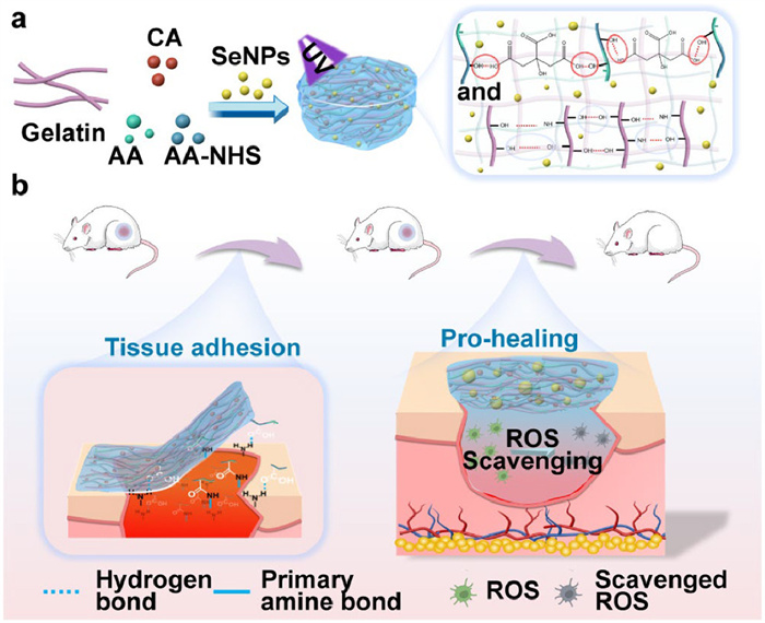
In this context, we developed a multifunctional gelatin-based nanocomposite hydrogel adhesive wound dressing that incorporated SeNPs as the redox regulator for wound management (Fig. 1). Gelatin, derived from hydrolyzed collagen, exhibits excellent biocompatibility and non-immunogenicity in wound application. This nanocomposite hydrogel adhesive was composed of N-hydroxysuccinimide (NHS)-grafted polyacrylic acid (PAA) and gelatin, where citric acid (CA) served as a supramolecular crosslinking agent to facilitate the connection between PAA chains and to increase the density of NHS and carboxyl groups on the surface of PAA. By integrating SeNPs synthesized through chemical reduction, the resulting SeNPs/CA/gelatin/NHS-PAA nanocomposite hydrogel adhesive (SCA) exhibited good biocompatibility, antioxidant properties, and favorable mechanical characteristics, including high elongation at break and tissue-matched elastic modulus. SCA also demonstrated a notable wet adhesion property. In vivo evaluations using a rat model with full-thickness skin defects confirmed the anti-inflammatory and wound healing efficacy of SCA. These findings suggest that SCA possesses the desirable attributes of an advanced wound dressing, positioning it as a potential candidate for the treatment of skin wounds and enhancing healing outcomes.
Figure 1
Figure 1.
Schematic diagram of preparation of the SCA and its application in full-thickness skin injury. (a) The schematic illustration of the fabrication of the nanocomposite hydrogel adhesive. (b) The nanocomposite hydrogel adheres to the damaged skin and promotes wound healing.
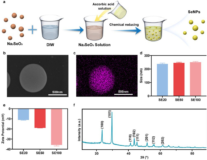
Fig. 2a illustrates the fabrication of SeNPs via a chemical reduction route. Sodium selenite serves as the selenium source, providing the essential selenium atoms for the formation of the nanoparticles. Whereas ascorbic acid plays a crucial role as the reducing agent, facilitating the conversion of selenium ions into elemental selenium, thus enabling the growth of SeNPs. Scanning electron microscope (SEM) observation shows a spherical morphology and a remarkably smooth surface of the prepared SeNPs (Fig. 2b). The energy dispersive spectrometer (EDS) mapping, displayed in Fig. 2c, illustrates the uniform distribution of the selenium on the spherical particle, further corroborating the successful fabrication of SeNPs. To ascertain the size distribution of SeNPs, DLS measurement was performed. An average diameter of approximately 240 nm with a narrow size distribution is observed for SE20, SE50, and SE100, indicating a high degree of morphology uniformity (Fig. 2d). The zeta potential result confirms the presence of a negative charge on the surface of SeNPs (Fig. 2e). An increase in the amount of SeNPs in different solutions lead to the increase in the negative charge. The X-ray diffraction (XRD) profile of the as-prepared SeNPs is illustrated in Fig. 2f. The peaks observed at 2θ values of 23.5°, 29.2°, 41.4°, 43.3°, 45.4°, 52.5°, 55.7°, and 62.7° correspond to the (100), (101), (110), (102), (111), (201), (112), and (202) crystal planes, respectively, as per the JCPDS card No. 06–362 standard. The presence of sharp and well-defined peaks in the XRD profile indicates that the SeNPs are highly crystalline.
Figure 2
Figure 2.
Fabrication and characterization of SeNPs. (a) The illustration showing the fabrication process of SeNPs. (b) SEM image, (c) element mapping, (d) average size, (e) zeta potential and (f) XRD profile. SE20, SE50, and SE100 correspond to the molar concentrations of selenium added during preparation. Data are presented as mean ± standard deviation (SD) (n = 3).
The adhesion in wet environments hinges on the repulsion of the hydration layer present on the substrate surface, since the hydration layer impedes the direct contact between the adhesive and the substrate [42,43]. Developing the hydrogel as the wet adhesive is of great significance for wound healing applications. In this work, a one-step crosslinking strategy was employed to fabricate the gelatin-based nanocomposite hydrogel adhesive loaded with SeNPs. The precursor solution was prepared by dissolving acrylic acid (AA), gelatin, CA, acrylic acid NHS ester (1 wt%), and ultraviolet (UV) initiator α-ketoglutaric acid (0.2 wt%) in DIW. Then, the SeNPs solution was added and UV curing was performed to construct the main crosslinked network comprised of gelatin and PAA-NHS. Introduction of CA is capable of actively modulating the supramolecular network and enhancing the wet adhesiveness of the adhesive by reorganizing the surficial NHS and carboxyl functional groups. SeNPs serve as physical crosslinkers to interlink gelatin chains through electrostatic interactions.
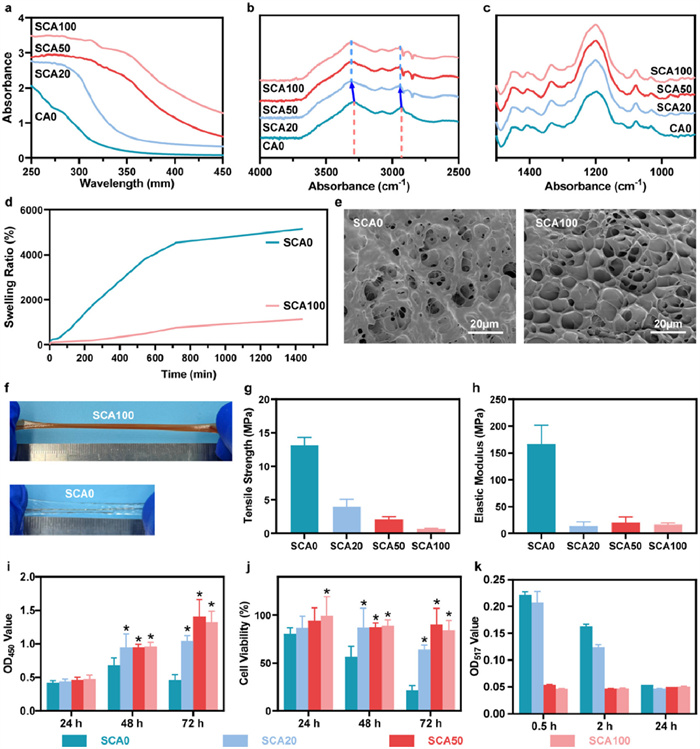
We confirmed the existence of SeNPs in SCA through the UV-vis spectroscopy. All the SCAs exhibit prominent UV light absorption peaks at approximately 270 nm (Fig. 3a), and the peak intensity is strongly correlated with the SeNP content. The SeNP amounts in SCA were evaluated via inductively coupled plasma‒optical emission spectrometry (ICP‒OES). The contents of SeNPs in SCA increase with the SeNP concentration of the SeNP solutions (Table S1 in Supporting information). It is observed from the FTIR spectra that incorporation of SeNPs leads to a blue shift in the O-H stretching peak at 3294 cm−1 and the C-H stretching peak at 2929–3310 cm−1 and 2945 cm−1, respectively (Fig. 3b). It may be attributed to the van der Waals interactions between the gelation in SCA and SeNPs. Meanwhile, the other characteristic peaks show no shift or obvious change in intensity, indicating a consistent chemical structure after the addition of SeNPs (Fig. 3c).
Figure 3
Figure 3.
Structure and properties of the adhesive nanocomposite hydrogel. (a) UV-vis spectra in the range of 250–450 mm. FTIR spectra (b) from 4000 cm−1 to 2500 cm−1 and (c) from 1500 cm−1 to 900 cm−1. (d) Swelling ratio curves and (e) SEM images of SCA0 and SCA100. (f) Digital photos of the stretched SCA0 and SCA100 (n = 3). (g) Tensile strength and (h) elastic modulus of different hydrogels (n = 3). (i) Optical density at 450 nm and (j) cell viability according to the CCK-8 assay after incubation with L929 fibroblasts for 24, 48, and 72 h (n = 5). (k) Optical density of DPPH after 0.5, 2 and 24 h of incubation with SCA (n = 5). Data are presented as mean ± SD. *P < 0.05 comparing with SCA0 at the same time.
Hydrogel wound dressings should possess superior water absorption capabilities to remove wound exudates during the healing process [44]. The swelling ratio was determined as shown in Fig. 3d. SCA0 exhibits an excessively high swelling rate, compromising its shape retention, whereas SCA100 demonstrates a promising swelling rate of approximately 1000%. The swelling rate of hydrogel is affected by the crosslinking network. However, SEM results confirm that SeNPs reduce the cohesion of SCA, where SCA100 shows the sparser porous structure than SCA0 (Fig. 3e). Therefore, it is reasonable to speculate that the decrease of the water absorption of SCA is due to the increase of the insolubility caused by SeNPs. The tensile behavior of SCA is displayed in Fig. 3f. SCA0 can be stretched to a certain extent, while SCA100 shows an excellent ductility. The elongation at break is significantly increased after adding SeNPs. The elongation at break increases from 276% ± 32% for SCA0 to 662% ± 39% for SCA100 (Fig. 3g). The elastic modulus shows a decreasing trend with the addition of SeNPs (Fig. 3h). According to the SEM results (Fig. 3e), the doping of SeNPs breaks the internal high crosslinking of SCA, facilitating the slippage of molecular chains of SCA. Nevertheless, it is interesting to observe SCA100 has an elastic modulus of ~17 MPa, the value of which falls within the range of that of human skin [45]. Such a high elongation at break and skin-matched elastic modulus make SCA a good candidate for treating open wounds.
Biocompatibility is a basic premise of wound dressings. A cell counting kit-8 (CCK-8) cytotoxicity assay was used to evaluate the biocompatibility of SCA. The OD450 values show a significant increase in cell proliferation from 24 h to 72 h by co-culturing L929 cells with the SCA, extracted culture medium (Fig. 3i). Although SCA0 exhibits toxicity, the addition of SeNPs significantly enhances the cell viability of SCA (Fig. 3j), demonstrating the improvement of biocompatibility. The antioxidant capability of wound dressings is valuable for facilitating wound healing by balancing the oxidative stress response at the wound site [46]. As shown in Fig. 3k, the 1,1-diphenyl-2-picrylhydrazyl (DPPH) free radical scavenging efficiency of SCA is remarkably high due to the presence of SeNPs. SCA50 and SCA100 are capable of eliminating free radicals in the first 0.5 h, indicating the good antioxidant property of SCA.
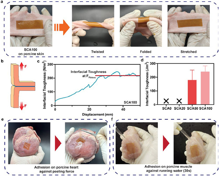
Water-absorbing hydrogels with wet adhesive properties can quickly close wounds and stop bleeding in emergency bleeding situations. At the same time, they can simplify surgical procedures, shorten recovery time, and improve the quality of patient care. Owing to the similarity between porcine skin and human skin, we used pig skin as a model tissue to simulate human skin. To visualize the wet adhesion ability of SCA100, it was adhered to the porcine skin as shown in Fig. 4a. Even under the large deformations, such as twisting, bending, and stretching, SCA100 maintains good adhesion to skin tissue without detachment. Fig. 4b shows the T-peeling test of SCA on the porcine skin, and the interfacial toughness-displacement curve of SCA100 is plotted in Fig. 4c. The results of the interfacial toughness of SCA are shown in Fig. 4d. SCA0 and SCA20 are unable to adhere to the moist pig skin surface, possibly because their excessive swelling results in the failure of adhesion. Further increasing the content of SeNPs imparts the wet adhesion characteristic of SCA. Especially the interfacial toughness of SCA100 reaches 240.6 ± 42.0 J/m2. It emphasizes the role of SeNPs in control over the swelling rate of SCA. And the insolubility of the SeNPs is also beneficial for interface drainage to improve the wet adhesion characteristic of SCA. To simulate the adhesion under harsh conditions, we tried to peel off SCA100 using a tweezer and wash it using running water. As photographed in Figs. 4e and f, SCA100 can firmly adhere to the heart tissue under large deformation, and resist running water without debonding. The above findings prove the excellent wet adhesion property of SCA100.
Figure 4
Figure 4.
Adhesion performance of the wet-adhesive nanocomposite hydrogel. (a) Photographs of flexible deformation of SCA100 adhered on the porcine skin. (b) Illustration of T-peeling test. (c) The interfacial toughness-displacement curve of SCA100. (d) The interfacial toughness of SCA. (e) Resistance to peel SCA100 from the porcine heart. (f) Resistance to running water of SCA100 on the porcine muscle. Data are presented as mean ± SD (n = 3).
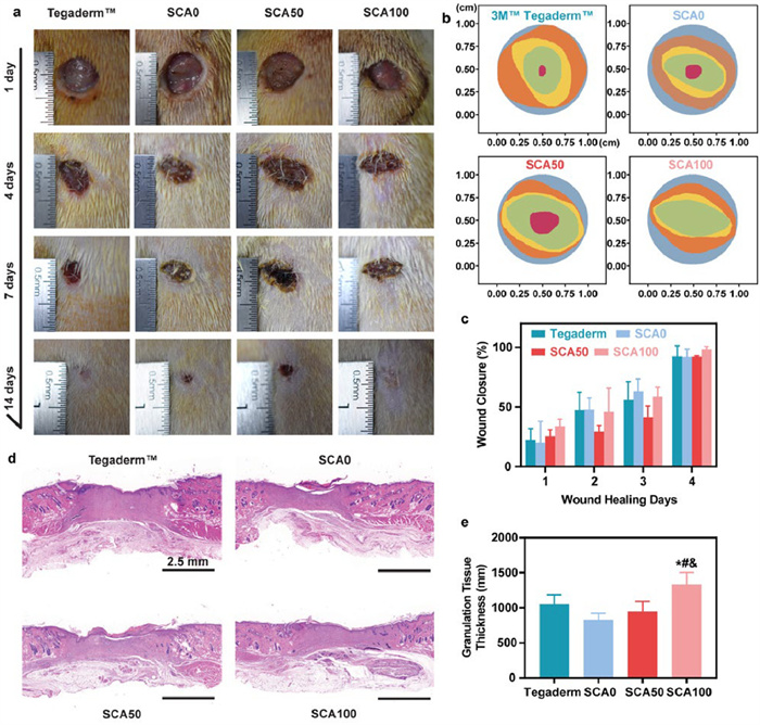
To assess the in vivo wound healing performance of SCA as a potential wound dressing, a full-thickness rat skin defect model was performed. For this assessment, we selected SCA0, SCA50, and SCA100 for wound repair performance testing. For comparison, 3M TegadermTM sterile dressing, which is widely used in clinical practice, was used as a control. All of the animal experiments were approved by the Institutional Review Board of Sichuan University (IACUC No. SCU46-2312-02). Representative photographs of comparing wound healing among the four groups are shown in Fig. 5a. The changes in the wound size and the average degree of wound closure are quantitatively shown in Fig. 5b. The wound areas of all the four groups decrease over time (Fig. 5b). The SCA100 group shows the comparable wound area to the 3M TegadermTM group. After 14 days of treatment, the SCA100 group exhibits the best treatment effect, with a wound closure rate close to 100% among the four groups (Fig. 5c). The thickening of the granulation tissue in the wound is an important indicator of the repair effect, since it stimulates the migration of distal keratinocytes to the wound area for the formation of a new epidermis. As shown in Fig. 5d, the SCA100 group displays a marked proliferation of the granulation tissue on day 14. Statistical analysis reveals a significant thickening in the granulation tissue between the SCA100 group and the other three groups (Fig. 5e, P < 0.05).
Figure 5
Figure 5.In vivo wound healing performance of SCA in a full-thickness rat skin defect model. (a) Photographs of wounds after treatment with different dressings on days 1, 4, 7, and 14. The wound dressings are 3M TegadermTM dressing, SCA0, SCA50, and SCA100. (b) Schematic diagram of the wound area for each group treated for 14 days, with x and y axis indicating the relative geometry of the wound size in cm. (c) Quantitative measurement of the ratio of wound closure on different days after surgery (n = 3). (d) H&E staining of granulation tissues on day 14 (n = 5). (e) Measurement of the granulation tissue thickness (n = 5). *, # and & indicate significant differences (P < 0.05) compared with the 3M TegadermTM, SCA0 and SCA50 groups. Data are presented as mean ± SD.
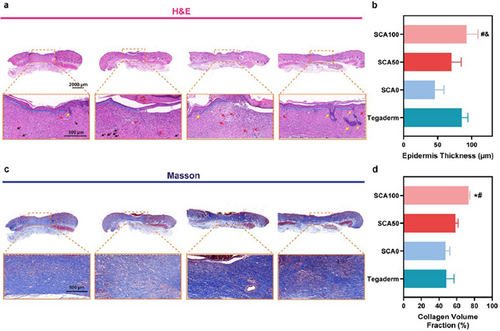
To ascertain the wound healing from a histomorphological perspective, hematoxylin and eosin (H&E) and Masson staining assays were performed. On day 14 of treatment, the wounds treated by TegadermTM and SCA0 presents a large number of inflammatory cells (black arrows), whereas few inflammatory cells are observed in the SCA50 and SCA100 groups (Fig. 6a). Additionally, more blood vessels are observed around the damaged area in the SCA50 and SCA100 groups than the other two groups. The occurrence of vascularization is favor of fibroblast recruitment to the wound and to supply oxygen, nutrients, and growth factors for tissue regeneration. Excitingly, the SCA100 group shows the most mature structures such as hair follicles and sebaceous glands. Fig. 6b shows the statistical analysis of the average new epidermal thickness formed in the treated groups. The antioxidant effect of SeNPs enables SCA to effectively attenuate the inflammatory response via generation of ROS. The re-epithelialization of the SCA100-treated wound is markedly improved, leading to the highest average new epidermal thickness (92.6 ± 16.5 µm).
Figure 6
Figure 6.
Histomorphological evaluation of the regenerated cutaneous tissues treated by SCA. (a) H&E staining of the treated wounds on day 14 (blood vessels: red arrows; hair follicles: yellow arrows; inflammatory cells: black arrow; boundary of epithelium: blue lines). (b) Quantitative analysis of epidermal thickness. (c) Masson’s trichrome staining of the treated wounds on day 14. (d) Quantitative analysis of the collagen volume fraction. *, # and & indicate significant differences (P < 0.05) compared with the 3M TegadermTM group, SCA0 group and SCA50 group, respectively. Data are presented as mean ± SD (n = 5).
Collagen deposition occurs throughout the wound healing process and plays a crucial role in skin tissue remodeling. Masson staining was conducted to detect the deposition of newly formed collagen on day 14 after treatment (Fig. 6c). Compared to the TegadermTM group, the SCA group present higher collagen deposition. The collagen fibers are more mature and organized in the SCA100 group than in the other groups. Mature collagen fibers have been recognized to facilitate ECM reconstruction and subsequently the skin tissue regeneration. Quantitative analysis reveals that the SCA100 group has the highest collagen volume fraction, followed by the SCA50 group, then the TegadermTM group (Fig. 6d). These outcomes exemplify the effectiveness of SCA100 in relieving the inflammatory response and promoting collagen deposition, thus remarkably accelerating wound healing.
In summary, a multifunctional gelatin-based nanocomposite hydrogel adhesive was fabricated with accelerated wound healing capacity enabled by SeNPs. The SeNPs were obtained through chemical reduction of sodium selenite. The hydrogel matrix consists of NHS-grafted PAA with added CA and gelatin, which exhibited good biocompatibility, mechanical properties, and tissue adhesion ability. Amongst all the SCA hydrogels, SCA100 exhibited the best antioxidation and wet adhesion abilities. The incorporation of SeNPs provided not only the adhesive with tissue-matching elastic modulus, but also wounding healing properties in the full-thickness skin defect model of rat, which was enhanced through their intrinsic ROS-scavenging ability. Overall, these properties suggest that SCA100 is a promising alternative to current wound dressings for clinical wound management.
Declaration of competing interest
The authors declare that they have no known competing financial interests or personal relationships that could have appeared to influence the work reported in this paper.
CRediT authorship contribution statement
Zhi-Peng Zhou: Writing – original draft, Visualization, Validation, Investigation. Xin Wei: Validation, Investigation. Ming Yan: Writing – review & editing, Visualization. Zhi-Guo Wang: Validation, Data curation. Rui Hong: Writing – review & editing, Funding acquisition, Data curation. Jia-Zhuang Xu: Writing – original draft, Supervision, Funding acquisition, Conceptualization.
Acknowledgments
This work was supported by National Natural Science Foundation of China (No. 52403042) and China Postdoctoral Science Foundation (No. 2023M742472).
Supplementary materials
Supplementary material associated with this article can be found, in the online version, at doi:10.1016/j.cclet.2025.111400.
[1]
S. Shang, K. Zhuang, J. Chen, et al., Bioact. Mater. 34 (2024) 298–310.
Figure 1
Schematic diagram of preparation of the SCA and its application in full-thickness skin injury. (a) The schematic illustration of the fabrication of the nanocomposite hydrogel adhesive. (b) The nanocomposite hydrogel adheres to the damaged skin and promotes wound healing.
Figure 2
Fabrication and characterization of SeNPs. (a) The illustration showing the fabrication process of SeNPs. (b) SEM image, (c) element mapping, (d) average size, (e) zeta potential and (f) XRD profile. SE20, SE50, and SE100 correspond to the molar concentrations of selenium added during preparation. Data are presented as mean ± standard deviation (SD) (n = 3).
Figure 3
Structure and properties of the adhesive nanocomposite hydrogel. (a) UV-vis spectra in the range of 250–450 mm. FTIR spectra (b) from 4000 cm−1 to 2500 cm−1 and (c) from 1500 cm−1 to 900 cm−1. (d) Swelling ratio curves and (e) SEM images of SCA0 and SCA100. (f) Digital photos of the stretched SCA0 and SCA100 (n = 3). (g) Tensile strength and (h) elastic modulus of different hydrogels (n = 3). (i) Optical density at 450 nm and (j) cell viability according to the CCK-8 assay after incubation with L929 fibroblasts for 24, 48, and 72 h (n = 5). (k) Optical density of DPPH after 0.5, 2 and 24 h of incubation with SCA (n = 5). Data are presented as mean ± SD. *P < 0.05 comparing with SCA0 at the same time.
Figure 4
Adhesion performance of the wet-adhesive nanocomposite hydrogel. (a) Photographs of flexible deformation of SCA100 adhered on the porcine skin. (b) Illustration of T-peeling test. (c) The interfacial toughness-displacement curve of SCA100. (d) The interfacial toughness of SCA. (e) Resistance to peel SCA100 from the porcine heart. (f) Resistance to running water of SCA100 on the porcine muscle. Data are presented as mean ± SD (n = 3).
Figure 5In vivo wound healing performance of SCA in a full-thickness rat skin defect model. (a) Photographs of wounds after treatment with different dressings on days 1, 4, 7, and 14. The wound dressings are 3M TegadermTM dressing, SCA0, SCA50, and SCA100. (b) Schematic diagram of the wound area for each group treated for 14 days, with x and y axis indicating the relative geometry of the wound size in cm. (c) Quantitative measurement of the ratio of wound closure on different days after surgery (n = 3). (d) H&E staining of granulation tissues on day 14 (n = 5). (e) Measurement of the granulation tissue thickness (n = 5). *, # and & indicate significant differences (P < 0.05) compared with the 3M TegadermTM, SCA0 and SCA50 groups. Data are presented as mean ± SD.
Figure 6
Histomorphological evaluation of the regenerated cutaneous tissues treated by SCA. (a) H&E staining of the treated wounds on day 14 (blood vessels: red arrows; hair follicles: yellow arrows; inflammatory cells: black arrow; boundary of epithelium: blue lines). (b) Quantitative analysis of epidermal thickness. (c) Masson’s trichrome staining of the treated wounds on day 14. (d) Quantitative analysis of the collagen volume fraction. *, # and & indicate significant differences (P < 0.05) compared with the 3M TegadermTM group, SCA0 group and SCA50 group, respectively. Data are presented as mean ± SD (n = 5).

 DownLoad:

 下载:
下载:


 下载:
下载:
