Scheme 1.
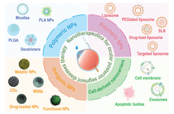
Schematic summary of versatile nanocarriers for drug delivery to OPS.
Nanotherapeutics for ocular posterior segment diseases therapy: Towards its advances and challenges
Mengdie Li , Shundong Cai , Hongjin Li , Yuhang Cheng , Jinfa Ye , Lang Ke , Yun Han , Min Su , Gang Liu , Chengchao Chu

Globally, about 206 million people have moderate to severe vision impairment, including 33 million with blindness [1]. Ocular posterior segment diseases (OPSDs), like glaucoma, diabetic retinopathy (DR), and age-related macular degeneration (AMD), severely threaten vision. Their increasing global prevalence imposes significant individual and societal burdens, necessitating urgent efforts in prevention, early detection, and novel therapies.
The human eye comprises an anterior segment (cornea, iris, lens) and a posterior segment (retina, choroid, vitreous humor) [2]. Located outside the cranium, the eyes allow diverse drug administration routes. Traditional ocular drug delivery includes non-invasive methods (eye drops, hydrogels) and invasive approaches (intravitreal (IVT) injections, implants) [3]. However, physiological barriers, such as tear drainage, lipid layers, and the blood-aqueous barrier (BAB) in the anterior segment, hinder posterior segment drug delivery. In the posterior segment, vitreous viscosity, aqueous humor flow, and the inner limiting membrane (ILM) further impede drug penetration [4]. Conventional methods (eye drops, injections, implants) suffer from poor bioavailability and risks like hemorrhage and fibrosis [5]. To overcome these challenges, innovative nanotherapeutics using nanocarriers have been developed.
Nanotherapeutics provide small size, high surface area, tunable properties, and biocompatibility, improving drug permeability, stability, and targeted ocular delivery. Major nanocarriers include polymeric nanoparticles (NPs), lipid NPs (LNPs), inorganic NPs, and cell-derived biomimetic systems. In vision science, they enable: (1) Targeted fundus therapy via surface functionalization, (2) prolonged ocular/vitreous retention, (3) reduced administration frequency/dosage, and (4) enhanced penetration across ocular barriers [6].
This review explores novel nanomaterials for ocular posterior segment (OPS) drug delivery (Scheme 1). It covers: (1) Posterior ocular diseases, their pathogenesis and treatments; (2) conventional drug delivery challenges; (3) nanomaterial characteristics and applications; and (4) clinical translation limitations and future prospects. The review provides insights for next-generation ocular drug delivery systems (DDS) design.
Glaucoma, a chronic disease characterized by retinal ganglion cell degeneration and optic nerve atrophy, causes irreversible vision loss. Affecting 70 million people globally in 2020, cases may surpass 1 billion by 2040. The primary risk factor is elevated intraocular pressure (IOP) due to impaired aqueous humor drainage. Untreated acute glaucoma can cause permanent blindness within days. Current treatments focus on IOP control through medications (prostaglandin analogs, β-blockers, cholinergic/α-adrenergic agonists), laser therapy, or surgery [7]. While topical medications remain the mainstay, long-term use can cause ocular side effects including dry eye, allergic conjunctivitis, and meibomian gland dysfunction [8].
Uveitis, an inflammatory condition of the highly vascular uvea, is classified into four types (anterior, intermediate, posterior, panuveitis) [9]. Ocular inflammation typically progresses from anterior to posterior segments, potentially involving the entire uvea and surrounding tissues. Consequently, uveitis management primarily focuses on controlling intraocular inflammation. Treatment strategies combine local and systemic approaches, particularly corticosteroids and immunosuppressants [10]. Local delivery methods include IVT injections (triamcinolone acetonide, dexamethasone) and implants (fluocinolone acetonide, suprachoroidal triamcinolone) [11,12]. While these interventions have been clinically established over 50 years, their repeated or prolonged use may lead to complications such as cataracts and elevated IOP [13], potentially compromising retinal protection [14].
Inherited retinal diseases (IRDs) are genetically diverse disorders, with RP being the most common—affecting 1.5 million people worldwide and involving mutations in over 60 genes [15]. RP causes progressive retinal degeneration, beginning with rod photoreceptor loss and night blindness, followed by peripheral vision impairment, and ultimately central vision loss and blindness [16]. Previously considered incurable, emerging therapies—including neuroprotective drugs [17], gene therapy [18], cell therapy [19], and laser treatment—now offer potential to slow photoreceptor degeneration and partially restore vision. Nanotherapeutics for ocular posterior segment diseases therapy are summarized in Table S1 (Supporting information) [20-114].
RB is the most common primary intraocular malignancy in children and second overall after uveal melanoma [115]. About 90% of cases occur before age three. Hereditary RB involves RB1 gene mutations in somatic cells, with secondary mutations potentially triggering malignancy due to environmental factors [116]. This form often presents as bilateral, multifocal tumors with increased risk of secondary cancers. Current treatments (enucleation, laser/cryotherapy, radiotherapy, chemotherapy) may cause systemic toxicity and vascular complications [117].
AMD is a progressive degenerative disease. Its main characteristic is the chronic degenerative changes in the macula. AMD is the leading cause of vision loss, impacting over 30 million individuals globally. It is estimated that by 2050, AMD will affect more than 5 million Americans [118]. The disease is divided into two types: dry AMD (dAMD) and wet AMD (wAMD). dAMD pathogenesis centers on oxidative stress, where metabolic ROS damage retinal pigment epithelial (RPE) cells, disrupting lipid metabolism, altering extracellular matrix, and recruiting macrophages. These changes promote pigmentation, RPE atrophy, and irreversible neovascularization [119]. dAMD is managed with antioxidant supplements (vitamins, zinc, lutein/zeaxanthin). While wAMD represents only 10% of cases, it causes 90% of severe vision loss. Current standard care for wAMD relies exclusively on anti-vascular endothelial growth factor (VEGF) therapies (e.g., ranibizumab, bevacizumab).
Pathological angiogenesis in FNDs appears as choroidal neovascularization (CNV) or retinal neovascularization (RNV), both caused by vascular endothelial growth factor A (VEGFA) overexpression [120]. RNV mainly stems from retinopathy of prematurity (ROP), DR, and retinal vein occlusion, while CNV primarily occurs in wAMD and high myopia [121]. Notably, DR affects 463 million diabetics worldwide and is the leading cause of blindness in working-age adults [122]. DR has two main stages: non-proliferative stage (early phase) featuring basement membrane thickening, cellular connectivity loss, and blood-retina barrier (BRB) impairment, causing microaneurysms, hemorrhages, and cotton wool spots; and proliferative stage, where progressive hypoxia upregulates VEGF, leading to neovascularization, vitreous hemorrhage, fibrous proliferation, and eventual retinal detachment/vision loss [123]. Although anti-VEGF therapy is first-line for early DR [124], many patients respond poorly, and repeated IVT injections may cause complications like hemorrhage and fibrosis [5].
Currently, drug delivery to OPS mainly includes three routes: topical administration, periocular injection, and intraocular injection (Fig. S1 in Supporting information). More specifically, the administration routes can be broadly categorized as non-invasive administration and invasive administration. Although great advancements have been achieved in OPS drug delivery, many challenges still remain.
Local ocular drug delivery to the posterior segment occurs via two primary pathways: (1) The corneal route (vitreous/uveoscleral pathways) and (2) the periocular route (conjunctival-scleral diffusion) [125]. However, multiple barriers limit efficacy: the lipid-rich corneal epithelium with tight junctions blocks hydrophilic drugs; the trilayered tear film (lipid/aqueous/mucin) creates a static barrier, while tear reflux and nasolacrimal drainage form dynamic clearance [126]. Although the conjunctiva is 15× more permeable than the cornea, its vasculature causes significant drug loss [127]. Additionally, the blood-aqueous barrier (BAB) restricts large and hydrophilic drugs via tight junctions [128], further hindering posterior delivery.
Functionalized NPs improve ocular permeability more effectively than conventional drugs. Sub-100 nm particles can directly penetrate corneal tight junctions, while larger ones require surface modifications. Current modification strategies involve three principal approaches: (1) Cationic coatings (e.g., chitosan) bind negatively charged mucin via electrostatic interactions, prolonging corneal contact [26]. (2) Polyethylene glycol (PEG) conjugation improves bioavailability through mucin hydrogen bonding and enhanced solubility [36]. (3) Targeted systems (peptides/antibodies) enable tissue-specific delivery for optimized drug utilization [42].
Notably, chitosan- or PEG-modified nanocarriers and liposomes show high efficacy in posterior drug delivery, enabling sustained drug release (3–6 months) for chronic conditions like AMD and macular edema. These systems overcome key ocular barriers, including rapid clearance and restricted penetration.
IVT and implants, while common for posterior drug delivery, carry risks of complications such as leakage, bleeding, and fibrosis [5]. The vitreous humor's viscosity impedes large molecule diffusion (e.g., proteins), with drug clearance occurring via anterior diffusion into aqueous humor or posterior permeation into retinal vessels [129]. Additionally, the ILM, a fibrous Müller cell network, further restricts retinal drug penetration (Fig. S2 in Supporting information) [130,131].
Nanomaterial-based ocular delivery exhibits size-dependent penetration: Sub-200 nm particles passively diffuse through vitreous collagen to reach retinal/choroidal tissues [100], while larger particles require surface modification for BRB crossing. The negatively charged hyaluronic acid-collagen mesh in vitreous gel impedes cationic drug diffusion [132]. Anion-coated NPs resist hydrodynamic clearance, prolonging OPS retention. For retinal targeting, cell-penetrating peptides (CPPs) or anti-VEGF peptides enable receptor-mediated transcytosis across retinal barriers, enhancing accumulation and extending therapeutic effects via optimized intracellular trafficking.
Notably, currently marketed nanomedicines are still mainly based on liposomes, micelles and PLGA as carriers, suggesting that although all nanocarriers can be modified via special process, the specific effectiveness for treating OPSD deserves more basic researches and clinical trials.
Polymer NPs are prepared by polymerizing monomers or dispersing natural macromolecules. Drugs can be either adsorbed on the surface of NPs or dispersed within the core of the NPs. Commonly utilized polymers for creating these NPs include polylactic acid (PLA), polylactic-co-glycolic acid (PLGA), micelles, and dendrimers (Table S2 in Supporting information) [133].
PLA, a lactic acid-based polymer derived from renewable sources like wheat, straw, and agricultural waste (e.g., bagasse, olive pits) [134], is biodegradable—breaking down into lactic acid, then carbon dioxide and water via metabolic pathways [135]. Its biocompatibility makes it ideal for ocular drug delivery. Sakai et al. developed PLA NPs loaded with betamethasone phosphate (BP) using an oil-in-water solvent diffusion method, showing rapid and sustained anti-inflammatory effects in treating autoimmune uveoretinitis (EAU) [136]. These NPs achieved efficacy equivalent to a fivefold BP dose and effectively suppressed glial activation, even post-EAU onset.
Previous studies have shown that PLA NPs can be internalized by phagocytes [137] and intestinal epithelial cells [138]. Bourges et al. injected PLA NPs loaded with Rh-6 G and Nile Red into the vitreous body, tracking their intraocular distribution via microscopy [139]. Rh-6G diffused from the NPs, staining retina and RPE cells, while NPs persisted in RPE for four months, demonstrating sustained drug delivery potential.
PLGA, an amphiphilic biodegradable polymer with ester linkages, combines hydrophilic glycolic acid for enhanced drug absorption and hydrophobic lactic acid for prolonged degradation, making its NPs ideal carriers for ocular posterior segment drug delivery [140].
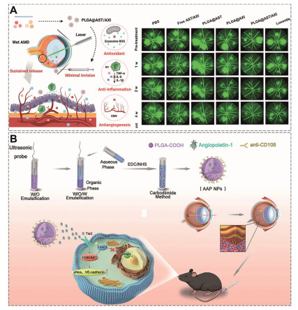
PLGA NPs effectively deliver anti-angiogenic drugs for treating FNDs. Chang et al. [141] developed PLGA@AST/AXI NPs, co-loaded with astaxanthin (AST) and axitinib (AXI), which demonstrated sustained two-month release after conjunctival injection. These NPs exhibited dual antioxidant and anti-angiogenic effects, reducing Bruch's membrane edema, CNV, and retinal damage in wAMD models (Fig. 1A). Yao et al. [142] further enhanced targeting by developing CD105-specific PLGA NPs (AAP NPs) for Ang1 delivery, which selectively accumulated in neovascular areas, suppressing CNV progression and modulating inflammation (Fig. 1B). Beyond small molecules, PLGA also shows promise for delivering exosomes (Exos), offering a "pseudo-cellular" therapeutic approach for posterior segment ocular diseases.
PLA/PLGA, a Food and Drug Administration (FDA)-approved biodegradable polymer, safely metabolizes into lactic acid (processed via the tricarboxylic acid cycle) and renally excreted glycolic acid. While over a dozen IVT drugs exist, only two PLGA-based formulations are currently approved for OPSD: Ozurdex® (dexamethasone-PLGA) for 3–6 months treatment of retinal vein occlusions and diabetic macular edema, and GB-102 (sunitinib-PLGA/mPEG-PLGA) providing 4–6 months IOP control in glaucoma. Ongoing clinical trials continue to demonstrate PLGA's potential as an effective sustained-release carrier for OPSD therapies (Table S3 in Supporting information).
Micelles (10–100 nm) are self-assembled amphiphilic structures (e.g., surfactants, polymers) that form above a critical concentration. In water, they arrange into hydrophobic-core/hydrophilic-shell monolayers, encapsulating proteins to mask surface charge and enhance hydrophobicity, facilitating cell membrane penetration [143-147].
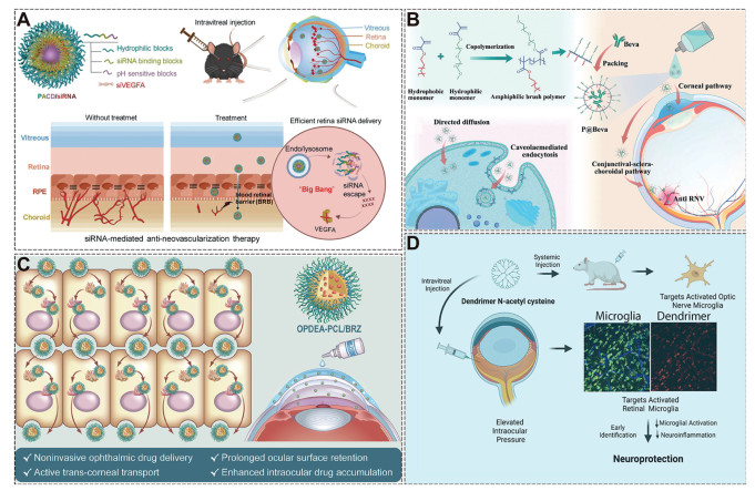
By adjusting the different combinations and ratios of the hydrophobic/hydrophilic components, the micelles can be customized to act as carriers for drug delivery [148]. Guo et al. [149] designed pH-responsive PACD micellar polymers (PEG (A)/siRNA-binding (B)/pH-sensitive blocks (C)) for siRNA delivery in RNV. At 100 nmol/L, PACD micelles (~100 nm) optimally inhibited angiogenesis in human umbilical vein endothelial cells (HUVECs) and achieved broad retinal distribution via hydrophilic/compact/cationic properties, suppressing RNV in oxygen-induced retinopathy (OIR) models (Fig. 2A). Cationic LNPs (lipid nanoparticles, < 100 nm) also enabled non-invasive eyedrop delivery for OPSD by transiently opening corneal tight junctions. Yang et al. [150] created 11 nm micelles for bevacizumab delivery via eye drops, achieving 23× greater posterior segment penetration than free drug through dual corneal/conjunctival-choroidal routes. In OIR models, they effectively suppressed neovascularization and choroidal sprouting while preserving anti-angiogenic efficacy (Fig. 2B). Wei et al. [151] developed zwitterionic micelles (OPDEA-PCL) for brinzolamide (BRZ) eye drops, improving ocular retention and corneal penetration via adsorption-mediated transcytosis. The formulation enhanced intraocular BRZ delivery and showed efficacy in glaucoma models (Fig. 2C).
Only two FDA-approved micellar ophthalmic drugs exist: Cequa® (dry eye) and Xelpros® (glaucoma). Micelle research lags behind other nanocarriers, with clinical trials mainly targeting ocular surface diseases, underscoring the need for more posterior segment studies.
Dendrimers are precisely structured, branched polymers with core-backbone-branch architecture, enabling uniform molecular distribution. Over 200 types exist, including widely used polyamidoamine (PAMAM), poly(l-lysine), polyethyleneimine (PEI), and polypropyleneimine (PPI) [152,153]. Their hollow interiors and amine/hydroxyl/carboxyl-rich surfaces [154] allow versatile drug/targeting/imaging agent loading while maintaining biocompatibility [155].
Hydroxylated PAMAM dendrimers selectively target retinal/optic nerve microglia via endocytosis in NAION, DR, AMD, and glaucoma models, enabling localized therapy with rapid clearance [156]. Pitha et al.'s fluorochrome-labeled PAMAM-NAC conjugates (D-NAC) achieved sustained retinal release (> 28 days) after IVT/systemic administration, reducing inflammation, suppressing A1 astrocyte activation, and promoting retinal ganglion cell (RGC) survival (Fig. 2D) [152]. Current dendrimer modifications (glycosylation, acetylation, PEGylation, peptides) reduce cytotoxicity via cation neutralization or enhance biodegradability [157]. Zhao et al.'s PAMAM-PVL-PEG unimNPs feature a hydrophobic drug core and PEG shell, showed 14-day RGC retention and neuroprotection post-IVT when conjugated with CTB/Cy5.5 [37].
While FDA-approved dendrimers carry small-molecule drugs, only one micellar polymer (Restasis® for dry eye) is used ophthalmologically. Dendrimer research lags behind other nanocarriers, with trials mainly focused on dry eye, necessitating more studies for OPSD applications.
LNPs consist of phospholipid bilayers forming spherical vesicles with uniform lipid cores [158], including solid LNPs (SLNs), nanostructured lipid carriers, lipid-drug conjugates, and polymer-lipid hybrid NPs [159].
SLNs, as first-generation biodegradable lipid carriers, offer excellent biocompatibility, drug-loading capacity, and sustained release [160]. Although effective for OPSD gene delivery, their retinal penetration is hindered by vitreoretinal and ILM barriers. Studies show sub-50 nm liposomes achieve superior retinal penetration, attributed to structural similarities between vitreous/ILM and mucus (a negatively charged polysaccharide network) [161]. PEG-modified LNPs effectively overcome these barriers, enabling efficient retinal distribution for small molecules and gene therapies [162]. Herrera-Barrera et al. [163] developed peptide-modified LNPs (0.15% MH42) for neuro-retinal mRNA delivery, using phage-derived targeting peptides (MH42/MH50) to achieve Cre mRNA-induced GFP expression in photoreceptors, RPE, and Müller cells within 48 h in Ai9 mice (Fig. 3A).
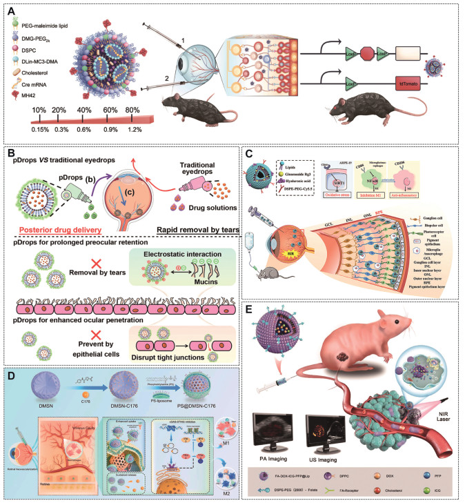
Structurally, LNPs have multilayered cores with nucleic acid-lipid complexes, whereas liposomes are bilayer vesicles carrying both hydrophilic and hydrophobic drugs. Liposomes reduce toxicity, enable sustained release, and enhance ocular drug bioavailability by prolonging surface/posterior retention [164-167], primarily via improved corneal/conjunctival permeability for OPS delivery. Wang et al. [164] developed core-shell pDrops NPs encapsulating hydrophilic ganciclovir (GCV) and hydrophobic curcumin (CUR) in a liposomal core with cationic chitosan coating. The chitosan enhanced mucin electrostatic interactions, opened corneal/conjunctival tight junctions, prolonged retention, and improved GCV/CUR delivery to OPS (Fig. 3B). Secondly, targeted liposome modification enables precise retinal drug localization and prolonged retention during invasive OPS administration. Huang et al. [165] developed CD44-targeted liposomes loaded with ginsenoside (Gg3) for retinal ischemia-reperfusion injury (Fig. 3C). These liposomes efficiently localized in the RPE layer, sustaining Gg3 release throughout the retina for 21 days. Third, studies confirmed their ability to preserve retinal structure and visual function by reducing oxidative stress and promoting M2 microglia/macrophage polarization. Shao et al. [166] developed PS-modified liposomal NPs (PS@DMSN–C176) co-loaded with STING inhibitor C176 and DMSNs for RNV treatment. The system enabled: (1) Macrophage-targeted delivery via PS recognition, (2) enhanced ocular stability (DMSNs), and (3) dual therapeutic effects-romoting M2 polarization while inhibiting cGAS-STING pathway. In OIR models, it reduced neovascularization, vascular leakage, and retinal inflammation (Fig. 3D).
By integrating with imaging modalities such as photoacoustic (PA), ultrasound (US), magnetic resonance (MR), and radionuclide tumor imaging, liposomes have secured a distinct role in the treatment of RB. Li et al. [74] created a FR-targeted liposomal platform (FA-DOX-ICG-PFP@Lip) co-loading doxorubicin (DOX), photothermal agent indocyanine green (ICG), and liquid perfluoropentane (PFP) for combined tumor therapy. The system achieved tumor-specific accumulation via enhanced permeability and retention effect (EPR) effects and folate receptor targeting, where near infrared (NIR) laser irradiation induced PFP phase transition to enhance ultrasound imaging and trigger DOX release, resulting in synergistic photothermal-chemotherapy with potent antitumor effects (Fig. 3E).
Liposomes, composed of natural phospholipids (e.g., phosphatides, cholesterol), offer excellent biocompatibility and metabolic clearance, reducing toxicity risks. These properties facilitate their clinical use as safe nanocarriers for small-molecule drugs. Over 20 FDA/European Medicines Agency (EMA)-approved liposomal drugs exist, mainly for fungal infections and cancer, with Visudyne® being the sole ophthalmic-approved product (for AMD, pathologic myopia, and ocular histoplasmosis). However, POSD research continues, showing promise with triamcinolone acetonide- (TNT) [168-171], GCV- [172], and dorzolamide- [173] loaded liposomes in clinical trials (Table S4 in Supporting information). Key challenges include formulation purity, stability, characterization methods, and pharmacokinetic profiling. Advanced analytics and engineering approaches may drive future OPSD liposomal therapy innovations.
Metal NPs typically have a core/shell structure: the metal core defines their properties, while the shell (metals/organic polymers) shields the core and enables biomacromolecule conjugation [174]. Noble metal NPs (e.g., Au, Ag, Pt) outperform transition metals in corrosion resistance, antioxidant capacity, and biocompatibility. Au NPs are especially useful in ophthalmology due to easy thiol-based surface modification. First, Au NPs effectively deliver bioactive molecules (proteins, peptides, DNA, oligonucleotides) [175]. Du et al. [176] engineered Au NPs functionalized with hexapeptides to generate drug-free peptide-based nanohybrids (P12). These nanohybrids exhibited pronounced anti-inflammatory activity in HUVECs in vitro. Moreover, following intravitreal administration, P12 efficiently reached the retina and markedly reduced vascular leakage and pericyte loss in streptozotocin-induced diabetic mice. Ag NPs also have a certain anti-angiogenic effect. They inhibit angiogenesis via phosphatidylinositol-3-kinase (PI3K)/Akt pathway activation and counteract advanced glycation end-products (AGEs) effects [177], reducing vascular permeability. Additionally, Au NPs serve dual roles as PA contrast agents and stimulus-activated drug carriers for targeted retinal delivery, leveraging their tunable electromagnetic/thermal properties. Daria et al. [178] developed polymer microcapsule NPs (MC) co-loaded with resveratrol and Au NPs. These enabled fluorescence-tracked RPE delivery and laser-triggered drug release, showing DR therapeutic potential (Fig. 4A). Younghoon et al. [179] optimized focused ultrasound (US) parameters with size/shape-tuned Au NPs, enabling intravenous dextran/Au NPs to noninvasively cross the BRB for retinal delivery, offering a targeted OPS drug delivery strategy (Fig. 4B).
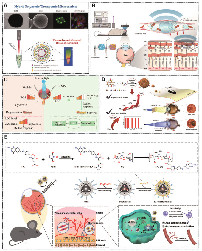
Platinum (Pt), cerium, and fullerene NPs exhibit multi-enzyme mimetic activity (peroxidase, catalase, and superoxide dismutase), enabling catalytic decomposition of H2O2 and conversion of O2− to O2/H2O2 [180]. These nanozymes show therapeutic potential against oxidative stress-induced photoreceptor degeneration. Su et al. [58] demonstrated that IVT administered Pt NPs effectively scavenged ROS in H2O2 and light-induced retinal damage models, protecting retinal cells through metabolic reprogramming that restored redox balance (Fig. 4C).
Cobalt, nickel, iron, and iron oxides are ideal for medical applications due to their superparamagnetism, enabling uses in cell separation, therapy, drug delivery, and magnetic resonance imaging (MRI) [181]. Bae et al. [182] developed PEG-coated Fe3O4 nanofluids for glaucoma treatment, using magnetic heating to upregulate HSP72 while precisely controlling RGC survival via adjustable electromagnetic parameters.
The application of metal NPs and metal oxides NPs are mainly focused on animal research, and the translation to the clinic is relatively difficult. First, complex synthesis requires precise size/crystal control and expensive equipment. Second, potential metal ion leakage causes ocular toxicity. Third, there are cumbersome surface modifications with limited drug loading. Future solutions may involve biodegradable carriers to reduce toxicity and multimodal therapies (e.g., photothermal/US) to enhance delivery.
CDs are spherical nanomaterials (< 10 nm) with a carbon core and functional groups (-OH, -COOH, -NH2), known for their biocompatibility, water solubility, and conjugation ability [183]. Their small size (~10 nm) has drawn interest in ophthalmology. CDs include carbon quantum dots (CQDs), carbon nanodots (CNDs), and polymer dots (PDs), with CQDs and graphene quantum dots (GQDs) being widely used in this field.
PEI-functionalized CDs (CD-PEI) enable efficient CRISPR delivery through clathrin- and caveolae-mediated endocytosis, enhancing gene transfer [184]. Wang et al. [185] developed chitosan-rapamycin CDs (CRCD, 3.7 nm) that induce M2 microglial polarization via autophagy, reducing oxidative stress and preserving retinal function in glaucoma. Additionally, CDs exhibit anti-angiogenic properties. Zhao et al. [106] demonstrated that GQDs suppress abnormal vessel growth in an OIR model by downregulating p-STAT3 and periostin in neovascularized retinas. CQDs, another key CD type, show promise for OPSD. White light-emitting diodes (WLEDs) emit harmful blue light (400–480 nm) that risks retinal damage. CQDs (< 5 nm), with anti-aggregation properties, enable blue light-blocking films. Guo et al. [186] synthesized stable Y-CQDs via alkali-catalyzed fusion, embedding them in PVA films to convert blue light to safer wavelengths while maintaining visibility. In LED-exposed rats, Y-CQD/PVA films preserved retinal structure by mitigating photochemical damage (Fig. 4D).
Mesoporous silica NPs (MSNs) feature high surface area (> 1000 m2/g) and adjustable pores (2–50 nm) [187], allowing stable encapsulation of large therapeutics (proteins, nucleic acids) with minimal leakage. Their high drug-loading capacity and surface modifiability (e.g., chitosan, folic acid) enable targeted delivery, enhancing bioavailability while reducing retinal toxicity. ROS-responsive MSNs coated with TK-argon-mPEG demonstrated sustained retinal activity for over 7 days, effectively treating retinopathy [188]. Additionally, large-pore MSNs protect nucleotides and modulate inflammation by suppressing M1 microglial polarization [189]. Huang et al. [190] developed folate-chitosan coated MSNs delivering miR-223 that selectively target activated microglia, promoting M2 polarization while reducing pro-inflammatory cytokines (IL-6, TNF-α) and increasing anti-inflammatory factors (IL-4, IL-10) in retinopathy models (Fig. 4E).
MSNs show promise for OPSD therapy due to high drug-loading capacity, controlled release, and modifiability, but challenges remain: (1) Developing degradable formulations to enhance safety; (2) improving targeting via multimodal strategies; (3) overcoming clinical translation barriers through simplified production and standardized quality control.
Overcoming immune system clearance is a significant challenge for NP drug delivery. Biomimetic nanocarriers from cell membranes evade immune clearance, prolong drug retention, and leverage homing proteins for targeted delivery [191], overcoming key limitations of conventional NPs.
CMVs, derived from stem cells, erythrocytes, or engineered cells, effectively deliver organic/inorganic NPs while preserving native membrane properties [192,193]. Their surface can be modified to enhance stability, reduce tear washout and protein-induced aggregation [194], and prolong intraocular retention. Although CMVs degrade slowly via ocular enzymes, their IVT dosage requires optimization to avoid oxidative stress from metabolite accumulation [195].
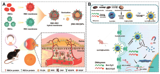
CMVs are prepared via gradient centrifugation, with co-extrusion fusing membranes to drug carriers while preserving targeting proteins, or engineered to boost targeting and lower immunogenicity [196]. However, their low drug-loading efficiency—particularly for proteins/nucleic acids—requires gradient loading or covalent coupling optimization. Li et al. created RBC-REC hybrid NPs by coating PLGA NPs with combined red blood cell and retinal endothelial cell membranes for CNV treatment [197]. These NPs avoid macrophage clearance through self-recognition while specifically targeting RECs to inhibit migration/proliferation (Fig. 5A), showing reduced vascular leakage and lesion area in CNV models. Similarly, Hu et al. [198] developed erythrocyte-based nanocomplexes using AS1411 aptamer-conjugated MnO₂ nano-sponges to target RB cells, achieving dual fluorescence (FL)/MRI while enhancing gene silencing and inducing apoptosis (Fig. 5B).
While CMVs show promise in ocular surface diseases (e.g., CNV [199], dry eye [200]), their OPSD applications remain limited due to large size and aggregation tendencies. Future studies should focus on multimodal CMVs or gene editing for precise immune-modulating therapy.
EVs, such as Exos, microvesicles, and apoptotic bodies, are nanoscale particles crucial for intercellular communication [201]. Among these, Exos are the most extensively studied due to their therapeutic potential and unique biological properties [202]. For OPSD therapy, Exos are derived from diverse sources, including stem cells [203], immune cells [204], organoids [205], retinal [206], and neural sources [207]. Packed with biomolecules like proteins, lipids, and nucleic acids [208], they facilitate targeted therapy by modulating cellular pathways, regulating gene expression, and inhibiting apoptosis. However, Exos production faces challenges, including complex isolation methods (e.g., ultracentrifugation and immunomagnetic beads), specialized equipment requirements, and low yields.
Stem cell-derived Exos present a safer and more effective alternative to stem cell therapy for eye diseases, offering advantages like low immunogenicity, stability, and therapeutic efficacy [209]. First, stem cells-derived Exos can regulate signaling pathway to protect retinal cells. Hwang et al. [210] found adipose stem cell Exos activate Nrf2 signaling, reducing RPE cell apoptosis and oxidative stress in RCS rats, attenuating RD and promoting retinal repair. Additionally, Exos deliver therapeutic RNAs to regulate gene expression and inflammation. Bone marrow mesenchymal stem cells (BMSCs)-derived Exos carrying miR-133b-3p inhibit angiogenesis and oxidative stress in DR by targeting human fibrillin-1 (FBN1) [211], while miR-22–3p-loaded Exos suppress NLRP3 inflammasome activation to mitigate retinal degeneration. Engineered Exos demonstrate enhanced efficacy through targeted modifications-cyclic arginylglycylaspartic acid (cRGD) peptide-functionalized Exos delivering IL-1 receptor antagonists showed superior microglia targeting and photoreceptor protection compared to natural Exos (Fig. 6A) [212-215].
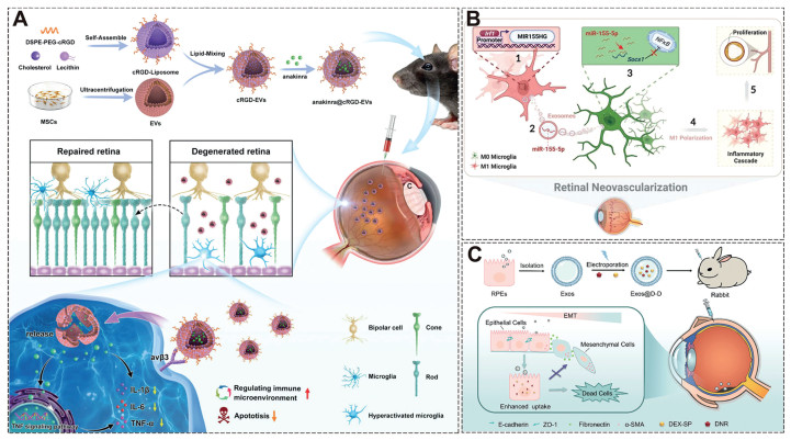
The retina's ten-layer structure makes different layers/cells susceptible to various diseases. Retinal-derived Exos (from Müller cells, RPE, and endothelial progenitors) possess target-specific membrane proteins but face research challenges. Zhao et al. [214] demonstrated RPE-derived Exos co-delivering dexamethasone sodium phosphate (DEX-SP) and daunorubicin hydrochloride (DNR) effectively treated PVR-Exos@D-D inhibited RPE proliferation/migration and epithelial-mesenchymal transition (EMT) markers while preventing retinal detachment, unlike controls (Fig. 6B).
Studies emphasize the immune system's key role in retinal diseases [216]. Microglia, the retina's primary immune cells, regulate metabolite clearance [217] and exhibit dual activation states: pro-inflammatory M1 and anti-inflammatory M2 phenotypes [218]. Chen et al. [213] found that Exos released by M1 microglia can stimulate resting microglia activation and amplify pro-angiogenic effects in the retina via the Irf1/miR-155–5p/Socs1 pathway (Fig. 6C).
Despite Exos' therapeutic potential, clinical challenges remain. Current mechanistic and safety data mainly come from animal studies, highlighting the need for technological improvement and clinical validation. Key hurdles include sourcing suitable cell lines for precise therapy, high production costs, contamination risks, and storage difficulties, underscoring the need for standardized Exos production.
Nanotherapeutics show great promise for OPSD treatment, offering advantages over traditional drugs through their small size, stability, and multifunctional modifications that improve drug permeability and ocular retention. These nanomaterials include polymeric/lipid/inorganic NPs and cell-derived carriers, with certain types (e.g., Pt/Au NPs, Exos) exhibiting inherent therapeutic properties.
While nanotherapeutics show promise for OPSD treatment, current research remains largely preclinical. Marketed nanomedicines primarily use polymeric NPs and liposomes (Table S5), with only PLGA/PLA, micelles, and modified liposomes clinically applied for OPSD. HA and dendrimers serve as corneal protectants but lack OPSD validation. Inorganic NPs face synthesis challenges, toxicity risks, and limited drug-loading capacity. Cell-derived carriers (CMVs/Exos) encounter clinical hurdles including size limitations, production costs, and storage issues-highlighting key areas for future research advancement. Firstly, future research should focus on elucidating nanotherapeutic delivery routes and metabolic behavior in OPS through real-time NP imaging, while critically evaluating long-term nanomaterial accumulation toxicity. Secondly, the difficulties in quality control and storage of the nanotherapeutics are other problems should be taken into consideration. Finally, the administration routes and frequency of nanotherapeutics should be rationally designed to improve patient compliance and reduce side effects. We anticipate that nanotherapeutics will benefit more and more patients with eye diseases in the future.
All authors disclosed no relevant relationships. This article have not been published previously, and are not under consideration for publication elsewhere. All co-authors have seen and approved the submission of the manuscript.
Mengdie Li: Writing – original draft, Conceptualization. Shundong Cai: Writing – original draft. Hongjin Li: Writing – review & editing. Yuhang Cheng: Writing – review & editing. Jinfa Ye: Writing – review & editing. Lang Ke: Writing – review & editing. Yun Han: Writing – review & editing. Min Su: Writing – review & editing. Gang Liu: Writing – review & editing. Chengchao Chu: Writing – review & editing, Conceptualization.
This work was funded by the National Natural Science Foundation of China (NSFC, Nos. 32271447, 32401176), the Science Foundation of Fujian Province (Nos. 2022J01021, 2024J08337, 2024J09005), Natural Science Foundation of Xiamen (No. 3502Z202371026), the Xiamen Medical College Research Fund (No. K2023–17), Educational Research Project for Middle-aged and Young Teachers in Fujian Province (No. JAT231135), Fujian Province Joint Funding Project for Scientific and Technological Innovation (No. 2024Y9727).
Supplementary material associated with this article can be found, in the online version, at doi:
GBD 2019 Blindness and Vision Impairment Collaborators, Vision loss expert group of the Global burden of disease study, Lancet Glob. Health 9 (2021) e144–e160. doi: 10.1016/S2214-109X(20)30489-7
A. Patel, K. Cholkar, V. Agrahari, A.K. Mitra, World J. Pharmacol. 2 (2013) 47–64. doi: 10.5497/wjp.v2.i2.47
M. Whalen, M. Akula, S.M. McNamee, et al., Bioengineering 11 (2024) 179. doi: 10.3390/bioengineering11020179
B. Mahaling, S.W.Y. Low, S. Ch, et al., Pharmaceutics 15 (2023) 2005. doi: 10.3390/pharmaceutics15072005
E.M. Becerra, F. Morescalchi, F. Gandolfo, Curr. Drug Targets. 12 (2011) 149–172. doi: 10.2174/138945011794182746
D.A. Srinivasarao, G. Lohiya, D.S. Katti, Wiley. Interdiscip. Rev. Nanomed. Nanobiotechnol. 11 (2019) e1548. doi: 10.1002/wnan.1548
H. Jayaram, M. Kolko, D.S. Friedman, G. Gazzard, Lancet 402 (2023) 1788–1801. doi: 10.1016/S0140-6736(23)01289-8
F. Fineide, N. Lagali, M.Y. Adil, et al., Ocul. Surf. 26 (2022) 19–49. doi: 10.1016/j.jtos.2022.07.007
D.A. Jabs, P. McCluskey, A.G. Palestine, J.E. Thorne, Clin. Exp. Ophthalmol. 50 (2022) 991–1000. doi: 10.1111/ceo.14175
J.R. Smith, J.E. Thorne, C.J. Flaxel, et al., Ophthalmology 131 (2024) 1107–1120. doi: 10.1016/j.ophtha.2024.02.019
T. Zhang, Z. Liu, N. Li, Front. Med. 11 (2024) 1402396. doi: 10.3389/fmed.2024.1402396
S. Fung, Y.Y. Syed, Drugs 82 (2022) 1403–1410.
S. Zeng, X.L. Liu, Eur. Rev. Med. Pharmacol. Sci. 27 (2023) 1743–1758.
Y.C. Chang, T.E. Kao, C.L. Chen, et al., Ann. Med. 56 (2024) 2352019. doi: 10.1080/07853890.2024.2352019
N. Schneider, Y. Sundaresan, P. Gopalakrishnan, et al., Prog. Retin. Eye Res. 89 (2022) 101029. doi: 10.1016/j.preteyeres.2021.101029
J.K. Phelan, D. Bok, Mol. Vis. 6 (2000) 116–124.
J. Li, Z. Huang, Y. Jin, et al., Curr. Neuropharmacol. 22 (2024) 1374–1390. doi: 10.2174/1570159x21666230907152207
Y. Liu, X. Zong, W. Cao, et al., Biomolecules 14 (2024) 903. doi: 10.3390/biom14080903
M. Radu, D.C. Brănișteanu, R.A. Pirvulescu, et al., Life 14 (2024) 668. doi: 10.3390/life14060668
D. Huang, P. Norat, L. Qi, et al., Adv. Sci. 11 (2024) e2401648. doi: 10.1002/advs.202401648
F.A. Maulvi, A.R. Patel, K.H. Shetty, et al., Drug Deliv. Transl. Res. 14 (2024) 3212–3224. doi: 10.1007/s13346-024-01543-8
C.F. Lai, F.J. Shiau, ACS Appl. Mater. Interfaces. 15 (2023) 18630–18638. doi: 10.1021/acsami.2c22860
Q. Jin, H. Li, Z. Jin, et al., Int. J. Pharm. 553 (2018) 21–28. doi: 10.1016/j.ijpharm.2018.10.033
P.B. Pisal, M.A. Joshi, M.N. Padamwar, et al., Int. J. Pharm. 461 (2014) 82–88. doi: 10.1016/j.ijpharm.2013.11.034
S. Schnichels, J. Hurst, J.W. de Vries, et al., ACS Appl. Mater. Interfaces. 13 (2021) 9445–9456. doi: 10.1021/acsami.0c18626
B.N. Kumara, R. Shambhu, A. Prabhu, K.S. Prasad, Carbohydr. Polym. 289 (2022) 119426. doi: 10.1016/j.carbpol.2022.119426
S. Schnichels, J. Hurst, J.W. de Vries, et al., Nanomedicine 29 (2020) 102260. doi: 10.1016/j.nano.2020.102260
S.D. Satyanarayana, A.S. Abu Lila, A. Moin, et al., Pharmaceuticals 16 (2023) 1001. doi: 10.3390/ph16071001
D.D. Nguyen, L.J. Luo, J.Y. Lai, Acta Biomater. 111 (2020) 302–315. doi: 10.1016/j.actbio.2020.04.055
M. Song, L. Li, J. Liu, et al., ACS Nano 17 (2023) 20979–20990. doi: 10.1021/acsnano.3c02685
H.R. Seong, C.H. Noh, S. Park, et al., Int. J. Mol. Sci. 24 (2023) 8073. doi: 10.3390/ijms24098073
K. Yao, X. Liang, G. Zhang, et al., Polymers 14 (2022) 3265. doi: 10.3390/polym14163265
S.E. Radwan, R.M. El-Moslemany, R.A. Mehanna, et al., Drug Deliv. 29 (2022) 1150–1163. doi: 10.1080/10717544.2022.2058648
L.J. Luo, D.D. Nguyen, J.Y. Lai, Theranostics. 11 (2021) 5447–5463. doi: 10.7150/thno.54525
T. Stack, M. Vincent, A. Vahabikashi, et al., Small 16 (2020) e2004205. doi: 10.1002/smll.202004205
E. Sánchez-López, M.A. Egea, B.M. Davis, et al., Small 14 (2018) 1701808. doi: 10.1002/smll.201701808
L. Zhao, G. Chen, J. Li, et al., J. Control. Release 247 (2017) 153–166. doi: 10.1016/j.jconrel.2016.12.038
M. Jeun, J.W. Jeoung, S. Moon, et al., Biomaterials 32 (2011) 387–394. doi: 10.1016/j.biomaterials.2010.09.016
W. Jiang, D. Xiao, C. Wu, et al., Mol. Ther. Nucleic. Acids. 35 (2024) 102258. doi: 10.1016/j.omtn.2024.102258
M.A. Mujtaba, H. Desai, A. Ambekar, et al., Biomed. Mater. 19 (2024) 065016. doi: 10.1088/1748-605x/ad7e6b
K. Imamachi, E. Stefánsson, A. Ohira, et al., Acta Ophthalmol. 97 (2019) 824–827. doi: 10.1111/aos.14119
X. Yu, R. Zhang, L. Lei, et al., Int. J. Nanomed. 14 (2019) 591–603. doi: 10.2147/IJN.S179118
T. Sakai, T. Ishihara, M. Higaki, et al., Invest. Ophthalmol. Vis. Sci. 52 (2011) 1516–1521. doi: 10.1167/iovs.10-5676
F. Cao, K. Liang, W.W. Tang, et al., J. Control. Release 372 (2024) 551–570. doi: 10.1016/j.jconrel.2024.06.047
R. Ganugula, M. Arora, M.A. Lepiz, et al., Sci. Adv. 6 (2020) eabb7878. doi: 10.1126/sciadv.abb7878
Y. Jin, D. Cai, L. Mo, et al., Biomaterials 309 (2024) 122617. doi: 10.1016/j.biomaterials.2024.122617
M. Shareef Khan, P. Rao Ravi, D. Shrikant Dhavan, Int. J. Pharm. 647 (2023) 123530. doi: 10.1016/j.ijpharm.2023.123530
M.L. Formica, G.V. Ullio Gamboa, L.I. Tártara, et al., Int. J. Pharm. 573 (2020) 118795. doi: 10.1016/j.ijpharm.2019.118795
A. Sabzevari, K. Adibkia, H. Hashemi, et al., Invest. Ophthalmol. Vis. Sci. 54 (2013) 5520–5526. doi: 10.1167/iovs.13-12296
Y. Zhu, J. Zhou, Y. Hu, et al., Int. J. Pharm. 622 (2022) 121836. doi: 10.1016/j.ijpharm.2022.121836
H. Li, Z. Zhang, Y. Li, et al., Front. Immunol. 13 (2022) 864956. doi: 10.3389/fimmu.2022.864956
L. Rebibo, C. Tam, Y. Sun, et al., J. Control. Releasee 333 (2021) 283–297. doi: 10.1016/j.jconrel.2021.03.035
M. Kasper, D. Gabriel, M. Möller, et al., Mol. Pharm. 15 (2018) 2539–2547. doi: 10.1021/acs.molpharmaceut.8b00014
M. Kasper, D. Gabriel, M. Möller, et al., Exp. Eye Res. 168 (2018) 49–56. doi: 10.1016/j.exer.2018.01.003
D.V. Pereira, F. Petronilho, H.R. Pereira, et al., Invest. Ophthalmol. Vis. Sci. 53 (2012) 8036–8041. doi: 10.1167/iovs.12-10743
E. Strettoi, C. Gargini, E. Novelli, et al., Proc. Natl. Acad. Sci. U. S. A. 107 (2010) 18706–18711. doi: 10.1073/pnas.1007644107
S.P. Read, S.M. Cashman, R. Kumar-Singh, Mol. Ther. 18 (2010) 1917–1926. doi: 10.1038/mt.2010.167
L. Su, X. Gong, R. Fan, et al., Redox. Biol. 65 (2023) 102836. doi: 10.1016/j.redox.2023.102836
A. Tzameret, H. Ketter-Katz, V. Edelshtain, et al., J. Nanobiotechnol. 17 (2019) 3. doi: 10.1186/s12951-018-0438-y
R. Iezzi, B.R. Guru, I.V. Glybina, et al., Biomaterials 33 (2012) 979–988. doi: 10.1016/j.biomaterials.2011.10.010
S. Francia, D. Shmal, S. Di Marco, et al., Nat. Commun. 13 (2022) 3677. doi: 10.1038/s41467-022-31368-3
R.N. Mitra, M.J. Merwin, Z. Han, et al., Free Radic. Biol. Med. 75 (2014) 140–148. doi: 10.1016/j.freeradbiomed.2014.07.013
P. Cao, Y. Cheng, Z. Li, et al., J. Nanobiotechnology. 21 (2023) 44. doi: 10.1186/s12951-023-01794-6
Y. Wang, W. Liu, B. Yuan, et al., Int. J. Nanomed. 14 (2019) 9763–9776. doi: 10.2147/ijn.s225992
M. Sen, M. Al-Amin, E. Kicková, et al., J. Control. Releasee 339 (2021) 307–320. doi: 10.1016/j.jconrel.2021.09.039
L. Valdés-Sánchez, S. Borrego-González, A. Montero-Sánchez, et al., J. Clin. Med. 11 (2022) 2170. doi: 10.3390/jcm11082170
R.N. Mitra, M. Zheng, E.R. Weiss, Z. Han, Biomaterials 157 (2018) 26–39. doi: 10.1016/j.biomaterials.2017.12.004
A. Koirala, R.S. Makkia, M.J. Cooper, M.I. Naash, Biomaterials 32 (2011) 9483–9493. doi: 10.1016/j.biomaterials.2011.08.062
S. Wu, Y. Mao, Q. Liu, et al., Front. Med. 8 (2021) 794299. doi: 10.3389/fmed.2021.794299
R.C. Cooper, J. Wang, H. Yang, Biomacromolecules 25 (2024) 5928–5937. doi: 10.1021/acs.biomac.4c00597
S.N. Tabatabaei, R.M. Derbali, C. Yang, et al., J. Control. Release 298 (2019) 177–185. doi: 10.1016/j.jconrel.2019.02.014
S. Wang, Y. Zhao, F. Yao, et al., Biomed. Pharmacother 174 (2024) 116437. doi: 10.1016/j.biopha.2024.116437
B. Jin, K. Lu, W. Gao, et al., Int. J. Nanomed. 19 (2024) 7799–7816. doi: 10.2147/ijn.s467949
M. Li, X. Bian, X. Chen, et al., Drug Deliv. 29 (2022) 519–533. doi: 10.1080/10717544.2022.2032876
W. Zheng, X. Li, H. Zou, et al., Int. J. Nanomed. 17 (2022) 3217–3237. doi: 10.2147/ijn.s364264
H. Zou, M. Li, X. Li, et al., Drug Deliv. 29 (2022) 1785–1799. doi: 10.1080/10717544.2022.2081379
S. Yan, J. Yan, D. Liu, et al., Theranostics. 11 (2021) 6833–6846. doi: 10.7150/thno.59020
E. Delrish, M. Jabbarvand, F. Ghassemi, et al., Exp. Eye Res. 204 (2021) 108423. doi: 10.1016/j.exer.2020.108423
H.S. Faizi, M.I. Nasiri, Y. Wu, et al., Int. J. Pharm. 664 (2024) 124614. doi: 10.1016/j.ijpharm.2024.124614
K. Huang, H. Deng, S. Wang, et al., Adv. Heal. Mater. 13 (2024) e2401613. doi: 10.1002/adhm.202401613
Y.S. Kwon, J. Munsoor, M. Kaufmann, et al., Adv. Sci. 11 (2024) e2400230. doi: 10.1002/advs.202400230
D. Sun, W. Sun, S.Q. Gao, et al., Pharm. Res. 41 (2024) 807–817. doi: 10.1007/s11095-024-03679-1
K. Liu, X. Zhang, R. Liu, et al., J. Agric. Food Chem. 72 (2024) 6347–6359. doi: 10.1021/acs.jafc.3c09054
S. Cupini, S. Di Marco, L. Boselli, et al., ACS Nano 17 (2023) 22800–22820. doi: 10.1021/acsnano.3c07517
K. Huang, X. Liu, Z. Lv, et al., Small 19 (2023) e2207335. doi: 10.1002/smll.202207335
D. Sun, W. Sun, S.Q. Gao, et al., J. Control. Release 330 (2021) 329–340. doi: 10.1016/j.jconrel.2020.12.010
G. Xin, M. Zhang, Z. Zhong, et al., ACS Appl. Mater. Interfaces 12 (2020) 57710–57720. doi: 10.1021/acsami.0c17296
M. Yadav, N. Schiavone, A. Guzman-Aranguez, et al., Drug Deliv. Transl. Res. 10 (2020) 919–944. doi: 10.1007/s13346-020-00733-4
R. Sun, A. Zhang, Y. Ge, et al., Expert. Opin. Drug Deliv. 17 (2020) 1305–1320. doi: 10.1080/17425247.2020.1783236
N. Elsaid, T.L. Jackson, Z. Elsaid, et al., Mol. Pharm. 13 (2016) 2923–2940. doi: 10.1021/acs.molpharmaceut.6b00335
J. Liu, X. Zhang, G. Li, et al., Int. J. Nanomed. 14 (2019) 8819–8834. doi: 10.2147/ijn.s217038
R. Suri, Y.R. Neupane, N. Mehra, et al., Int. J. Biol. Macromol. 191 (2021) 548–559. doi: 10.1016/j.ijbiomac.2021.09.069
P. Bhatt, G. Fnu, D. Bhatia, et al., Adv. Mater. 35 (2023) e2302069. doi: 10.1002/adma.202302069
W. Li, L. Chen, Z. Gu, et al., J. Control. Release 355 (2023) 358–370. doi: 10.1016/j.jconrel.2023.01.080
D.D. Nguyen, L.J. Luo, C.J. Yang, J.Y. Lai, ACS Nano 17 (2023) 168–183. doi: 10.1021/acsnano.2c05824
S.P. Kambhampati, I.A. Bhutto, T. Wu, et al., J. Control. Release 335 (2021) 527–540. doi: 10.1016/j.jconrel.2021.05.035
J. Pandit, Y. Sultana, M. Aqil, Carbohydr. Polym. 267 (2021) 118217. doi: 10.1016/j.carbpol.2021.118217
J.F. Grondek, K. Huffman, E.J. Lee, et al., Nanomedicine 17 (2022) 2089–2108. doi: 10.2217/nnm-2022-0255
F. Qiu, T. Meng, Q. Chen, et al., Mol. Pharm. 16 (2019) 1958–1970. doi: 10.1021/acs.molpharmaceut.8b01319
V. Dave, R. Sharma, C. Gupta, S. Sur, Colloids Surf. B 194 (2020) 111151. doi: 10.1016/j.colsurfb.2020.111151
Q. Chen, F.S. Qiu, W. Xie, et al., Int. J. Pharm. 666 (2024) 124758. doi: 10.1016/j.ijpharm.2024.124758
T. Niu, X. Shi, X. Liu, et al., Mol. Med. 30 (2024) 24.
S. Madhusudhan, N.V. Gupta, M. Rahamathulla, et al., Pharmaceutics 15 (2023) 2419. doi: 10.3390/pharmaceutics15102419
M. Bohley, A.E. Dillinger, B.M. Braunger, et al., Drug Deliv. Transl. Res. 13 (2023) 2807–2818. doi: 10.1007/s13346-023-01350-7
S.L. Taheri, M. Rezazadeh, F. Hassanzadeh, et al., Int. J. Biol. Macromol. 220 (2022) 1605–1618. doi: 10.1016/j.ijbiomac.2022.09.101
N. Zhao, X. Gui, Q. Fang, et al., J. Nanobiotechnol. 20 (2022) 174. doi: 10.1186/s12951-022-01362-4
S.E. Radwan, A. El-Kamel, E.I. Zaki, et al., Int. J. Nanomed. 16 (2021) 4481–4494. doi: 10.2147/ijn.s316564
R. Amato, M. Giannaccini, M. Dal Monte, et al., Front. Bioeng. Biotechnol. 8 (2020) 144. doi: 10.3389/fbioe.2020.00144
L. Zeng, W. Ma, L. Shi, et al., Int. J. Nanomed. 14 (2019) 6357–6369. doi: 10.2147/ijn.s214727
L. Xu, W. Li, Q. Shi, et al., J. Photochem. Photobiol. B 196 (2019) 111502. doi: 10.1016/j.jphotobiol.2019.04.011
Y. Dong, G. Wan, P. Yan, et al., J. Photochem. Photobiol. B 195 (2019) 51–57. doi: 10.1016/j.jphotobiol.2019.04.012
X. Rong, Y. Ji, X. Zhu, et al., Int. J. Nanomed. 14 (2019) 45–55.
R.N. Mitra, C.A. Nichols, J. Guo, et al., J. Control. Release 236 (2016) 31–37. doi: 10.1016/j.jconrel.2016.06.020
E.J. Oh, J.S. Choi, H. Kim, et al., Biomaterials 32 (2011) 3115–3123. doi: 10.1016/j.biomaterials.2011.01.003
A. Pareek, D. Kumar, A. Pareek, et al., Heliyon. 10 (2024) e32844. doi: 10.1016/j.heliyon.2024.e32844
X. Ma, X. Li, Q. Sun, et al., Curr. Issues. Mol. Biol. 46 (2024) 5307–5321. doi: 10.3390/cimb46060317
R. Manukonda, R.V. Narayana, S. Kaliki, et al., Expert. Opin. Ther. Targets. 26 (2022) 937–947. doi: 10.1080/14728222.2022.2158812
J.B. Jonas, C.M.G. Cheung, S. Panda-Jonas, Asia Pac. J. Ophthalmol. 6 (2017) 493–497.
D. Śpiewak, Ł. Drzyzga, M. Dorecka, D. Wyględowska-Promieńska, J. Clin. Med. 13 (2024) 4227. doi: 10.3390/jcm13144227
Y. Zhou, M. Xu, W. Shen, et al., Adv. Heal. Mater. 13 (2024) e2304626. doi: 10.1002/adhm.202304626
A.R. Fielder, G.E. Quinn, P.K. Shah, et al., Arch. Dis. Child Fetal. Neonatal. Ed. 110 (2024) 8–9.
M. Kropp, O. Golubnitschaja, A. Mazurakova, et al., EPMA J. 14 (2023) 21–42. doi: 10.1007/s13167-023-00314-8
N. Cheung, P. Mitchell, T.Y. Wong, Lancet 376 (2010) 124–136. doi: 10.1016/S0140-6736(09)62124-3
X. Liu, K. Huang, F. Zhang, et al., J. Nanobiotechnol. 22 (2024) 354. doi: 10.1186/s12951-024-02614-1
Y. Jiang, X. Fu, M. Shao, et al., Biomaterials 305 (2024) 122429. doi: 10.1016/j.biomaterials.2023.122429
C. Linsen, L. Missotten, Bull. Soc. Belge Ophtalmol. 238 (1990) 35–44.
B. Silva, B. São Braz, E. Delgado, L. Gonçalves, et al., Int. J. Pharm. 606 (2021) 120873. doi: 10.1016/j.ijpharm.2021.120873
R. Varela-Fernández, V. Díaz-Tomé, A. Luaces-Rodríguez, et al., Pharmaceutics 12 (2020) 269. doi: 10.3390/pharmaceutics12030269
M. El Sanharawi, L. Kowalczuk, E. Touchard, et al., Prog. Retin. Eye Res. 29 (2010) 443–465. doi: 10.1016/j.preteyeres.2010.04.001
K.Y. Zhang, T.V. Johnson, Exp. Eye Res. 206 (2021) 108545. doi: 10.1016/j.exer.2021.108545
L. Samoilă, O. Voștinaru, E. Dinte, et al., Int. J. Mol. Sci. 24 (2023) 8045. doi: 10.3390/ijms24098045
E. Ramsay, T. Lajunen, M. Bhattacharya, et al., J. Control. Releasee 361 (2023) 1–19.
U.B. Kompella, A.C. Amrite, R. Pacha Ravi, S.A. Durazo, Prog. Retin. Eye Res. 36 (2013) 172–198. doi: 10.1016/j.preteyeres.2013.04.001
J. Rydz, W. Sikorska, M. Kyulavska, D. Christova, Int. J. Mol. Sci. 16 (2014) 564–596. doi: 10.3390/ijms16010564
A. Kumari, S.K. Yadav, S.C. Yadav, Colloids Sur. B: Biointerfaces 75 (2010) 1–18.
T. Sakai, H. Kohno, T. Ishihara, Exp. Eye Res. 82 (2006) 657–663. doi: 10.1016/j.exer.2005.09.003
S. Moeller, R. Kegler, K. Sternberg, R.G. Mundkowski, Nanomedicine 8 (2012) 1293–1300. doi: 10.1016/j.nano.2012.01.011
S. Sun, X. Du, M. Fu, et al., Int. J. Pharm. 595 (2021) 120227. doi: 10.1016/j.ijpharm.2021.120227
J.L. Bourges, S.E. Gautier, F. Delie, et al., Invest. Ophthalmol. Vis. Sci. 44 (2003) 3562–3569. doi: 10.1167/iovs.02-1068
N. Kamaly, B. Yameen, J. Wu, O.C. Farokhzad, Chem. Rev. 116 (2016) 2602–2663. doi: 10.1021/acs.chemrev.5b00346
W. Chang, X. Lv, J. Zhu, et al., ACS Nano 18 (2024) 19649–19662.
H. Yao, H. Xu, M. Wu, et al., Acta Biomater. 166 (2023) 536–551. doi: 10.1016/j.actbio.2023.05.021
F. Paganelli, J.A. Cardillo, L.A. Melo, et al., Invest. Ophthalmol. Vis. Sci. 50 (2009) 3041–3047. doi: 10.1167/iovs.08-2920
B.D. Kuppermann, M.S. Blumenkranz, J.A. Haller, et al., Arch. Ophthalmol. 125 (2007) 309–317. doi: 10.1001/archopht.125.3.309
D.T. Tan, S.P. Chee, L. Lim, et al., Ophthalmology 108 (2001) 2172–2181. doi: 10.1016/S0161-6420(01)00839-9
D.T. Tan, S.P. Chee, L. Lim, et al., Ophthalmology 106 (1999) 223–231. doi: 10.1016/S0161-6420(99)90060-X
K. Cholkar, A. Patel, A.D. Vadlapudi, A.K. Mitra, Adv. Drug Deliv. Rev. 122 (2017) 31–64. doi: 10.1016/j.addr.2017.04.001
J.J. López-Cano, A. Sigen, V. Andrés-Guerrero, et al., Pharmaceutics. 13 (2021) 234. doi: 10.3390/pharmaceutics13020234
S. Guo, C. Li, C. Wang, et al., Acta Pharm. Sin. B 14 (2024) 781–794. doi: 10.1016/j.apsb.2023.09.001
R. Yang, S. Tang, X. Xie, et al., Adv. Mater. 36 (2024) e2314126. doi: 10.1002/adma.202314126
Q. Wei, C. Zhu, G. Yuan, et al., J. Control. Release 377 (2025) 578–590. doi: 10.1016/j.jconrel.2024.11.050
I. Pitha, S. Kambhampati, A. Sharma, et al., Biomacromolecules. 24 (2023) 1355–1365. doi: 10.1021/acs.biomac.2c01381
S. Maheshwari, A. Singh, Recent. Pat. Nanotechnol. 19 (2025) 3560363.
M. Kandeel, A. Al-Taher, B.K. Park, et al., J. Med. Virol. 92 (2020) 1665–1670. doi: 10.1002/jmv.25928
D. Kim, K. Javius-Jones, N. Mamidi, et al., Nanoscale 16 (2024) 10208–10220. doi: 10.1039/d4nr00635f
R.A. Alshammari, F.S. Aleanizy, A. Aldarwesh, et al., Pharmaceutics. 14 (2022) 1444. doi: 10.3390/pharmaceutics14071444
H. Wang, S. Li, X. Wang, et al., J. Sep. Sci. 47 (2024) e2400154. doi: 10.1002/jssc.202400154
X. Hou, T. Zaks, R. Langer, Y. Dong, Nat. Rev. Mater. 6 (2021) 99. doi: 10.1038/s41578-021-00281-4
F. Fathi, T.O.X. Machado, A.C.K.H. de, et al., Expert. Opin. Drug Deliv. 21 (2024) 1479–1490. doi: 10.1080/17425247.2024.2410951
A.A.A. Alsaad, A.A. Hussien, M.M. Gareeb, Sys. Rev. Pharm 11 (2020) 259–273.
S. Tavakoli, K. Peynshaert, T. Lajunen, et al., J. Control. Release 328 (2020) 952–961. doi: 10.1016/j.jconrel.2020.10.028
H.A. Liu, Y.L. Liu, Z.Z. Ma, et al., Invest. Ophthalmol. Vis. Sci. 52 (2011) 4789–4794. doi: 10.1167/iovs.10-5891
M. Herrera-Barrera, R.C. Ryals, M. Gautam, et al., Sci. Adv. 9 (2023) eadd4623. doi: 10.1126/sciadv.add4623
Y. Wang, Y. Hu, J. An, et al., Adv. Funct. Mater. 34 (2024) 2403142. doi: 10.1002/adfm.202403142
Y. Huang, J. Lu, L. Zhao, et al., Free Radic. Biol. Med. 206 (2023) 162–179. doi: 10.1016/j.freeradbiomed.2023.06.024
A. Shao, L. Jin, Y. Ge, et al., Bioact. Mater. 39 (2024) 392–405.
S. Zeng, Y. Chen, F. Zhou, et al., Adv. Drug Deliv. Rev. 199 (2023) 114965. doi: 10.1016/j.addr.2023.114965
J. Navarro-Partida, J.C. Altamirano-Vallejo, A. Gonzalez-De la Rosa, et al., Pharmaceutics. 13 (2021) 322. doi: 10.3390/pharmaceutics13030322
J. Navarro-Partida, J.C. Altamirano-Vallejo, E.J. Lopez-Naranjo, et al., J. Ocul. Pharmacol. Ther. 36 (2020) 393–403. doi: 10.1089/jop.2019.0143
A. Gonzalez-De la Rosa, J. Navarro-Partida, J.C. Altamirano-Vallejo, et al., J. Ocul. Pharmacol. Ther. 35 (2019) 106–115. doi: 10.1089/jop.2018.0101
A. Gonzalez-De la Rosa, J. Navarro-Partida, J.C. Altamirano-Vallejo, et al., J. Ocul. Pharmacol. Ther. 35 (2019) 512–521. doi: 10.1089/jop.2019.0032
M. Díaz-Llopis, M.J. Martos, E. España, et al., Doc. Ophthalmol. 82 (1992) 297–305. doi: 10.1007/BF00161017
M. Kouchak, M. Malekahmadi, N. Bavarsad, et al., Drug Dev. Ind. Pharm. 44 (2018) 1239–1242. doi: 10.1080/03639045.2017.1386196
P.T. Villalobos Gutiérrez, J.L. Muñoz Carrillo, C. Sandoval Salazar, et al., Pharmaceutics 15 (2023) 1932. doi: 10.3390/pharmaceutics15071932
T.N. Elizarova, M.L. Antopolsky, D.O. Novichikhin, et al., Molecules 28 (2023) 3318. doi: 10.3390/molecules28083318
M. Du, X. Zhao, M. Guo, et al., Theranostics 15 (2025) 3943–3960. doi: 10.7150/thno.102775
S. Gurunathan, K.J. Lee, K. Kalishwaralal, et al., Biomaterials 30 (2009) 6341–6350. doi: 10.1016/j.biomaterials.2009.08.008
D. Stoia, R. Pop, A. Campu, et al., Colloids. Surf. B: Biointerfaces. 220 (2022) 112915. doi: 10.1016/j.colsurfb.2022.112915
Y. Park, J. Shin, J. Park, et al., Transl. Vis. Sci. Technol. 13 (2025) 5.
M. Alrobaian, Saudi. Pharm. J. 31 (2023) 101761. doi: 10.1016/j.jsps.2023.101761
M.K. Khalid, M. Asad, P. Henrich-Noack, et al., Int. J. Mol. Sci. 19 (2018) 2613. doi: 10.3390/ijms19092613
S. Bae, J.W. Jeoung, M. Jeun, et al., Biomaterials 101 (2016) 165–175. doi: 10.1016/j.biomaterials.2016.05.049
N. Mauro, M.A. Utzeri, A. Sciortino, et al., ACS Appl. Mater. Interfaces. 14 (2022) 2551–2563. doi: 10.1021/acsami.1c19599
M.R. Biswal, S. Bhatia, Nanomaterials 11 (2021) 935. doi: 10.3390/nano11040935
Q. Wang, J. Dong, M. Du, et al., Int. J. Nanomed. 19 (2024) 2265–2284. doi: 10.2147/ijn.s440025
H. Guo, X. Zhang, Z. Chen, et al., Carbon. N. Y. 199 (2022) 431–438. doi: 10.1016/j.carbon.2022.08.003
F.C. Lin, J.I. Zink, J. Am. Chem. Soc. 142 (2020) 5212–5220. doi: 10.1021/jacs.9b13082
A.M. Elbedwehy, J. Wu, H.K. Na, et al., J. Control. Release 373 (2024) 224–239. doi: 10.1016/j.jconrel.2024.07.022
X. Zhang, Q. He, J. Sun, et al., Adv. Heal. Mater. 11 (2022) e2200255. doi: 10.1002/adhm.202200255
K. Huang, Z. Lin, Y. Ge, et al., J. Control. Release 350 (2022) 789–802. doi: 10.1016/j.jconrel.2022.08.006
H. Wang, Y. Liu, R. He, et al., Biomater. Sci. 8 (2020) 552–568. doi: 10.1039/c9bm01392j
Q. Zhang, D. Dehaini, Y. Zhang, et al., Nat. Nanotechnol. 13 (2018) 1182–1190. doi: 10.1038/s41565-018-0254-4
A. Li, Y. Zhao, Y. Li, et al., Drug Deliv. 28 (2021) 1237–1255. doi: 10.1080/10717544.2021.1938757
H. Wu, J. Ye, M. Zhang, et al., J. Nanobiotechn. 22 (2024) 290. doi: 10.1186/s12951-024-02510-8
R. Pamplona, BBA-Bioenerg. 1777 (2008) 1249–1262. doi: 10.1016/j.bbabio.2008.07.003
S. Bai, Z. Lu, Y. Jiang, et al., ACS Nano 16 (2021) 997–1012.
M. Li, Z. Xu, L. Zhang, et al., ACS Nano 15 (2021) 9808–9819. doi: 10.1021/acsnano.1c00680
X. Hu, D. Zhang, L. Huang, et al., ACS Biomater. Sci. Eng. 10 (2024) 1830–1842. doi: 10.1021/acsbiomaterials.3c01972
X. Liu, Y. Bi, C. Wei, et al., Adv. Mater. 37 (2025) e2411030. doi: 10.1002/adma.202411030
L. Wang, X. Wang, Q. Chen, et al., Ocul. Surf. 28 (2023) 131–140. doi: 10.1016/j.jtos.2023.03.002
A. Aghakhani, P.S. Pezeshki, N. Rezaei, Expert. Opin. Investig. Drugs 33 (2024) 721–740. doi: 10.1080/13543784.2024.2360209
D. Rafieezadeh, A. Rafieezadeh, Int. J. Physiol. Pathophysiol. Pharmacol. 16 (2024) 1–9. doi: 10.62347/qpag5693
Z.Q. Peng, X.H. Guan, Z.P. Yu, et al., Exp. Eye Res. 244 (2024) 109919. doi: 10.1016/j.exer.2024.109919
X. Wang, C. Xu, C. Bian, et al., J. Nanobiotechn. 22 (2024) 56. doi: 10.1186/s12951-024-02330-w
S. Huang, Y. Zeng, Q. Guo, et al., J. Control. Release 370 (2024) 405–420. doi: 10.3390/healthcare12030405
M.S. Gad, N.M. Elsherbiny, D.R. El-Bassouny, et al., Microvasc. Res. 154 (2024) 104695. doi: 10.1016/j.mvr.2024.104695
S. Zhu, L. Chen, M. Wang, et al., J. Control. Releasee 363 (2023) 641–656. doi: 10.1016/j.jconrel.2023.10.012
H. Yu, J. Wu, G. Pan, Aging Dis. 16 (2025) 1225–1241. doi: 10.14336/AD.2024.0535
G. Zanirati, P.G. Dos Santos, A.M. Alcará, et al., Int. J. Mol. Sci. 25 (2024) 7371. doi: 10.3390/ijms25137371
J.S. Hwang, H.B. Song, G. Lee, et al., J. Ocul. Pharmacol. Ther. 40 (2024) 688–701. doi: 10.1089/jop.2024.0064
G. Liang, Z. Qin, Y. Luo, et al., Gene Ther. 29 (2022) 710–719. doi: 10.1038/s41434-021-00310-5
Y. Liu, P. Xia, F. Yan, et al., Small 19 (2023) e2302962. doi: 10.1002/smll.202302962
X. Chen, X. Wang, Z. Cui, et al., Int. J. Biol. Sci. 19 (2023) 1791–1812. doi: 10.7150/ijbs.79784
P. Zhao, J. Wang, H. Huang, et al., Regen. Biomater. 11 (2024) rbae081. doi: 10.1093/rb/rbae081
Y. Chen, G. Yao, J. Tong, et al., Stem Cells. 42 (2024) 64–75. doi: 10.1093/stmcls/sxad078
H. Li, B. Li, Y. Zheng, Int. J. Mol. Med. 53 (2024) 45. doi: 10.3892/ijmm.2024.5369
A. Hu, M.H.H. Schmidt, N. Heinig, Angiogenesis. 27 (2024) 311–331. doi: 10.1007/s10456-024-09911-1
M.M. Anwar, L. Pérez-Martínez, G. Pedraza-Alva, Immunol. Invest. 53 (2024) 891–946. doi: 10.1080/08820139.2024.2358446

Figure 1 (A) Diagram of PLGA@AST/AXI reducing CNV leakage and neovascularization via antioxidant, anti-inflammatory, and anti-angiogenic effects. Reproduced with permission [141]. Copyright 2024, American Chemical Society. (B) Diagram showing the AAP NPs preparation and the process of treating CNV. Reproduced with permission [142]. Copyright 2023, Elsevier.

Figure 2 (A) Diagram showing the preparation of PACD polymeric micelles and their delivery pathway for treating OIR mice. Reproduced with permission [149]. Copyright 2024, Elsevier. (B) Diagram of P@Beva eye drop preparation for OIR treatment via corneal/conjunctival-choroidal delivery. Reproduced with permission [150]. Copyright 2024, Wiley‐VCH GmbH. (C) Diagram of OPDEA-PCL/BRZ eye drops enhancing ocular retention, corneal transport, and intraocular drug accumulation. Reproduced with permission [151]. Copyright 2024, Elsevier B.V. (D) Diagram showing the D-NAC can target ON cells, realize sustained drug release, and protect RGCs. Reproduced with permission [152]. Copyright 2023, American Chemical Society.

Figure 3 (A) Preparation method of peptide-LNP conjugated NPs and Cre mouse model for two administration routes. Reproduced with permission [163]. Copyright 2023, American Academy of Arts and Sciences. (B) Diagram of liposomal pDrops enhancing ocular retention/penetration for GCV and CUR delivery to OPS. Reproduced with permission [164]. Copyright 2024, Wiley-VCH GmbH. (C) Schematic representation of Rg3@HA-Lips as a treatment for RIR. Reproduced with permission [165]. Copyright 2023, Elsevier. (D) Schematic of PS@DMSN—C176 synthesis and RNV treatment via M2 polarization and cGAS-STING inhibition. Reproduced with permission [166]. Copyright 2024, Elsevier. (E) Schematic of FA-DOX-ICG-PFP@Lip: RB-releasing nanoplatform combining photothermal-chemotherapy for enhanced antitumor efficacy. Reproduced with permission [74]. Copyright 2022, Taylor & Francis.

Figure 4 (A) Au-BP-triggered MC imaging post-NIR irradiation and confocal microscopy images of MC localization in live RPE cells. Reproduced with permission [178]. Copyright 2022, Elsevier B.V. (B) Experimental setup for US-guided delivery of Au NPs in retina. Reproduced with permission [179]. Copyright 2024, Association for Research in Vision and Ophthalmology. (C) Schematic illustration of IVT Pt NPs administration to reduce apoptosis, maintain retinal structure and preserve retinal function. Reproduced with permission [58]. Copyright 2023, Elsevier. (D) Schematic diagram of Y-CQDs/PVA films as a therapeutic agent for blue LED light-induced photochemical damage. Reproduced with permission [186]. Copyright 2022, Elsevier. (E) Schematic illustration of CS/PMSN/miR-223 NPs preparation and the main mechanism pathways of CS/PMSN/miR-223 to inhibit the RNV. Reproduced with permission [190]. Copyright 2022, Elsevier.

Figure 5 (A) Schematic diagram of the RBC-REC NPs preparation and the process of treating CNV via intravenous injection. Reproduced with permission [197]. Copyright 2021, American Chemical Society. (B) Schematic diagram of the Apt-EG@Dual/hMNs complex NPs preparation and the targeted treatment of RB. Reproduced with permission [198]. Copyright 2024, American Chemical Society.

Figure 6 (A) Schematic diagram of the cRGD-EVs formulation and its protective effect on RD through targeting hyperactivated microglia. Reproduced with permission [212]. Copyright 2023, Wiley‐VCH GmbH. (B) Schematic diagram of the Exos derived from M1 microglia can stimulate resting microglia activation and amplify pro-angiogenic effects in the retina via the Irf1/miR-155–5p/Socs1 pathway. Reproduced with permission [213]. Copyright 2023, Ivyspring International Publisher. (C) Schematic diagram of the Exon@D-D formulation and its inhibitory effect on proliferative vitreoretinopathy (PVR) development. Reproduced with permission [214]. Copyright 2024, Oxford University Press.

 扫一扫看文章

扫一扫关注我们
 DownLoad:

 下载:
下载:
 下载:
下载: