Guizhou University of Traditional Chinese Medicine, Guiyang 550025, China
b.
Department of Orthopedics, Guizhou Provincial People's Hospital, Guiyang 550000, China
c.
Wuhan Kangchuang Biotechnology, Wuhan 430074, China
* Corresponding author. E-mail address: longchen@gzu.edu.cn (L. Chen). 1 These authors contributed equally to this work.
Received Date:
08 March 2025 Accepted Date:
08 May 2025 Revised Date:
04 May 2025 Available Online:
15 May 2026
Abstract:
Decellularized amniotic membrane (dAM) holds significant potential in tissue engineering; however, its inherent mechanical limitations and rapid degradation hinder its clinical translation. This study integrates dAM with high molecular weight polymer polycaprolactone (PCL) and natural gelatin (Gel) nanofibers using electrospinning technology and a 1-ethyl-3-(3-dimethylaminopropyl) carbodiimide/N-hydroxysuccinimide (EDC/NHS) covalent crosslinking system to produce two composite biomaterials. Both PCL-dAM and Gel-dAM composites demonstrate enhanced strain, tensile strength, and elasticity compared to pure dAM, showcasing improved mechanical properties and significantly reduced degradation rates, with Gel-dAM exhibiting superior overall performance. Gel-dAM also shows considerably better compatibility with fibroblasts, macrophages, and tendon stem cells than PCL-dAM, suggesting that it more effectively supports cell adhesion, proliferation, and differentiation, thus providing a more favorable microenvironment for tissue repair. In macrophage immune modulation, Gel-dAM significantly promotes the polarization of macrophages toward the M2 phenotype, exhibiting potential anti-inflammatory and repair-enhancing effects, thereby offering new insights into the use of dAM in tissue regeneration. These advancements open new possibilities for the clinical application of dAM, particularly in tissue repair and wound dressing.
Decellularized amniotic membrane (dAM) has gained significant attention as an emerging natural scaffold in tissue engineering due to its wide availability, low immunogenicity, inherent extracellular matrix (ECM) structure, and abundance of growth factors [1,2]. Compared to amniotic membranes that have not undergone decellularization, dAM significantly reduces immunogenicity while preserving laminin, fibronectin, collagen, and other bioactive molecules [3,4]. dAM is widely utilized in corneal surface repair and reconstruction, where its basement membrane structure facilitates the adhesion and proliferation of corneal stem cells and suppresses local inflammation, creating a favorable microenvironment for corneal epithelial regeneration [5–7]. In tissue engineering applications in mechanically demanding regions, such as the periosteum, Ghanmi et al. studied a rabbit tibial segmental defect model and employed fresh human amniotic membrane transplantation as a periosteum substitute. Their findings demonstrated that human amniotic membrane implantation significantly promoted trabecular formation and accelerated bone healing. Although the bone repair effect has not yet fully reached the level of the natural periosteum, this study offers valuable insights into the use of human amniotic membrane as a substitute for the periosteum in repairing large bone defects [8]. In wound healing applications, Karmakar et al. developed a hydrogel system based on sodium alginate and carboxymethyl cellulose crosslinked composites, incorporating dAM into the formulation. In vitro fibroblast assays in rats and inflammation models in zebrafish revealed the significant antibacterial and anti-inflammatory effects of this composite material [9]. Furthermore, the incorporation of dAM promoted cell adhesion and proliferation, significantly accelerating the wound healing process and offering new directions for the development of dressings for difficult-to-heal wounds. Our research suggests that dAM regulates macrophage phenotypic polarization through the epidermal growth factor (EGF)/phosphatidylinositol 3-kinase (PI3K)/protein kinase B (AKT)/hypoxia-inducible factor-1 alpha (HIF-1α) signaling pathway, creating a microenvironment conducive to tissue regeneration, exhibiting unique biological activity in promoting tissue healing, inhibiting scar formation, and inducing regeneration [10]. However, in practical applications, issues such as rapid degradation and insufficient mechanical properties of dAM persist [11]. Often, when tissue healing is incomplete, dAM degrades prematurely, weakening its support for new tissue repair [12,13]. This study aims to explore methods to enhance the mechanical properties of dAM and optimize its degradation rate by incorporating appropriate composite materials or modification strategies, while preserving its inherent biological advantages, such as immune regulation and repair promotion, thus offering new approaches for the repair of various tissue defects.
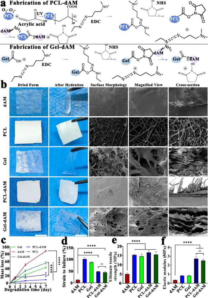
We prepared randomly oriented polycaprolactone (PCL) and gelatin (Gel) nanofiber membranes by electrospinning, activated the carboxyl groups on the membrane surface using the 1-ethyl-3-(3-dimethylaminopropyl) carbodiimide/N-hydroxysuccinimide (EDC/NHS) system to promote condensation with the amine groups on the dAM, and employed mechanical pressing to ensure sufficient contact between them, resulting in two composite membranes: PCL-dAM and Gel-dAM (Fig. 1a). Pure dAM tends to curl into clusters after hydration and is difficult to maintain in its original shape. In contrast, both Gel-dAM and PCL-dAM maintained their integrity after hydration and exhibited improved flexibility and extensibility (Fig. 1b). Scanning electron microscopy (SEM) results revealed that the surface of dAM was smooth and even, with a porous structure visible in the cross-section. In the PCL-dAM composite membrane, PCL nanofibers were tightly bonded with dAM, and due to the tight binding, its internal pores were fewer, presenting a sheet-like structure [14,15]. The Gel-dAM composite membrane, utilizing the excellent interface compatibility of the natural material Gel, formed an organic composite with dAM. This significantly enhanced the flexibility and stability of the Gel-dAM membrane while retaining the natural structure of dAM (Fig. 1b). We placed dAM, PCL nanofiber membrane, Gel nanofiber membrane, PCL-dAM, and Gel-dAM in a buffer solution containing 1 U/mL trypsin to assess their degradation rates by measuring mass loss at 1, 3, and 7 days. The results showed that dAM degraded relatively quickly and was almost completely degraded by the 7th day. After composite with Gel and PCL nanofiber membranes, the degradation rates of Gel-dAM and PCL-dAM significantly decreased, with PCL-dAM showing the lowest degradation rate, with only about 20% degradation by the 7th day (Fig. 1c). Mechanical testing indicated that Gel-dAM and PCL-dAM significantly outperformed dAM in strain, ultimate tensile strength, and elastic modulus (Figs. 1d and f), suggesting that their mechanical strength greatly enhanced that of dAM. Notably, Gel-dAM, based on the natural source of gelatin, not only maintained mechanical strength similar to PCL-dAM but also preserved the natural structure of dAM and exhibited an appropriate degradation rate. For tissue repair, the slow degradation of PCL-dAM can lead to prolonged foreign body stimulation of local tissues, potentially causing fibrous adhesion [16]. Therefore, the natural composite material Gel-dAM demonstrated superior overall performance.
Figure 1
Figure 1.
Preparation and characterization of the PCL-dAM and Gel-dAM composites. (a) Preparation process of PCL-dAM and Gel-dAM. (b) Gross appearance and SEM observation of dAM, PCL, Gel, PCL-dAM and Gel-dAM. Scale bar: 5 mm (dried form), 5 mm (after hydration), 10 µm (surface morphology), 1 µm (magnified view), 3 µm (cross-section). (c) Degradation profiles of dAM, PCL, Gel, PCL-dAM and Gel-dAM. (d–f) Comparison of strain to failure, ultimate tensile strength and elastic modulus for dAM, PCL, Gel, PCL-dAM and Gel-dAM. Data are presented as mean ± standard deviation (SD) (n = 3). ****P < 0.0001. ns: no significance.
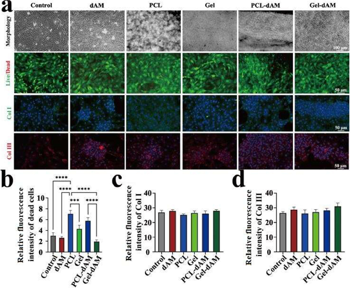
Fibroblasts were co-cultured with dAM, PCL nanofiber membranes, Gel nanofiber membranes, PCL-dAM, and Gel-dAM for 48 h. Microscopic observation revealed that cells in the PCL and PCL-dAM groups exhibited a looser arrangement and an uneven distribution, whereas those in the dAM and Gel-dAM groups displayed tighter adhesion and spreading (Fig. 2a). To assess cell viability, calcein acetoxymethyl ester/propidium iodide (calcein-AM/PI) dual staining was performed, revealing that red fluorescence (indicative of dead cells) was significantly higher in the PCL and PCL-dAM groups compared to the control, dAM, and Gel-dAM groups (Figs. 2a and b). This finding suggests that PCL and PCL-dAM have lower cell compatibility than dAM and Gel-dAM. To evaluate the effects of these materials on fibroblast biological activity, immunofluorescence staining for collagen type Ⅰ (Col Ⅰ) and Col Ⅲ was conducted (Fig. 2a), followed by fluorescence intensity quantification (Figs. 2c and d). While no significant differences were observed between groups, the Gel-dAM group exhibited slightly higher expression levels of Col Ⅰ and Col Ⅲ than the other groups. This result aligns with the cell viability and death staining findings, further suggesting that Gel-dAM supports fibroblast adhesion and growth more effectively while maintaining mechanical properties. Consequently, Gel-dAM demonstrates superior short-term biocompatibility. These findings further highlight the advantages of natural composite materials in enhancing biocompatibility.
Figure 2
Figure 2.
Cell compatibility of dAM, PCL, Gel, PCL-dAM and Gel-dAM co-cultured with fibroblasts. (a) Morphology, calcein-AM/PI staining, immunofluorescence staining of Col Ⅰ and Col Ⅲ for fibroblasts co-cultured with membranes. Scale bar: 100 µm (morphology), 50 µm (calcein-AM/PI, Col Ⅰ and Col Ⅲ). (b–d) Dead cells, Col Ⅰ and Col Ⅲ expression for fibroblasts co-cultured with membranes. Data are presented as mean ± SD (n = 3). ***P < 0.001, ****P < 0.0001.
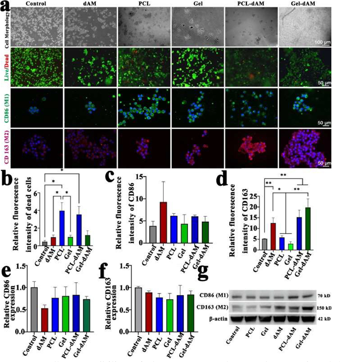
Macrophages play a pivotal role in regulating inflammation and tissue repair. To evaluate the impact of various materials on macrophages, we co-cultured macrophages with dAM, PCL nanofiber membranes, Gel nanofiber membranes, PCL-dAM, and Gel-dAM. Microscopic observations revealed that, compared to the PCL group, cells in the dAM and Gel-dAM groups exhibited more uniform adhesion and more rounded morphologies. Notably, the Gel-dAM group demonstrated the most compact cell distribution and the most favorable overall condition after 48 h of co-culture. While PCL-dAM partially alleviated the excessive aggregation of cells observed in the PCL group, its cell distribution and morphology still lagged behind those in the dAM and Gel-dAM groups (Fig. 3a). Results from calcein-AM/PI dual fluorescence staining further confirmed that the fluorescence expression of dead cells was significantly higher in the PCL and PCL-dAM groups compared to the dAM and Gel-dAM groups (Figs. 3a and b), indicating that Gel-dAM offers a superior cell compatibility profile. Immunofluorescence and western blot analyses demonstrated that both dAM and Gel-dAM significantly upregulated the M2 macrophage surface marker CD163, with Gel-dAM most effectively inducing the transition of macrophages to the M2 phenotype (Figs. 3a, c, d and g), consistent with our previous findings [10]. Notably, there were no significant differences in the expression levels of the CD86 and CD163 genes across groups (Figs. 3e and f), suggesting that the relevant regulatory processes likely occur post-transcriptionally. Collectively, these findings suggest that Gel-dAM preserves the regulatory effects of dAM on macrophages to the greatest extent, thereby enhancing tissue repair.
Figure 3
Figure 3.
Cell compatibility of dAM, PCL, Gel, PCL-dAM and Gel-dAM co-cultured with macrophages. (a) Morphology, calcein-AM/PI staining, immunofluorescence staining of CD86 and CD163 for macrophages co-cultured with membranes. Scale bar: 100 µm (cell morphology), 50 µm (calcein-AM/PI, CD86 and CD 163). (b–d) Dead cells, CD86 and CD163 expression for macrophages co-cultured with membranes. (e, f) Relative expression levels of CD86 and CD163 in macrophages cultured on membranes. (g) Western blot analysis for CD86 and CD163 in macrophages cultured on membranes. *P < 0.05, **P < 0.01. Data are presented as mean ± SD (n = 3).
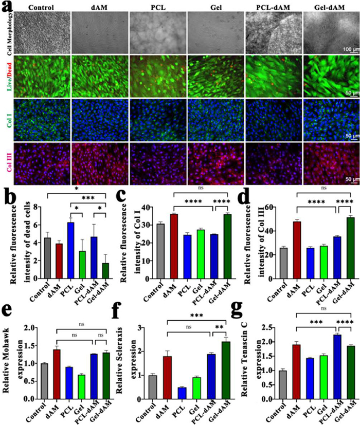
Stem cells play a crucial role in tissue repair. In this study, we co-cultured tendon stem cells with dAM, PCL nanofiber membranes, Gel nanofiber membranes, PCL-dAM, and Gel-dAM to evaluate the effects of different materials on stem cells. Our results demonstrated that tendon stem cells could adhere to the surfaces of all materials and maintain proliferative activity. Notably, the Gel-dAM group exhibited a more spread-out cell morphology and enhanced proliferation. In contrast, PCL and PCL-dAM showed poorer cell adhesion, resulting in reduced proliferation compared to dAM and Gel-dAM. This was further confirmed by calcein-AM/PI staining, which revealed a higher number of dead cells (indicated by red fluorescence) in the PCL and PCL-dAM groups, suggesting that Gel-dAM offers superior cell compatibility (Figs. 4a and b). Immunofluorescence analysis indicated that dAM and Gel-dAM promoted higher synthesis of Col Ⅰ and Col Ⅲ in tendon cells compared to PCL nanofiber membranes, Gel nanofiber membranes, and PCL-dAM, highlighting the potential of the natural structure and growth factors retained in dAM, in combination with gelatin, to create a more conducive microenvironment for tendon stem cell ECM synthesis and deposition (Figs. 4a, c and d). Reverse transcription quantitative polymerase chain reaction (RT-qPCR) results showed no significant differences in Mohawk gene expression among the groups; however, the expression of Scleraxis and Tenascin C was significantly higher in the dAM, PCL-dAM, and Gel-dAM groups compared to the PCL and Gel groups. Notably, Gel-dAM exhibited the highest Scleraxis expression, while PCL-dAM showed increased Tenascin C expression (Figs. 4e–g). These findings suggest that the introduction of nanofiber membranes to enhance the biomechanics and degradation properties of dAM can effectively pres erve its biological activity, offering a novel approach for tissue repair.
Figure 4
Figure 4.
Cell compatibility of dAM, PCL, Gel, PCL-dAM and Gel-dAM co-cultured with tendon stem cells. (a) Morphology, calcein-AM/PI staining, immunofluorescence staining of Col Ⅰ and Col Ⅲ for tendon stem cells co-cultured with membranes. Scale bar: 100 µm (cell morphology), 50 µm (calcein-AM/PI, Col Ⅰ and Col Ⅲ). (b–d) Dead cells, Col Ⅰ and Col Ⅲ expression for tendon stem cells co-cultured with membranes. (e–g) Relative expression levels of Mohawk, Scleraxis and Tenascin C in tendon stem cells cultured on membranes. *P < 0.05, **P < 0.01, ***P < 0.001, ****P < 0.0001. Data are presented as mean ± SD (n = 3).
The advantages of dAM in tissue repair primarily arise from its abundant natural components, including collagen, laminin, and growth factors, which create an optimal biological environment for cells and promote tissue regeneration. However, the deformability and rapid degradation of dAM upon hydration limit its clinical utility. Consequently, enhancing its mechanical properties and degradation characteristics has become a major focus of contemporary research. In this study, the incorporation of PCL and Gel nanofiber membranes into dAM significantly improved both its mechanical properties and degradation rate. The composite PCL-dAM and Gel-dAM exhibited superior strain, tensile strength, and elastic modulus compared to pure dAM, showcasing enhanced mechanical performance and a notably reduced degradation rate. Gel-dAM, in particular, demonstrated more favorable overall performance. As a natural material, Gel not only enhanced the flexibility and stability of dAM but also preserved its biocompatibility. These improvements open new possibilities for the clinical use of dAM, particularly in tissue injury repair and wound dressing. Furthermore, the study revealed that Gel-dAM exhibited superior compatibility with fibroblasts, macrophages, and stem cells compared to PCL-dAM, suggesting that Gel-dAM better supports cell adhesion, proliferation, and differentiation, thereby providing a more ideal microenvironment for tissue repair. Notably, in terms of macrophage immune regulation, Gel-dAM significantly promoted the transformation of macrophages into the M2 phenotype, demonstrating potential anti-inflammatory and repair-enhancing effects, thus offering new perspectives for the application of dAM in tissue regeneration. While this study presents an effective strategy to improve dAM's performance, further research is necessary to precisely control the degradation rate of the composite material, enhance its long-term efficacy, and strengthen its immune-regulatory functions to meet more complex clinical needs. Additionally, the combined application of stem cells and dAM may offer novel approaches to tissue repair, providing a robust theoretical foundation and experimental support for future clinical treatments.
Declaration of competing interest
The authors declare the following financial interests/personal relationships which may be considered as potential competing interests: Dr. Juan Wu (a co-author) is an employee of Wuhan Kangchuang Biotechnology Limited that funded this study.
This work was partially supported by the National Natural Science Foundation of China (No. 82302772) to L.C.; Guizhou Basic Research Project (No. ZK [2023] General 201) to L.C.; Medical Research Union Found for High-quality health development of Guizhou Province (No. 2024GZYXKYJJXM0041) to L.C.; Science and Technology Fund of Guizhou Provincial Health Commission (No. gzwkj2025–353) to X.S.; and partially supported by Wuhan Kangchuang Biotechnology Co., Ltd. to J.W.
[1]
R.A. Salah, I.K. Mohamed, N. El-Badri, J. Mol. Histol. 49 (2018) 289–301. doi: 10.1007/s10735-018-9768-1
[2]
A. Firouzeh, I. Shabani, R. Karimi-Soflou, A. Shabani, Colloids Surf. B 240 (2024) 113974. doi: 10.1016/j.colsurfb.2024.113974
Figure 1
Preparation and characterization of the PCL-dAM and Gel-dAM composites. (a) Preparation process of PCL-dAM and Gel-dAM. (b) Gross appearance and SEM observation of dAM, PCL, Gel, PCL-dAM and Gel-dAM. Scale bar: 5 mm (dried form), 5 mm (after hydration), 10 µm (surface morphology), 1 µm (magnified view), 3 µm (cross-section). (c) Degradation profiles of dAM, PCL, Gel, PCL-dAM and Gel-dAM. (d–f) Comparison of strain to failure, ultimate tensile strength and elastic modulus for dAM, PCL, Gel, PCL-dAM and Gel-dAM. Data are presented as mean ± standard deviation (SD) (n = 3). ****P < 0.0001. ns: no significance.
Figure 2
Cell compatibility of dAM, PCL, Gel, PCL-dAM and Gel-dAM co-cultured with fibroblasts. (a) Morphology, calcein-AM/PI staining, immunofluorescence staining of Col Ⅰ and Col Ⅲ for fibroblasts co-cultured with membranes. Scale bar: 100 µm (morphology), 50 µm (calcein-AM/PI, Col Ⅰ and Col Ⅲ). (b–d) Dead cells, Col Ⅰ and Col Ⅲ expression for fibroblasts co-cultured with membranes. Data are presented as mean ± SD (n = 3). ***P < 0.001, ****P < 0.0001.
Figure 3
Cell compatibility of dAM, PCL, Gel, PCL-dAM and Gel-dAM co-cultured with macrophages. (a) Morphology, calcein-AM/PI staining, immunofluorescence staining of CD86 and CD163 for macrophages co-cultured with membranes. Scale bar: 100 µm (cell morphology), 50 µm (calcein-AM/PI, CD86 and CD 163). (b–d) Dead cells, CD86 and CD163 expression for macrophages co-cultured with membranes. (e, f) Relative expression levels of CD86 and CD163 in macrophages cultured on membranes. (g) Western blot analysis for CD86 and CD163 in macrophages cultured on membranes. *P < 0.05, **P < 0.01. Data are presented as mean ± SD (n = 3).
Figure 4
Cell compatibility of dAM, PCL, Gel, PCL-dAM and Gel-dAM co-cultured with tendon stem cells. (a) Morphology, calcein-AM/PI staining, immunofluorescence staining of Col Ⅰ and Col Ⅲ for tendon stem cells co-cultured with membranes. Scale bar: 100 µm (cell morphology), 50 µm (calcein-AM/PI, Col Ⅰ and Col Ⅲ). (b–d) Dead cells, Col Ⅰ and Col Ⅲ expression for tendon stem cells co-cultured with membranes. (e–g) Relative expression levels of Mohawk, Scleraxis and Tenascin C in tendon stem cells cultured on membranes. *P < 0.05, **P < 0.01, ***P < 0.001, ****P < 0.0001. Data are presented as mean ± SD (n = 3).

 DownLoad:

 下载:
下载:


 下载:
下载:
