Citation:
Sajid Hussain, Omer M.A. Dagah, Essam A.M.S Obaid, Peng Jin, Ovas Ahmed Dar, Muhammd Irfan, Yiming Qi, Qinghua Wu, Ming Jin, Tengli Zhang, Lei Luo. Chitosan as oral absorption enhancer and inhibitor: A comprehensive review[J]. Chinese Chemical Letters,
2026, 37(1): 111273.
doi:
10.1016/j.cclet.2025.111273
Chitosan as oral absorption enhancer and inhibitor: A comprehensive review
English
Chitosan as oral absorption enhancer and inhibitor: A comprehensive review
Received Date:
01 November 2024 Accepted Date:
28 April 2025 Revised Date:
25 April 2025 Available Online:
15 January 2026
Abstract:
Chitosan (CS), a natural polymer derived from chitin found in the exoskeletons of crustaceans, has garnered significant interest in the pharmaceutical field due to its unique properties, including biocompatibility and biodegradability. In recent years, various studies have reported that CS can affect drug bioavailability, and interestingly, it works as an oral absorption enhancer and inhibitor. This review offers an in-depth analysis of the mechanisms underlying such a phenomenon and supports its application as a pharmaceutical excipient. CS enhances oral drug absorption through various mechanisms, such as interaction with the intestinal mucosa, tight junction modulation, inhibition of efflux transporters, enzyme inhibition, solubility and stability enhancement, and complexation. On the other side, CS exhibits the ability to inhibit the absorption of certain drugs by adsorbing to lipids and sterols, modulating bile acids and gut microbiota, altering drug-cell interaction at the polar interface, and mucus-mediated entrapment and interference. Future potential pharmaceutical research in this field includes elucidating the underneath absorption relevant mechanisms, rational use in formulations as excipient, exploring functional CS derivatives, and developing CS-based drug delivery systems. This comprehensive review highlights CS’s versatile and significant role in enhancing and inhibiting oral drug absorption, providing insights into the complexities of drug delivery and the potential of CS to improve therapeutic outcomes.
Oral drug absorption is the process by which a drug is absorbed into the bloodstream after being ingested orally. The oral route normally involves the drug passing through the mouth to the stomach and then into the small intestine. Most of the absorption occurs in the small intestine. Then, the absorbed drugs enter the bloodstream through the intestinal wall and are transported to various tissues and organs. Numerous factors can affect the absorption of drugs, including the physicochemical properties of the drug, such as molecular weight, solubility, lipophilicity, ionization, stability, the formulation of the drug product, and the physiological conditions of the gastrointestinal tract (GIT) [1,2]. The bioavailability of an orally administered drug can be low due to poor absorption or rapid metabolism and elimination [3,4]. The drugs encounter several physiological barriers when taken orally that can inhibit their absorption or lower their bioavailability [5,6]. Salivary enzymes in the buccal cavity, like amylase, can cause drug degradation. However, since the drug stays in the oral cavity for a short time, the impact of these enzymatic barriers is generally low [7]. Once swallowed, the esophagus helps move the drug to the stomach through peristalsis but it does not play a role in drug absorption or digestion.
Oral drugs face physiological barriers in the stomach before entering the small intestine (Fig. S1 in Supporting information). The presence of enzymes like lipase in the stomach, which digests fat, can cause drug hydrolysis. Enzymatic degradation can hinder drug dissolution, alter effective drug concentration, and affect drug absorption [8]. Supersaturation or precipitation will likely occur in the stomach if the drug has altered solubility at different pH values. After the drug crosses these biochemical barriers, its intestinal permeability determines its fate [9]. Pancreatic enzymes in the duodenum can lead to first-pass metabolism, reducing drug bioavailability. The small intestinal mucosa has villi that increase surface area and aid drug absorption. Drugs can be absorbed through transcellular or paracellular pathways, with hydrophobic drugs preferring the transcellular pathway and hydrophilic drugs the paracellular pathway. The lipid bilayer of the GIT’s biological membrane creates a barrier that can hinder drug movement. Molecular weight and charge can also affect drug absorption, with positively charged molecules adhering to negatively charged mucins [10].
Drug absorption in the colon is restricted by solubility and non-specific interactions, where drugs can adhere to feces, mucus, or other secretions. Hydrophilic drugs exhibit greater absorption compared to hydrophobic ones, owing to the colon’s capacity to absorb water. The primary problem in oral drug administration is the formulation of macromolecules, which frequently have inadequate solubility, are prone to enzymatic breakdown, and demonstrate restricted oral absorption. These factors can affect the drug’s ability to traverse the intestinal membrane and enter the bloodstream, ultimately impacting its safety and efficacy. Therefore, a thorough understanding of a drug’s properties is essential for optimizing its formulation and predicting its behavior in the body [11].
Absorption enhancers can be used as one of the easiest ways to improve the bioavailability of orally administered drugs. Absorption enhancers are functional excipients that can increase the intestinal epithelium’s permeability or alter the drug’s physicochemical properties to enhance its absorption. Absorption enhancers’ examples include surfactants, bile salts, cholesterol, glycerides, salicylates, chelating agents. The use of surfactants as an absorption enhancer in one study increased the bioavailability of a poorly soluble drug by over 200%. Conversely, absorption inhibitors can be used to decrease the absorption of drugs [12-15]. Absorption inhibitors are functional excipients that can decrease the permeability of the intestinal epithelium or change the physicochemical properties of the drug to reduce its oral absorption. Among the absorption inhibitors include polymers, transporter and enzyme inhibitors as well as some dietary fibers like glucomannan [16]. Absorption enhancers and inhibitors can benefit drug development to improve the bioavailability and efficacy of orally administered drugs. However, careful consideration must be given to their safety and potential adverse effects on the GIT and the whole body.
Chitosan (CS) is a versatile natural polymer obtained from chitin, a complex carbohydrate present in the outer shells of several crustaceans, including crabs, lobsters, and shrimps [17-19]. The key component of chitin is N-acetyl glucosamine (GlCN), a molecule that links through β-1, 4-glycosidic linkages. Contrasting to chitin, CS features a combination of N-acetyl-D-GlCN and D-GlCN, making it a distinctive copolymer evident in its structural representation (Fig. S2 in Supporting information). The conversion of chitin into CS includes a process known as partial deacetylation, the removal of acetyl groups from chitin, which can be achieved through either chemical means, using strong alkaline substances, or enzymatically, with specific enzymes like chitin deacetylase facilitating the process. The subsequent CS polymer is noteworthy for its solubility in acidic milieus, along with its commendable characteristics, like being biocompatible, biodegradable, and non-toxic to biological systems [20-23]. CS has attracted attention for potential uses across various fields due to its exceptional properties [18,24-26]. Among other applications, it demonstrates potential in facilitating drug delivery, enhancing wound healing processes, inflammatory bowel disease, and extending the shelf life of food products [19,27-33]. This exploration into CS’s capabilities underscores its importance in advancing both food preservation and medical technologies [27,28,34,35]. One of its most promising applications is as an oral absorption enhancer. CS has exhibited the ability to enhance the oral absorption of certain drugs by improving their solubility, enhancing their permeability across the intestinal membrane, and reducing their metabolism and elimination [36-40]. The CS’s mucoadhesive properties make it an effective absorption enhancer, which permits it to bind to the intestinal epithelium and increase drug residence time, and its ability to tight junctions opening between epithelial cells, allowing for enhanced paracellular transport [9]. CS has also demonstrated interaction with certain transporters and enzymes in the intestinal epithelium, causing altered drug pharmacokinetics and increased absorption [41-43]. Overall, these unique properties of CS make it an ideal candidate to improve oral drug delivery.
On the other hand, CS has also been exposed to inhibit the oral absorption of certain drugs, which can have implications for the efficacy of drug therapy. This review will summarize the current literature on CS as an oral absorption enhancer and inhibitor, focusing on the mechanisms and underlying its effects. We will also explore the latest developments in CS-based oral drug delivery systems and discuss the future directions for research in this field.
2.
CS as an oral absorption enhancer
The mechanism of action of CS as an absorption enhancer is believed to be multi-functional and involves several factors. Numerous mechanisms have been proposed to explain CS’s ability to enhance oral absorption, including interaction with the intestinal mucosa, tight junction modulation, inhibition of efflux transporters and enzymes, solubility and stability enhancement, and complexation.
2.1
Interaction with the intestinal mucosa
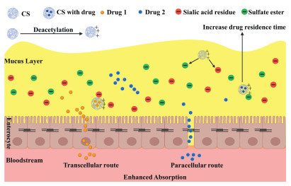
CS has gained significant attention in the pharmaceutical field due to its exceptional mucoadhesive properties. This cationic polymer demonstrates a remarkable ability to interact with negatively charged mucosal surfaces found in various body parts, including the GIT, nasal passages, ocular surfaces, and other mucosal linings. The mechanism underlying this mucoadhesion primarily revolves around electrostatic forces. The mucoadhesive characteristics of CS enhance with the degree of deacetylation, as increased deacetylation leads to a bigger quantity of free amino groups in the polymer. The positively charged amino groups in CS engage in electrostatic interactions with the negatively charged sialic acid residues and sulfate esters found in mucosal glycoproteins. Hence, the higher the number of amino groups, the stronger the adhesion will be [44,45]. Additionally, hydrogen bonding and hydrophobic interactions play a crucial role in this process, enhancing the stability of the CS-drug complex at the mucosal surface. One of the most significant benefits of CS’s mucoadhesive properties is the increased residence time of drugs at their absorption sites (Fig. 1). By adhering to mucosal surfaces, CS prolongs the presence of the drug, allowing for an extended absorption period, which is crucial for improving the drug’s bioavailability [46-49]. This feature is especially beneficial for drugs that require prolonged contact with mucosal surfaces to achieve effective absorption. In addition to prolonging residence time, CS protects drugs from enzymatic degradation within the mucosa. It also facilitates the drug’s penetration through the mucosal barrier, further enhancing its absorption. This dual role of protection and enhanced absorption makes CS invaluable in various drug delivery systems [50].
Figure 1
Figure 1.
CS interaction with intestinal mucosa. The positively charged amino groups of CS interact with the negatively charged sialic acid residues (red) and sulfate esters (green) in the mucosal surface. Mucoadhesion improves with the degree of deacetylation; therefore, the greater the number of amino groups, the stronger the adhesion.
In nasal drug delivery, for instance, CS was extensively researched, particularly for drugs targeting the central nervous system (CNS). It enhances the nasal absorption of drugs like insulin, sumatriptan, and vaccines by adhering to the nasal mucosa and increasing their permeation [51,52]. Amin et al. developed CS nanoparticles (NPs) whose mucoadhesive properties were markedly improved by applying alginate (ALG) coatings. This modification resulted in greater mucin attachment, reflecting enhanced mucoadhesion. Such mucoadhesive NPs can adhere to the mucosal linings of the GIT, extending their stay at the absorption sites. CS’s mucoadhesive properties allow it to be closely associated with the mucosal surface of the GIT. This close contact can facilitate the passage of drug molecules via both transcellular (through cells) and paracellular (between cells) routes. Additionally, CS’s ability to form complexes with negatively charged molecules, such as drug molecules, can enhance their transport across the intestinal epithelium. This prolonged presence at the absorption site potentially leads to more efficient uptake of the encapsulated drugs or vaccines [53]. Furthermore, CS’s role extends to transdermal and topical drug delivery. It is used in transdermal patches and topical formulations to enhance the absorption of drugs through the skin or mucosal tissues. This application is particularly relevant for drugs like lidocaine and diclofenac, providing a more effective and targeted delivery mechanism [54].
Several studies have illuminated the dynamics between CS microspheres and mucin. Research by He et al. unveiled that mucin is highly adsorptive to CS surfaces, showcasing a notable affinity [55]. Similarly, Dhawan et al. have confirmed that the interaction is mainly due to the opposite charges of CS and mucin; CS carries a positive charge, while mucin is negatively charged. Their methodology spanned both in vitro assays and in vivo experiments using animal models to establish these findings [56]. Furthermore, Lehr et al. added that CS’s mucoadhesive capabilities are directly correlated with its molecular weight, observing that even with repeated application, CS films maintain their adhesive properties [57]. The fundamental mechanism of this adhesion was identified as electrostatic attraction, particularly potent in acidic conditions. Moreover, the degree of mucoadhesion was positively correlated with CS’s degree of deacetylation and inversely with its level of cross-linking [58].
The concept of thiomers introduces a novel angle to enhancing mucoadhesion. Thiomers, polymers modified to feature a thiol group (-SH) on their surface, exhibit enhanced mucoadhesive properties. This enhancement in mucoadhesion upon integrating a thiol moiety into CS, leading to the creation of thiolated CS (CS-SH), marks a significant advancement [58]. Among the developed CS-SHs, the CS-thioglycolic acid (CS-TGA) conjugate, CS-4-thiobutyl-amidine (CS-TBA) conjugate, CS-cysteine (CS-Cys) conjugate, and CS-thioethylamidine (CS-TEA) conjugate stand out [58]. The introduction of thiolation enhances mucoadhesion by promoting stronger interactions through disulfide bonds between the thiol groups of CS and the cysteine-rich regions in mucosal glycoproteins [56].
The enhancement in mucoadhesive strength due to covalent bonding in thiolated CS surpasses the initial adhesive interactions facilitated by the charge differences between CS and mucin. Thiolation primarily targets the amino group located at the second position on the GlCN unit within CS, enabling the attachment of a thiol group. This process results in the formation of either amide or amidine bonds, significantly amplifying the mucoadhesive properties of CS. Evidence of this enhanced interaction is observed with the CS-TGA derivative, which exhibits a tenfold increase in mucoadhesion compared to its non-thiolated counterpart. The CS-TBA conjugate further advances these adhesive capabilities [59]. However, the susceptibility of thiol groups to oxidative processes necessitates careful reaction conditions, either within an inert atmosphere or under acidic conditions (pH < 5), to minimize the formation of disulfide bonds. Thiolation introduces covalent bonds at the amino group of the GlCN unit, using carbodiimides like 1-ethyl-3-(3-dimethylaminopropyl)carbodiimide (EDC) for amide bond creation and 2-iminothiolane for amidine bonds. The latter method is preferred due to its ability to maintain a higher presence of free thiol groups, thereby reducing undesired disulfide bonding [60]. As a result, CS-SH demonstrates an impressive increase in mucoadhesive strength, up to 150 times that of original CS [59].
Trimethyl CS (TMC) represents another notable advancement in CS modification. Produced through the reductive methylation of CS, TMC is a partially quaternized derivative that exhibits improved mucoadhesion. This modification broadens the applicability and effectiveness of CS in biomedicine, particularly in enhancing mucosal delivery systems [61,62]. Recently, a study by Jalal et al. focused on developing mucoadhesive NPs for ciprofloxacin delivery using methacrylated chitosan (MeCS) through an ionic gelation technique involving sodium tripolyphosphate (TPP). They compared the performance of these NPs with those made from unmodified CS. The study included mucoadhesion tests on sheep abomasum mucosa, revealing that the MeCS NPs, when optimized with TPP, exhibited superior retention capabilities compared to their unmodified CS counterparts, with adherence rates of 96% and 88%, respectively. This demonstrates the significant promise of MeCS NPs in drug delivery applications [63]. In a separate research effort, Wen and colleagues explored the critical molecules involved in chitooligosaccharide (COS) transport by conducting transcriptome and proteome analyses. Their enrichment analysis revealed that the differentially expressed genes in the duodenum of COS-treated mice were mainly associated with transmembrane and immune functions, with notable upregulation of B2m, Itgb2, and Slc9a1. Notably, inhibition of Slc9a1 reduced COS transport efficiency both in vitro (MODE-K cells) and in vivo mice, whereas Slc9a1 overexpression in MODE-K cells significantly enhanced fluorescein isothiocyanate (FITC)-COS transport compared to controls. Molecular docking analysis suggested a stable interaction between COS and Slc9a1 via hydrogen bonds, underscoring Slc9a1′s vital role in COS transport within mice. This discovery is crucial for enhancing COS’s efficacy as a drug adjuvant [64].
2.2
Tight Junctions modulation
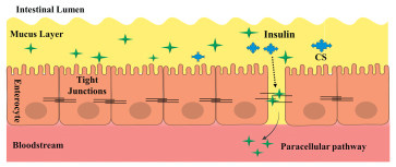
CS’s role in opening tight junctions is a pivotal aspect of its utility in drug delivery, especially for facilitating the transport of larger and more hydrophilic drugs across epithelial barriers. CS can modulate the tight junctions between epithelial cells in the intestinal mucosa, leading to increased paracellular transport of drugs and other molecules, facilitating the paracellular transport of drug molecules and nutrients. This effect is thought to be due to the ability of CS to interact with the negatively charged components of the tight junctions, such as claudins, zonola occludin-1 (ZO-1), and occludin, and disrupt their assembly and stability [65-68]. The interaction between CS and these tight junction proteins leads to the temporary modulation or opening of these junctions. This modulation reduces the barrier function of the tight junctions, thereby rendering the paracellular route, typically restrictive to larger molecules, more permeable (Fig. 2). This is particularly significant in the pharmaceutical field, as many drugs, including macromolecules like peptides, proteins, and nucleic acids, require effective transport mechanisms to cross these barriers and reach their target sites within the body. One of the most notable benefits of this mechanism is the enhanced paracellular transport it facilitates. By opening tight junctions, CS allows larger and more hydrophilic drugs, which usually cannot permeate through the cell membrane via the transcellular pathway, to be transported across epithelial barriers effectively [37,69].
Figure 2
Figure 2.
Modulation of tight junctions by CS. CS interacts with the tight junction’s proteins, such as claudins, ZO-1, and occludin, causing temporary opening of these junctions, thus rendering the paracellular route.
For instance, insulin, a peptide hormone crucial for regulating blood glucose levels, is a prime example of a macromolecule whose absorption is significantly enhanced by CS. This enhancement occurs via the paracellular route, allowing insulin to bypass the usual barriers it faces in traditional drug delivery methods. A study conducted by Prego et al. highlighted this capability, displaying that insulin-loaded CS NPs enhanced the absorption of insulin across the intestinal epithelium, indicating the opening of tight junctions [70]. A recent study by Pratap-Singh et al. demonstrates MNA-TG-CS as a promising material for improved peptide oral delivery [71]. Additionally, CS’s application extends to the delivery of vaccines and nucleic acids like DNA, siRNA and miRNA [72]. These applications are particularly important in the context of gene therapy and vaccination, where efficient and targeted delivery of nucleic acids is paramount. CS facilitates the transport of these substances across epithelial barriers, opening new avenues in drug delivery and therapeutic interventions. The potential of CS in enhancing the paracellular transport of nucleic acids has also been discussed in the literature. For example, Illum et al. explored the capabilities of CS as a pharmaceutical excipient, particularly in the context of nucleic acid delivery across mucosal barriers [73].
2.3
Inhibition of efflux transporters
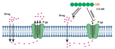
CS can inhibit certain efflux transporters, such as P-glycoprotein (P-gp), which are responsible for pumping drugs out of cells and reducing their absorption. By inhibiting these transporters, CS can increase the intracellular drug concentration, ultimately leading to enhanced oral absorption. CS and its derivatives, particularly CS-SH, have been studied for their potential to inhibit efflux transporters like P-gp [74-76]. These transporters play a crucial role in multi-drug resistance (MDR), where they efflux various drugs out of cells, thereby reducing their effectiveness. CS-SH enhances the transmucosal transport of substrates by inhibiting the efflux action of P-gp, potentially improving the bioavailability and therapeutic efficacy of various drugs (Fig. 3). This inhibition mechanism is believed to involve the binding of CS’s SH groups to P-gp, which can interfere with its efflux function [77].
Figure 3
Figure 3.
CS causing inhibition of P-gp efflux transporter. CS-SH inhibits the P-gp efflux transporter, thereby increasing the bioavailability of drugs.
Multi-drug-resistant proteins (MRPs) such as P-gp and MRP1, which play a crucial role in decreasing the effectiveness of various active pharmaceutical ingredients (APIs) and in making tumors and bacteria resistant to chemotherapy and antibiotics, respectively, have cysteine components in their membrane-channel structures. CS-SHs can interact with these efflux pumps by forming disulfide bonds, effectively inhibiting their function [78-81]. This interaction mechanism was validated by Sakloetsakun et al. who demonstrated that the extent of P-gp inhibition is significantly influenced by the number of thiol groups bonded to CS, with a higher concentration of thiol groups leading to more significant inhibition [82]. In a recent study by Kim et al. a PTX-loaded nanostructured lipid carriers (P-NLC) was prepared and coated with positively charged CS to create P-NLC-CS, which was subsequently conjugated to siRNA (P-NLC-CS-siRNA). The study investigates the use of CS-coated NLC to enhance drug delivery by reducing P-gp-mediated efflux in MCF-7/ADR cells. When P-NLC-CS-siRNA was injected subcutaneously into BALB/c nude mice with tumors, it resulted in a decrease in tumor size and significantly lowered programmed cell death ligand-1 (PD-L1) mRNA levels in both in vitro and in vivo experiments. Furthermore, the in vivo study indicated an increase in tumor-specific CD4+ and CD8+ T cell responses within the tumor tissue after treatment. These results suggest that CS-coated NLC for the co-delivery of paclitaxel (PTX) and PD-L1 siRNA could be a promising strategy for chemoimmunotherapy [83]. Furthermore, enhancing the reactivity of thiol groups in the CS-SH by introducing pyridyl derivatives to create S-protected forms has been exposed to further intensify the inhibition of P-gp [84]. Huo et al. demonstrated the effectiveness of an efflux pump inhibitor by initially verifying the P-gp inhibition capability of N-mercaptoacetyl-N′-octyl-O, N″-glycol CS using Rhodamine-123 as a P-gp substrate. Subsequently, this modified CS was utilized to create micelles encapsulating PTX, a chemotherapeutic agent. The use of this delivery system resulted in a significant enhancement of PTX bioavailability in rats [85]. Kang et al. developed a cancer therapy approach using polyethylene glycol (PEG)-enhanced doxorubicin (Dox)/carboxymethyl chitosan (CMC)/gold NPs (AuNPs). CMC acts as a reducing agent and stabilizer in creating AuNPs, onto which Dox is loaded. PEG is bonded to these AuNPs to extend systemic exposure and circulation time. PEGylated Dox/CMC-AuNPs (Dox/CMC-AuNPs-PEG) showed significant improvements over non-PEGylated versions, including smaller hydrodynamic size, less negative zeta potential, and higher Dox loading efficiency. Both NP types effectively inhibited A549 cancer cell proliferation and reduced efflux ratio, suggesting the ability to counteract P-gp-mediated MDR. In vivo studies disclosed a lower drug clearance rate and longer half-life in rats compared to non-PEGylated counterparts. This suggests Dox/CMC-AuNPs-PEG as a promising nanotherapeutic strategy for bypassing MDR in cancer treatments and enhancing drug presence in the bloodstream [86].
2.4
Enzyme inhibition
CS has the ability to inhibit the activity of various enzymes in the intestinal lumen, such as lipases and α-glucosidases, which can degrade and metabolize drugs. This effect is thought to be due to the cationic nature of CS, which enables it to bind and form complexes with the negatively charged active sites of these enzymes. By reducing drug metabolism, CS can prolong the presence of the drug in systemic circulation, leading to improved bioavailability [87]. Enzyme inhibition has also been demonstrated by CS-SH. This effect appears to be caused by the development of disulfide bridges between the cysteine substructures of enzymes and the thiol groups of CS. This impact is further enhanced by CS-SHs’ capacity to chelate divalent cations, which are necessary cofactors for the majority of enzymes to remain active. As a result, CS-thioglycolic acid was found to be an inhibitor of several enzymes, including trypanothione reductase, an essential enzyme for parasitic protozoa like leishmania and trypanosomes, and the drug-metabolizing CYP3A4 and CYP2A6, which together contribute to the metabolism (about 60%) of administered APIs [88]. Additionally, an inhibitory action against myeloperoxidase and metalloproteinases, which are crucial enzymes for wound healing, was discovered for CS 4-thiobutylamidine. On the other hand, overexpression of these enzymes can impede the healing process and result in chronic wounds [89,90].
2.5
Solubility and stability enhancement
CS’s capacity to enhance the solubility and stability of drugs significantly contributes to its suitability in pharmaceutical applications. This biopolymer’s special chemical properties allow it to interact with various drugs, particularly those with poor water solubility, improving their overall effectiveness and bioavailability. This property is especially beneficial for drugs with poor water solubility, a common challenge that limits the bioavailability of many therapeutic agents. The mechanism by which CS enhances solubility and stability lies in its cationic nature. CS can form complexes with drugs through various interactions, including ionic bonding, hydrogen bonding, and hydrophobic interactions. These interactions can lead to the formation of drug-CS complexes or micelles, effectively increasing the water solubility of poorly soluble drugs. Additionally, CS can modify the chemical environment surrounding a drug molecule, which can be crucial in preventing the drug’s degradation and improving its stability over time. This modification is key to maintaining the drug’s efficacy, especially in aqueous environments where many drugs are unstable [91-93].
In oral drug delivery, the solubility of a drug is a determining factor for its absorption in the gastrointestinal tract. CS’s ability to enhance the solubility of drugs plays a pivotal role in this route of administration. For instance, curcumin, renowned for its antioxidant and anti-inflammatory properties, struggles with poor water solubility. Research by Anitha et al. demonstrated that CS could be used to significantly enhance the solubility and oral bioavailability of curcumin, thereby facilitating its therapeutic application and improving its efficacy. In their study, curcumin-O-CMC NPs were developed to increase oral bioavailability. These NPs were characterized using various methods, and they exhibited 87% efficiency in entrapping curcumin in O-CMC. Cytotoxicity studies revealed curcumin-O-CMC NPs were toxic to cancer cells but non-toxic to normal cells. Overall, O-CMC is a promising nanomatrix for drug delivery applications [94]. Stability is a critical aspect of injectable drugs, where maintaining the active form of the drug is essential for efficacy. CS finds a significant application here as well. PTX, an anticancer drug with low solubility in water, exemplifies this application. Research by Zhang et al. presented that CS NPs could improve the solubility and stability of PTX, enhancing its effectiveness in cancer treatment [91]. This application is particularly important in chemotherapeutic agents, where the efficacy of the drug is paramount.
2.6
Complexation
CS’s cationic nature enables ionic complex formation with anionic drugs, enhancing oral absorption and improving drug bioavailability. The mechanism and application of these ionic interactions have been explored in several significant studies, demonstrating CS’s versatility and effectiveness in drug delivery. CS is inherently cationic due to its amino groups, which become positively charged in acidic environments. This allows CS to readily interact with negatively charged components, including cell membranes and anionic drugs. The primary mode of interaction is electrostatic, leading to the formation of stable complexes with these anionic entities. The formation of such complexes is crucial in enhancing the solubility, stability, and bioavailability of drugs, particularly those with poor solubility or stability under physiological conditions [95-99].
The study by Grozdova et al. showcases CS’s role in creating multi-liposomal complexes (MLCs) for intracellular drug delivery. Here, CS enhances the drug’s absorption and delivery efficiency by encapsulating it within these complexes, facilitating its transport and uptake by cells. This approach leverages CS’s ionic interaction capabilities to improve drug delivery to targeted intracellular sites. CS NPs cross-linked with sulfate-anions form CS NPs, used as scaffolds for negatively charged cardiolipin/egg lecithin liposomes loaded with Dox. These MLCs effectively transmit Dox into the cytoplasm of cells, increasing efficiency by 4–5 times. MLCs showed effects on mouse fibroblasts, MCF-7 cells, and OVCAR-8 cells, demonstrating their potential for effective drug delivery [95]. The research by Bezrodnykh et al. highlights how CS forms complexes with anionic molecules such as sodium dodecyl sulfate (SDS), enhancing its antimicrobial properties. This property broadens CS’s applications in developing antimicrobial agents. This study demonstrated that shorter CS chains interact more preferentially with SDS and generate bigger microparticles. The study reveals that microparticle formation in CS is a cooperative electrostatic interaction, primarily due to hydrogen bonding, and can be used for pharmaceutical hydrocolloids, cosmetic products, and Pickering emulsions with strong anionic surfactants, influenced by ionic strength, urea, and temperature [98]. In the study by Shao et al. CS was utilized to develop controlled-release tablet formulations. The ionic interactions between CS and anionic polymers are key in modulating the drug release rate, enabling sustained release and improved bioavailability of water-soluble drugs [99]. The application of CS in cancer therapy is highlighted by Montero et al. who demonstrate the use of ionic-crosslinked CS in forming biocompatible NPs for efficient delivery of antitumor drugs. CS’s ability to form complexes with cancer drugs enhances their delivery to tumor sites, offering a promising strategy in cancer treatment [100].
These mechanisms highlight the potential of CS as an oral absorption enhancer. A summary of CS’s role as an oral absorption enhancer can be found in Table S1 (Supporting information), which outlines how CS and its derivatives influence the absorption of various drugs. The continued investigation of these mechanisms and the development of new CS derivatives can help improve the efficacy of CS-based drug delivery systems, ultimately leading to better therapeutic outcomes for patients. It is important to note that the effectiveness of CS as an oral absorption enhancer can vary based on factors such as CS characteristics (molecular weight, degree of deacetylation), drug properties, and formulation design. Our group is also discovering the fascinating aspect of CS in oral drug absorption. In a recent study, we inspected COS as an effective, safe, and low-cost oral drug absorption enhancer. We exposed the hydrodynamic law of interaction between COS and the intestinal mucus layer, which was related to absorption promoting mucus structural reconstruction [101]. Additionally, further research is still ongoing to optimize the use of CS in enhancing oral drug absorption.
3.
CS as an oral absorption inhibitor
CS has an exceptional ability to act as an oral absorption inhibitor in addition to its role as an enhancer. There are also reports on CS’s effectiveness in inhibiting oral absorption of drugs. CS exhibits the ability to inhibit the absorption of certain drugs by adsorbing lipids and sterols, modulating bile acids and gut microbiota, altering drug-cell interaction at the polar interface, and mucus-mediated entrapment and interference. This dual functionality allows for the modulation of drug absorption, which can be useful in controlling drug release, reducing drug-drug interactions, and managing nutrient absorption.
3.1
Lipids and sterols adsorption
CS exerts its beneficial effects on cholesterol and fat metabolism through several key mechanisms [102]. Firstly, it creates films with negatively charged surfaces, enabling interactions with various molecules. Importantly, CS is resistant to specific hydrolysis by human digestive enzymes, but it can undergo limited digestion due to the activity of bacterial flora and nonspecific enzymes. In the stomach, CS selectively binds to negatively charged molecules, such as fatty acids and bile acids, thus reducing the absorption of fats from the gastrointestinal tract. Additionally, it disrupts the emulsification of neutral lipids, including cholesterol, by forming hydrophobic bonds with them [103-105]. These combined actions contribute to CS’s potential cholesterol-lowering and fat-reduction effects, making it a promising candidate for dietary interventions to manage cholesterol levels and obesity [106]. This suggests that CS might potentially interfere with the absorption of steroid and lipid-based medications [107]. Additionally, it has been observed that CS can lead to a reduction in the absorption of fat-soluble vitamins such as vitamins A, D, E, and K. This is primarily due to CS’s ability to bind with negatively charged lipids and bile acids in the gastrointestinal tract, potentially affecting the bioavailability of these essential vitamins [108-110]. In a case report authored by Shao-Sung Huang and colleagues, the study highlighted the potential of CS to enhance the effects of warfarin. This enhancement is believed to occur through CS’s interference with the absorption of vitamin K. Warfarin is a well-known anticoagulant that exerts its effects by hindering the function of vitamin K-dependent coagulation factors. When combined with CS, which has the capacity to disrupt vitamin K absorption, there is a likelihood of intensifying the anticoagulation effects of warfarin. This observation underscores the importance of considering the interaction between CS and warfarin in clinical settings and the need for careful monitoring when these substances are used concomitantly [111].
Unlike CS, CS sulfate is known for its anticoagulant properties, primarily due to its sulfate groups, which are crucial for this activity [112]. CS sulfate notably impacts the intrinsic coagulation pathway and the final stages of the coagulation cascade, as identified by Hirano et al. who found that it extends the duration of both activated partial thromboplastin time and thrombin time. On the other hand, it rarely affects prothrombin time [113]. In a case where a patient consumed CS instead of CS sulfate, it is important to note that CS, being positively charged in the pH environment of the gastrointestinal tract, attaches to negatively charged lipids and bile acids. This interaction can hinder the absorption of fat-soluble vitamins such as A, D, E, and K. Research in animals has demonstrated that CS reduces lipid levels, a finding also observed in human studies. This lipid-lowering capability of CS might enhance the anticoagulant effect of warfarin, which operates by blocking vitamin K–dependent clotting factors [114,115]. The adverse reaction experienced by the patient is likely linked to CS. Nevertheless, definitive proof would require well-designed clinical trials. While variations in vitamin K consumption can affect warfarin response, this patient did not alter their diet, indicating another mechanism might be at play. One theory was that CS could potentially inhibit CYP2C9, a key enzyme in metabolizing warfarin. However, most CS passes through the body without being absorbed, making CYP2C9 inhibition by CS unlikely, and no studies have supported this hypothesis [116].
3.2
Bile acids modulation
Bile acids are ionic amphiphilic compounds characterized by their steroid-like structure. Their primary function lies in the solubilization and transportation of lipids and certain medications across hydrophobic barriers in the body. The complex mechanism through which bile salts enhance absorption by facilitating the permeation of various molecules and drugs across epithelial barriers remains an area of ongoing research, with their tendency to agglomerate at concentrations surpassing the critical micelle concentration being a notable characteristic [117]. Additionally, bile acids exhibit the capacity to surmount gastrointestinal challenges, facilitate the absorption of physically complexed or chemically conjugated drugs through transporters, improve drug bioavailability, even at submicellar levels, by enhancing solubility and dissolution rates, and influence membrane permeability [118]. Furthermore, the effects induced by bile acids are frequently mediated through nuclear receptors, which in turn substantially impact the expression of genes, including those associated with membrane transport proteins. Bile acids can also engage in ion-pairing interactions with drug molecules, forming complexes with varying polarities. The versatility of bile acids in enhancing drug delivery has led to their application in a wide range of drug delivery systems, encompassing traditional dosage forms, as well as newer approaches like micellar, vesicular, and polymer-based systems [118].
The distinctive physical and chemical characteristics of CS, along with the steroidal structure of bile acids, facilitate a synergistic interaction between them [117]. Research conducted by Fukada et al. revealed that adding CS to the diets of rats, even when consuming a diet low in cholesterol, can substantially reduce serum cholesterol levels, suggesting its utility in managing cholesterol levels. Furthermore, this study indicated that CS prevents the transformation of cholesterol into coprostanol without affecting the overall excretion rates of neutral sterols. A significant alteration was observed in the composition of fecal bile acids following CS administration. There was an increase in the levels of lithocholic and deoxycholic acids, whereas there was a decrease in hyodeoxycholic acid and its 6β-isomer, along with the 5-epimeric 3α-hydroxy-6-keto-cholanoic acid(s). This shift likely results from the increased pH levels in the cecum and colon due to CS, which influences the conversion processes of primary to secondary bile acids in the large intestine. Additionally, the study noted changes in the concentration of bile acids and their derivatives within the cecum. These observations underscore the role of CS not only as an agent for cholesterol reduction but also as a potential influencer of intestinal health through the modulation of bile acid profiles. The interaction between CS and bile acids, rooted in their respective physicochemical and steroidal characteristics, suggests a mutual enhancement of their biological roles [119]. This interaction appears to be a key mechanism through which CS exerts its effects on cholesterol and bile acid metabolism, pointing to its potential benefits for cholesterol management and intestinal health [117].
Statins serve as a notable illustration of medications that can be significantly impacted by impaired bile acid function within the gastrointestinal tract. Statins are a widely prescribed class of drugs known for their cholesterol-lowering properties and their essential role in the management of hyperlipidemia. To effectively achieve their therapeutic effects, statins depend on the presence and proper functioning of bile acids. When the function of bile acids is impaired, such as through disruptions in secondary bile acid production due to changes in gut microbiota or related factors, it can lead to alterations in the absorption and bioavailability of statin medications [120]. Itraconazole serves as another example of medications that depend on bile acids for efficient absorption in the gastrointestinal tract. These drugs require bile acids to dissolve and be absorbed effectively. Impairments in bile acid function can lead to reduced drug absorption, potentially necessitating dose adjustments for optimal treatment outcomes [121,122]. Furthermore, in the absorption of cyclosporin (CyA), essential factors such as bile composition and bile flow significantly affect the extent to which this drug is absorbed in the gastrointestinal tract [123].
3.3
Gut microbiota modulation
CS possesses the capability to influence gut microbiota composition, which in turn can have implications for the absorption and metabolism of certain drugs. The alterations in gut microbiota induced by CS can potentially impact drug interactions within the gastrointestinal tract and affect the bioavailability and metabolism of these drugs [124]. CS and its derivatives exert antimicrobial effects through diverse mechanisms on both Gram-positive and Gram-negative bacteria [24,125-129]. The primary, widely accepted model involves electrostatic interactions between the positively charged CS and the negatively charged cell surface components of bacteria, leading to the disruption of cell membranes and subsequent leakage of intracellular content [130], ultimately causing the death of microbe (Fig. S3 in Supporting information). In Gram-positive bacteria, this interaction involves teichoic acids, while in Gram-negative bacteria, it neutralizes the negative charges from lipopolysaccharides (LPS). Additionally, CS’s antimicrobial actions include the formation of a dense polymer film by high-molecular-weight CS, inhibiting nutrient exchange in Gram-negative bacteria. Low-molecular-weight CS and its hydrolysis products penetrate cell walls, influencing DNA/RNA and protein synthesis. CS also exhibits chelation properties, interacting with metal ions on the bacterial surface, destabilizing cell surface potential, and causing membrane rupture. The effectiveness of these mechanisms is influenced by pH levels and the presence of bivalent cations [131].
In the realm of exerting its antifungal activity, CS strategically targets the intricate composition of the fungal cell wall. This structural defense, characterized by chitin, β-d-glucans, and mannoproteins or mannan in its outer layer, becomes the focal point for CS’s potent antifungal activity [132]. CS disrupts the plasma membrane by binding to the phosphorylated mannosyl side in fungi, causing the leakage of intracellular materials [133-135]. Its impact on DNA/RNA expression and protein biosynthesis is notable, particularly with smaller oligo-CS crossing the cell wall and membrane to bind intracellular DNA/RNA [136]. The antifungal efficacy of CS is intricately linked to its molecular weight and degree of acetylation, where fungicidal activity correlates positively with acetylation and inversely with molecular weight. CS-resistant fungi, characterized by a plasma membrane barrier, exhibit fewer polyunsaturated fatty acids and reduced membrane fluidity, while CS-sensitive fungi demonstrate increased fluidity and negative charges. Beyond disrupting the cell wall and membrane, CS inhibits spore germination, mycelium growth, and ribosome biogenesis. Electrostatic interactions occur between positively charged amino groups of CS and the negatively charged fungal cell surface, complemented by the chelation of metal cations. High-molecular-weight CS obstructs nutrient exchange in Gram-negative bacteria, while low-molecular-weight CS and oligo-chitosan induce mitochondrial dysfunction in fungi, collectively showcasing the diverse and comprehensive antifungal mechanisms of CS. These multifaceted interactions contribute to the antimicrobial efficacy of CS against a range of bacterial species [137].
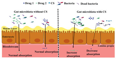
Given CS’s capacity to modulate the gut microbiome, it may have significant implications for various pharmaceutical agents’ activation, absorption, and subsequent pharmacological effects. Prodrugs such as prontosil and sulfasalazine, which rely on gut microbiota-mediated activation for their conversion into active forms, may exhibit altered bioavailability when administered in the presence of CS (Fig. 4). This alteration in bioavailability could stem from changes in the gut microbial ecosystem, potentially impacting the efficiency of drug conversion and subsequent absorption. Likewise, lipid-lowering drugs like lovastatin, cardiovascular drugs such as amlodipine and nifedipine, and anti-inflammatory drugs like aspirin, all of which are influenced by the gut microbiota, may experience changes in their pharmacokinetics, absorption, and bioavailability in the presence of CS [138].
Figure 4
Figure 4.
Modulation of gut microbiota. CS interacts with the gut bacteria and inhibits the activation of certain prodrugs, thereby affecting the metabolism and bioavailability of drugs. The image illustrates the influence of CS on gut microbiota and the subsequent effects on drug absorption and metabolism. The left side shows the gut microbiota without CS, where two drugs are being normally absorbed through the mucus layer, enterocytes, and into the bloodstream. The bacteria present appear active and unaffected by CS. The right side of the image presents the gut microbiota with CS. Here, we see CS interacting with the bacteria, causing some to become inactive (dead bacteria) and affecting others, as indicated by dashed lines. The presence of CS leads to decreased absorption of one drug (Drug 1) while increased absorption of another (Drug 2). The mucus layer shows changes in permeability, as indicated by the different directions of the arrows.
Additionally, research indicates that the elimination of H. pylori enhances the absorption and pharmacokinetic properties of levodopa, highlighting the potential impact of gut bacteria on the absorption of this essential drug for Parkinson’s disease treatment [139,140]. CS’s potential to modify gut microbial communities can lead to alterations in drug metabolism, impacting the amount of active drug available for absorption into the systemic circulation. While these specific interactions between CS, gut microbiota, drug metabolism, and absorption necessitate further investigation, the dynamic interplay among these factors holds promise for novel therapeutic strategies, personalized medicine approaches, and a deeper understanding of how CS may modulate the effects of various drugs within the gastrointestinal tract.
3.4
Alteration of drug-cell interaction at the polar interface
CS demonstrates the ability to interact with and influence the uptake of certain compounds, potentially altering their absorption processes. This modulation involves factors such as pH adjustments, mucoadhesive properties that enable interactions with biological membranes, and disruption of electrostatic interactions, leading to changes in the charged surfaces of the involved components. In a study led by Iwazaki et al. CS was observed to decrease losartan uptake in Caco-2 cells. Under experimental conditions maintained at 37 ℃ and a pH of 6.0, mimicking the apical side of Caco-2 cells, CS exhibited a significant capacity to reduce the uptake of losartan. This decrease was partially attributed to a minor pH elevation (pH 6.7) within the CS-losartan mixture, which contributed to the observed decline in losartan uptake. The mucoadhesive nature of CS added an additional layer of intricacy to the mechanism, suggesting interactions with cell membranes, further contributing to the decreased uptake of losartan. Moreover, the well-known affinity of losartan for the polar interface, driven by electrostatic interactions involving its negatively charged tetrazole group and choline groups of phospholipids, typically enhances its absorption. However, the presence of CS seemed to disrupt this electrostatic interaction, potentially resulting in alterations to the charged surfaces of losartan, CS, and cell membranes. These dynamic changes likely play a pivotal role in the reduction of losartan uptake. The intricate interplay between CS and losartan underscores the pressing need for comprehensive investigations to elucidate the underlying mechanisms and their broader implications for drug absorption and transport [141]. The mechanism highlights the need for further investigation into its implications for drug absorption and transport.
3.5
Mucus-mediated entrapment and absorption interference
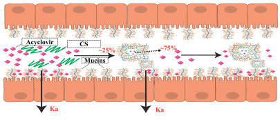
CS can hinder a drug’s diffusion through dense mucus, reducing its concentration near the intestinal wall and potentially impeding absorption. This effect is achieved by increasing mucus viscosity without altering the drug’s diffusion coefficient. The interaction between CS and mucus diminishes the free fraction of the drug available for absorption, leading to drug entrapment. Consequently, a portion of the drug forms coacervates with mucus and CS in the intestinal lumen, further decreasing its concentration and hindering its absorptive flow. A study by García et al. has revealed an intriguing phenomenon related to the interplay between CS and acyclovir during intestinal drug absorption. CS’s introduction obstructs acyclovir’s diffusion through dense mucus, leading to a reduction in its concentration near the intestinal wall, potentially inhibiting absorption [139]. CS remains unable to access the epithelium due to its interaction with loosely attached mucus, resulting in the formation of coacervates that entrapped approximately 75% of the acyclovir dose within the absorption window of the small intestine (Ka). The acyclovir trapped within these coacervates remains unreleased throughout the transit time in the small intestine and eventually reaches the large intestine, where absorption is no longer feasible (Fig. 5). Interestingly, while CS increases mucus viscosity, it does not affect acyclovir’s diffusion coefficient. The interaction between CS and mucus is pivotal as it diminishes the free fraction of acyclovir available for absorption, indicating that some of the drug gets trapped. This leads to a fraction of the acyclovir dose forming coacervates with mucus and CS within the intestinal lumen, further reducing its concentration and hindering its absorptive flow [139,142,143].
Figure 5
Figure 5.
Proposed Mechanism of interaction between CS and mucins causing the decreased bioavailability of acyclovir. CS increases mucus viscosity and entraps about 75% of acyclovir in the mucus, reducing the drug’s available concentration for absorption and forming coacervates that hinder its passage to the intestinal wall. Only about 25% of acyclovir approaches the wall for potential absorption.
3.6
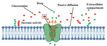
GlCN activates intestinal P-gp inhibiting drug absorption
GlCN, a naturally occurring compound extensively present in the human body, especially in joint fluid, serves a pivotal role in the synthesis of cartilage, the dense connective tissue that cushions joints. This compound is also widely consumed as a dietary supplement, obtained either from shellfish or through synthetic production methods. Its primary use is to alleviate the discomfort associated with osteoarthritis and joint pain, attributed to its anti-inflammatory properties and its ability to assist in the regeneration of cartilage [144-147]. CS, closely related to GlCN, is comprised of GlCN and N-acetyl GlCN molecules, making it essentially a GlCN-based substance. This connection underlines the versatility and wide-ranging applications of GlCN in biomedical research and dietary supplementation. A particularly compelling aspect of GlCN’s functionality is its interaction with P-gp, a critical drug transporter protein that is ubiquitous in the GIT. P-gp plays an indispensable role in minimizing the absorption of drugs in the GIT and in the expulsion of potentially harmful foreign substances from the body, thus preventing their accumulation in cells and tissues [148-150]. This mechanism is essential for staving off pathological changes by precluding the buildup of xenobiotics. GlCN has been identified by our lab as a potent and direct activator of P-gp, enhancing the protein’s efflux capacity, which consequently diminishes the absorption of drugs by cells lining the intestine (Fig. 6). This reduces the overall availability of the drug in the body, without altering the levels of the P-gp protein itself. This unique attribute of GlCN positions it as an influential factor in the modulation of drug absorption and bioavailability.
Figure 6
Figure 6.
Schematic representation of GlCN as a direct activator of P-gp, enhancing the protein’s efflux capacity and consequently decreasing drug absorption by cells lining the intestine.
Our laboratory is at the forefront of exploring this intriguing aspect of GlCN’s interaction with P-gp and its implications for drug absorption and metabolism. We are delving into the specifics of how GlCN influences P-gp activity and the potential therapeutic benefits this interaction may offer, especially in terms of enhancing the efficacy and safety profile of various drugs. Our research is focused on elucidating the molecular mechanisms underlying this process, with an aim to provide a detailed understanding that could inform the development of new therapeutic strategies. We are currently compiling our findings and insights into a comprehensive research paper. This document will present a thorough analysis of GlCN’s role as a modulator of drug absorption through its action on P-gp, alongside a discussion of the potential implications for pharmaceutical science and medicine. Our team is excited about the opportunity to contribute to the body of knowledge in this area and anticipates the publication of our research in the near future. This work promises to shed light on a novel approach to optimizing drug therapies and improving patient outcomes by leveraging the natural properties of compounds like GlCN.
A summary of CS’s role as an oral absorption inhibitor can be found in the Table S2 (Supporting information), which outlines how CS and its derivatives influence the absorption of various drugs and vitamins. It highlights the mechanisms through which CS affects drug bioavailability, including modulation of lipid and sterol adsorption, gut microbiota modulation, and changes in drug-cell interactions. Table S2 also provides information on how different molecular weights and modifications of CS impact drug absorption, making it a valuable tool for controlling the bioavailability of certain therapeutic agents.
4.
Future directions
CS has shown promise as an oral absorption enhancer, however, it is important to acknowledge some of its limitations. CS’s absorption-enhancing properties are highly dependent on its pH, molecular weight, degree of deacetylation, and concentration. Finding the optimal CS formulation for a specific drug can be challenging due to these variations. Furthermore, its efficiency as an oral absorption enhancer may differ among individuals, possibly resulting in uneven outcomes. It may not be compatible with all drugs since some drugs may chemically interact with CS, leading to reduced drug stability or altered drug release profiles. Compatibility issues can limit the range of drugs that can effectively utilize CS as an absorption enhancer. Although CS is generally considered safe, the long-term effects on the GIT still need to be fully understood. There have been reports of gastrointestinal side effects, such as nausea, vomiting, and diarrhea, associated with its use. It is crucial to consider the potential adverse effects of CS when evaluating its suitability for oral drug delivery.
Here are some future directions for CS research working as an oral absorption enhancer and inhibitor:
(1) Exploration into novel CS derivatives with improved solubility, stability, and compatibility with a broader range of drugs. This includes synthesizing CS with different degrees of deacetylation and molecular weights to tailor its interaction with drugs and the gastrointestinal environment.
(2) Deeper investigation into the molecular mechanisms underlying CS’s role both as enhancer and inhibitor of drug absorption. This entails studying the interaction of CS with the intestinal mucosa, tight junctions, efflux transporters, and enzymes in more detail, using advanced molecular biology techniques.
(3) Since CS can modulate the gut microbiota, research into how these changes affect drug metabolism and absorption could yield valuable insights. This might lead to the development of CS-based prebiotics or synergistic therapies for managing conditions like dysbiosis.
(4) Patient-friendly CS formulations, such as orally disintegrating tablets or films, should be developed to improve patient compliance, especially in populations with swallowing difficulties.
(5) Investigating blends of CS with polymers that respond to specific external stimuli, such as temperature, light, or magnetic fields. These blends could be used to create smart, controlled-release systems that target drug delivery to specific sites or at precise times.
(6) Designing CS-based carriers for delivering gene-editing tools like CRISPR/Cas9 directly to target cells in the GIT. Such carriers could revolutionize treatment strategies for genetic disorders and certain cancers.
(7) Developing formulations where CS gradually releases inhibitory agents, offering sustained inhibition in areas where absorption must be controlled.
(8) Tailoring CS formulations to selectively inhibit absorption in conditions where over-absorption of specific nutrients or drugs exacerbates the disease (e.g., hyperglycemia or iron overload).
(9) Exploring the use of CS as an absorption inhibitor to mitigate drug-drug interactions, potentially reducing adverse effects by selectively blocking certain medications’ uptake.
(10) Addressing regulatory challenges by establishing clear guidelines and standards for the production, characterization, and clinical use of CS in drug delivery systems.
(11) By focusing on these areas, the research community can overcome current limitations and harness CS’s full potential.
5.
Conclusion
In conclusion, this comprehensive review has effectively highlighted the dual functionality of CS, both as an oral absorption enhancer and an inhibitor. Its interactions with the intestinal mucosa, tight junctions’ modulation, inhibition of enzyme and efflux transporters, etc., demonstrate its potential as an enhancer in oral drug absorption. On the other hand, its ability to adsorb lipids, interact with bile acids, modulate gut microbiota, and activate the efflux transporters like P-gp presents a novel method to modulate and potentially inhibit the absorption of certain drugs, thereby extending its utility in pharmaceutical formulations. This contrast not only strengthens the complexities of CS’s interaction with the gastrointestinal milieu but also opens up new avenues for controlling and fine-tuning the bioavailability of drugs. Future research should continue to explore and refine the use of CS, focusing on understanding the nuances of its interactions within the GIT and developing novel CS-based formulations that leverage its dual capabilities. Such advancements could lead to more targeted and effective therapies, reducing the variability in drug absorption and improving therapeutic outcomes.
Declaration of competing interest
The authors declare that they have no known competing financial interests or personal relationships that could have appeared to influence the work reported in this paper.
This research was financially supported by National Key Research and Development Program of China (No. 2021YFD1800900), National Natural Science Foundation of China (No. 82073790), Special Fund for Youth Team of Southwest University (No. SWU-XJLJ202306), Chongqing Science and Technology Commission (Nos. CSTB2022TIAD-LUX0001, CSTB2023NSCQ-JQX0002), Innovation Research 2035 Pilot Plan of Southwest University (No. SWU-XDPY22007).
Supplementary materials
Supplementary material associated with this article can be found, in the online version, at doi:10.1016/j.cclet.2025.111273.
[1]
M.S. Alqahtani, M. Kazi, M.A. Alsenaidy, et al., Front. Pharmacol. 12 (2021) 618411. doi: 10.3389/fphar.2021.618411
L. Zhu, L. Lu, S. Wang, et al., Oral absorption basics: Pathways and physicochemical and biological factors affecting absorption, in: Y. Qiu, Y. Chen, G.G.Z. Zhang, L. Yu, R.V. Mantri (Eds. ), Developing Solid Oral Dosage Forms (Second Edition), Elsevier, 2017, pp. 297-329.
H.M. Ibrahim, E.M.R. El-Zairy, Chitosan as a biomaterial–structure, properties, and electrospun nanofibers, in: V. Bobbarala (Eds. ), Concepts, Compounds and the Alternatives of Antibacterials, InTechOpen, 2015, pp. 81-101.
S. Rodrigues, M. Dionísio, C.R. López, et al., J. Funct. Biomater. 3 (2012) 615–641. doi: 10.3390/jfb3030615
[46]
I.A. Sogias, A.C. Williams, V.V. Khutoryanskiy, Int. J. Pharm. 436 (2012) 602–610. doi: 10.1016/j.ijpharm.2012.07.007
[47]
K. Al Rubeaan, M. Rafiullah, S. Jayavanth, Expert Opin. Drug Deliv. 13 (2016) 223–237. doi: 10.1517/17425247.2016.1107543
[48]
N. Morin-Crini, E. Lichtfouse, G. Torri, et al., Environ. Chem. Lett. 17 (2019) 1667–1692. doi: 10.1007/s10311-019-00904-x
[49]
K. Pal, B.K. Pradhan, D. Kim, M. Jarzębski, Chitosan-based nanoparticles for ocular drug delivery, in: K. Pal, S. Verma, P. Datta, et al., Advances In Biomedical Polymers And Composites, Elsevier, 2023, pp. 247-263.
I. Furda, Interaction of dietary fiber with lipids–mechanistic theories and their limitations, in: I. Furda, C.J. Brine (Eds. ), New Developments in Dietary Fiber. Physiological, Physicochemical, and Analytical Aspects, Springer, Boston, 1990, pp. 67–82.
R. Ylitalo, S. Lehtinen, E. Wuolijoki, et al., Arzneimittelforschung 52 (2002) 1–7.
[107]
G. Maleki, J.M. Milani, Functional properties of chitin and chitosan-based polymer materials, in: S. Gopi, S. Thomas, A. Pius (Eds. ), Handbook of Chitin and Chitosan, Elsevier, 2020, pp. 177–198.
K. Deuchi, O. Kanauchi, M. Shizukuishi, et al., Biosci. Biotechnol. Biochem. 59 (1995) 1211–1216. doi: 10.1271/bbb.59.1211
[110]
M. Gallo, D. Naviglio, A. Armone Caruso, L. Ferrara, Applications of chitosan as a functional food, in: A.M. Grumezescu (Eds. ), Novel Approaches of Nanotechnology in Food, Elsevier, 2016, pp. 425–464.
H. Ma, X. Li, T. Zhou, et al., Diabetes Care 43 (2020) 719–725. doi: 10.2337/dc19-1836
[146]
K. Azuma, T. Osaki, T. Wakuda, et al., Inflammation 35 (2012) 1462–1465. doi: 10.1007/s10753-012-9459-0
[147]
M. Derwich, B. Górski, E. Amm, et al., Int. J. Mol. Sci. 24 (2023) 4925. doi: 10.3390/ijms24054925
[148]
M. Kataoka, T. Yokoyama, Y. Masaoka, et al., Eur. J. Pharm. Sci. 44 (2011) 544-551. doi: 10.1016/j.ejps.2011.09.007
[149]
P. Anderle, E. Niederer, W. Rubas, et al., J. Pharm. Sci. 87 (1998) 757–762. doi: 10.1021/js970372e
[150]
K. Fenner, M. Troutman, S. Kempshall, et al., Clin. Pharmacol. Ther. 85 (2009) 173–181. doi: 10.1038/clpt.2008.195
Figure 1
CS interaction with intestinal mucosa. The positively charged amino groups of CS interact with the negatively charged sialic acid residues (red) and sulfate esters (green) in the mucosal surface. Mucoadhesion improves with the degree of deacetylation; therefore, the greater the number of amino groups, the stronger the adhesion.
Figure 2
Modulation of tight junctions by CS. CS interacts with the tight junction’s proteins, such as claudins, ZO-1, and occludin, causing temporary opening of these junctions, thus rendering the paracellular route.
Figure 4
Modulation of gut microbiota. CS interacts with the gut bacteria and inhibits the activation of certain prodrugs, thereby affecting the metabolism and bioavailability of drugs. The image illustrates the influence of CS on gut microbiota and the subsequent effects on drug absorption and metabolism. The left side shows the gut microbiota without CS, where two drugs are being normally absorbed through the mucus layer, enterocytes, and into the bloodstream. The bacteria present appear active and unaffected by CS. The right side of the image presents the gut microbiota with CS. Here, we see CS interacting with the bacteria, causing some to become inactive (dead bacteria) and affecting others, as indicated by dashed lines. The presence of CS leads to decreased absorption of one drug (Drug 1) while increased absorption of another (Drug 2). The mucus layer shows changes in permeability, as indicated by the different directions of the arrows.
Figure 5
Proposed Mechanism of interaction between CS and mucins causing the decreased bioavailability of acyclovir. CS increases mucus viscosity and entraps about 75% of acyclovir in the mucus, reducing the drug’s available concentration for absorption and forming coacervates that hinder its passage to the intestinal wall. Only about 25% of acyclovir approaches the wall for potential absorption.
Figure 6
Schematic representation of GlCN as a direct activator of P-gp, enhancing the protein’s efflux capacity and consequently decreasing drug absorption by cells lining the intestine.

 DownLoad:

 下载:
下载:


 下载:
下载:
