Figure 1.
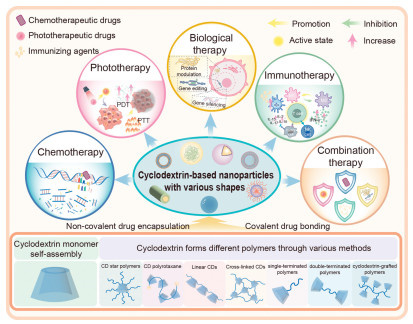
The design and antitumor application of CD-based nanotherapeutics.
Cyclodextrin-based nanotherapeutics: A promising strategy for enhanced cancer therapy
Menglin Zhang , Fanpeng Ran , Yun Zhang , Xiaoli Zhang , Zhigang Xu , Xiaoxiao Shi

Cancer is a high-mortality disease threatening human life today. Conventional non-surgical cancer therapy strategies, with the aim of inhibiting the proliferation of tumor cells or inducing apoptosis, lack precise specificity and selectivity [1]. In view of the extremely similarity of tumor cells to normal cells and higher vitality, conventional cancer therapy has inferior antitumor efficacy and severe side effects [2-4]. Besides, most chemotherapeutic drugs are characterized by poor water solubility and difficult to reach the concentration required for systemic circulation, thereby greatly reducing their bioavailability [5]. Notably, specificity and solubility are main challenges in current cancer treatment. Nanomedicine, with the emergence of highly effective treatment strategies such as tumor-targeted therapy and immunotherapy based on nanotherapeutics, shows promise in enhancing anticancer efficacy [6-10]. The design and development of nanomaterial-based drug delivery systems have been garnered increasing attention, due to their ability to encapsulate hydrophobic drugs within water-soluble nanocarriers, thereby imparting desirable pharmacokinetics and facilitating drug interactions with target specific tissues [11-15]. These systems, typically ranging in diameter from 10 nm to 200 nm, can extend circulation time and preferentially accumulate at the tumor site through the enhanced permeability and retention (EPR) effect, which contributes to improving anticancer efficiency while alleviating damage to healthy tissues [16-18]. Compared with conventional cancer therapy, nanotherapeutics do show unique merits. In the past decades, nanotherapeutics have been rationally applied in synthesis of various multifunctional nanocarriers to cater to specific drug delivery requirements. Moreover, nanotherapeutics can also be engineered to accommodate multiple therapies and achieve synergistic treatment, thereby enhancing the therapeutic efficacy [19,20].
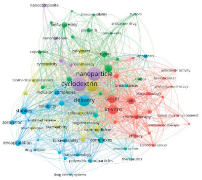
Cyclodextrins (CDs), a class of cyclic oligosaccharides obtained by enzymatic degradation of starch, are extremely versatile building blocks for the design of drug delivery systems (Fig. 1) [21-23]. Typical CDs can be categorized as α-, β-, and γ-CD based on the number of D-glucose units linked by α−1, 4-glycosidic bonds within a cyclic structure [24]. The CD interior is hydrophobic cavity, which is lined with carbons and ethereal oxygen of the glucose residues. The CD exterior has a large number of hydroxyls groups, leading to a hydrophilic surface [25]. CDs are therefore amphiphilic molecules owning a hydrophobic core and a hydrophilic shell. This property endows them with sufficient water solubility and the ability to serve as a host to accommodate a wide range of hydrophobic guest molecules within their cavity via host-guest interactions [26]. Additionally, the hydroxyl groups on the outer surface of CDs can be classified as primary hydroxyls located on the upper rim and secondary hydroxyls situated on the lower rim [27]. Therefore, CDs can be used to generate polymers with different structures and functions through chemical reactions. Furthermore, CDs possess prominent biocompatibility so that they do not elicit immune responses and have low toxicities [28]. These superior properties of CDs render them extensively employed in drug delivery systems [29]. For instance, the hydrophobic cavities of CDs can bind various water-soluble low molecular weight drug molecules through multiple non-covalent interactions, such as electrostatic interactions, van der Waals forces, hydrogen bonds, π-π interactions, and hydrophobic interactions. At the same time, the CD’s multiple intricate hydroxyl sites also facilitate the covalent attachment of drugs, enabling precise modulation of anticancer agents and achieving targeted regulation of drug release. Therefore, CD-based nanoparticles (NPs), when combined with a variety of drugs, can be effectively utilized to treat various types of tumors, including but not limited to breast cancer, colorectal cancer, lung cancer, liver cancer, bladder cancer and cervical cancer [30-33]. In recent decades, extensive research has been conducted on CD-based nanotherapeutics for diverse cancer therapy strategies due to their unique chemical structure and properties, yielding enhanced anticancer efficacy. The co-occurrence keywords of CD-based nanotherapeutics for tumor therapy over the past decade were visually presented by using the VOSviewer 1.6.20 software (Fig. 2) [34].
In this review, we focus on discussing and summarizing the recent advancements in CD-based nanotherapeutics for enhanced cancer therapy in recent years. Firstly, we introduce different types of CD polymers. Subsequently, the design and application of CD-based NPs in drug delivery are presented. Afterwards, we emphasize various aspects associated with the utility of CD-based NPs in the most recent achievements as anticancer agents for enhanced cancer therapy. In the last part, we discuss the outlooks and challenges of CD-based nanotherapeutics.
Due to the limitations of CD monomers, such as their low solubility, poor acid-base stability, and rapid clearance, various CD polymers have been developed. Compared to CD monomers, CD polymers possess numerous advantages. For instance, CD polymers have good stability and a large surface area, which can improve drug metabolism. Moreover, CD polymers can increase the solubility of CD by copolymerizing with other hydrophilic monomers. In addition to that, the complex NP systems formed by CD polymers can enhance the targeting of drug delivery.
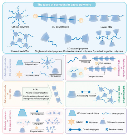
CDs possess multiple intricate hydroxyl sites, and through the processes of polymerization, substitution, and graft modification, diverse types of CD polymers (pCDs) can be synthesized. The currently researched pCDs can be categorized into five main types: CD star polymers, CD polyrotaxane, linear CDs, cross-linked CDs, and CD-capped polymers. Among these, CD-capped polymers can be further divided into single-terminated polymers, double-terminated polymers, and CD-grafted polymers [35-37].
There are numerous techniques available for the modification of CDs to generate pCDs, including reversible addition-fragmentation chain transfer polymerization (RAFT) [38], ring-opening polymerization (ROP) [39], free radical polymerization (FRP) [40], anionic polymerization [41], cationic polymerization [42], click reactions [43], and depolymerization polymerization [44]. This section will introduce the types and design of CD-based polymers (Fig. 3).
By using a CD as the core and connecting polymer arms to its upper and lower edges, CD star polymers can be obtained. This distinctive architecture confers them with a large surface area and diverse functional sites. Additionally, star-shaped block copolymers are widely recognized as versatile and stable single-molecule micelle templates [45], rendering them highly suitable for drug delivery applications. CD is frequently used as an initiator in polymerization techniques such as RAFT polymerization, ROP, and atom transfer radical polymerization (ATRP), allowing for the controlled synthesis of CD star polymers.
It is well known that CD has two types of hydroxyl groups, and it is feasible to functionalize all primary hydroxyl groups of β-CD. For example, Yu et al. designed and synthesized a star-shaped cationic polymer with a β-CD core (β-CD-g-PSSn) through a mild and controllable ROP reaction, which can facilitate caveolin-mediated endocytosis. In their study, thiol-CD (β-CD-SH) was employed as an initiator, wherein the thiol group of β-CD-SH undergoes a reversible dissociation reaction to transform into an active thio-nucleophile that attacks the disulfide bond in the lipoic acid derivative monomer (La-Arg), forming a new active thio-nucleophile and initiating a controlled and efficient polymerization reaction (Fig. 4A) [46]. The synthesis of CD star polymers via ATRP is more intricate compared to the ROP method, resulting in relatively more intricate polymers. For example, Ma et al. used CD-Br as an initiator to initiate the ATRP polymerization process with multiple initiation sites. They utilized CPTM as a hydrophobic block, and poly(ethylene glycol) methyl ether methacrylate (POEGMA) as a hydrophilic block. Additionally, they incorporated a dual-responsive CPT multi-drug (CDCPT) into their system to fabricate multi-stimuli responsive nanomedicines. The nanomedicines were utilized for the co-delivery of camptothecin, a chemotherapeutic drug, and chlorin e6 (Ce6), a photosensitizer (Fig. 4B) [47]. Yan et al. first used β-CD as an initiator for the synthesis of star-shaped PCL homopolymers via ROP. Then, 2-(n-butyltrithiocarbonate) propanoic acid was used as a chain transfer agent (CTA) to modify the chain ends of PCL. Subsequently, they extended the star-shaped PCL-b-(P(S-Cl)) by incorporating 4-chloromethylstyrene (S-Cl), leading to the preparation of star-shaped PCL-b-(P(S-Cl)) block copolymers (single molecular micelles) through photoinduced electron/energy transfer RAFT (PET-RAFT) polymerization [48].

Unlike other CD star polymers, CD polyrotaxane is a supramolecular polymer. The polymer chain is threaded through the cavities of multiple CDs, thereby interlocking them, and the ends of the polymer chain are capped with macromolecules, resulting in the formation of a CD polyrotaxane [49]. Due to its supramolecular structure, the CDs on the chain are not restricted by covalent bonds, enabling their unrestricted movement and rotation along the axis [50]. This characteristic renders CD polyrotaxane highly suitable for drug delivery. The synthesis of CD polyrotaxane typically involves initial formation of polypseudorotaxanes (PPR) from CDs and polymer chains in an aqueous medium, commonly utilizing PEG as the polymer chain. Subsequently, a capping reaction is performed in organic solvents such as dimethyl sulfoxide (DMSO) or N,N-dimethylformamide (DMF), frequently employing reactive functional groups like carboxyl and amino groups for end-capping [51,52].
For example, Zhang et al. synthesized pseudo-CD (PPRX) by reacting β-CD, N3-β-CD, LiCl, and H2N-PEG-b-PPG-b-PEG-NH2 in aqueous solution. Subsequently, they introduced N-(triphenylmethyl) glycine (Trt-Gly-OH) and 4-(4, 6-dimethoxy-1, 3, 5-triazin-2-yl)-4-methylmorpholinium chloride (DMT-MM), dissolved in a mixture of methanol (MeOH) and acetonitrile (MeCN), followed by dilution with DMF, and dissolution in water to obtain N3-PRX. This intermediate was further methylated and modified with the cRGDfK peptide to yield acid-degradable polyrotaxanes containing Me-β-CDs (Me-PRXs). The acid-sensitive Me-PRXs release the threaded Me-β-CDs in lysosomes while accumulating in the endoplasmic reticulum to induce autophagic cell death for malignant tumors treatment [53].
Although both linear CDs and CD polyrotaxane exhibit linear arrangements of CDs, they possess distinct characteristics. Linear CDs are polymer compounds made by covalently linking multiple CDs through alkene copolymerization, ring-opening reactions of epoxides, or other special functional group condensation. In contrast to CD polyrotaxane, the cavities within the structure of linear CDs remain unoccupied, enabling them to effectively encapsulate drugs through host-guest interactions, making them an excellent choice for drug delivery [54]. Yang et al. [55] developed a novel supramolecular delivery system composed of poly(β-CD) (PCD-PA) functionalized with benzoic acid groups and polyethylene glycol (PEG-AD) terminated with adamantyl groups (Fig. 4C). Under alkaline conditions, β-CD and epichlorohydrin (EPI) underwent a ring-opening reaction of epoxides to synthesize linear PCD, which subsequently reacts with the hydroxyl group of phthalic anhydride (PA) to obtain PCD-PA. mPEG2000 and 1-adamantyl carbonyl chloride were employed for the synthesis of PEG-AD. The resulting PCD-PA possesses multiple host cavities capable of interacting with PEG-AD through host-guest interactions, enhancing the drug loading capacity.
Cross-linked CDs differ from other CD polymers in that they require cross-linking agents to link the individual CDs together. These cross-linking agents possess bifunctional or multifunctional groups, which react with the hydroxyl groups of the CDs to facilitate cross-linking. Commonly used cross-linking agents include isocyanates and acid anhydrides [56]. In cross-linked CDs, the cavities of the CDs remain unoccupied, enabling drug encapsulation in a supramolecular form for drug delivery. However, due to the presence of cross-linking agents, mobility within cross-linked CDs may be impeded. Innovative modifications of cross-linking agents can also be utilized to develop drug delivery systems that specifically respond to the acidic and hypoxic microenvironments of tumors, achieving precise drug delivery effects. Hu et al. [57] synthesized a p-nitrophenyl carbonate groups-terminated crosslinker containing disulfide bonds (referred to as DBHD), which covalently crosslinks with the hydroxyl groups of β-CDs. To enhance stability, PEG-NH2 was also introduced. Doxorubicin (DOX) was encapsulated in the cavity of the CDs through host-guest interactions, forming glutathione (GSH)-responsive polycyclodextrin supramolecular nanocages (PDOP NCs) (Fig. 4D), achieving accurate drug delivery and enhancing the therapeutic effect against tumors.
CD-capped polymers refer to CD polymers in which CDs are covalently linked at the endpoints of the polymer chains. There are two methods for synthesizing CD-capped polymers: one involves extending from a CD-based large initiator to generate homo- and block copolymers, and the second method involves first polymerizing a linear polymer with reactive groups at the ends and then conjugating it with CD-based derivatives [58,59]. Although many CDs are connected through intermediates, CD-capped polymers differ from previously mentioned types of CD polymers in that their CDs are not linked through crosslinkers or functional group polymerization reactions, but rather directly bonded covalently to the linear polymer.
CD-capped polymers can be classified into three categories: single-terminated polymers, double-terminated polymers, and CD-grafted polymers. Single-terminated polymers refer to those with a CD cap at only one end. Han et al. [60] first polymerized aldehyde-containing hyaluronic acid (HA) and reacted it with NH2-β-CD to generate single-terminated polymers. Several single-terminated polymers assemble into an amphiphilic shell, where hexadecanol modified ferrocene (FC) is embedded within the CDs’ cavity through host-guest interactions. A hydrophobic core composed of benzyl modified poly(γ-glutamic acid) (BzPGA), LY2109761 (LY, a TGF-β1 inhibitor), and Ce6 (a classic type-Ⅱ photosensitizer) is encapsulated in NPs (Fig. 5A). The prepared NPs (LC@HCDFC NPs) exhibit enhanced tumor targeting, reactive oxygen species (ROS)-responsive drug release, and improved drug encapsulation efficiency, thereby augmenting anti-tumor effects. Double-terminated polymers are formed by conjugating CDs at both ends of the polymer chain. Wu et al. [61] were pioneers in synthesizing the polymer boron dipyrromethene (BODIPY) via ring-opening polymerization (Fig. 5B). Two NH2-β-CD host molecules were covalently conjugated onto a BODIPY theranostic agent (B-β-CD). The drug CPT was linked to the polyethylene glycol (PEG) chain via a thiol bond, enabling responsiveness to ROS. Through the synergistic effects of host-guest molecular recognition between β-CD and CPT, π-π stacking interactions involving CPT, multiple hydrogen bonds formed with β-CD, and the protein absorption resistance provided by the PEG shell, supramolecular self-assembly occurs to form NPs. The prepared nanodrug possesses capabilities for fluorescence imaging, photodynamic therapy (PDT), and photothermal therapy (PTT), enhancing drug solubility and effectively inducing immunogenic cell death (ICD) in tumor cells.
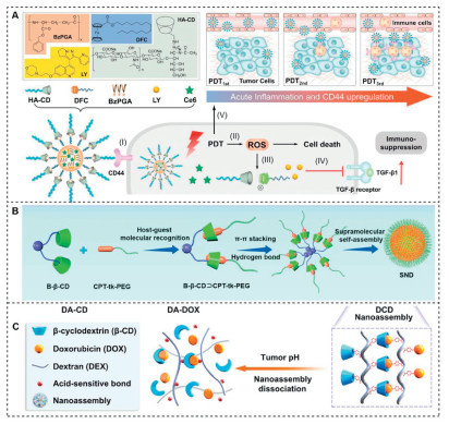
In addition, CD-grafted polymers can be classified as CD-capped polymers, characterized by the presence of CD on both ends of the polymer chain. Liang et al. [62] synthesized DA polymer chains by esterifying polyhydroxy dextran (DEX) and 4-carboxybenzaldehyde acid (4-CBA). Mono(6-amino-6-deoxy)-β-CD (β-CD-NH2) was connected to the DA polymer chain through an amine-aldehyde reaction, resulting in the formation of DA−CD conjugates. They also modified DOX to generate DA−DOX. The host-guest interactions between DA−CD and DA−DOX formed size-transformable supramolecular nanoassemblies (DCD SNs) (Fig. 5C), achieving enhanced antitumor therapy by increasing drug circulation time and penetration depth.
As excellent building blocks, CDs pave the way for designing effective drug delivery carriers. Both native CDs and modified CDs are promising units for constructing multifunctional drug delivery systems due to their superiority in structures and properties. As mentioned above, CDs have been widely applied as an excipient to compensate for the shortcomings of chemotherapeutic drug molecules [63]. Due to their intrinsic hydrophobic cavity, both native and modified CDs are fascinating hosts, enabling them to effectively encapsulate a variety of drugs to form noncovalent host-guest inclusion complexes. Alternatively, harnessing the abundant hydroxyl groups, CDs can be chemically modified by various functional groups. Drug molecules can therefore be chemically bonded with CDs to generate prodrugs [64]. Moreover, through facile modification of CDs with specific stimuli-responsive functional moieties such as benzimidazole for pH responsiveness, azobenzene for light responsiveness, and disulfide for redox responsiveness, drug release at desired sites can be triggered effectively [65]. The triggered drug delivery system not only ensures the relative stability of drugs within the bloodstream, but also achieves rapid release of active agents via stimulus-responsive mechanisms specifically at tumor sites, thereby enhancing anticancer efficiency.
The driving forces responsible for the physical embedding of chemotherapeutic agents within CDs primarily include electrostatic interactions, van der Waals forces, hydrogen bonding and hydrophobic interactions [66]. It is noteworthy that the drug molecules embedded in CDs acquire significantly altered physical, chemical and pharmaceutical characteristics compared with their free counterparts. For instance, Patel et al. investigated the role of β-CD and its derivative, hydroxypropyl β-CD (HP-β-CD), in improving the solubility and stability of rigosertib, an anti-cancer drug [67]. The results demonstrated that CDs and their analogues significantly improved the poor solubility and chemical instability of the drugs in acidic solutions. Also, dissolution profiles indicated that the inclusion compounds exhibited a higher drug release effect compared to free drug molecules. Similarly, Lodagekar et al. employed the freeze-drying method to incorporate the anticancer drug niclosamide into HP-β-CD [68]. The dissolution study revealed a nearly threefold increase in the drug release rate of the inclusion compound compared to that of the free drug molecule alone in the initial 30 min. Furthermore, cytotoxicity assays indicated that HP-β-CD-incorporated Niclosamide exhibited enhanced efficacy against cancer cells. In vivo experiments confirmed superior pharmacokinetic properties of the inclusion complex. It was verified that CDs play a significant role in improving the solubility and bioavailability of anticancer drugs, thereby enhancing cancer therapy.
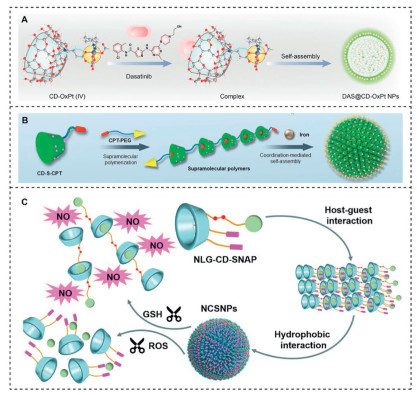
In addition, Ye et al. developed pH/redox dual-activated supramolecular DAS@CD-OxPt (Ⅳ) NPs (Fig. 6A) [69]. They first synthesized the CD-OxPt (Ⅳ) supramolecular prodrug as a compatible platform for dasatinib (DAS) encapsulation through host-guest interactions. Compared to the pure drug, these NPs exhibited ideal blood circulation and effective accumulation in tumor tissues. Under the acidic tumor environment, the morphology of the dynamic DAS@CD-OxPt (Ⅳ) NPs transformed into spatial helical nanofibers through in situ assembly, leading to the release of DAS molecules from the DAS@CD-OxPt (Ⅳ) NPs. Meanwhile, the coupled toxic OxPt (Ⅱ) was further activated by high levels of GSH within tumor cells, achieving precise tumor delivery and inducing a cascade effect of drug penetration and apoptosis.
Diverse supramolecular systems can be constructed by means of the host-guest interactions between CDs and specific guest molecules. Adamantane (Ada) is a typical guest molecule known for its strong interaction with CDs [70]. Therefore, chemotherapeutic agents conjugated with Ada can dramatically interact with CDs to form a stable host-guest inclusion complex. For example, Zhang et al. fabricated a supramolecular nanoassembly by exploiting the robust host-guest interaction between β-CD-decorated hyaluronic acid (HACD) and disulfide-linked Ada-naphthalimide CPT prodrug (AdaCPT) [71]. It was demonstrated that the inclusion complex enabled sustained release of CPT under reductive conditions, while the naphthalimide (NAP) molecule served as a fluorescence reporter to monitor drug release. The CPT-loaded supramolecular nanoassembly exhibited comparable toxicity against HCT-116 cancer cells compared to commercial CPT, but showed low toxicity towards NIH3T3 normal cells, suggesting enhanced bioavailability and antitumor activity of CD-based assembly.
Additionally, drugs can be covalently linked to CDs. The hydroxyl groups on CDs can react with a variety of chemical reagents under different conditions. Notably, the hydroxyl group at the sixth position displays high reactivity with chemical reagents, while the hydroxyl groups at positions 2 and 3 can be activated by highly active protonating reagents [72]. Consequently, numerous researchers have employed covalent bonding between drugs and CDs to modify drug characteristics.
Yang et al. developed a supramolecular polymer nanodrug (SNP) [73], by covalently coupling the drug CPT to CDs through a chemical reaction between CD-NH2COOH and CPT-S-OH. The resulting monomer can achieve supramolecular self-assembly through host-guest interactions. To control the morphology of the self-assembly and enhance stability, they introduced CPT-PEG during the supramolecular self-assembly process, ultimately forming SNPs (Fig. 6B). This approach significantly improved the solubility of CPT and stimulated an anti-tumor immune response, promoting the infiltration of cytotoxic T lymphocytes within tumors, thereby enhancing the efficacy of tumor immunotherapy.
Furthermore, CD-based prodrug molecules with stimulus-responsive capabilities provide a promising and effective strategy for cancer treatment. Hu et al. engineered a supramolecular nanodrug [74]. In this design, checkpoint blockade drugs (NLG) served as a ROS-triggered linker via its thioketal moiety, while nitric oxide (NO) was covalently attached to CDs through an S-nitrosothiol bond (-SNO) as a GSH-responsive linker, resulting in the formation of the NLG-CD-SNAP monomer. Leveraging microfluidic technology, NLG-CD-SNAP self-assembled into polymer nanodrugs (NCSNPs) (Fig. 6C), exhibiting remarkable advantages in terms of tumor accumulation and bioavailability.
To realize optimal antitumor efficacy while minimizing side effects, a plethora of emerging therapies are being explored to enhance cancer treatment, including tumor-targeting chemotherapy, PTT, immunotherapy, gene therapy, and multifunctional synergistic therapy. Benefiting from the unique properties and desired biocompatibility of CDs, CD-based nanotherapeutics have been extensively studied in diverse strategies for cancer therapy (Table 1 [75-96]). In this section, we focus on discussing the utility of CD-based NPs as anticancer agents to augment therapy based on the latest advanced findings.
 DownLoad:
CSV
DownLoad:
CSV
| Nanotherapeutics | Treatment strategy | Major merit | Ref. |
| polyCD-based ring-like colloids | Chemotherapy | Improving the stability of reversible cross-linked colloids and enhancing their drug loading capacity | [75] |
| MA-CD/Fc-CPT NP | Chemotherapy | Good biological safety, suitable for drug delivery in the TME, and improving cancer treatment outcomes | [76] |
| PTX/Fol-c1-ORCyD | Chemotherapy | Having a higher antitumor effect than a single drug and inhibiting the side effects of folic acid-modified CD | [77] |
| D-PTA-CD | Chemotherapy | Increasing drug loading capacity and achieving a significant synergistic effect in inhibiting cancer cell proliferation | [78] |
| OGPA/P-C | Chemotherapy | Specific targeting and overcoming chemotherapy resistance | [79] |
| CDTP@Fc-PEG NPs | Chemotherapy | Enhancing the chemosensitivity of cancer cells; high tumor accumulation; high tumor cytotoxicity | [80] |
| TPP-Ad2/CD2 | PDT | High tumor accumulation; enhanced drug solubility and targeting; improved PDT efficacy | [81] |
| TOCN/PVA7-PDA@DOX | PTT | Achieving controlled drug release; enhancing photothermal conversion efficiency; improving therapeutic effects; reducing side effects | [82] |
| ICD lipo | PTT | Outstanding resistance to solvents, heat, and light; high biocompatibility; strong PTT effect. | [83] |
| DOX@AuNCs | PTT+ chemotherapy | Improved efficacy of synergistic therapy | [84] |
| Supra-SNA | Gene therapy | Enhanced cellular uptake and gene modulation efficacy; effective inhibition of tumor cell proliferation | [85] |
| Ad-HSA/FA-MβCD | Protein therapy | Enhanced blood retention time; improved antitumor efficacy and increased safety | [86] |
| γCDP-(DMA/PEG-Tf) NPs | Protein therapy | Promoted drug absorption and enhanced tumor cytotoxicity | [87] |
| CICG@TA NPs | Gene therapy + PTT | Accurate tumor imaging and effective tumor ablation | [88] |
| NPs | Immunotherapy | Promoting the release of M1 factors and downregulating the levels of M2 factors by reprogramming TAM | [89] |
| RCNPDOX + MCNPR848 NPs | Immunotherapy + chemotherapy | Synergistic chemo-immunotherapy | [90] |
| DACss | Immunotherapy + PDT | Enhancing the anti-tumor response and reprograming the immunosuppressive tumor microenvironment | [91] |
| ZnPc-(PEG)5: Ac-CD: DOX | Chemotherapy + PDT/PTT | Excellent stability and pH-sensitive drug release | [92] |
| BODIY-CPT-NPs | Chemotherapy + PDT | The good complementarity of the two therapies in anti-tumor effects | [93] |
| SRN | Chemotherapy + gene therapy | Promoted inhibition of telomerase reverse transcriptase gene and excellent tumor-targeting effect | [94] |
| GNR-CDP8MA | Immunotherapy + PTT | Inducing ICD in primary tumors and activating antigen-presenting dendritic cells and tumor-specific effector T cells to enhance antitumor immunity | [95] |
| CD-PEG-FA.Rg3.QTN | Immunotherapy + chemotherapy | Extended blood circulation; enhanced tumor targeting; the transformation of immunosuppressive TME | [96] |
Chemotherapy, a conventional method for clinical cancer treatment, is widely used; however, it presents challenges such as poor drug specificity and poor water solubility, which can easily lead to systemic toxicity and harm to normal cells [97]. With the development of nanomedicine, efficient and precise delivery of chemotherapeutic and targeted drugs has been achieved [98]. CDs, commonly used as nanodrug carriers for drug delivery, possess a distinctive molecular structure that effectively addresses the shortcomings of chemotherapeutic drugs. Bai et al. designed a non-covalent self-assembled nanodrug based on CDs for enhanced drug delivery [99]. In this design, M-γ-CD was synthesized by conjugating mannoses at the C1′ position of γ-CD. By utilizing M-γ-CD as the core, regorafenib (Stivarga, RG), a chemotherapeutic drug, was encapsulated through co-crystallization to form the RG@M-γ-CD complex. This mannose-modified nanodrug exhibited remarkable improvements in biodistribution and pharmacokinetic properties of RG, demonstrating excellent targeting, safety profile, and potent anti-tumor effects, effectively inhibiting tumor cell proliferation.
CDs offer a captivating avenue for delivering phototherapeutic agents (e.g., photosensitizers (PS), photothermal agents) to achieve PDT and PTT [100]. In comparison to conventional cancer therapies, phototherapy has unique merits including low systemic toxicity, minimal invasion, and repeatable dosing. However, the limited solubility and tendency towards self-aggregation in aqueous media hinder the phototherapeutic efficacy of most phototherapy agents, resulting in suboptimal circulation performance and inadequate tumor-selective accumulation [101-103]. Studies have shown that CDs serve as exceptional containers capable of overcoming these challenges while enhancing anticancer efficiency.
For instance, Zagami et al. developed a ternary nanoassembly for PDT using a nonionic amphiphilic β-CD decorated with folic acid (FA) as targeting agents and encapsulated pheophorbide (Pheo) as a photosensitizer [104]. The release results of Pheo from the nanoassembly (~20% within 1 week), in comparison to the stability of free Pheo under physiological conditions, suggest the vital role of CDs in dispersing the PS and preventing its leakage. Mazzaglia et al. reported a novel photosensitizer for PDT therapy through the self-assembly of an amphiphilic cationic β-CD (CD-N) and an anionic porphyrin (TPPS) [105]. CD-N effectively enhanced the intracellular delivery of TPPS with HeLa cells. Comparative analysis of the PDT efficiency between free TPPS and TPPS/CD-N assemblies revealed that cells treated with the latter exhibited higher cytotoxicity, regardless of irradiation. It was suggested that CD-based carriers can promote cellular uptake of photosensitizers, thereby enhancing antitumor efficiency.
To enhance the efficiency of PDT, Dai et al. reported a targeted tumor self-luminescent supramolecular T-NPCe6-LN NP [106]. This NP was obtained through the self-assembly of amino-modified β-CD (β-CD-NH2) conjugated with the photosensitizer Ce6, luminol, and the NO donor S-nitrosoacetylpenicillamine (SNAP), followed by host-guest interaction with a benzimidazole (BM)-coated pancreatic cancer cell-targeting peptide (Fig. 7A). By triggering chemiluminescent resonance energy transfer (CRET) between luminol and Ce6 through endogenous H2O2, it is anticipated to overcome the inherent limitations of traditional PDT.
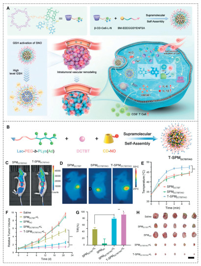
Hu et al. ingeniously designed a supramolecular drug delivery system that integrates NO release with PTT [107]. This system utilizes glycosylated poly(ethylene glycol) block polylysine (Lac-PEG-b-PLys (Ad)) and β-CD (CD-NO), loaded with NO donors, to spontaneously assemble into supramolecular polymer micelles through host-guest interactions. Furthermore, employing physical encapsulation technology, the photothermal agent DCTBT was enveloped within the micelles to successfully construct a supramolecular nanocarrier named T-SPMDCTBT/NO. Notably, DCTBT not only exhibits ROS generation capability but also possesses near-infrared-Ⅱ (NIR-Ⅱ) imaging functionality and outstanding photothermal conversion efficiency, enabling T-SPMDCTBT/NO to achieve mild PTT. Near-infrared fluorescence imaging technology revealed a significant enhancement of fluorescence in the tumor area of mice injected with T-SPMDCTBT/NO (Fig. 7B). Under laser irradiation, the T-SPMDCTBT/NO treatment group showed a more rapid rate of temperature elevation (Figs. 7C–E). This mild PTT approach demonstrated remarkable therapeutic efficacy in both subcutaneous and orthotopic hepatocellular carcinoma (HCC) animal models (Figs. 7F–H).
Moreover, CD-based supramolecular nanoassemblies have been successfully integrated with diverse biofunctional macromolecular agents such as proteins, plasmids, and nucleic acids, making them a highly promising platform for enhancing cancer therapy. For instance, Wan et al. developed a supramolecular nanoassembly composed of β-CD-grafted polyethyleneimine (CP) and disulfide-linked guanidyl Ada (Ad-SS-GD) to effectively deliver Cas9 ribonucleoprotein (RNP) for targeted genome editing of the oncogene KRAS in colorectal cancer cells (Fig. 8A) [108]. In this case, controlled release of Cas9 RNP was achieved through cleavage of disulfide bonds within the nanoassembly structure, resulting in significant antitumor efficiency and low systemic toxicity. The nanoassemblies could efficiently suppress tumor growth and inhibit lung metastasis in SW480 tumor-bearing nude mice.
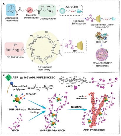
The disruption of actin cytoskeleton in tumor cells is a prospective strategy for cancer treatment, given its indispensable role in numerous cellular activities within tumor tissues [109]. Yu et al. described an actin cytoskeleton-targeting supramolecular nanofibers via self-assembly of β-CD-pendent HA (HACD) and actin-binding peptide-grafted magnetic NPs (MNP-ABP-Ada) (Fig. 8B) [110]. It was found that A549 tumor cells treated with the nanofibers experienced severe impairment when exposed to an alternating magnetic field (AMF). Remarkably, the application of MNP-ABP-Ad/HACD resulted in a remarkable apoptosis rate of 94% against A549 tumor cells with AMF assistance, while those treated with MNP-ABP-Ada only showed 46.2% apoptosis under identical conditions. In vivo behavior further demonstrated that MNP-ABP-Ad/HACD owned high-efficiency cancer treatment without lung metastasis.
Furthermore, Yang et al. reported supramolecular NPs (ADA2+@HACD) for efficient DNA delivery. In this system, the ADA2+@HACD was synthesized through strong host-guest interactions between β-CD-pendent HA (HACD) and Ada-conjugated ADA2+, which contains two positively charged chains [111]. Thus, electrostatic interaction facilitated the conjugation of negatively charged plasmid DNA (pDNA) with positively charged ADA2+. Likewise, Wen et al. constructed a supramolecular siRNA delivery platform based on the host-guest complexation between β-CD polymers and a library of guest polymers [112]. The poly(2-(dimethylamino) ethyl methacrylate) (pDMAEMA) arms were covalently linked to the β-CD core via disulfide bonds, forming the CD-SS-P host. The guests consisted of folic acid-functionalized PEG polymers with various shapes, sizes, and densities, wherein folic acid was attached at the distal end of PEG to enhance targeting efficiency. This carrier effectively encapsulated siRNA within its core to form PNP complexes. In vivo experiments verified that this carrier efficiently exhibited remarkable tumor cell-targeting ability and efficiently delivered therapeutic siRNA-Bcl2 to tumor cells in mouse animal models, effectively inhibiting tumor growth. The versatility and adaptability of this platform make it a promising candidate for the delivery of various nucleic acids and therapies, thereby demonstrating its potential in precision cancer therapy applications.
Immunotherapy has emerged as a highly efficient cancer treatment modality by regulating the immune system and augmenting immune responses for tumor eradication, which contributes to improving patient survival rates and preventing tumor recurrence and metastasis [113-115]. However, the clinical application of immune agonists is limited due to their associated toxic side effects [116]. CD-based nanocarriers have been identified as promising vehicles for targeted delivery of immune agonists to overcome these limitations.
For instance, Zhu et al. designed a hydrogel composed of in situ-formed β-CD-modified alginate (ALG-βCD), named the RER-T system. This hydrogel encapsulated the chemokine C—C motif chemokine ligand 25 (CCL25) through supramolecular interactions, which selectively recruits the CCR9+CD8+ T cells with potent anti-tumor activity. Moreover, the cavity of β-CD bonded to the adamantine-modified anti-PDL1 antibody (Ad-aPDL1) through host-guest interactions. Through intravenous injection of the anti-PD1 antibody (Ad-aPD1), it can be further anchored into the ALG-βCD hydrogel. Ad-aPDL1 and Ad-aPD1 could promote the binding between CD8+ T cells and tumor cells. This strategy enhances the activation and tumoricidal capabilities of T cells and the efficacy of T cell-mediated immunotherapy (Fig. 9A) [117]. After intratumoral injection (i.t.) of the RER-T system, a greater number of T cell subsets (CCR9+CD8+) were recruited to the tumor tissue (Figs. 9B–D). Consequently, there was an approximately twofold increase in the concentration of Ad-aPD1 within the tumor (Fig. 9E), enhancing the affinity of CD8+ T cells for tumor cells and consequently elevating interferon gamma (IFN-γ) production (increased by 5.14-fold, Fig. 9F) and granzyme B (increased by 2.27-fold, Fig. 9G), ultimately enhancing the cytotoxicity of CD8+ T cells against tumor cells.
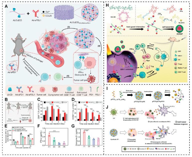
Immune checkpoint blockade (ICB) therapy, by means of immune checkpoint inhibitors, can restore the function of T cells and thus exert a crucial role in the treatment of tumors [118]. Hu et al. described an immune-photodynamic combination therapy where the indoleamine 2, 3-dioxygenase 1 (IDO-1) inhibitor NLG919 and photosensitizer pheophorbide A (PPa) were conjugated via disulfide and incorporated into the cavity of β-CD through host-guest interaction. It was discovered that the ROS produced by PPa could trigger ICD effect, leading to dendritic cell (DC) maturation. This contributed to proliferate cytotoxic T lymphocytes by presenting tumor-specific antigens to the naive T lymphocytes, thus enhancing immune response. Furthermore, these nanocarriers significantly reduced the Kyn to Trp ratio after irradiation, confirming their IDO-1 inhibition activity (Fig. 9H) [119]. The synergistic treatment efficiently suppressed both the primary and abscopal tumors in a CT26 tumor model. Besides, at 30 days post-treatment, there was a significant increase in effective memory T lymphocytes in the spleen, indicating that this synergistic immunotherapy has the potential to inhibit tumor recurrence and metastasis.
Moreover, Kim et al. designed a CD-based nanoassembly (pPTX/pCD-pSNO) to mimic oncolytic virus (OV) therapy for the treatment of locoregional melanoma (Figs. 9I and J) [120]. After endocytosis, the co-delivery system of PTX and NO presented synergistic cytotoxicity and induced systemic anti-tumor immunity by increasing ICD, activating DCs, and augmenting T cell response. Combination of pPTX/pCD-pSNO with cytotoxic T lymphocyte antigen-4 (CTLA-4) blockade resulted in prolonged survival and remarkable regression of both primary and abscopal tumors.
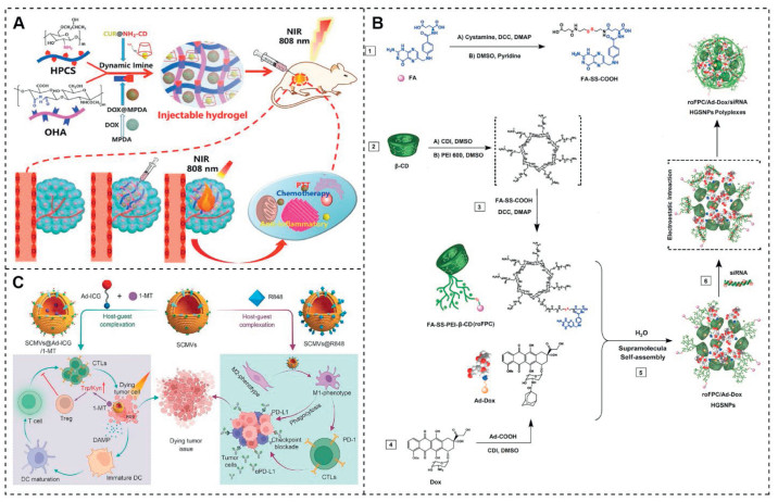
Combination therapy, by integration of multiple therapeutic agents in one shot, can present synergistic effects and enhance anticancer efficiency [121]. For example, Rong et al. prepared an injectable polysaccharide-based composite hydrogel, utilizing oxidized hyaluronic acid (OHA) and hydroxypropyl chitosan (HPCS) as the key components [122]. OHA and HPCS can form OHA-HPCS hydrogels through Schiff base reactions. The complexation of curcumin (CUR) with NH2CD was facilitated via supramolecular host-guest chemistry, while mesoporous polydopamine (MPDA) NPs acted as photothermal agents that can adsorb DOX through π-π stacking interactions (DOX@MPDA), which were then loaded into the hydrogel. A defined quantity of DOX@MPDA and CUR@NH2CD was dispersed and solubilized within an HPCS solution, which was then homogeneously blended with an equivalent mass of OHA solution to synthesize the composite hydrogel (DOX-MPDA & CUR@gel) (Fig. 10A). MPDA conferred superior photothermal conversion efficacy upon the hydrogel. The presence of CUR helped alleviate inflammation reactions caused by PTT. Moreover, in vivo treatment studies using the composite hydrogel have demonstrated its significant advantages in combined chemotherapy and photothermal treatment of tumors loaded with DOX, CUR, and MPDA.
The PTT can be combined not only with chemotherapy but also with PDT. Duy-Thuc Nguyen and colleagues reported a type of photobleaching-mediated charge-reversible NPs (P-ZWNIR NPs) that exhibit strong rectal tumor targeting and penetration capabilities [123]. NIR fluorophores and tumor-targeting moiety phenylboronic acid (PBA) were covalently linked to the CD core, resulting in the formation of P-ZWNIR CDs. By utilizing inclusion complexes formed between β-CD derivatives and the photosensitizer Pheo-Fc, P-ZWNIR NPs were constructed. Introduction of amphoteric ions within the CD units by heptamethine cyanine dye IR783 enables charge conversion through photobleaching, thereby enabling transformation of P-ZWNIR NPs into positively charged NPs upon irradiation with an 808 nm laser. Through extravasation and active targeting mechanisms, P-ZWNIR NPs enhanced tumor penetration, while their therapeutic efficacy for rectal cancer was further optimized by incorporating Pheo-Fc, making it a dual-functional agent specifically tailored for the combined photodynamic/chemodynamic therapy of rectal cancer.
In addition, the combination of gene therapy and chemotherapy has garnered widespread attention due to its enhanced antitumor effects. Mousazadeh and colleagues developed a type of supramolecular NPs (HGSNPs) capable of co-delivering Dox and hTERT siRNA, thereby enhancing therapeutic efficacy (Fig. 10B) [124]. To achieve this, they crosslinked β-CD with branched PEI, and subsequently modified the resulting PEI-β-CD (PC) polymer with a disulfide bond to form redox-sensitive folate-appended-polyethylenimine-β-CD (roFPC). By utilizing host-guest interactions, adamantane-conjugated DOX (Ad-Dox) was assembled with roFPC to generate roFPC/Ad-Dox HGSNPs. Electrostatic interactions between negatively charged hTERT siRNA and positively charged PEI-600 enabled the formation of a complex comprising roFPC/Ad-Dox/siRNA HGSNPs that synergistically delivered both drugs and genes. This complex exhibited reversible assembly-disassembly dynamics in response to the tumor microenvironment, leading to intracellular drug/gene release as well as carrier degradation and clearance. Co-delivery of hTERT siRNA and Dox demonstrated enhanced cytotoxic effects in vitro. Furthermore, effective transfection of hTERT siRNA resulted in significant gene suppression, as evidenced by real-time RT-PCR and western blot analysis. These findings highlight the superior therapeutic efficacy of combined gene-chemotherapy treatment.
Phototherapy, as an emerging clinical treatment for tumors, holds great potential as an adjunct for cancer immunotherapy [125]. Recently, numerous studies have focused on the combination of phototherapy with immunotherapetic strategies. For example, Qi et al. reported supramolecular cell membrane vesicles (SCMVs) (Fig. 10C) [126], that effectively encapsulated adamantane (Ad)-modified indocyanine green (Ad-ICG) within β-CD through host-guest interactions to achieve PDT. By co-delivering the indoleamine 2, 3-dioxygenase (IDO) inhibitor in the cavity and encapsulating the Toll-like receptor 7 and 8 (TLR7/8) agonist resiquimod (R848) within the SCMV, they were able to re-polarize tumor-associated macrophages (TAMs) towards the M1 phenotype. This innovative design empowers the vesicles synergistic effects of PDT and immunotherapy to suppress tumors, while significantly enhancing the therapeutic efficacy of immune checkpoint blockade therapy.
This review summarizes the recent advances in the development of CD-based nanotherapeutics for constructing drug delivery systems and enhancing cancer treatment. The unique structure and properties of CDs provide tremendous potential for the creation of efficient drug delivery systems. Moreover, chemical modifications based on CDs can be used to generate polymers with different structures and functions through chemical reactions. Both unmodified and modified CDs present intriguing prospects for formulating diverse multifunctional nanocarriers. Drugs can be effectively incorporated into CD through covalent or non-covalent interactions. The tailored design of CD-based NPs, endowed with desirable characteristics, facilitates their extensive utilization in various cancer therapies, including: (Ⅰ) Various antitumor agents, such as chemotherapeutic drugs, phototherapeutic agents, macromolecular agents, immune agents and other agents, can be integrated into CD-based NPs to build highly efficient therapeutic platforms. (Ⅱ) The utilization of CD-based NPs improves the solubility, stability and bioavailability of the encapsulated antitumor agents due to their superior biocompatibility and facile functionalization. (Ⅲ) Incorporating CD-based supramolecular assemblies for the functionalization of biomacromolecules, such as proteins, plasmids, and nucleic acids, effectively enhances their tumor-targeting ability and antitumor activity, presenting a promising strategy for cancer therapy. (Ⅳ) CD-based NPs with low toxicity and desirable loading capacity hold great potential as delivery systems for immune agents in constructing immunotherapy nanoplatforms.
The application of CD-based nanotherapeutic brings numerous benefits in the development of multifunctional cancer therapeutic platforms. However, there are still several considerations for clinical translation that warrant attention, including (Ⅰ) CD-based nanoassemblies formed by noncovalent host-guest interactions may exhibit instability on account of high dilution in vivo and potential interference from blood components. Thus, it is crucial to explore strategies for constructing more stable nanoarchitectures. (Ⅱ) Although a large number of CD-based NPs have been utilized for drug delivery, most of them still lack in-depth toxicology assessments. The advent of renal clearable CD-based nanotherapeutic capable of rapid excretion by the kidney addresses concerns regarding long-term toxicity. In the future, further efforts are anticipated to overcome the latent long-term toxicity. (Ⅲ) Despite the notable progress in CD-based nanodrug research, challenges persist, including suboptimal drug delivery efficiency and constraints in the clinical translation of nanocarriers. The swift evolution of artificial intelligence (AI) methodologies offers innovative strategies for the predictive performance assessment and theoretical design of medical nanomaterials, potentially mitigating the aforementioned challenges [127]. In prospective applications, the establishment of computational models for diverse CD-based nanodrugs is feasible. These models, informed by machine learning algorithms, can interrogate extensive datasets to screen drugs compatible with CD structures, considering molecular forces and performance metrics. Furthermore, this approach enables the direct engineering of rational CD nanodrug delivery systems, potentially enhancing the manufacturing efficiency and therapeutic efficacy of such nanodrugs [128,129]. Collectively, CD-based nanotherapeutics hold great promise for augmenting cancer treatment.
The authors declare that they have no known competing financial interests or personal relationships that could have appeared to influence the work reported in this paper.
Menglin Zhang: Writing – review & editing, Writing – original draft, Conceptualization. Fanpeng Ran: Writing – review & editing, Writing – original draft. Yun Zhang: Writing – original draft. Xiaoli Zhang: Writing – review & editing, Funding acquisition. Zhigang Xu: Writing – review & editing, Supervision, Funding acquisition. Xiaoxiao Shi: Writing – review & editing, Supervision, Funding acquisition, Conceptualization.
This work was financially supported by National Natural Science Foundation of China (No. 3240117, X.S), Sichuan Science and Technology Program (No. 2024YFFK0345, Z.X), Natural Science Foundation of Chongqing (No. CSTB2024NSCQ-MSX0046, F.R), Startup Fund of Chongqing Normal University (No. 23XLB036, F.R), National College Student Innovation and Entrepreneurship Program of Southwest University (No. 202410635109, Y.Z) and Guangdong High-level Hospital Construction Fund.
J.H. Zou, Y. Zhang, Y.B. Pan, et al., Chem. Soc. Rev. 53 (2024) 3224. doi: 10.1039/d3cs00162h
K.E.D. Visser, J.A. Joyce, Cancer Cell 41 (2023) 374–403. doi: 10.1364/ao.476868
D. Hanahan, R.A. Weinberg, Cell 144 (2011) 646–674. doi: 10.1016/j.cell.2011.02.013
L. Han, M.J. Zhang, M. Ye, et al., Adv. Funct. Mater. 35 (2025) 2417439. doi: 10.1002/adfm.202417439
B.L. Wang, S.Q. Hu, Y. Teng, et al., Signal Transduct. Target. Ther. 9 (2024) 200. doi: 10.1201/9781003483755-20
P.J. Gawne, F. Miguel, J.D. Paolo, Nat. Rev. Mater. 8 (2023) 783–798. doi: 10.1038/s41578-023-00581-x
M.M.T. Van Leent, B. Priem, D.P. Schrijver, et al., Nat. Rev. Mater. 7 (2022) 465–481. doi: 10.1038/s41578-021-00413-w
X. Ma, S.J. Li, Y. Liu, et al., Chem. Soc. Rev. 51 (2022) 5136–5174. doi: 10.1039/d2cs00247g
M.Y. Ma, Y.L. Zhang, K.Y. Pu, et al., Chem. Soc. Rev. 54 (2025) 653–714. doi: 10.1039/d4cs00679h
Y.D. Guo, P. Hu, J.L. Shi, J. Am. Chem. Soc. 146 (2024) 10217–102338. doi: 10.1021/jacs.3c14005
M.T. Manzari, Y. Shamay, H. Kiguchi, et al., Nat. Rev. Mater. 2 (2021) 351–370. doi: 10.1038/s41578-020-00269-6
X.D. Jia, Y. Wang, Y. Qiao, et al., Chem. Soc. Rev. 53 (2024) 11590–11656. doi: 10.1039/d4cs00404c
H. Cabral, J.J. Li, K. Miyata, et al., Nat. Rev. Bioeng. 2 (2024) 214–232.
E.P. Stater, A.Y. Sonay, C. Hart, et al., Nat. Nanotechnol. 16 (2021) 1180–1194. doi: 10.1038/s41565-021-01017-9
J. Wang, Y. Chen, L. Han, et al., Chin. Chem. Lett. 35 (2024) 109029. doi: 10.1016/j.cclet.2023.109029
P. Ma, G. Wang, K. Men, et al., Nano TransMed 3 (2024) 100036. doi: 10.1016/j.ntm.2024.100036
J. Wang, Y.Y. Li, G.J. Ni, Nat. Rev. Mater. 6 (2021) 766–783. doi: 10.1038/s41578-021-00315-x
A. Dasgupta, A.M. Sofias, F. Kiessling, et al., Nat. Rev. Bioeng. 2 (2024) 714–716. doi: 10.1038/s44222-024-00203-3
Q. Liu, Y.H. Duo, J.Y. Fu, et al., Nano Today 36 (2021) 101023. doi: 10.1016/j.nantod.2020.101023
J.M. Zhu, J. Wang, Y.P. Li, Biomed. Pharmacother. 159 (2023) 114227. doi: 10.1016/j.biopha.2023.114227
C.J. Hobbs, V. Novotný, M. Řezanka, Carbohyd. Polym. 348 (2025) 122926. doi: 10.1016/j.carbpol.2024.122926
M. Mohamadhoseini, Z. Mohamadnia, Coordin. Chem. Rev. 432 (2021) 213711. doi: 10.1016/j.ccr.2020.213711
C.O. Mellet, J.M.G. Fernández, J.M. Benito, Chem. Soc. Rev. 40 (2011) 1586–1608. doi: 10.1039/C0CS00019A
G. Narayanan, J. Shen, I. Matai, et al., Prog. Mater. Sci. 124 (2022) 100869. doi: 10.1016/j.pmatsci.2021.100869
P. Li, Z. Li, D. Zhang, et al., Chin. Chem. Lett. 34 (2023) 107619. doi: 10.1016/j.cclet.2022.06.042
F. Topuz, T. Uyar, Carbohyd. Polym. 297 (2022) 120033. doi: 10.1016/j.carbpol.2022.120033
Y.C. Li, F.F. Liu, T. Abdiryim, Coordin. Chem. Rev. 502 (2024) 215613. doi: 10.1016/j.ccr.2023.215613
J. Wankar, N.G. Kotla, S. Gera, et al., Adv. Funct. Mater. 30 (2020) 1909049. doi: 10.1002/adfm.201909049
Y.H. Zhang, L.J. Wang, J. Wang, et al., Chin. Chem. Lett. 32 (2021) 1902–1906. doi: 10.1016/j.cclet.2021.01.032
F. Trotta, T. Loftsson, R.S. Gaud, et al., Carbohyd. Polym. 295 (2022) 119880. doi: 10.1016/j.carbpol.2022.119880
X. Chen, T. Chen, L. Zhang, et al., Biomaterials 261 (2020) 120304. doi: 10.1016/j.biomaterials.2020.120304
M.J. Baek, D.T. Nguyen, D. Kim, et al., Nat. Nanotechnol. 18 (2023) 945–956. doi: 10.1038/s41565-023-01381-8
C. Zhang, J. Niu, J. Li, et al., Chin. Chem. Lett. 35 (2024) 108556. doi: 10.1016/j.cclet.2023.108556
L. Waltman, N.J. van Eck, E.C. Noyons, et al., J. Informetrics 4 (2010) 629–635. doi: 10.1016/j.joi.2010.07.002
X. Yao, P. Huang, Z. Nie, Prog. Polym. Sci. 93 (2019) 1–35. doi: 10.1016/j.progpolymsci.2019.03.004
Z. Liu, L. Ye, J. Xi, et al., Prog. Polym. Sci. 118 (2021) 101408. doi: 10.1016/j.progpolymsci.2021.101408
Q. You, P. Zhang, S. Bai, et al., Colloids Surf. A. 484 (2015) 130–135. doi: 10.1016/j.colsurfa.2015.07.059
Y. Li, D. Yang, A. Adronov, et al., Macromolecules 45 (2012) 4698–4706. doi: 10.1021/ma300432c
K.H. Mortell, R.V. Weatherman, L.L. Kiessling, J. Am. Chem. Soc. 118 (1996) 2297–2298. doi: 10.1021/ja953574q
S. Bernhardt, P. Glöckner, A. Theis, et al., Macromolecules 34 (2001) 1647–1649. doi: 10.1021/ma0016533
G. Pedehontaa-Hiaa, F. Gaudiere, R. Khelif, et al., Colloid. Surface. A 664 (2023) 131154. doi: 10.1016/j.colsurfa.2023.131154
Y.K. Monfared, M. Mahmoudian, C. Cecone, et al., Pharmaceutics 14 (2022) 2690. doi: 10.3390/pharmaceutics14122690
Y. Miao, P. Zinck, Polym. Chem. 3 (2012) 1119–1122. doi: 10.1039/c2py00567k
Q. Zhang, G.Z. Li, C.R. Becer, et al., Chem. Commun. 48 (2012) 8063–8065. doi: 10.1039/c2cc33742h
M. Zhang, Y. wen, Z. Zhang, et al., Nano Today 59 (2024) 102511. doi: 10.1016/j.nantod.2024.102511
D. Yu, Y. Wang, S. Qu, et al., Adv. Mater. 35 (2023) 2307190. doi: 10.1002/adma.202307190
X. Ma, W. Su, M. Ye, et al., Nano Today 48 (2023) 101727. doi: 10.1016/j.nantod.2022.101727
K. Yan, H. Kong, Z. Cui, et al., Langmuir 36 (2020) 6690–6697. doi: 10.1021/acs.langmuir.0c00673
G. Kali, S. Haddadzadegan, A. Bernkop-Schnürch, Carbohyd. Polym. 324 (2024) 121500. doi: 10.1016/j.carbpol.2023.121500
W.J. Wang, X.Y. He, X.J. Wang, et al., Chin. Chem. Lett. 35 (2024) 108656. doi: 10.1016/j.cclet.2023.108656
A. Harada, J. Li, M. Kamachi, Nature 356 (1992) 325–327. doi: 10.1038/356325a0
T. Higashi, T. Taharabaru, K. Motoyama, Carbohyd. Polym. 337 (2024) 122143. doi: 10.1016/j.carbpol.2024.122143
S. Zhang, A. Tamura, N. Yui, Biomolecules 14 (2024) 223. doi: 10.3390/biom14020223
Y. Liu, T. Lin, C. Cheng, et al., Molecules 26 (2021) 1090. doi: 10.3390/molecules26041090
Z. Yang, X. Shi, L. Qiu, Carbohyd. Polym. 347 (2025) 122743. doi: 10.1016/j.carbpol.2024.122743
B. Gidwani, A. Vyas, Colloids Surf. B 114 (2014) 130–137. doi: 10.1016/j.colsurfb.2013.09.035
J. Hu, M. Liang, M. Ye, et al., Carbohyd. Polym. 301 (2023) 120365. doi: 10.1016/j.carbpol.2022.120365
L. Ding, W. Song, R. Jiang, et al., Polym. Chem. 7 (2016) 6992–7001. doi: 10.1039/C6PY01509C
J. Willenbacher, B.V.K.J. Schmidt, D. Schulze-Suenninghausen, et al., Chem. Commun. 50 (2014) 7056–7059. doi: 10.1039/c4cc03218g
L. Han, X. Huang, H. Zhu, et al., Chem. Eng. J. 455 (2023) 140830. doi: 10.1016/j.cej.2022.140830
D. Wu, Z. Zhang, X. Li, et al., Acta Biomater. 168 (2023) 565–579. doi: 10.1016/j.actbio.2023.07.022
M. Liang, Y. Gao, W. Qiu, et al., ACS Appl. Mater. Interfaces 13 (2021) 37680–37692. doi: 10.1021/acsami.1c10064
N. Qiu, X. Li, J. Liu, J. Incl. Phenom. Macrocyc. Chem. 89 (2017) 229–246. doi: 10.1007/s10847-017-0752-2
B. Tian, S. Hua, J. Liu, Carbohyd. Polym. 232 (2020) 115805. doi: 10.1016/j.carbpol.2019.115805
L. Peng, S. Liu, A. Feng, et al., Mol. Pharmaceutics 14 (2017) 2475–2486. doi: 10.1021/acs.molpharmaceut.7b00160
L. Liu, Q.X. Guo, J. Incl. Phenom. Macrocyc. Chem. 42 (2002) 1–14. doi: 10.1023/A:1014520830813
H. Patel, M. Trivedi, M. Maniar, et al., J. Pharm. Res. Int. 21 (2018) 1–20.
A. Lodagekar, R.M. Borkar, S. Thatikonda, et al., Carbohyd. Polym. 212 (2019) 252–259. doi: 10.1016/j.carbpol.2019.02.041
M. Ye, J. Hu, L. Han, et al., Adv. Sci. 11 (2024) 2402809. doi: 10.1002/advs.202402809
H.W. Ooi, J.M.M. Kocken, F.L.C. Morgan, et al., Biomacromolecules 21 (2020) 2208–2217. doi: 10.1021/acs.biomac.0c00148
Y.H. Zhang, Y.M. Zhang, X. Sheng, et al., Chem. Commun. 56 (2020) 1042–1045. doi: 10.1039/c9cc08491f
B. Tian, Y. Liu, J. Liu, Carbohyd. Polym. 242 (2020) 116401. doi: 10.1016/j.carbpol.2020.116401
K. Yang, S. Qi, X. Yu, et al., Angew. Chem. Int. Ed. 61 (2022) e202203786. doi: 10.1002/anie.202203786
W. Hu, B. Ye, G. Yu, et al., Adv. Sci. 11 (2024) 2305382. doi: 10.1002/advs.202305382
M. Zhang, S. Li, J. Peng, et al., Mater. Today Chem. 26 (2022) 2468–5194. doi: 10.3390/w14162468
C. Xing, H. Chen, Y. Guan, et al., J. Colloid. Interf. Sci. 628 (2022) 864–876. doi: 10.1016/j.jcis.2022.08.110
S. Saito, Y. Koya, H. Kajiyama, et al., Cancer Sci., 111 (2020) 1794–1804. doi: 10.1111/cas.14379
K. Yang, B. Bai, F. Huang, et al., iScience 27 (2024) 109070. doi: 10.1016/j.isci.2024.109070
T. Wu, D. Jin, M. Wu, et al., Nano Today 56 (2024) 102288. doi: 10.1016/j.nantod.2024.102288
H. Chen, C. Xing, H. Lei, et al., J. Control. Release 368 (2024) 637–649. doi: 10.1016/j.jconrel.2024.03.015
J. Tian, L. Xia, J. Wu, et al., ACS Appl. Mater. Interfaces 12 (2020) 32352–32359. doi: 10.1021/acsami.0c07333
Z.J. Zhang, H.R. Cui, X. Wang, et al., Carbohyd. Polym. 332 (2024) 121931. doi: 10.1016/j.carbpol.2024.121931
M. Kim, J.E. Hwang, J.S. Lee, et al., ACS Appl. Mater. Interfaces 16 (2024) 32945–32956. doi: 10.1021/acsami.4c01078
P. Wei, Y. Li, Y. Wu, et al., J. Mater. Chem. B 12 (2024) 3521–3532. doi: 10.1039/d3tb02822d
M. Chen, S. Miao, Y. Zhang, et al., Angew. Chem. Int. Ed. 63 (2024) e202410744. doi: 10.1002/anie.202410744
A. Sakai, Y. Yamashita, S. Misumi, et al., FEBS Open Bio 13 (2023) 233–245. doi: 10.1002/2211-5463.13540
S. Yoon, Y. Kim, Y.S. Youn, et al., Pharmaceutics 12 (2020) 1109. doi: 10.3390/pharmaceutics12111109
C. Ren, F. Wang, X. Meng, et al., Adv. Healthc. Mater. 13 (2024) 2401885. doi: 10.1002/adhm.202401885
Y. Sun, M.F. Cronin, M.C.P. Mendonca, et al., Mol. Pharm. 20 (2023) 5921–5936. doi: 10.1021/acs.molpharmaceut.3c00769
Z. Chen, T. Wen, X. Wang, et al., Int. J. Biol. Macromol. 226 (2023) 1396–1410. doi: 10.3390/rs15051396
S. He, L. Wang, D. Wu, et al., Acta Pharm. Sin. B 14 (2024) 765–780. doi: 10.1016/j.apsb.2023.10.006
K. Zheng, X. Liu, H. Liu, et al., ACS Appl. Mater. Interfaces 13 (2021) 10674–10688. doi: 10.1021/acsami.0c19027
Y. Yin, P. Sun, H. Dong, et al., Chin. Chem. Lett. 34 (2023) 108594. doi: 10.1016/j.cclet.2023.108594
H. Mousazadeh, M. Khorsandi, N. Zarghami, et al., J. Drug Deliv. Sci. Technol. 88 (2023) 104940. doi: 10.1016/j.jddst.2023.104940
J. Liu, Y. Song, Y. Wang, et al., ACS Appl. Mater. Interfaces 14 (2022) 40612–40623. doi: 10.1021/acsami.2c09978
D. Sun, Y. Zou, L. Song, et al., Acta Pharm. Sin. B 12 (2022) 378–393. doi: 10.1016/j.apsb.2021.06.005
S. Guo, L. Miao, Y. Wang, et al., J. Control. Release 174 (2014) 137–142. doi: 10.1016/j.jconrel.2013.11.019
Y. Hong, Y. Rao, Biomed. Pharmacother. 114 (2019) 108764. doi: 10.1016/j.biopha.2019.108764
H. Bai, J. Wang, C.U. Phan, et al., Nat. Commun. 12 (2021) 759. doi: 10.1038/s41467-021-21071-0
X. Zhang, X. Ma, K. Wang, et al., Macromol. Rapid Commun. 39 (2018) 1800142. doi: 10.1002/marc.201800142
L. Wang, Na. Li, W. Wang, et al., ACS Nano 18 (2024) 4683–4703. doi: 10.1021/acsnano.3c12316
J. Voskuhl, U. Kauscher, M. Gruener, et al., Soft Matter 9 (2013) 2453–2457. doi: 10.1039/c2sm27353e
Zhang, C. Hu, W. Ran, et al., Theranostics 6 (2016) 948–968. doi: 10.7150/thno.15217
R. Zagami, V. Rapozzi, A. Piperno, et al., Biomacromolecules 20 (2019) 2530–2544. doi: 10.1021/acs.biomac.9b00306
A. Mazzaglia, N. Micali, V. Villari, et al., J. Porphyr. Phthalocyanines 21 (2017) 398–405. doi: 10.1142/S108842461750033X
W. Dai, X. Zhou, J. Zhao, et al., Biomaterials 312 (2025) 122743. doi: 10.1016/j.biomaterials.2024.122743
H. Hu, D. Li, W. Dai, et al., Adv. Funct. Mater. 33 (2023) 2213134. doi: 10.1002/adfm.202213134
T. Wan, Y. Chen, Q. Pan, et al., J. Control. Release 322 (2020) 236–247. doi: 10.1016/j.jconrel.2020.03.015
D. Kulms, H. Düssmann, B. Pöppelmann, et al., Cell Death. Differ. 9 (2002) 598–608. doi: 10.1038/sj.cdd.4401002
Q. Yu, B. Zhang, Y.M. Zhang, et al., ACS Appl. Mater. Interfaces 12 (2020) 13709–13717. doi: 10.1021/acsami.0c01762
Y. Yang, X. Jia, Y.M. Zhang, et al., Chem. Commun. 54 (2018) 8713–8716. doi: 10.1039/c8cc04783a
Y. Wen, H. Bai, J. Zhu, et al., Sci. Adv. 6 (2020) eabc2148. doi: 10.1126/sciadv.abc2148
X. Ma, M.J. Zhang, J. Wang, et al., Adv. Mater. 34 (2022) 2204034. doi: 10.1002/adma.202204034
D. Wang, T. Wang, H. Yu, et al., Sci. Immunol. 4 (2019) eaau6584. doi: 10.1126/sciimmunol.aau6584
N. Zhang, Y. Zhou, Z. Dai, Chin. Sci. Bull. 63 (2018) 535–546.
W. Song, M. Das, Y. Xu, et al., Mater. Today Nano 5 (2019) 100029. doi: 10.1016/j.mtnano.2019.100029
Y. Zhu, L. Jin, J. Chen, et al., Adv. Mater. 35 (2023) 2309667. doi: 10.1002/adma.202309667
C.B. Rodell, S.P. Arlauckas, M.F. Cuccarese, et al., Nat. Biomed. Eng. 2 (2018) 578–588. doi: 10.1038/s41551-018-0236-8
X. Hu, B. Hou, Z. Xu, et al., Adv. Sci. 7 (2020) 1903332. doi: 10.1002/advs.201903332
J. Kim, L.F. Sestito, S. Im, et al., Adv. Funct. Mater. 30 (2020) 1908788. doi: 10.1002/adfm.201908788
C.M.J. Hu, L. Zhang, Biochem. Pharmacol. 83 (2012) 1104–1111. doi: 10.1016/j.bcp.2012.01.008
L. Rong, Y. Liu, Y. Fan, et al., Carbohyd. Polym. 310 (2023) 120721. doi: 10.1016/j.carbpol.2023.120721
D.T. Nguyen, M.J. Baek, S.M. Lee, et al., Nat. Nanotechnol. 19 (2024) 1723–1734. doi: 10.1038/s41565-024-01757-4
H. Mousazadeh, E. Bonabi, N. Zarghami et al., Carbohyd. Polym. 276 (2022) 118747. doi: 10.1016/j.carbpol.2021.118747
L. Han, X. Huang, B. Zhao, et al., Cancer Immunol. Immunother. 72 (2023) 3079–3095. doi: 10.1007/s00262-023-03479-3
S. Qi, H. Zhang, X. Zhang, X. et al., Sci. Bull. 67 (2022) 1898–1909. doi: 10.1016/j.scib.2022.08.030
X.J. Gao, K. Ciura, Y. Ma, et al., Adv. Mater. 36 (2024) 2407793. doi: 10.1002/adma.202407793
M. Yan, S. Wu, Y. Wang, et al., Adv. Mater. 36 (2024) 2304249. doi: 10.1002/adma.202304249
J. Deng, G. Shi, Z. Ye, et al., Chin. Chem. Lett. 36 (2025) 110496. doi: 10.1016/j.cclet.2024.110496

Figure 2 Keyword co-occurrence network diagram of CD-based nanotherapeutics for tumor therapy.

Figure 4 (A) Schematic illustration of the preparation of β-CD-g-PSSn. Reproduced with permission [46]. Copyright 2023, Wiley-Blackwell. (B) The schematic diagram of CDCPT and Ce6 integrating into nano-composite particles (CCNP) during the co-assembly process. Reproduced with permission [47]. Copyright 2023, Elsevier. (C) Construction of PCD-PA/PEG-AD supramolecular self-assembly. Copied with permission [55]. Copyright 2025 Elsevier Ltd. (D) Schematic illustration of GSH-sensitive supramolecular nanocage (PDOP NCs). Reproduced with permission [57]. Copyright 2023, Elsevier Ltd.

Figure 5 (A) The composition and mechanism of action of LC@HCDFC NPs. Copied with permission [60]. Copyright 2023, Elsevier. (B) The construction of supramolecular nanomedicine. Reproduced with permission [61]. Copyright 2023, Elsevier BV. (C) The assembly and disassembly of DCD SNs. Reproduced with permission [62]. Copyright 2021, American Chemical Society.

Figure 6 (A) Schematic diagram illustrating the design of the supramolecular DAS@CD-OxPt (Ⅳ) NPs for CRC chemo-immunotherapy. Reproduced with permission [69]. Copyright 2024, Wiley-VCH Verlag. (B) Synthetic routes of CD-S-CPT and scheme illustration of the processes involved in supramolecular NPs (SNPs) preparation. Reproduced with permission [73]. Copyright 2022, John Wiley and Sons Ltd. (C) The synthesis pathway of NLG-CD-SNAP and the specific responsive activation of NCSNP. Reproduced with permission [74]. Copyright 2024, Wiley-VCH Verlag.

Figure 7 (A) Schematic diagram of the synthesis process of T-NPCe6-LN and the NP’s regulation of the tumor microenvironment. Copied with permission [106]. Copyright 2024, Elsevier BV. (B) Supramolecular self-assembly of T-SPMDCTBT/NO. (C) The in vivo accumulation of supramolecular drug nanocarriers in tumor tissues by fluorescent imaging. (D) Thermal images of tumor-bearing nude mice. (E) Temperature profiles of the tumor tissues. (F) The inhibition of tumor growth after different treatments by measuring the tumor volumes at different time intervals (n = 5). (G) The TIR of different treatments (n = 5). (H) Photograph of LM3 tumors harvested on day 21 after different treatments. Scale bar: 1 cm. Reproduced with permission [107]. Copyright 2023, Wiley-VCH Verlag.

Figure 8 (A) Schematic diagram of construction process of CP/Ad-SS-GD/RNP supramolecular nanoassemblies. Reproduced with permission [108]. Copyright 2020, Elsevier. (B) Schematic diagram for preparation of MNP-ABP-Ad/HACD supramolecular nanofibers for targeting actin cytoskeleton. Reproduced with permission [110]. Copyright 2020, American Chemical Society.

Figure 9 (A) Schematic illustrating the processes of recruitment, engagement, and rejuvenation facilitated by the RER-T system in T cell-based immunotherapy. (B) Experimental protocol for assessing T cell recruitment by the ALG-βCDCCL25 hydrogel. (C) Flow cytometry-based quantification of CD8+ T cells at different time points following hydrogel injection. (D) Flow cytometry-based quantification of CCR9+ T cells at different time points following hydrogel injection. (E) Normalized level of Ad-aPD1 within tumor tissue after injection. (F) Concentrations of IFN-γ. (G) Concentrations of granzyme B. Reproduced with permission [117]. Copyright 2023, Wiley-Blackwell. (H) Schematic diagram of construction of HCNSP supramolecular nanoassemblies by host-guest interaction and the mechanism of synergistic immunotherapy mediated by ICD induction and IDO-1 inhibition. Copied with permission [119]. Copyright 2020, Wiley-VCH Verlag. (Ⅰ) Schematic representation of the PTX and NO co-delivery system construction (pPTX/pCD-pSNO) and its release mechanism. (J) Schematic illustration of mimicking OV therapy preformed on pPTX/pCD-pSNO. Reproduced with permission [120]. Copyright 2020, Wiley-VCH Verlag.

Figure 10 (A) Schematic representation of the composite hydrogel design for tumor therapy using NIR irradiation. Copied with permission [122]. Copyright 2023, Elsevier Ltd. (B) Schematic illustration depicting the formation process of roFPC/Ad-Dox HGSNPs, as well as the concept behind roFPC/Ad-Dox/siRNA HGSNPs polyplexes. Copied with permission [124]. Copyright 2022, Elsevier Ltd. (C) SCMV NPs are prepared for delivering therapeutic drugs in cancer immunotherapy. Copied with permission [126]. Copyright 2022, Elsevier B.V.
Table 1. CD-based nanotherapeutics for enhancing cancer therapy.
| Nanotherapeutics | Treatment strategy | Major merit | Ref. |
| polyCD-based ring-like colloids | Chemotherapy | Improving the stability of reversible cross-linked colloids and enhancing their drug loading capacity | [75] |
| MA-CD/Fc-CPT NP | Chemotherapy | Good biological safety, suitable for drug delivery in the TME, and improving cancer treatment outcomes | [76] |
| PTX/Fol-c1-ORCyD | Chemotherapy | Having a higher antitumor effect than a single drug and inhibiting the side effects of folic acid-modified CD | [77] |
| D-PTA-CD | Chemotherapy | Increasing drug loading capacity and achieving a significant synergistic effect in inhibiting cancer cell proliferation | [78] |
| OGPA/P-C | Chemotherapy | Specific targeting and overcoming chemotherapy resistance | [79] |
| CDTP@Fc-PEG NPs | Chemotherapy | Enhancing the chemosensitivity of cancer cells; high tumor accumulation; high tumor cytotoxicity | [80] |
| TPP-Ad2/CD2 | PDT | High tumor accumulation; enhanced drug solubility and targeting; improved PDT efficacy | [81] |
| TOCN/PVA7-PDA@DOX | PTT | Achieving controlled drug release; enhancing photothermal conversion efficiency; improving therapeutic effects; reducing side effects | [82] |
| ICD lipo | PTT | Outstanding resistance to solvents, heat, and light; high biocompatibility; strong PTT effect. | [83] |
| DOX@AuNCs | PTT+ chemotherapy | Improved efficacy of synergistic therapy | [84] |
| Supra-SNA | Gene therapy | Enhanced cellular uptake and gene modulation efficacy; effective inhibition of tumor cell proliferation | [85] |
| Ad-HSA/FA-MβCD | Protein therapy | Enhanced blood retention time; improved antitumor efficacy and increased safety | [86] |
| γCDP-(DMA/PEG-Tf) NPs | Protein therapy | Promoted drug absorption and enhanced tumor cytotoxicity | [87] |
| CICG@TA NPs | Gene therapy + PTT | Accurate tumor imaging and effective tumor ablation | [88] |
| NPs | Immunotherapy | Promoting the release of M1 factors and downregulating the levels of M2 factors by reprogramming TAM | [89] |
| RCNPDOX + MCNPR848 NPs | Immunotherapy + chemotherapy | Synergistic chemo-immunotherapy | [90] |
| DACss | Immunotherapy + PDT | Enhancing the anti-tumor response and reprograming the immunosuppressive tumor microenvironment | [91] |
| ZnPc-(PEG)5: Ac-CD: DOX | Chemotherapy + PDT/PTT | Excellent stability and pH-sensitive drug release | [92] |
| BODIY-CPT-NPs | Chemotherapy + PDT | The good complementarity of the two therapies in anti-tumor effects | [93] |
| SRN | Chemotherapy + gene therapy | Promoted inhibition of telomerase reverse transcriptase gene and excellent tumor-targeting effect | [94] |
| GNR-CDP8MA | Immunotherapy + PTT | Inducing ICD in primary tumors and activating antigen-presenting dendritic cells and tumor-specific effector T cells to enhance antitumor immunity | [95] |
| CD-PEG-FA.Rg3.QTN | Immunotherapy + chemotherapy | Extended blood circulation; enhanced tumor targeting; the transformation of immunosuppressive TME | [96] |
 下载: 导出CSV

 扫一扫看文章

扫一扫关注我们


 下载:
下载: