College of Chemistry and Materials Science, Guangdong Provincial Key Laboratory of Supramolecular Coordination Chemistry, Jinan University, Guangzhou 510632, China
Received Date:
26 December 2024 Accepted Date:
14 April 2025 Revised Date:
09 April 2025 Available Online:
15 September 2025
Abstract:
Directly occluding polymer nanoparticles into growing host crystals provides a versatile pathway for synthesizing polymer-inorganic composite crystals, where guest nanoparticles are distributed within the crystal matrix. However, systematically controlling the extent of nanoparticle occlusion within a host crystal remains a significant challenge. In this study, we employ a one-step, soap-free emulsion polymerization method to synthesize polyethyleneimine-functionalized poly(tert‑butyl methacrylate) (PtBMA/PEI) nanoparticles. These cationic nanoparticles are subsequently modified using formaldehyde to systematically tune the content of surface amine group via the Eschweiler-Clarke reaction. This approach yields a series of model nanoparticles that allow us to investigate how surface chemistry influences the extent of nanoparticle occlusion within calcite crystals. Our findings reveal that the extent of nanoparticle occlusion within calcite crystals is proportional to the surface amine group content. This study offers a new design rule for creating composite crystals with tailored compositions through a nanoparticle occlusion strategy.
The occlusion of nanoparticles into inorganic single crystals represents a transformative strategy for designing nanocomposite materials with tailored properties [1,2]. Traditionally, crystallization has been viewed as a purification process; however, the incorporation of guest nanoparticles during crystal growth challenges this paradigm and enables the creation of diverse hybrid materials [3-8]. This strategy has been applied to various host crystals, facilitating the construction of functional materials by combining different guest-host pairs [9-19]. For example, occlusion of polymer-functionalized gold nanoparticles endows zinc oxide (ZnO) crystals with promoted performance in photocatalytic degradation of rhodamine B dye [20], while the incorporation of polymer-modified MOF crystals into calcite (CaCO3) improves fluoride removal capabilities [21]. Furthermore, incorporating anionic diblock copolymers into silver phosphate (Ag3PO4) enables systematic tuning of its band gap, broadening its functional applications [22].
Over the past decade, substantial efforts have focused on elucidating the design rules for efficient nanoparticle occlusion [9,23-27]. Nanoparticles functionalized with anionic surface groups, such as carboxylate [27-29], phosphonate [30], sulfonate [31], or sulfate [25], have been incorporated into calcite crystals, resulting in improved physical properties [32-36]. In addition to surface chemistry, factors such as steric stabilizer length and chain density play critical roles in determining occlusion efficiency [25,37]. Among these, anionic polymer nanoparticles, particularly polycarboxylates, have been extensively studied due to their strong affinity with calcite crystals [38-40]. However, challenges such as limited colloidal stability at high salt concentrations or supersaturations restrict their application [41]. These limitations have spurred interest in non-ionic additives. For instance, poly(glycerol monomethacrylate)-functionalized gold nanoparticles have been successfully occluded into calcite and ZnO crystals, likely due to interactions facilitated by cis-diol groups [20,42]. Recently, the Meldrum research group has demonstrated that poly(ethyleneimine)-functionalized poly(methyl methacrylate) nanoparticles could achieve dense occlusion within calcite crystals, highlighting the potential of cationic surface chemistry for driving nanoparticle occlusion [43].
Despite the progress in identifying design rules, precisely controlling the extent of nanoparticle occlusion within crystals remains a significant challenge. To address this challenge, we herein systematically investigate the effect of amine group on nanoparticle occlusion using a series of PEI-functionalized model nanoparticles with varying content of amine groups (Fig. 1). This study aims to elucidate the relationship between surface functional groups and extent of nanoparticle occlusion, providing essential design principles for constructing novel composite crystals with tunable compositions.
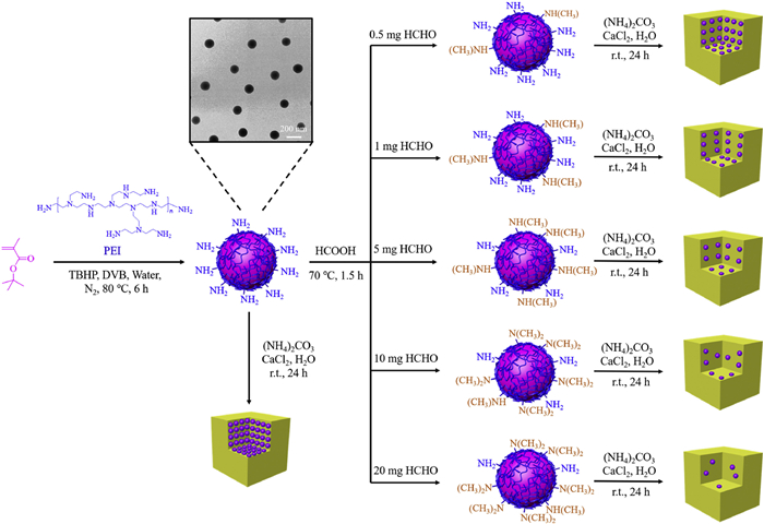
Figure 1
Figure 1.
Synthesis of poly(ethyleneimine)-functionalized poly(tert‑butyl methacrylate) (PtBMA/PEI) nanoparticles by soap-free emulsion polymerization at 80 ℃ for 24 h. Such nanoparticles were subsequently reacted with varying amounts of formaldehyde, producing model nanoparticles with tunable content of surface primary amines, which influence the degree of nanoparticle occlusion within calcite crystals.
PtBMA/PEI nanoparticles were synthesized via soap-free emulsion polymerization, using tert‑butyl hydroperoxide (TBHP) as the initiator and divinylbenzene (DVB) as the cross-linking agent. The tBMA/PEI mass ratio was a critical factor influencing the nanoparticle size. Nanoparticles prepared with a tBMA/PEI mass ratio of 4:1 were designated as BP10K-41, where "10K" refers to the molecular weight of PEI and "41" represents the mass ratio of tBMA/PEI. The detailed synthetic procedures are provided in Table S1 (Supporting information), and the formation mechanism was illustrated in Fig. S1 (Supporting information). Under the initiation of TBHP, PEI chains generated a redox pair. An electron transferred from the amino group of PEI to TBHP produced amino cation radicals and tert‑butoxy radicals (Fig. S1). The amino cation radicals initiated the homopolymerization of tert‑butyl methacrylate, while tert‑butoxy radicals abstracted hydrogen atoms from PEI, forming PEI macro-radicals. The PtBMA homopolymer grafted onto these macro-radicals [44]. Owing to differences in hydrophilicity, the resulting graft copolymer self-assembled, forming nanoparticles with hydrophobic PtBMA cores coated by hydrophilic PEI shells.
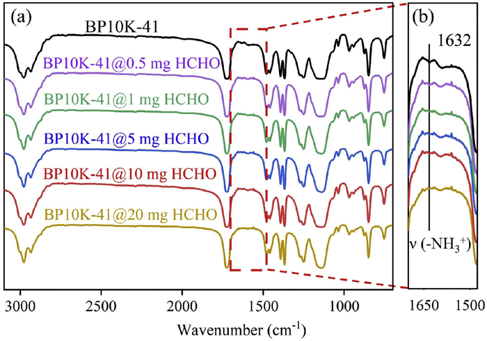
Potentiometric titration confirmed the presence of PEI on the surface of PtBMA/PEI nanoparticles, as the titration curves for both PEI and BP10K-41 nanoparticles showed similar responses to either acid titration or base titration (Fig. S2 in Supporting information). This observation reflects PEI's broad buffering capacity, a characteristic commonly referred to as the "proton sponge" effect [45-47]. Fourier transform infrared (FTIR) spectroscopy further supported the successful grafting of PEI onto PtBMA nanoparticles. The spectra revealed characteristic PtBMA peaks: carbonyl (C=O) stretching at 1725 cm−1 and tert‑butyl (-C(CH3)3) vibrations at 1394 cm−1 and 1367 cm−1 (Fig. S3 in Supporting information) [48-50]. Peaks associated with PEI were also evident, including N–H stretching at 3430 cm−1, CH₂ asymmetric and symmetric stretching at 2977 cm−1 and 2936 cm−1, respectively, and -NH3+ vibrations at 1632 cm−1 [51-54].
Transmission electron microscopy (TEM) revealed that the size of PtBMA/PEI nanoparticles increased with the tBMA/PEI mass ratio, ranging from 99 ± 6 nm to 143 ± 8 nm (Fig. S4 in Supporting information). Dynamic light scattering (DLS) confirmed their unimodal size distribution and excellent uniformity, with a dispersity index below 0.1 (Fig. S5 in Supporting information). The zeta potential of PtBMA/PEI nanoparticles, ranging from 25 mV to 60 mV, further confirmed the presence of PEI on the nanoparticle surface (Fig. S6 in Supporting information). To regulate the surface amine content on BP10K-41 nanoparticles, a series of samples were synthesized by reacting BP10K-41 with varying amounts of formaldehyde (0.5, 1, 5, 10, and 20 mg). Nanoparticles treated with 0.5 mg formaldehyde were labeled BP10K-41@0.5 mg HCHO. The reaction, known as the Eschweiler-Clarke reaction, involves the conversion of primary or secondary amines into methylated tertiary amines in the presence of formaldehyde and excess formic acid [55,56]. This modification was designed to adjust the proportions of primary, secondary, and tertiary amines on the nanoparticle surface. Hydrodynamic diameters of the six types of nanoparticle samples, measured at pH 9.5, showed no significant size changes following the formaldehyde reaction, indicating excellent colloidal stability (Fig. S7 in Supporting information). This stability is critical for ensuring effective nanoparticle occlusion in subsequent experiments.
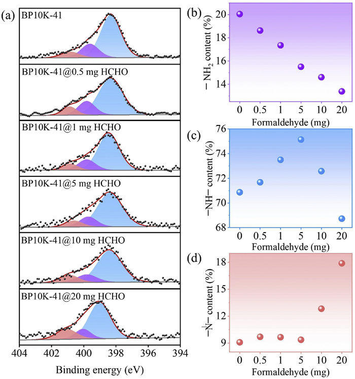
FTIR analysis revealed that the -NH3+ vibration peak at 1632 cm−1 in BP10K-41 nanoparticles diminished with increasing formaldehyde, indicating the reaction of primary amines in PEI with formaldehyde (Fig. 2). This was corroborated by X-ray photoelectron spectroscopy (XPS) (Fig. 3a), which showed that the primary amine content decreased as formaldehyde usage increased (Fig. 3b). Secondary amines initially increased, peaking at 5 mg formaldehyde, before declining (Fig. 3c). In contrast, tertiary amines remained stable at lower formaldehyde concentrations but increased at higher levels (Fig. 3d). These findings confirm the occurrence of the Eschweiler-Clarke reaction between formaldehyde and PEI on the BP10K-41 nanoparticle surface. At lower formaldehyde levels (0.5–5 mg), primary amines were primarily converted to secondary amines, while tertiary amine levels remained unchanged. At higher formaldehyde levels, secondary amines were further converted to tertiary amines, resulting in a decrease in secondary amines and an increase in tertiary amines.
Figure 2
Figure 2.
(a) FT-IR spectra of pure BP10K-41 nanoparticles and the corresponding BP10K-41 nanoparticle reacted with various amounts of formaldehyde. (b) Enlarged view of the region highlighted by the dashed red box in (a).
Figure 3.
(a) High resolution X-ray photoelectron spectroscopy (XPS) recorded for BP10K-41, BP10K-41@0.5 mg HCHO; BP10K-41@1 mg HCHO; BP10K-41@5 mg HCHO; BP10K-41@10 mg HCHO; BP10K-41@20 mg HCHO and (b-d) Content of primary, secondary and tertiary amines of BP10K-41 nanoparticles reacted with different amounts of formaldehyde.
The well-established ammonia diffusion method (ADM) was used to precipitate CaCO3 crystals in the presence of various nanoparticles at different Ca2+ concentrations at 25 ℃ for 24 h (Fig. S8 in Supporting information) [57]. To determine optimal conditions for nanoparticle occlusion experiments, CaCO3 crystals were grown in the presence of BP10K-41 nanoparticles (0.25–0.5 wt%) at Ca2+ concentrations ranging from 1.5 mmol/L to 5 mmol/L (Figs. S9 and S10 in Supporting information). An initial Ca2+ concentration of 2.5 mmol/L and a nanoparticle concentration of 0.25 wt% were selected for subsequent experiments, as these conditions allowed for uniform occlusion of BP10K-41 nanoparticles within CaCO3 crystals.
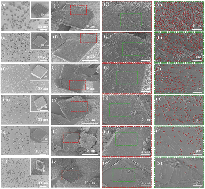
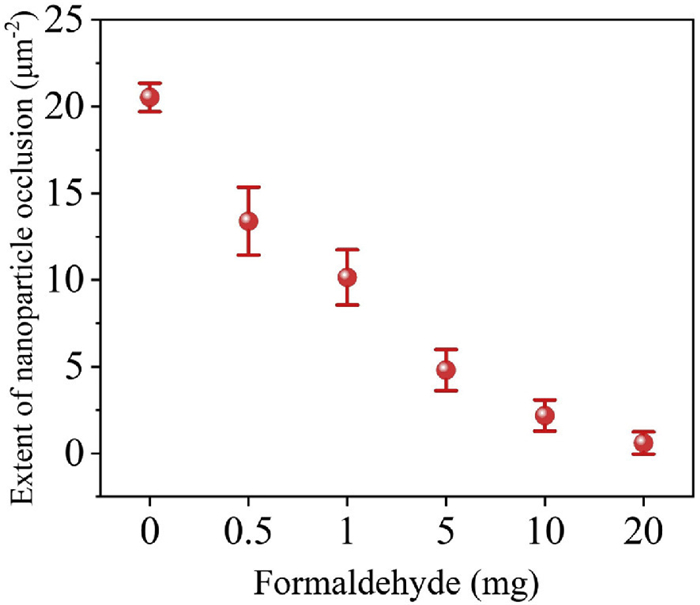
Optical microscopy (OM) revealed that perfect rhombohedral, transparent CaCO3 crystals formed in the absence of additives (Fig. S8). In contrast, translucent CaCO3 crystals with rough surfaces were obtained in the presence of various nanoparticles, suggesting successful nanoparticle occlusion (Figs. S9–S11 in Supporting information). Visualization of the cross-sections of randomly-fractured composite crystals provided direct evidences showing the successful occlusion of nanoparticles within CaCO3 crystals (Fig. 4). Notably, BP10K-41 nanoparticles exhibited uniform and dense occlusion. When BP10K-41 nanoparticles with surface PEI modified by varying formaldehyde amounts were tested, occlusion efficiency systematically decreased with increasing formaldehyde usage and the surface of the composite crystals become smoother (Fig. S11). Quantification of occluded nanoparticles per unit area in the cross-sections revealed that the extent of occlusion declined with reduced primary amine content on the nanoparticle surface (Fig. 5).
Figure 4
Figure 4.
Representative FE-SEM images recorded for calcium carbonate crystals precipitated in the presence of 0.25 wt% various nanoparticles at 2.5 mmol/L Ca2+. (a-d) BP10K-41; (e-h) BP10K-41@0.5 mg HCHO; (i-l) BP10K-41@1 mg HCHO; (m-p) BP10K-41@5 mg HCHO; (q-t) BP10K-41@10 mg HCHO; (u-x) BP10K-41@20 mg HCHO. The left column presents low-magnification images of composite crystals, with insets in (a), (e), (i), (m), (q), and (u) showing the surfaces of individual crystals. The second column displays cross-sections of randomly fractured the composite crystals. The third column provides enlarged SEM images, showing the red-boxed regions indicated in the second column, while the fourth column shows magnified SEM images of the dashed green-boxed regions labelled in the third column. To aid comparison, the occluded nanoparticles were red-labelled in the fourth column.
Figure 5.
Extent of nanoparticle occlusion versus the usage of formaldehyde. The extent of nanoparticle occlusion was determined by counting the number of nanoparticles within a 5 µm × 3 µm area of a cross-section of composite crystals, with the final results averaged from measurements taken across three parallel samples.
FTIR analysis confirmed the incorporation of nanoparticles within the CaCO3 crystals. This was evidenced by the presence of -CH₂- stretching at 2979 cm−1, attributed to PEI, and an ester carbonyl stretching at 1725 cm−1, corresponding to PtBMA, observed in the composite crystals (Fig. S12 in Supporting information). Raman spectroscopy further demonstrated that the polymorph of the crystals remained calcite, as indicated by characteristic bands at 1086 cm−1 (ν1), 712 cm−1 (ν2), 282 cm−1 and 155 cm−1 (lattice modes, Fig. S13 in Supporting information) [58,59]. Additionally, powder X-ray diffraction (PXRD) confirmed the crystal structure of the composite crystals as calcite, verifying that the incorporation of nanoparticles did not alter the crystallography of the host calcite crystals (Fig. S14 in Supporting information).
Previous studies have identified the mechanisms behind efficient nanoparticle occlusion within calcite crystals using in situ atomic force microscopy [38,39,60,61]. The surface chemistry of the guest nanoparticles plays a crucial role, as the surface stabilizers must interact closely with the growing calcite crystals. Without this intimate interaction, nanoparticles are excluded by advancing crystal steps [39]. Only nanoparticles with suitable surface chemistries can be efficiently occluded, with adsorbed particles gradually embedded by the crystal growth. Additionally, the colloidal stability of nanoparticles is important, as high calcium ion concentrations can cause aggregation of nanoparticles bearing carboxylic acid groups [31]. In this study, all tested nanoparticles exhibited high colloidal stability, confirmed by calcium titration experiments (Fig. S15 in Supporting information).
For anionic nanoparticles, divalent calcium ions bridge the particles and the growing crystal surface, promoting occlusion [25]. In our study, cationic amine groups on the surface of the nanoparticles provided binding sites for direct interaction with the negatively charged calcite surface via electrostatic interaction (Fig. S16 in Supporting information) [43]. Our previous investigations demonstrated that the solution pH during calcite crystal growth was maintained at approximately 9.8 [41]. At this pH, the secondary and tertiary amines of PEI were deprotonated, as their pKa values are below 8. In contrast, the primary amines of PEI on the surface of BP10K-41 nanoparticles remained partially protonated, given their pKa is approximately 9–10 [46]. The Eschweiler–Clarke reaction in this study converts primary amines to secondary or tertiary amines, thereby reduced the number of protonated primary amines on the BP10K-41 nanoparticle surface. Consequently, the available binding sites for interactions with the growing calcite surface decreased (Fig. S16), leading to reduced nanoparticle occlusion. This finding is in good agreement with prior studies, which revealed that sufficient binding sites are important for promoting nanoparticle occlusion within calcite crystals [30]. Previous studies have shown that altering nanoparticle surface chemistry can create patterned occlusion, where nanoparticles are preferentially occluded into specific regions of the crystal [27,41]. This patterning occurs because poly(methacrylic acid) functionalized nanoparticles are colloidally instable at a high concentration of calcium ions. Therefore, the occlusion cannot occur at the early stage of the crystallization. Occlusion occurs only at reduced calcium ion concentrations at the later stages of crystallization [41]. In contrast, cationic nanoparticles in this study maintained high colloidal stability even at high calcium concentrations. As a result, nanoparticles with fewer amine groups did not interact closely with the growing calcite surface, leading to sparse occlusion rather than patterned occlusion. This is significant for creating polymer-organic nanocomposite crystals with uniformly distributed guest nanoparticles.
In summary, we investigated the impact of surface amine content on nanoparticle occlusion within calcite crystals. A series of cationic PEI-functionalized nanoparticles were synthesized through soap-free emulsion polymerization, and their amine content was systematically adjusted by reacting with formaldehyde. Using calcite as a host crystal, our findings revealed that the amine content plays a crucial role in determining the extent of nanoparticle occlusion within the crystals. This work enhances the understanding of how primary amine groups influence nanoparticle occlusion and provides valuable design guidelines for constructing composite crystals with tunable guest species content.
Declaration of competing interest
The authors declare that they have no known competing financial interests or personal relationships that could have appeared to influence the work reported in this paper.
Y. Ning is grateful for the financial supports from the National Natural Science Foundation of China (Nos. 22475084 and 22101100), Guangdong Basic and Applied Basic Research Foundation (Nos. 2024A1515012114 and 2025A1515012931), and College Students' Innovation and Entrepreneurship Training Program. The Analysis and Testing Center of Jinan University is thanked for TEM/SEM support.
Supplementary materials
Supplementary material associated with this article can be found, in the online version, at doi:10.1016/j.cclet.2025.111205.
S. Yamabe, N. Tsuchida, S. Yamazaki, J. Phys. Org. Chem. 34 (2021) e4253.
[57]
J. Ihli, P. Bots, A. Kulak, et al., Adv. Funct. Mater. 23 (2012) 1965–1973.
[58]
C. Gabrielli, R. Jaouhari, S. Joiret, et al., J. Raman Spectrosc. 31 (2000) 497–501.
[59]
U. Wehrmeister, A.L. Soldati, D.E. Jacob, et al., J. Raman Spectrosc. 41 (2009) 193–201.
[60]
Y. Dong, J. Chi, Z. Ren, et al., Angew. Chem. Int. Ed. 62 (2023) e202300031.
[61]
T. Ye, X.Y. Jin, L. Chen, et al., Chin. Chem. Lett. 28 (2017) 857–862.
Figure 1
Synthesis of poly(ethyleneimine)-functionalized poly(tert‑butyl methacrylate) (PtBMA/PEI) nanoparticles by soap-free emulsion polymerization at 80 ℃ for 24 h. Such nanoparticles were subsequently reacted with varying amounts of formaldehyde, producing model nanoparticles with tunable content of surface primary amines, which influence the degree of nanoparticle occlusion within calcite crystals.
Figure 2
(a) FT-IR spectra of pure BP10K-41 nanoparticles and the corresponding BP10K-41 nanoparticle reacted with various amounts of formaldehyde. (b) Enlarged view of the region highlighted by the dashed red box in (a).
Figure 3
(a) High resolution X-ray photoelectron spectroscopy (XPS) recorded for BP10K-41, BP10K-41@0.5 mg HCHO; BP10K-41@1 mg HCHO; BP10K-41@5 mg HCHO; BP10K-41@10 mg HCHO; BP10K-41@20 mg HCHO and (b-d) Content of primary, secondary and tertiary amines of BP10K-41 nanoparticles reacted with different amounts of formaldehyde.
Figure 4
Representative FE-SEM images recorded for calcium carbonate crystals precipitated in the presence of 0.25 wt% various nanoparticles at 2.5 mmol/L Ca2+. (a-d) BP10K-41; (e-h) BP10K-41@0.5 mg HCHO; (i-l) BP10K-41@1 mg HCHO; (m-p) BP10K-41@5 mg HCHO; (q-t) BP10K-41@10 mg HCHO; (u-x) BP10K-41@20 mg HCHO. The left column presents low-magnification images of composite crystals, with insets in (a), (e), (i), (m), (q), and (u) showing the surfaces of individual crystals. The second column displays cross-sections of randomly fractured the composite crystals. The third column provides enlarged SEM images, showing the red-boxed regions indicated in the second column, while the fourth column shows magnified SEM images of the dashed green-boxed regions labelled in the third column. To aid comparison, the occluded nanoparticles were red-labelled in the fourth column.
Figure 5
Extent of nanoparticle occlusion versus the usage of formaldehyde. The extent of nanoparticle occlusion was determined by counting the number of nanoparticles within a 5 µm × 3 µm area of a cross-section of composite crystals, with the final results averaged from measurements taken across three parallel samples.

 DownLoad:

 下载:
下载:


 下载:
下载:
