Scheme 1.
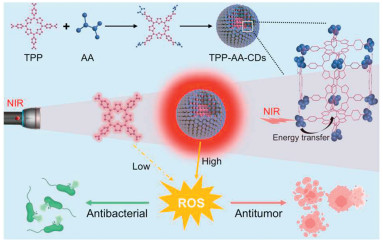
Schematic illustration of the development of light-harvesting pigment-binding protein-mimicking carbon dots for photodynamic therapy.
Light-harvesting pigment-binding protein-mimicking carbon dots for photodynamic therapy
Quanxin Ning , Yidan Zhang , Huayi Sun , Xin Zhao , Haodong Zhang , Feng Cui , Xiaochun Xie , Fangman Chen , Wen Sun , Hong Zhang

Photodynamic therapy (PDT), which leverages photosensitizers to harvest light for the generation of reactive oxygen species (ROS), has garnered significant attention due to its spatiotemporal precision and non-invasive nature [1–5]. Porphyrins, characterized by their large π-conjugated planar structures comprising four pyrrole subunits linked via methine bridges, are among the most studied photosensitizers [6,7]. Their absorption spectra typically exhibit a prominent Soret band around 400–450 nm and a series of weaker Q-bands between 500 nm and 700 nm. These molecules are known for their excellent photostability and distinct photophysical properties, making them widely used in fluorescence imaging and PDT. Several porphyrin-based photosensitizers have even been approved for clinical use [8,9]. However, their clinical potential is limited by issues such as extreme hydrophobicity, aggregation-caused quenching (ACQ) due to π-π stacking, and weak absorption in the near-infrared (NIR) region, all of which hinder efficient ROS generation [10,11]. Although modifications to porphyrins can enhance their phototherapeutic efficacy, these approaches are often labor-intensive and time-consuming [12,13]. Therefore, there is a pressing need for innovative strategies that not only improve the solubility and mitigate the ACQ effect of traditional photosensitizers but also enhance light-harvesting efficiency within the NIR region, thereby improving PDT efficacy.
Photosynthesis, one of nature's most efficient sunlight energy conversion systems, relies heavily on pigment-binding proteins that serve as light-harvesting antennas [14,15]. These proteins are integral to the high efficiency of natural photosynthesis, as they migrate excited energy and transfer harvested energy to charge-separating systems [16]. Inspired by the unique supramolecular structures and optical properties of pigment-binding proteins, the use of light-harvesting scaffolds has emerged as a promising approach for enhancing the PDT efficacy of photoactive nanoparticles [17–20]. Carbon dots (CDs), a new class of fluorescent nanomaterials, offer numerous advantages, including tunable optical properties, excellent water dispersibility, biocompatibility, and rapid renal excretion due to their small size [21–24]. These properties make CDs highly versatile, with applications spanning biosensing [25,26], imaging [27,28], biomedical diagnostics [29,30], and drug delivery [31,32]. Their broad absorption range, spanning from visible to NIR light, positions CDs as nearly ideal building blocks for constructing light-harvesting scaffolds [33,34]. Consequently, the development of simple porphyrin-doped CDs that integrate the superior light-harvesting capabilities of CDs and effectively transfer the excitation energy to the porphyrin core is highly desirable for enhancing PDT.
Inspired by the highly efficient light-harvesting capabilities of pigment-binding proteins, we have developed carbon dots derived from porphyrin and amino acid mixture (TPP-AA-CDs) to enhance light-harvesting efficiency and improve PDT (Scheme 1). The amino acids (AA) serve as the foundational building blocks for these CDs, scaffolding porphyrin centre within the frameworks through a four-stage process involving dehydration, polymerization, passivation, and carbonization [35]. The side chains of the AA precursors, featuring functional groups such as -NH2 and -OH, offer potential for covalent conjugation with porphyrin via dehydration reactions [36]. As a result, the TPP-AA-CDs exhibit significantly higher light-harvesting efficiency in the range of 500–700 nm (corresponding to the Q-bands) within the "phototherapeutic window" compared to free porphyrin pigments. Notably, TPP-AA-CDs exhibit characteristic red fluorescence from the porphyrin and enhanced fluorescence emission dependent on the AA precursors [37]. Furthermore, these TPP-AA-CDs generate markedly higher ROS yields than free porphyrin, indicating that the amino acid-derived CDs (AA-CDs) scaffolds effectively harness photo and transfer light energy to the porphyrin centre, thereby amplifying the photodynamic effect. Among the various TPP-AA-CDs, the carbon dots derived from porphyrin and histidine mixture (TPP-H-CDs) demonstrate the most optimal performance and superior PDT efficacy. Additionally, TPP-H-CDs exhibit high biocompatibility in cellular and in vivo studies, showing the capability to generate ROS for antibacterial activity to promote wound healing and antitumor effect to inhibit tumor growth under NIR light irradiation. These findings highlight a promising approach to enhancing PDT efficacy of porphyrin in the NIR region through the use of amino acid-derived CDs scaffolds as light-harvesting antennas [38].
To systematically investigate and optimize the formation of TPP-AA-CDs using various amino acid precursors: leucine (L, nonpolar, aliphatic, neutral), tyrosine (Y, nonpolar, aromatic, neutral), serine (S, polar, aliphatic, neutral), lysine (K, polar, aliphatic, positive), histidine (H, polar, aromatic, positive), glutamic acid (E, polar, aliphatic, negative), and methionine (M, nonpolar, thioether, neutral), we employed a facile and sustainable one-step hydrothermal synthesis method, as illustrated in Fig. 1a. UV–vis absorption spectroscopy, visual analysis, and fluorescence studies were conducted to assess the impact of different amino acids on the formation of TPP-AA-CDs from the TPP and AA mixture. The resulting TPP-AA-CDs demonstrated characteristic UV-visible spectral features similar to porphyrins, including the Soret band (a prominent absorption peak at 414 nm) and Q-bands (four distinct peaks in the 500–700 nm range) (Fig. 1b). Specifically, the absorption spectra of TPP-H-CDs, TPP-K-CDs, TPP-Y-CDs, and TPP-S-CDs exhibited a pronounced increase in the Q-band absorption between 500 nm and 700 nm compared to free TPP under identical conditions, while no significant absorbance changes were observed in TPP-L-CDs, TPP-E-CDs, and TPP-M-CDs. Visual inspection of the aqueous suspensions of the synthesized TPP-AA-CDs revealed a predominantly dark yellow color, in contrast to the light pale-yellow color of the pure TPP solutions (Fig. 1c). These observations confirm the successful formation of TPP-AA-CDs using the one-step hydrothermal method, with amino acid-derived scaffolds enhancing the light-harvesting capacity in the range of visible to NIR region. Among the TPP-AA-CDs, TPP-H-CDs, TPP-K-CDs, and TPP-S-CDs exhibited notably brighter red fluorescence compared to the free TPP solution, while other TPP-AA-CDs showed no significant increase in fluorescence emission (Fig. 1c). The enhanced light-harvesting efficiency of these porphyrin-doped carbon dots, particularly those derived from hydrophilic or basic AA, is likely attributable to the -NH2 or -OH groups on the AA side chains, which facilitate the integration of porphyrin into the framework through the stages of dehydration, polymerization, passivation, and carbonization. Conversely, TPP-Y-CDs displayed significantly lower fluorescence intensity, likely due to the aggregation-caused quenching effect resulting from the hydrophobic nature of tyrosine [39].
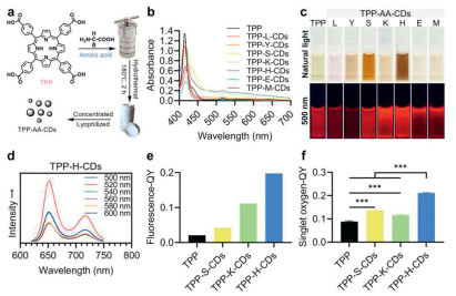
Photoluminescence spectroscopy was employed to investigate the photophysical properties of TPP-H-CDs, TPP-K-CDs, and TPP-S-CDs, and to assess their potential for PDT [40]. These materials exhibited two distinct emission peaks around 650 nm and 710 nm, corresponding to fluorescence and phosphorescence emissions, respectively, which are similar to the emission wavelengths observed for TPP upon excitation in 500–600 nm range (Fig. 1d and Fig. S2 in Supporting information). The strongest fluorescence emission was recorded at an excitation wavelength of 520 nm. Notably, the fluorescence quantum yields (QY) of TPP-H-CDs (0.1977), TPP-K-CDs (0.1115), and TPP-S-CDs (0.0420) were significantly higher compared to those of free TPP (0.0207), correlating with the enhanced features observed in the UV–vis absorption spectra (Fig. 1e). On the contrary, AA-CDs also showed a wide range of UV-visible light absorption, but it was worse than the corresponding TPP-AA-CDs, which might be because the hybrid of TPP increases the degree of conjugation of carbon dots and promotes light absorption (Fig. S1 in Supporting information). At the same time, the fluorescence emissivity of AA-CDs under visible light excitation was completely different from that of TPP-AA-CDs due to lack of efficient TPP as sensitization center (Fig. S2 in Supporting information). This increase in quantum yield suggests that the amino acid-derived CDs scaffold in these TPP-AA-CDs functions effectively as a light-harvesting antenna, transferring energy to the TPP centre [41]. To evaluate the singlet oxygen (1O2) generation capacity of TPP-AA-CDs, we employed 1,3-diphenylisobenzofuran (DPBF) as a specific indicator [42]. The observed reduction in absorbance at 410 nm corresponds to oxidative degradation, highlighting the efficiency of 1O2 production. As illustrated in Fig. 1f and Fig. S3 (Supporting information), TPP-H-CDs, TPP-K-CDs, and TPP-S-CDs demonstrated markedly enhanced 1O2 generation efficiencies upon irradiation when compared to free TPP, with TPP-H-CDs exhibiting the highest singlet oxygen quantum yield. This quantitative measure of the singlet oxygen quantum yield was consistent with the data obtained from UV–vis absorption spectroscopy and the fluorescence quantum yield. These findings underscored that the TPP-AA-CDs possess enhanced light-harvesting efficiency conducive to ROS generation. Among these, TPP-H-CDs, with their optimal optical performance, exhibited the most pronounced absorption features, facilitating greater photon capture and ROS production.
After optimizing the formation of TPP-AA-CDs with various amino acid precursors, the morphology of the TPP-H-CDs was characterized using transmission electron microscopy (TEM). As depicted in Figs. 2a and c, the histidine-derived carbon dots (H–CDs) used as a control exhibited good dispersion and uniform spherical morphology, with an average particle size of approximately 2.73 nm and a zeta potential of -20.4 mV. The morphology of the synthesized TPP-H-CDs was similar to that of the H–CDs (Fig. 2b). However, the increase in particle size and the decrease in zeta potential indicate successful hybrid of TPP within the AA-CDs (Fig. 2c). The chemical structure and composition of TPP-H-CDs were further analyzed using X-ray photoelectron spectroscopy (XPS), revealing typical peaks for C 1s (285.08 eV, 65.56%), N 1s (400.08 eV, 12.92%), and O 1s (531.08 eV, 21.52%) (Fig. 2d). The C 1s spectrum displayed three main peaks at 288.23 eV, 285.80 eV, and 284.70 eV, corresponding to C═O/C═N, C–O/C–N, and C═C/C–C bonds, respectively. The N 1s spectrum showed signals at 399.8 eV and 398.3 eV, attributed to N═C and N–C bonds. The O 1s spectrum exhibited two peaks at 534.4 eV and 531.4 eV, corresponding to C═O and C–OH groups, respectively (Fig. S4 in Supporting information). Fourier transform infrared (FT-IR) spectroscopy was employed to further elucidate the functional groups in TPP-H-CDs (Fig. 2e). The FT-IR spectrum showed vibrational peaks at 2900 cm-1 (C–H), 1603 cm-1 (C═O), 1541 cm-1 (C═N), 1391 cm-1 (C–N), and 1100 cm-1 (C–O). Broad absorption bands at 3418 cm-1 and 3200 cm-1 were assigned to the stretching vibrations of hydrophilic O–H and N–H groups, respectively [43]. The XPS and FT-IR data are consistent, indicating the presence of numerous hydrophilic units, such as amino and carboxyl groups, in TPP-H-CDs. Consequently, the aqueous solution of TPP-H-CDs demonstrated long-term stability at room temperature without noticeable precipitation, reflecting high colloidal stability in physiological conditions (Fig. S5 in Supporting information).
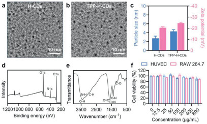
Before proceeding with in vivo and in vitro evaluations of photodynamic therapy, it was crucial to assess the biocompatibility of TPP-H-CDs [44]. Cytotoxicity was evaluated using the CCK-8 assay in human umbilical vein endothelial cells (HUVECs) and RAW 264.7 macrophages. Both types of cells maintained viability greater than 80% after 24 h of incubation, even at TPP-H-CDs concentrations up to 800 µg/mL (Fig. 2f). Hemolysis tests were conducted on TPP-H-CDs to determine their hemolysis rate. The results obtained from these tests confirmed the hemolysis rate of TPP-H-CDs remained below 5% even at a concentration of 1000 µg/mL, demonstrating its good blood compatibility (Fig. S6 in Supporting information). Additionally, blood biochemical assays were conducted to assess standard parameters, including alanine aminotransferase (ALT), aspartate aminotransferase (AST), creatinine (CREA), urea (UREA), and blood urea nitrogen (BUN). No significant variations were observed across all groups (Fig. S7 in Supporting information). Histopathological examination of primary organs (heart, liver, spleen, lung, and kidney) from mice, conducted 7 days after intravenous injection of TPP-H-CDs, revealed no statistically significant changes (Fig. S8 in Supporting information). These findings collectively indicate that TPP-H-CDs exhibit favorable biocompatibility and low cytotoxicity.
Porphyrin-based PDT is a non-contact, convenient, and effective therapeutic approach with a low risk of inducing drug resistance, making it a valuable clinical treatment for bacterial infections [45]. Leveraging the histidine-derived CDs scaffold to enhance photon harvesting and facilitate energy transfer to the TPP centre, we investigated the antibacterial activity of TPP-H-CDs via PDT [46].
After incubating Gram-negative Escherichia coli (E. coli) and Gram-positive Staphylococcus aureus (S. aureus) with TPP-H-CDs for 24 h, we observed a significant increase in antibacterial activity under near-infrared (NIR) irradiation at 700 nm (20 mW/cm2) for 10 min (Fig. 3a). In contrast, the control group treated with free TPP under the same irradiation conditions exhibited only a slight inhibition of bacterial growth. Notably, all treatments conducted without irradiation displayed minimal inhibition of bacterial growth, confirming the broad-spectrum antibacterial efficacy of TPP-H-CDs through PDT. This trend was further corroborated by the number of bacterial colonies observed on LB agar plates (Fig. 3b and Fig. S9 in Supporting information).
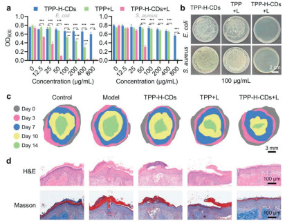
Encouraged by the promising in vitro photodynamic antibacterial activity, we further investigated the potential of TPP-H-CDs as a photodynamic therapeutic agent for enhancing wound healing in the context of bacterial infection [47]. To develop a model for bacterial-infected wounds, 10 mm full-thickness dorsal skin punch wounds were created on the backs of mice and subsequently infected with Pseudomonas aeruginosa (P. aeruginosa). The mice were then divided into groups: those receiving PBS (as the model group), TPP with NIR irradiation, TPP-H-CDs alone, and TPP-H-CDs with NIR irradiation. Uninfected wounds served as the control group. The wound healing process was monitored and assessed through representative images (Fig. S10 in Supporting information), schematic diagrams (Fig. 3c), and quantitative analysis of wound contraction (Fig. S11 in Supporting information). Wounds infected with P. aeruginosa showed a slower healing rate compared to the control group, accompanied by minor yellow pus exudate. Remarkably, wounds treated with TPP-H-CDs plus NIR irradiation exhibited significantly faster healing compared to all other groups at multiple time points. By day 14, epithelialization was evident in all groups, with the TPP-H-CDs plus NIR irradiation group demonstrating the most advanced epithelialization and hair follicle formation. Masson's trichrome staining further revealed superior collagen deposition in the TPP-H-CDs group on day 14 compared to the other groups (Fig. 3d). These findings indicate that TPP-H-CDs, when activated by NIR irradiation, effectively accelerate wound healing and prevent scar formation in vivo, likely due to their enhanced light-harvesting capacity and subsequent photodynamic efficacy.
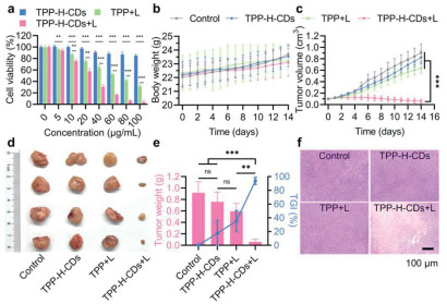
Porphyrin-based PDT has emerged as a promising clinical approach for tumor treatment [48]. Given the superior ROS generation capabilities of TPP-H-CDs under NIR light irradiation, we further explored their potential in antitumor applications. After incubating CT26.WT cells at 37 ℃ with TPP-H-CDs, clusters of bright intensities were observed inside all the cells imaged. This observation occurred in a time-dependent manner, and after 4 h of incubation, a plateau period was reached. These findings suggest a high efficiency of cellular uptake for TPP-H-CDs (Fig. S12 in Supporting information). The cytotoxicity of TPP-H-CDs on the mouse colon cancer CT26.WT cell line were further conducted using the CCK-8 assay. Upon NIR light exposure (700 nm, 20 mW/cm2 for 30 min), TPP-H-CDs exhibited significant light-triggered cytotoxicity against CT26.WT cells, resulting in markedly lower cell viability compared to free TPP. Conversely, TPP-H-CDs showed minimal cytotoxic effects on CT26.WT cells in the absence of NIR light, across the tested concentrations (Fig. 4a). To evaluate the in vivo PDT efficacy, we conducted studies on syngeneic CT26.WT tumor-bearing mice [49]. To establish the optimal irradiation time, we investigated the biodistribution of TPP-H-CDs in CT26-tumor-bearing mice at several time intervals following administration (Fig. S13 in Supporting information). The results indicated that the average fluorescence intensity of TPP-H-CDs at the tumor site peaked at 6 h post-intravascular administration, after which a decline in intensity was observed by the 12 h mark. Therefore, we concluded that 6 h post-administration is the optimal time point for NIR irradiation. The mice were subjected to four different treatment regimens: PBS only, TPP-H-CDs only, TPP with NIR light, and TPP-H-CDs with NIR light. Body weight fluctuations within the four groups suggest that the treatment did not induce any significant adverse effects (Fig. 4b). Tumors in the PBS, TPP-H-CDs only and TPP with NIR light groups grew rapidly, while significant tumor growth inhibition was observed in the TPP-H-CDs with NIR light groups (Figs. 4c and d). Notably, the TPP-H-CDs with NIR light group demonstrated a higher tumor growth inhibition (TGI) rate than the TPP with NIR light group, underscoring the potent photodynamic antitumor efficacy of TPP-H-CDs under NIR light irradiation (Fig. 4e). Histological analysis using hematoxylin and eosin (H&E) staining further revealed extensive apoptosis and necrosis in tumor sections from the TPP-H-CDs with NIR light group, which was significantly more pronounced than in the other treatment groups (Fig. 4f). These results highlight the exceptional PDT efficacy of TPP-H-CDs in suppressing tumor growth, suggesting that the amino acid-derived carbon dots scaffold enhances the NIR light-mediated photodynamic effects of TPP [50].
In summary, inspired by the unique supramolecular structures and optical properties of pigment-binding proteins, we have developed carbon dots derived from porphyrin and amino acid mixture for NIR light-mediated PDT. This system is designed to accelerate the healing of infected wounds and enhance antitumor effects. Utilizing the spontaneous conjugation between amino acids and porphyrin, TPP-AA-CDs are synthesized through a four-stage process involving dehydration, polymerization, passivation, and carbonization in a hydrothermal reactor. The amino acid-derived framework acts as a light-harvesting antenna that scaffolds the porphyrin centre. Moreover, the optical properties of TPP-AA-CDs can be finely tuned by selecting different amino acid precursors, leading to enhanced light-harvesting efficiency for improved PDT effects within the red-NIR light range. This enhancement is likely due to the presence of -NH2 or -OH groups on the amino acid side chains, which facilitate the incorporation of porphyrin within the framework. The photosensitive framework scaffolding the porphyrin presents a promising strategy for developing more efficient NIR-absorbing porphyrin-based materials, with significant potential for future clinical applications.
The authors declare that they have no known competing financial interests or personal relationships that could have appeared to influence the work reported in this paper.
Quanxin Ning: Writing – original draft, Investigation. Yidan Zhang: Methodology, Investigation. Huayi Sun: Methodology. Xin Zhao: Investigation. Haodong Zhang: Methodology. Feng Cui: Investigation. Xiaochun Xie: Investigation. Fangman Chen: Writing – original draft, Conceptualization. Wen Sun: Writing – review & editing, Conceptualization. Hong Zhang: Writing – review & editing, Funding acquisition.
This work was supported by the Key Research and Development Joint Fund of the Department of Science and Technology of Liaoning Province (No. 2021JH2/1030005), the General Program of the Education Department of Liaoning Province (No. JYTMS20230101).
Supplementary material associated with this article can be found, in the online version, at doi:
T.J. Dougherty, C.J. Gomer, B.W. Henderson, et al., J. Natl. Cancer Inst. 90 (1998) 889–905. doi: 10.1093/jnci/90.12.889
Z. Zhou, J. Song, L. Nie, X. Chen, Chem. Soc. Rev. 45 (2016) 6597–6626. doi: 10.1039/C6CS00271D
J. Xu, J. Xu, T. Shi, et al., Adv. Mater. 35 (2023) e2207890. doi: 10.1002/adma.202207890
M. Dong, R. Tang, J. Li, et al., Chin. Chem. Lett. 35 (2024) 108539. doi: 10.1016/j.cclet.2023.108539
F. Ruan, H. Fang, F. Chen, et al., Angew. Chem. Int. Ed. 63 (2024) e202317943. doi: 10.1002/anie.202317943
M. Ethirajan, Y. Chen, P. Joshi, R.K. Pandey, Chem. Soc. Rev. 40 (2011) 340–362. doi: 10.1039/B915149B
R. Hu, X. Zhai, Y. Ding, et al., Chin. Chem. Lett. 33 (2022) 2715–2720. doi: 10.1016/j.cclet.2021.08.110
S.S. Lucky, K.C. Soo, Y. Zhang, Chem. Rev. 115 (2015) 1990–2042. doi: 10.1021/cr5004198
Y. Zhou, X. Liang, Z. Dai, Nanoscale 8 (2016) 12394–12405. doi: 10.1039/C5NR07849K
M.A. Rajora, J.W.H. Lou, G. Zheng, Chem. Soc. Rev. 46 (2017) 6433–6469. doi: 10.1039/C7CS00525C
J. Tian, B. Huang, M.H. Nawaz, W. Zhang, Coordin. Chem. Rev. 420 (2020) 213410. doi: 10.1016/j.ccr.2020.213410
J. Chen, Y. Zhu, S. Kaskel, Angew. Chem. Int. Ed. 60 (2021) 5010–5035. doi: 10.1002/anie.201909880
J.M. Park, K.I. Hong, H. Lee, W.D. Jang, Acc. Chem. Res. 54 (2021) 2249–2260. doi: 10.1021/acs.accounts.1c00114
E.J. Boekema, B. Hankamer, D. Bald, et al., Proc. Natl. Acad. Sci. U. S. A. 92 (1995) 175–179. doi: 10.1073/pnas.92.1.175
A. Ben-Shem, F. Frolow, N. Nelson, Nature 426 (2003) 630–635. doi: 10.1038/nature02200
X. Li, O. Björkman, C. Shih, et al., Nature 403 (2000) 391–395. doi: 10.1038/35000131
J. Sun, J. Zhang, M. Zhang, et al., Nat. Commun. 3 (2012) 1139. doi: 10.1038/ncomms2152
C. Fiankor, J. Nyakuchena, R.S.H. Khoo, et al., J. Am. Chem. Soc. 143 (2021) 20411-20148. doi: 10.1021/jacs.1c10291
Z. Wang, F. Chen, Y. Cao, et al., Adv. Mater. 36 (2024) e2314197. doi: 10.1002/adma.202314197
F. Chen, F. Ruan, X. Xie, et al., Adv. Mater. 36 (2024) e2402966. doi: 10.1002/adma.202402966
D. Shao, F. Zhang, F. Chen, et al., Adv. Mater. 32 (2020) e2004385. doi: 10.1002/adma.202004385
M. Fang, B. Wang, X. Qu, et al., Chin. Chem. Lett. 35 (2024) 108423. doi: 10.1016/j.cclet.2023.108423
L. Cao, M. Zan, F. Chen, et al., Carbon 194 (2022) 42–51. doi: 10.1016/j.carbon.2022.03.058
H. Cai, X. Wu, L. Jiang, et al., Chin. Chem. Lett. 35 (2024) 108946. doi: 10.1016/j.cclet.2023.108946
M. Pourmadadi, E. Rahmani, M. Rajabzadeh-Khosroshahi, J. Drug Deliv. Sci. Tec. 80 (2023) 104156. doi: 10.1016/j.jddst.2023.104156
F. Du, L. Yang, L. Wang, J. Mater. Chem. B 11 (2023) 8117–8135. doi: 10.1039/d3tb01329d
J. Zhong, X. Chen, M. Zhang, et al., Chin. Chem. Lett. 31 (2020) 769–773. doi: 10.1016/j.cclet.2020.01.007
L. Wang, B. Wang, E. Liu, et al., Chin. Chem. Lett. 33 (2022) 4111–4115. doi: 10.1016/j.cclet.2022.01.042
C. Li, J. Huang, L. Yuan, et al., Theranostics 19 (2023) 3064–3102. doi: 10.7150/thno.80579
M.M. Hussain, W.U. Khan, F. Ahmed, et al., Chem. Eng. J. 465 (2023) 143010. doi: 10.1016/j.cej.2023.143010
J. Lu, T. Shi, C. Shi, et al., Research 6 (2023) 0204. doi: 10.34133/research.0204
Q. Li, J. Fan, H. Mu, et al., Chin. Chem. Lett. 35 (2024) 108947. doi: 10.1016/j.cclet.2023.108947
R. Pathak, V.D. Punetha, S. Bhatt, M. Punetha, J. Mater. Sci. 58 (2023) 6419–6443. doi: 10.1007/s10853-023-08408-4
H. Liu, X. Zhong, Q. Pan, et al., Coordin. Chem. Rev. 498 (2024) 215468. doi: 10.1016/j.ccr.2023.215468
J. Yang, W. Chen, X. Liu, et al., Mater. Res. Bull. 89 (2017) 26–32. doi: 10.1016/j.materresbull.2017.01.013
K.D.W. Vollett, H.M. Cheng, Org. Biomol. Chem. 22 (2024) 6308–6320. doi: 10.1039/d4ob00704b
H. Ding, S.B. Yu, J.S. Wei, H.M. Xiong, ACS Nano 10 (2016) 484–491. doi: 10.1021/acsnano.5b05406
F. Chen, H. Huang, F. Zhang, et al., Adv. Mater. (2024) e2413385.
L. Ai, Z. Song, M. Nie, et al., Angew. Chem. Int. Ed. 62 (2023) e202217822. doi: 10.1002/anie.202217822
B. Wang, S. Lu, Matter 5 (2022) 110–149. doi: 10.1016/j.matt.2021.10.016
V.B. Kumar, S.K. Mirsky, N.T. Shaked, E. Gazit, ACS Nano 18 (2024) 2421–2433. doi: 10.1021/acsnano.3c10792
Z. Wu, F. Cui, H. Li, et al., Sensor. Actuat. B: Chem. 398 (2024) 134684. doi: 10.1016/j.snb.2023.134684
F. Arcudi, L. Đorđević, M. Prato, Angew. Chem. Int. Ed. 55 (2016) 2107–2112. doi: 10.1002/anie.201510158
N. Parvin, V. Kumar, S.W. Joo, T.K. Mandal, Nanomaterials 14 (2024) 1085. doi: 10.3390/nano14131085
B.M. Amos-Tautua, S.P. Songca, O.S. Oluwafemi, Molecules 24 (2019) 2456. doi: 10.3390/molecules24132456
K.A.D.F. Castro, N.M. M Moura, F. Figueira, et al., Int. J. Mol. Sci. 20 (2019) 2522. doi: 10.3390/ijms20102522
F. Cui, F. Chen, X. Xie, et al., Chin. Chem. Lett. 35 (2024) 108681. doi: 10.1016/j.cclet.2023.108681
R. Boscencu, N. Radulea, G. Manda, et al., Molecules 28 (2023) 1149. doi: 10.3390/molecules28031149
L. Li, S. He, B. Liao, et al., Research 7 (2024) 0364. doi: 10.34133/research.0364
Z. Tian, H. Li, Z. Liu, et al., Curr. Treat. Option. On. 24 (2023) 1274–1292. doi: 10.1007/s11864-023-01120-0

Scheme 1 Schematic illustration of the development of light-harvesting pigment-binding protein-mimicking carbon dots for photodynamic therapy.

Figure 1 Synthesis and screening of TPP-AA-CDs. (a) Synthetic route of TPP-AA-CDs. (b) UV–vis spectra of TPP-AA-CDs. (c) Photographs of TPP-AA-CDs solutions under natural light and 500 nm light. (d) Fluorescence spectra of TPP-H-CDs. (e) Fluorescence quantum yields of TPP-AA-CDs. (f) The singlet oxygen quantum yield of TPP-AA-CDs, n = 3. Data are shown as mean ± SD. (***P < 0.001).

Figure 2 Characterization of TPP-H-CDs. TEM images of (a) H–CDs and (b) TPP-H-CDs. (c) DLS analysis of H–CDs and TPP-H-CDs, n = 3. (d) XPS survey spectra of TPP-H-CDs. (e) FT-IR spectra of TPP-H-CDs. (f) Relative cell viability of HUVECs and RAW 264.7 cells incubated with TPP-H-CDs in the dark for 24 h, n = 3. Data are shown as mean ± SD.

Figure 3 Antibacterial activity and bacterial-infected wound healing efficacy of TPP-H-CDs. (a) Optical density at 600 nm of E. coli and S. aureus after incubation with TPP or TPP-H-CDs for 24 h, with or without NIR light irradiation, n = 3. (b) Agar plate photos of E. coli and S. aureus incubated with 100 µg/mL TPP-H-CDs or 10 µg/mL TPP for 12 h, with or without NIR light irradiation, n = 3. (c) Diagrams illustrating temporal changes in wound area, n = 3. (d) Representative histological images of regenerated wound tissues on day 14, stained with H&E and Masson's trichrome staining, n = 3. Data are shown as mean ± SD (***P < 0.001).

Figure 4 Antitumor activity of TPP-H-CDs. (a) Relative cell viability of CT26.WT cells treated with different concentrations of TPP or TPP-H-CDs, with or without 30 min of NIR light irradiation, n = 3. (b) Body weight and (c) tumor volume over time in CT26-tumor-bearing BALB/c mice, n = 4. (d) Photographs of excised tumors on day 14, n = 4. (e) Tumor weight and growth inhibition rate, n = 4. (f) Representative H&E staining images of from each treatment group, n = 3. Data are shown as mean ± SD. **P < 0.01, ***P < 0.001.

 扫一扫看文章

扫一扫关注我们
 DownLoad:
 下载:
下载:
 下载:
下载: