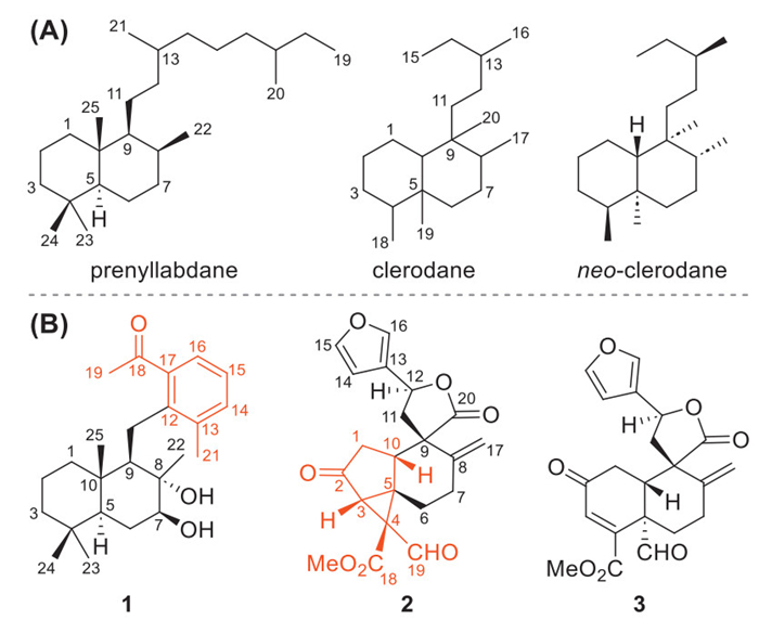
Figure 1.
(A) Structures of prenyllabdane and (neo)clerodane. (B) Structures of compounds 1–3.
Isolation of two novel terpenoid skeletons from Croton laui, an aromatic norsesterterpenoid and a highly rearranged neo-clerodane diterpenoid
Zong-Yi Zhang , Xin Wang , Ying Li , Yuan Gao , Yao-Yue Fan , Jian-Min Yue , Jin-Xin Zhao

New-skeleton terpenoids are highly valued for their potential to broaden the chemical landscape, unlock novel opportunities within biological and pharmaceutical fields, and furnish synthetic chemists with fresh structural templates [1–6], thus having garnered widespread interest and enthusiasm. Sesterterpenoids are amongst the rarest class of terpenoids with approximately 1400 members [3,7,8], of which ca. 200 ones were isolated from terrestrial plants [9–12]. Prenyllabdane sesterterpenoids, existing in some plant species of the genus Salvia [3], are derived from a similar cyclization as labdane diterpenoids. To date, only six prenyllabdane norsesterterpenoids have been documented [13–15], yet no biological evaluation has been reported for these compounds. Clerodane diterpenoids are a large group of secondary metabolites found in a wide range of organisms, such as plants, microorganisms, and marine sponges [16]. Despite the prevalence in nature, variations (rearrangement, elimination, or cyclization) in the skeletal structures of clerodanes, particularly those involving the decalin core [17–22], remain uncommon and underreported. These clerodane variants often exhibit diverse bioactivities, such as inflammasome activation [17] and inhibition [18], anti-Alzheimer's disease [20], and modulatory activity of multidrug resistance in MCF-7 cancer cells [22].
Croton laui Merrill & F. P. Metcalf (Euphorbiaceae), an indigenous perennial shrub in Hainan Province of China, has been utilized as a folk remedy for treating headaches, stomachaches, and diphtheria [23]. Previous investigations have demonstrated that it is a prolific source of labdane and neoclerodane diterpenoids [24–30]. In connection with our ongoing efforts to identify bioactive natural products from ethnomedicinal plants [31–34], two new-skeleton terpenoids, including an aromatic 12,17-cyclo20-nor phenyllabdane sesterterpenoid, crolatinoid A (1), and a 19(5→4)-abeo-3,5-cycloneoclerodane diterpenoid, crolatinoid B (2), along with its plausible biosynthetic precursor crolatinoid C (3) (Fig. 1), were isolated from the bark of Croton laui. Furthermore, the multidrug resistant reversal and antiadipogenic activities were evaluated for these terpenoids. Herein, the isolation, structural elucidation, and biological evaluation of these new-skeleton terpenoids are described.
Crolatinoid A (1), colorless gum, was assigned a molecular formula of C24H36O3 incorporating seven degrees of unsaturation (DOUs), as supported from its positive high-resolution electrospray ionization mass spectrometry [(+)-HRESIMS] ion peak at m/z 395.2555 [M + Na]+ (calcd. 395.2557). Its one-dimensional nuclear magnetic resonance (1D NMR) data (Table S1 in Supporting information), with the aid of the 1H–13C heteronuclear single quantum coherence (HSQC) and distortionless enhancement by polarization transfer-135 (DEPT-135) experiments, displayed typical signals for one 1,2,3-trisubstituted phenyl ring (δH 7.13, t, J = 7.6 Hz, 7.22, d, J = 7.6 Hz, and 7.36, d, J = 7.6 Hz; δC 125.5, 126.5, 133.9, 138.1, 140.2, and 141.1), six methyls, one oxymethine (δH 7.13, δC 81.0), one oxygenated tertiary carbon (δC 77.7), and one carbonyl group (δC 206.4). These functionalities occupied five out of the seven DOUs. The remaining two ones required the presence of two additional rings in 1.
The planar structure of 1 was constructed by the two-dimensional (2D) NMR experiments. Specifically, 1H–13C heteronuclear multiple bond correlation (HMBC) signals (Fig. 2A) from H3–22 to C-7, C-8, and C-9, from H3–23 and H3–24 to C-3, C-4, and C-5, and from H3–25 to C-1, C-9, and C-10, in conjunction with 1H–1H homonuclear correlation spectroscopy (COSY) cross-peaks of H2–1/H2–2/H2–3 and H-5/H2–6/H-7, delineated a bicyclic decalin framework (A- and B-ring) adorned with four methyls (Me-22, Me-23, Me-24, and Me-25). The 7,8-vicinal diol moiety was confirmed by the chemical shift values of CH-7 and C-8. Additionally, HMBC correlations from H2–11 to C-13 and C-17 and the COSY cross-peak of H-9/H2–11, verified the connection of the phenyl ring with B-ring via the C-9–C-11–C-12 sequence. The presence of an acetyl group at C-17 and a methyl at C-13 was confirmed by the HMBCs from H-16 to C-12 and C-18, H3–19 to C-17 and C-18, and H3–21 to C-12, C-13, and C-14. Therefore, compound 1 was established as a new-skeleton aromatic norsesterterpenoid.
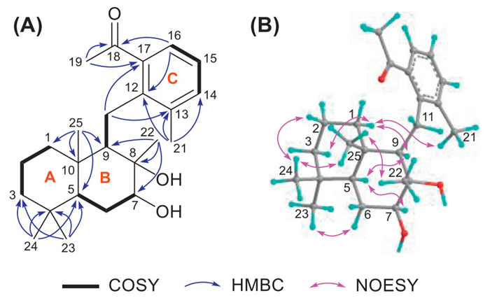
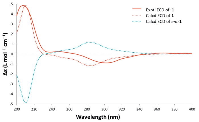
The relative configuration of 1 was determined by the nuclear Overhauser effect spectroscopy (NOESY) experiment (Fig. 2B). In the chair conformation of the decalin core, the observed NOE correlations of H3–25 with H3–24 and H3–22 revealed that they were cofacial and were assigned arbitrarily as β-oriented [24]. Consequently, the NOE correlations of H-5 with H-7 and H-9 suggested that H-5, H-7, and H-9 were α-oriented. The absolute configuration of 1 was determined via quantum chemical electronic circular dichroism (ECD) calculation, by using time-dependent density functional theory (TDDFT) method at the ωB97XD/6–311G**//B3LYP/6–31G* level [35–38]. As shown in Fig. 3, the experimental ECD curve matched well with the calculated one in the region of 200–400 nm, establishing its absolute configuration as 5S,7S, 8S,9R,10S.
Crolatinoid B (2) was obtained as colorless crystals with a negative optical rotation in acetonitrile (
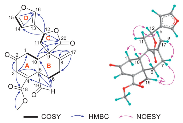
Further analysis of the HMBC spectra (Fig. 4A) established the planar structure of 2. Four different spin coupling fragments of CH2–1/CH-10, CH2–6/CH2–7, CH2–11/CH-12, and CH-14/CH-15 were readily identified by interpretation of the 1H–1H COSY spectrum. HMBC correlations from H2–1 to C-2 (δC 206.4), and from H-3 to C-1, C-5, and C-10 delineated a cyclopentanone moiety (A2-ring), which was fused to the six-membered B-ring, as inferred from HMBCs from H-10 to C-8 and C-9, and from H2–6 to C-8 and C-10. The HMBCs from H2–17 to C-7, C-8, and C-9 anchored the location of the Δ8(17) double bond. Additionally, the presence of a cyclopropane A1-ring that was fused to A2-ring was assigned by HMBC correlations from H-3, H2–6, and from H-10 to C-4. HMBC correlations from H-3 and CH3O-18 to C-18 indicated that a methoxycarbonyl group was attached to the C-4 quaternary carbon. By comparing the NMR data with those of methyl exo-6-formylbicyclo[3.1.0]hexane-6-carboxylate [39], a formyl group was assigned to C-4, which was supported by the NOESY correlation between H-19 and CH3O-18. Furthermore, HMBC correlations from H2–11 to C-9 and C-20 (δC 175.3), and from H-12 to C-20 confirmed the existence of 9-spiroγ-butyrolactone (C-ring). The attachment of the β-substituted furan (D-ring) to C-12 was evidenced by HMBC correlations from H-12 to C-14 and C-16. Therefore, compound 2 was established as a new-skeleton diterpenoid featured by a unique 3/5/6-fused tricyclic framework.
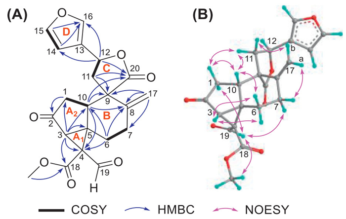
The relative configuration of 2 was mainly established by the NOESY experiment (Fig. 4B). The NOE correlations of H-1α with H-19 and H-7α with H-19 indicated that H-1α, H-7α, and the 19-formyl group were co-facial and arbitrarily designated as α-oriented. Consequently, the H-1β/H-10 and H-10/H-6β correlations indicated that all these protons were β-oriented. The NOE correlation between H-3 and H2–6 suggested a cis-fusion between A1- and A2-ring, and H-3 was thus assigned as β-oriented. In addition, the NOE correlations of 11α/H-1β and 11α/H-10 suggested a 9S* configuration. However, the C-12 configuration remained undetermined due to insufficient reliable NOE data. Fortunately, this limitation was overcome by a successful single-crystal X-ray diffraction (XRD) experiment, which not only corroborated the above assignments, but also established its absolute configurations as 3R,4S,5S,9S,10S,12S [Fig. 5, Flack parameter = 0.09(11)] [40].
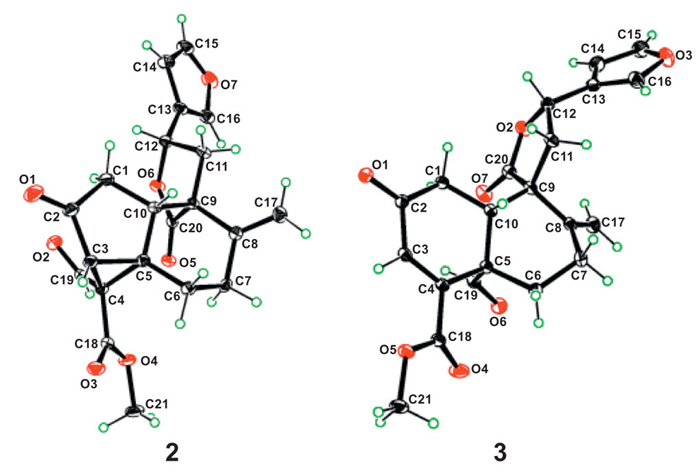
Crolatinoid C (3), which took the form of colorless crystals, possesses the molecular formula C21H20O7, based on analysis of its (+)-HRESIMS and the 13C NMR data. Its NMR data were highly similar to that of a known neoclerodane diterpenoid, mangelonine H [41], indicating that they are structural analogs. The only difference between them was the presence of a C-2 keto carbonyl group (δC 197.1) conjugated with the Δ3(4) double bond in 3 instead of the C-2 methylene group in the latter. This assignment was verified by scrutiny of the HMBC and 1H–1H COSY correlations (Fig. 6). Further interpretation of the NOESY data revealed that compound 3 shared the same relative configuration as mangelonine H. Lastly, based on XRD analysis [Fig. 5, Flack parameter = 0.01(9)] [40], the absolute configuration of 3 was unambiguously determined to be 5S,9S,10S,12S as depicted. Crolatinoids A (1) and B (2) both exhibit novel skeletal structures, leading us to undertake an investigation into their biosynthetic origins. From a structural and biogenetic perspective, compound 1 likely originated from a phenyllabdane sesterterpenoid (Scheme 1), e.g., ⅰ that was formed through enzyme-catalyzed cyclization and elimination of geranylfarnesyl diphosphate (GFDP). Subsequent oxidation would give intermediate ⅱ, which undergoes a dehydration process to produce polyene ⅲ. A subsequent 6π electrocyclization connects C-12 and C-17, affording cyclohexadiene ⅳ. Oxidation at the C-7, C-8, C-19, and C-20 positions would result in intermediate ⅴ, featuring a vicinal diol and β-keto carboxylic acid group. Then, this intermediate would undergo an intramolecular decarboxylation to form enol vi, which would transform into compound 1 via enol-keto tautomerism and oxidative aromatization.

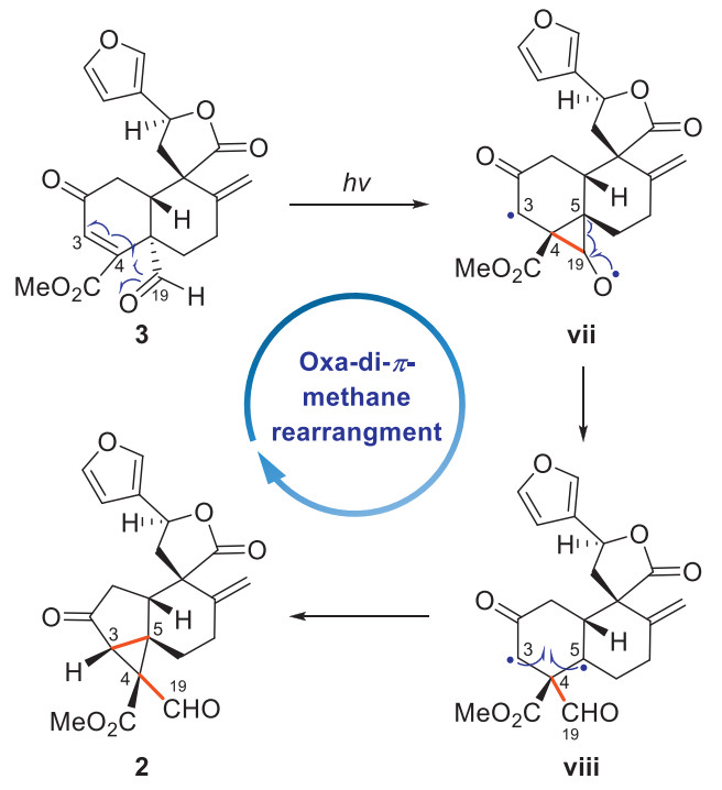
Compound 2 is characterized by an unprecedented 19(5→4)-abeo-3,5-cycloneoclerodane skeleton, which is hypothesized to be biogenetically derived from compound 3 through an oxa-di-π-methane rearrangement (Scheme 2) [42]. Specifically, upon photochemical irradiation, compound 3 would initially undergo homolytic cleavage of both the Δ3(4) double bond and the C-19 aldehyde carbonyl group, which is followed by the formation of the C-4–C-19 bond, yielding the diradical intermediate ⅶ. Subsequent C-5–C-19 bond cleavage would generate the intermediate ⅷ, which would be converted into compound 2 via the C-3–C-5 bond formation. To validate the biosynthetic hypothesis, a transformation from 3 to 2 was envisaged. However, the minute isolated quantity of compound 3 (1.0 mg) hindered this endeavor, necessitating additional verification through a synthetic approach.
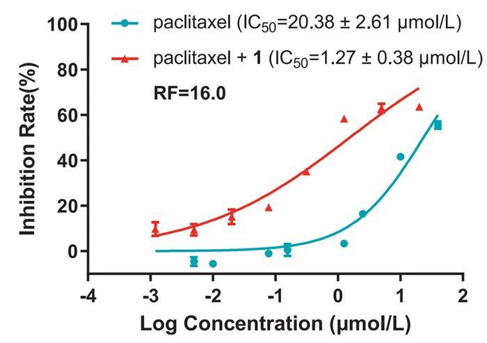
At this stage, we moved forward to explore the biological potential of these structurally fascinating scaffolds. A significant challenge in cancer treatment arises from the frequent development of resistance in cancer cells to both chemotherapeutic agents and targeted medications. Natural products play a pivotal role as a promising resource for combating cancer multidrug resistance (MDR) [43–46]. In the present study, the cytotoxicity and MDR reversal activity of compound 1 were tested in paclitaxel-resistant colorectal cancer cells (HCT-15/Taxol). The findings indicated that compound 1 exhibited no cytotoxicity to these cell lines at a concentration of 20 µmol/L. Moreover, a combination therapy with compound 1 could significantly enhance the efficacy of paclitaxel in HCT-15/Taxol cell lines with a half maximal inhibitory concentration (IC50) value of 1.27 µmol/L, demonstrating a reversal fold (RF) value of 16 (Fig. 7).
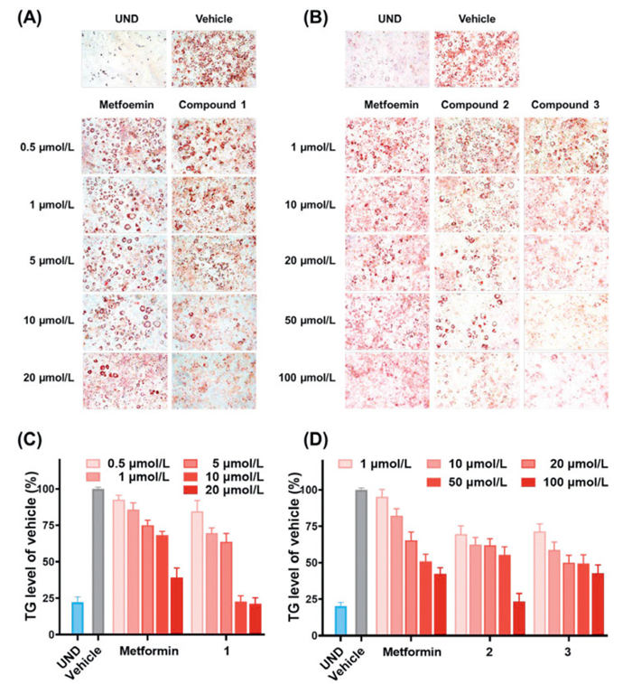
Obesity is a wide-spreading epidemic that significantly impacts quality of life, making medical treatment increasingly crucial [47]. In this study, compounds 1–3 were evaluated for their capacity to suppress lipid deposition in the 3T3-L1 adipocyte model [48–50], utilizing triglycerides (TG) levels as an indicator and oil red O (ORO) staining for visual verification (Fig. 8, Tables S4 and S5 in Supporting information), while metformin [51,52] served as the positive control. The results showed that compounds 1–3 demonstrated a dose-dependent reduction in TG levels with series doses, while exhibiting low cytotoxicity (Table 1). Significantly, compounds 1–3 all showed inhibition on the adipocyte differentiation of 3T3-L1 cells with half maximal effective concentration (EC50) values of 4.38, 32.62, and 18.08 µmol/L, respectively, which were either superior to or comparable with the efficacy of metformin.
 DownLoad:
CSV
DownLoad:
CSV
| Compounda | EC50 (µmol/L) | IC50 (µmol/L) |
| 1b | 4.38 ± 0.64 | >100 |
| 2c | 32.62 ± 3.96 | >100 |
| 3c | 18.08 ± 3.60 | >100 |
| a Bioassay data were obtained from two separate batches, and metformin was used as the positive control. b IC50 value of positive control was tested as 16.45 ± 1.21 µmol/L. c IC50 value of positive control was tested as 36.08 ± 6.75 µmol/L. |
||
In conclusion, three novel terpenoids including a 12,17-cyclo20-nor phenyllabdane sesterterpenoid (1), a 19(5→4)-abeo-3,5-cycloneoclerodane diterpenoid (2), and a neoclerodane diterpenoid (3), were isolated from the bark of Croton laui in the present study. Compounds 1 and 2 exhibit two unprecedented carbon skeletons. Notably, compound 1 marks the first discovery of a 12,17-cyclostructure within the phenyllabdane sesterterpenoid class. Biological evaluation revealed that compound 1 could significantly reverse multidrug resistance in paclitaxel-resistant HCT-15 cells. Additionally, all compounds 1–3 exhibited antiadipogenic activity in 3T3-L1 adipocytes. This study not only enhances the diversity of terpenoid carbon skeletons but also offers new valuable resources for research in multidrug resistance reversal and anti-adipogenesis.
The authors declare that they have no known competing financial interests or personal relationships that could have appeared to influence the work reported in this paper.
Zong-Yi Zhang: Writing – original draft, Visualization, Validation, Software, Resources, Investigation, Data curation. Xin Wang: Resources, Investigation, Data curation. Ying Li: Visualization, Resources, Investigation, Data curation. Yuan Gao: Resources, Investigation, Data curation. Yao-Yue Fan: Supervision, Resources, Investigation, Data curation. Jian-Min Yue: Writing – review & editing, Supervision, Project administration, Investigation, Funding acquisition, Data curation. Jin-Xin Zhao: Writing – review & editing, Visualization, Validation, Supervision, Resources, Project administration, Funding acquisition, Data curation, Conceptualization.
The financial support from the National Key Research and Development Program of China (No. 2023YFE0206100), the National Natural Science Foundation of China (Nos. 22237007 and T2192972), the Youth Innovation Promotion Association of Chinese Academy of Sciences (CAS) (No. 2022282), and Shanghai Institute of Materia Medica of CAS (No. SIMM0120231002) is gratefully acknowledged. We thank Prof. Shi-Man Huang, Department of Biology, Hainan University, People's Republic of China, for the identification of the plant material.
Supplementary material associated with this article can be found, in the online version, at doi:
Y. Cheng, D. Qin, Novel Plant Natural Product Skeletons. Discoveries from 1999 to 2021, Springer, Singapore, 2024.
Z. Hu, Y. Ye, Y. Zhang, Nat. Prod. Rep. 38 (2021) 1775–1793. doi: 10.1039/d0np00069h
K. Guo, Y. Liu, S.H. Li, Nat. Prod. Rep. 38 (2021) 2293–2314. doi: 10.1039/d1np00021g
Z. Wang, D.R. Nelson, J. Zhang, et al., Nat. Prod. Rep. 40 (2023) 452–469. doi: 10.1039/d2np00054g
Z.J. Zhan, S. Li, W. Chu, et al., Nat. Prod. Rep. 39 (2022) 2132–2174. doi: 10.1039/d2np00047d
Y. Wang, P. Tang, W. Tu, et al., Chin. Chem. Lett. 36 (2025) 109955. doi: 10.1016/j.cclet.2024.109955
L. Wang, B. Yang, X.P. Lin, et al., Nat. Prod. Rep. 30 (2013) 455–473. doi: 10.1039/c3np20089b
J.R. Hanson, Nat. Prod. Rep. 3 (1986) 123–132. doi: 10.1039/np9860300123
K. Guo, T.T. Zhou, S.H. Luo, et al., J. Med. Chem. 67 (2023) 513–528.
L.L. Teng, R.F. Mu, Y.C. Liu, et al., Org. Lett. 23 (2021) 2232–2237. doi: 10.1021/acs.orglett.1c00369
S.X. Jing, R. Fu, C.H. Li, et al., J. Org. Chem. 86 (2021) 11169–11176. doi: 10.1021/acs.joc.1c00374
C.Y. Zheng, J.X. Zhao, C.H. Yuan, et al., Chem. Sci. 14 (2023) 13410–13418. doi: 10.1039/d3sc04238c
A. Bisio, A.M. Schito, F. Pedrelli, et al., J. Nat. Prod. 83 (2020) 1027–1042. doi: 10.1021/acs.jnatprod.9b01024
A. Ulubelen, G. Topcu, U. Sönmez, et al., Phytochemistry 43 (1996) 431–434. doi: 10.1016/0031-9422(96)00248-8
F.M. Moghaddam, M.M. Farimani, M. Seirafi, et al., J. Nat. Prod. 73 (2010) 1601–1605. doi: 10.1021/np1002516
R. Li, S.L. Morris-Natschke, K.H. Lee, Nat. Prod. Rep. 33 (2016) 1166–1226. doi: 10.1039/C5NP00137D
D.W. Bi, F. Xiong, B. Cheng, et al., J. Nat. Prod. 85 (2022) 2675–2681. doi: 10.1021/acs.jnatprod.2c00568
D.B. Pu, X.J. Zhang, D.-W. Bi, et al., J. Nat. Prod. 83 (2020) 2191–2199. doi: 10.1021/acs.jnatprod.0c00288
M. Fan, X.J. Chen, X.D. Wu, et al., Tetrahedron Lett. 59 (2018) 3065–3068. doi: 10.1016/j.tetlet.2018.06.015
Z.X. Zhang, P.Q. Wu, H.H. Li, et al., Org. Biomol. Chem. 16 (2018) 1745–1750. doi: 10.1039/c7ob02991h
L.B. Zhang, H.B. Liao, H.Y. Zhu, et al., Tetrahedron 72 (2016) 8036–8041. doi: 10.1016/j.tet.2016.10.034
E. Bautista, M. Fragoso-Serrano, R.A. Toscano, et al., Org. Lett. 17 (2015) 3280–3282. doi: 10.1021/acs.orglett.5b01320
H.F. Dai, X.L. Zheng, F.W. Xing, et al., Records of Li Folk Medicine, China Science & Technology Press, Beijing, 2014, pp. 115–116.
Z.Y. Zhang, Y. Li, J.H. Yu, et al., Phytochemistry 223 (2024) 114138. doi: 10.1016/j.phytochem.2024.114138
C.P. Liu, J.B. Xu, J.X. Zhao, et al., J. Nat. Prod. 77 (2014) 1013–1020. doi: 10.1021/np500042c
L. Yang, Y.B. Zhang, Z.N. Wu, et al., Chem. Lett. 45 (2016) 1235–1237. doi: 10.1246/cl.160632
F. Li, D.B. Zhang, J.T. Li, et al., Nat. Prod. Res. 35 (2019) 2849–2857. doi: 10.3390/ijms20112849
L. Yang, Z.N. Wu, Y.B. Zhang, et al., Nat. Prod. Res. 31 (2017) 1028–1033. doi: 10.1080/14786419.2016.1266350
C. Yang, H. Chen, S. Gao, et al., Phytochem. Lett. 53 (2023) 37–41. doi: 10.1109/dsde58527.2023.00014
L. Yang, Y.B. Zhang, L.F. Chen, et al., Bioorg. Med. Chem. Lett. 26 (2016) 4687–4691. doi: 10.1016/j.bmcl.2016.08.052
J.S. Zhou, L. Cheng, Y. Gao, et al., Engineering 38 (2024) 144–154. doi: 10.1016/j.eng.2023.09.015
C.L. Wang, Y. Dai, Q. Zhu, et al., J. Nat. Prod. 86 (2023) 1345–1359. doi: 10.1021/acs.jnatprod.3c00173
J. Qi, Y. Zhang, Q. Liu, et al., Chin. J. Chem. 39 (2021) 1891–1897. doi: 10.1002/cjoc.202100117
X.H. Gao, Y.Y. Fan, Q.F. Liu, et al., Org. Lett. 21 (2019) 7065–7068. doi: 10.1021/acs.orglett.9b02630
T.D. Crawford, M.C. Tam, M.L. Abrams, J. Phys. Chem. A 111 (2007) 12057–12068. doi: 10.1021/jp075046u
N. Berova, L.D. Bari, G. Pescitelli, Chem. Soc. Rev. 36 (2007) 914–931. doi: 10.1039/b515476f
C. Diedrich, S. Grimme, J. Phys. Chem. A 107 (2003) 2524–2539. doi: 10.1021/jp0275802
A. Fu, C. Chen, Q. Li, et al., Chin. Chem. Lett. 35 (2024) 109100. doi: 10.1016/j.cclet.2023.109100
A. de Meijere, V. Bagutski, F. Zeuner, et al., Eur. J. Org. Chem. 2004 (2004) 3669–3678. doi: 10.1002/ejoc.200400132
H.D. Flack, G. Bernardinelli, Chirality 20 (2008) 681–690. doi: 10.1002/chir.20473
Z.Y. Jiang, Q. Niu, H.X. Wang, et al., Phytochemistry 226 (2024) 114206. doi: 10.1016/j.phytochem.2024.114206
H.E. Zimmerman, D. Armesto, Chem. Rev. 96 (1996) 3065–3112. doi: 10.1021/cr910109c
T. Chen, Z. Xiao, X. Liu, et al., Pharmacol. Res. 202 (2024) 107099. doi: 10.1016/j.phrs.2024.107099
A. Kumar, V. Jaitak, Eur. J. Med. Chem. 176 (2019) 268–291. doi: 10.1504/ijef.2019.104071
S. Long, E. Sousa, A. Kijjoa, et al., Molecules 21 (2016) 892. doi: 10.3390/molecules21070892
C.P. Liu, C.Y. Xie, J.X. Zhao, et al., J. Am. Chem. Soc. 141 (2019) 6812–6816. doi: 10.1021/jacs.9b02259
P. González-Muniesa, M.A. Mártinez-González, F.B. Hu, et al., Nat. Rev. Dis. Primers 3 (2017) 17034. doi: 10.1038/nrdp.2017.34
Y.Y. Chi, J.L. Shen, J. Zhang, et al., Food Sci. Biotechnol. 25 (2016) 1147–1153. doi: 10.1007/s10068-016-0183-7
Z.Y. Zhang, X.H. Gao, Y. Huang, et al., J. Nat. Prod. 87 (2024) 1441–1453. doi: 10.1021/acs.jnatprod.4c00246
Q. Tan, R.Z. Fan, W. Yang, et al., Chin. Chem. Lett. 35 (2024) 109390. doi: 10.1016/j.cclet.2023.109390
S.C. Chen, R. Brooks, J. Houskeeper, et al., Mol. Cell. Endocrinol. 440 (2017) 57–68. doi: 10.1016/j.mce.2016.11.011
L. Ma, C. Xie, Y. Ran, et al., J. Med. Chem. 55 (2012) 9958–9972. doi: 10.1021/jm301164y

Figure 1 (A) Structures of prenyllabdane and (neo)clerodane. (B) Structures of compounds 1–3.

Figure 5 X-ray ORTEP drawings of compounds 2 and 3 with 20% thermal ellipsoid probability.

Figure 7 Multidrug resistance reversal activity of compound 1 against the HCT-15/Taxol cells. The RF is determined as the ratio of the IC50 value of paclitaxel to the IC50 value of paclitaxel combined with compound 1. Data represent mean ± standard deviation (SD) of three independent experiments.

Figure 8 Antiadipogenic activity of metformin and compounds 1–3 in 3T3-L1 adipocytes. (A, B) Representative microscopy images were captured after ORO staining (×200). UND: undifferentiated group. (C, D) TG levels under the interference of indicated compounds. TG levels were tested by ORO staining and TG assay. The TG level of differentiated group (vehicle) without compounds treatment was viewed as 100%. Data represent mean ± SD of three independent experiments.
Table 1. Antiadipogenic effects and cytotoxicity evaluation of metformin and compounds 1–3 in 3T3-L1 adipocytes.
| Compounda | EC50 (µmol/L) | IC50 (µmol/L) |
| 1b | 4.38 ± 0.64 | >100 |
| 2c | 32.62 ± 3.96 | >100 |
| 3c | 18.08 ± 3.60 | >100 |
| a Bioassay data were obtained from two separate batches, and metformin was used as the positive control. b IC50 value of positive control was tested as 16.45 ± 1.21 µmol/L. c IC50 value of positive control was tested as 36.08 ± 6.75 µmol/L. |
||
 下载: 导出CSV

 扫一扫看文章

扫一扫关注我们


 下载:
下载: