Department of Mathematics and Physics, Luoyang Institute of Science and Technology, Luoyang 471023, China
b.
State Key Laboratory of Chemo/Biosensing and Chemometrics, College of Chemistry and Chemical Engineering, Hunan Provincial Key Laboratory of Biomacromolecular Chemical Biology, Hunan University, Changsha 410082, China
c.
College of Chemical and Biological Engineering, Shandong University of Science and Technology, Qingdao 266590, China
kunli@hnu.edu.cn (K. Li). 1 These authors contributed equally to this work.
Received Date:
27 September 2024 Accepted Date:
04 February 2025 Revised Date:
29 December 2024 Available Online:
15 November 2025
Abstract:
Nanozymes, characterized by their stability, cost-effectiveness, and tunable catalytic activity, are promising alternatives to natural enzymes. However, specifically mimicking a single natural enzyme's activity presents a challenge. By exploiting the catalytic selectivity derived from the valence-band hybridization of noble metal nanoalloys, we introduce an alloying strategy to modulate the reaction specificity of metallic nanozymes. AgPd nanoalloy exhibits enhanced peroxidase-like activity and eliminated oxidase-like activity by adjusting the Ag content. The introduction of Ag changes the hybrid d band energy of the alloyed metal and inhibits the O2 adsorption and decomposition on Pd, while improving the peroxidase mimicry by allowing for the H2O2 activation. By exemplifying the construction of a highly sensitive and selective colorimetric glucose detection platform with its practicality validated in serum samples, this strategy pioneers a multi-noble metal nanozyme with tailored peroxidase activity based on the chemical structure engineering and would advance the development of single-catalytic function nanozymes for building exclusively specific biosensors through reducing substrate competition.
As nanomaterial-based enzyme mimics, nanozymes, which possess natural enzyme mimicking activity, are very suitable substitutes of enzymes because of their low cost, high stability, and conveniences of preparation [1–3]. Until now, a large number of nanomaterials that imitate multienzymes were studied, including noble metals [4–6], metal oxides [7,8], metal-organic frameworks (MOFs) [9–12], single atom-based materials [13,14], and carbon-based materials [15,16]. These nanomaterials with multienzymes-mimicking activity have been extensively applied to biosensors [17,18], organic degradation [19,20], disease treatments [21,22], and antibacterial materials [23,24]. Notably, the mimic of natural oxidoreductase is the most widely studied type of nanozymes, including oxidase (OXD), peroxidase (POD), catalase (CAT), and superoxide dismutase (SOD). Many nanomaterials have the activity of two, three, or even four natural enzymes [25,26], and the catalytic reaction produces a variety of oxygen-related free radicals, which are beneficial to be used for sterilization or killing cancer cells [2,27,28]. Characteristically, when applied to biosensing, however, nanozymes need to be catalytically specific like natural enzymes that catalyze only one kind of substrate, so as to reduce substrate competition and ensure detection accuracy [3]. In particular, nanozymes possessing POD-like activity that is employed in hydrogen peroxide (H2O2) detection must exclusively use H2O2 as the substrate without initiating the dissociation of oxygen. Therefore, it is of significant essence to modulate the possible reactions that one nanozyme may catalyze so that it can only simulate a single natural enzyme and possess a highly specific catalytic function [29].
To advance the catalytic performance of nanozymes, versatile methods have been developed, including tuning the valency of metals [30], adjusting the pH of the solution [31], covering with molecularly imprinted polymers [32], regulating the coordination number [33,34], doping with other elements [35], and stimulating with light [36–38]. However, the preparation procedures of these methods are complicated, and the prepared nanomaterials usually need to be further modified. More importantly, the regulation mechanism of nanozyme catalysis is not always clear, hindering the improvement of the specificity of catalytic functions. Notably, metal alloying is an emerging method to regulate the catalytic performance of metal-based nanozymes. For example, the formation of citric acid modified rhodium-platinum bimetal nanoparticles can enhance the POD-like activity, but its catalytic specificity was not studied [39]. Accordingly, creating new type of nanozymes with POD-like reaction specificity in a simple way is still a challenge.
Pd nanozymes have been reported to exhibit both OXD- and POD-like activities through thermal-driven catalysis [40]. Interestingly, our previous research revealed that ambient oxygen can only be photoactivated via plasmon-coupled valence-band hybridization under visible light irradiation but cannot be in the dark [41]. This exciting finding inspired us to investigate the suppression of the thermal-driven OXD-like activity of Pd nanozymes by alloying with Ag. Given the thermal-driven POD-like activity of Ag NPs [42], we hypothesized that AgPd alloy nanoparticles (AgPd ANPs) can exclusively activate H2O2 in the dark. Thus, by preparing AgPd ANPs with zirconia as the inert support through the impregnation-reduction method, we propose an effective strategy for modulating the reaction specificity of nanozymes. The introduction of Ag results in the elimination of the OXD-like activity of Pd NPs, which we attribute to the inability of AgPd ANPs to activate O2. The prepared AgPd ANPs, which retain a highly specific POD-like activity, demonstrate that the specificity regulation on catalytic functions of metal-based nanozymes can be achieved by alloying. Ultimately, the consequently constructed colorimetric biosensor for glucose detection exhibits enhanced sensing performance, with noted assay reliability successfully realized in the complicated matrix of human serum.
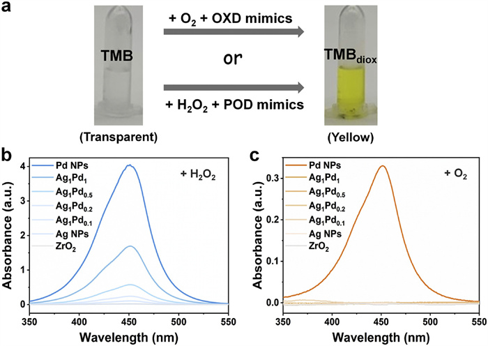
The synthesis of AgPd ANPs with varied elemental proportions was achieved through the impregnation-reduction method employing different precursor ratios (Ag/Pd = 0:1, 1:1, 1:0.5, 1:0.2, 1:0.1, and 1:0) (Fig. S1 in Supporting information). In order to prevent the agglomeration of AgPd ANPs nanoparticles and the consequent influence on their catalytic activity, ZrO2 was introduced during the preparation of AgPd ANPs as inert support load AgPd ANPs to improve the stability of AgPd ANPs [41]. UV−vis spectra (Fig. S2 in Supporting information) of silver nitrate (AgNO3), palladium(Ⅱ) sodium chloride (Na2PdCl4), and the aqueous dispersion of various ANPs confirm the preliminary synthesis of ANPs. ICP-MS determined the metal contents of Ag and Pd (Table S1 in Supporting information), validating the approximate alignment of theoretical and actual molar ratios. The catalytic performance of OXD and POD were assessed using the oxidation reaction of 3, 3′, 5, 5′-tetramethylbenzidine (TMB) (Fig. 1a), monitored by UV–vis absorbance changes. In the TMB oxidation process, TMB was initially converted to a single-electron product (TMBox), and subsequent addition of terminator (H2SO4) transformed TMBox into a yellow product of two electrons (TMBdiox) [43].
Figure 1
Figure 1.
(a) Schematic diagram of the formation of TMBdiox (yellow) from TMB (transparent) in the presence of OXD or POD. (b) UV–vis absorption spectra of TMBdiox with pure ZrO2 or different AgPd ANPs loaded on ZrO2 in H2O2 solution (corresponding to POD-like activity). (c) UV–vis absorption spectra of TMBdiox with pure ZrO2 or different AgPd ANPs loaded on ZrO2 in aqueous solution (corresponding to OXD-like activity).
Interestingly, subsequent investigation into the OXD- and POD-like activities of AgPd ANPs with different proportions reveals intriguing findings. Fig. 1b indicates that all kinds of AgPd ANPs primarily exhibit POD-like activity, which diminishes with increasing Ag content. Notably, the introduction of Ag results in the loss of OXD-like activity, even if only a small amount of Ag exists (Fig. 1c). Meanwhile, the inert support, ZrO2, does not exhibit any POD-like activity or OXD-like activity as expected. This suggests that Ag incorporation may influence the OXD-like activity of Pd NPs, showcasing metal alloying as a tool to modulate the catalytic reaction specificity of nanozymes. For subsequent experiments, AgPd ANPs synthesized with a molar ratio of 1:0.5 were selected. Above results demonstrate that ZrO2 is retained in the AgPd ANPs system but does not affect the enzyme-mimicking activities. For simplicity, these ZrO2 supported Ag1Pd0.5 alloying nanozymes are hereinafter referred to as AgPd ANPs.
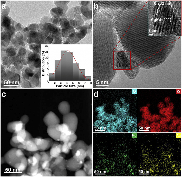
The morphological characteristics of the as-prepared nanomaterials, specifically the AgPd ANPs, were elucidated through transmission electron microscopy (TEM). In Fig. 2a, the size of AgPd ANPs is determined to be 4 (±0.5) nm. High-resolution TEM (HRTEM) further reveals the single crystal properties of AgPd ANPs, with well-resolved lattice fringes indicating the face-centered cubic (fcc) AgPd alloy (Fig. 2b). The (111) plane spacing of fcc Pd and fcc Ag is 0.22 nm and 0.24 nm, respectively [44]. The lattice fringe spacing of 0.232 nm corresponds to the (111) plane of fcc AgPd alloy. High-angle annular dark field scanning TEM (HAADF-STEM) image (Fig. 2c) and energy dispersive X-ray spectroscopy (EDX) elemental spectrum (Fig. 2d) provide additional evidence of the uniform distribution of Ag and Pd on the ZrO2 carrier, substantiating the successful preparation of AgPd ANPs.
Figure 2
Figure 2.
TEM (a), HRTEM (b), HAADF-STEM (c) and elemental mapping (d) images of AgPd ANPs loaded on ZrO2. Inset of panel (a) shows the size distribution of AgPd ANPs.
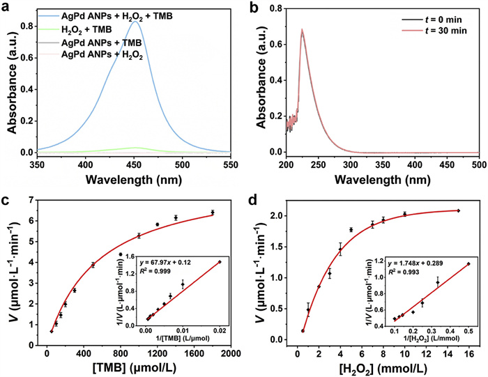
AgPd ANPs was expected to possess efficient and specific POD-like activity. Fig. 3a illustrates that the introduction of AgPd ANPs significantly increased the absorbance of TMBdiox, signifying excellent POD-like activity. Moreover, under ambient air conditions, no TMBdiox signal was detected in the system of AgPd ANPs and TMB, preliminary evidence ruling out OXD-mimicking activity. As a control, catalase-like activity of AgPd ANPs was also probed by monitoring H2O2 decomposition, revealing negligible change in absorbance intensity of the characteristic peak of H2O2 at 240 nm after 30 min (Fig. 3b), confirming the absence of catalase-like activity. These results confirm the specific POD-like activity of AgPd ANPs.
Figure 3
Figure 3.
(a) UV–vis absorption spectra of TMBdiox of different reaction systems. (b) The absorbance of TMBdiox representing catalase-like activity of AgPd ANPs. (c) Steady-state kinetic assay of AgPd ANPs by varying the concentration of TMB. Inset: Lineweaver−Burk plot of photocatalytic activity of the AgPd ANPs with H2O2 as the substrate. (d) Steady-state kinetic assay of AgPd ANPs by varying the concentration of H2O2. Inset: Lineweaver−Burk plot of photocatalytic activity of the AgPd ANPs with TMB as the substrate. In panels b, c, and d, the reactions were conducted with 208 µmol/L TMB, 10 mmol/L H2O2, and 100 µg/mL AgPd ANPs in 0.2 mol/L acetate buffer (pH 4.0). Error bars refer to the standard deviation of three replicates.
To investigate the catalytic performance of AgPd ANPs, steady-state kinetics was studied by varying the concentrations of TMB or H2O2 in acetate buffer (0.2 mol/L, pH 4.0). As shown in Figs. 3c and d, the absorbance of TMBdiox adheres well to the standard Michaelis-Menten equation with changing TMB or H2O2 concentration, illustrating characteristic enzymatic kinetic behavior for AgPd ANPs. The Michaelis-Menten constant (Km) and maximum initial velocity (Vmax) were derived from the Lineweaver−Burk plot via Michaelis-Menten curve [45]. Table 1 [1,46–50] summarizes that AgPd ANPs, functioning as a POD mimic for TMB, exhibit a Km value of 0.566 × 10⁻³ mol/L and a Vmax of 13.89 × 10⁻⁸ mol L−1 s−1, comparable to those of horse-radish peroxidase (HRP). This suggests a high affinity of AgPd ANPs for TMB. While the Km value for H2O2 surpasses that of HRP or other natural peroxidases, it remains close to most reported Pd-based nanozymes.
Table 1
Table 1.
Comparison of kinetic parameters (Km and Vmax) of different catalysts.
To affirm the substrate versatility of AgPd ANPs, 2, 2′-azino-bis(3-ethylbenzothiazoline-6-sulfonic acid) (ABTS) and 1, 2-diaminobenzene (OPD) were chosen as substrates to evaluate their POD-like activity (Fig. S3 in Supporting information). The kinetic parameters of ABTSox were similar to those of TMB (Table S2 in Supporting information), signifying that AgPd ANPs possess POD-like activity that facilitates the reaction of H2O2 with various co-substrates. Moreover, AgPd ANPs exhibit excellent reproducibility and stability within their POD-like activity (Fig. S4 in Supporting information).
We have proved in Fig. 1 that Pd NPs exhibit both OXD- and POD-like activities [51,52], while Ag NPs display no enzyme-mimicking activity. Upon introducing Ag into Pd, the resulting ANPs exclusively demonstrate POD-like activity. The catalytic activity gradually diminishes with an increase in Ag content. Meanwhile, the inclusion of Ag lead to the annulment of the OXD-like activity of Pd NPs. Previous studies indicate Pd NPs have significant OXD-like activity by activating O2 molecule through the generation of Oad atoms [53], then hydrogen is extracted from the substrate to realize the oxidation of the substrate. Interestingly, the formation of an AuPd metal alloy is reported to hinder the dissociation and adsorption of O2 on Pd (111) crystal plane because these processes are unfavorable in thermodynamics and kinetics, resulting in the loss of Pd's OXD-like activity [54]. Considering these findings, we hypothesize that the POD-like catalytic specificity of AgPd ANPs arises from the incorporation of Ag, preventing O2 dissociative adsorption on Pd (111) crystal plane, consequently leading to the disappearance of OXD-like activity.
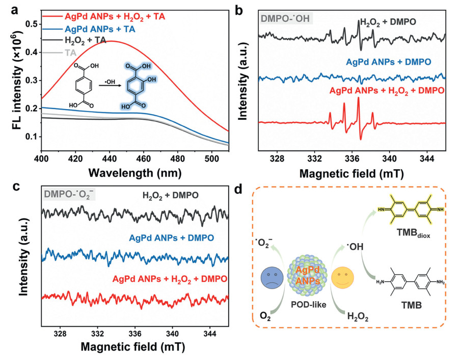
Although the widely accepted catalytic mechanism of POD-like nanozymes remains unclear, it is evident that two key steps characterize the POD-like pathway of nanozymes. The first involves the interaction between the nanozyme and H2O2 adsorbed on the surface, yielding •OH, and the second step generates the oxidation product of TMB through the interaction between •OH and TMB. Therefore, to detect the presence of •OH, terephthalic acid (TA) was employed as a fluorescent probe [55]. The TA solution exhibited fluorescence emission peak, but 2-hydroxybenzoic acid (TAOH) displayed a distinct fluorescence characteristic peak at 440 nm under excitation at 320 nm. In Fig. 4a, an obvious characteristic peak at 440 nm was observed in the mixed system of AgPd ANPs, H2O2 and TA. Conversely, no fluorescence characteristic peaks were observed in the mixed system of AgPd ANPs and TA or the mixed system of H2O2 and TA. These results confirm that AgPd ANPs catalyze the decomposition of H2O2 to form •OH.
Figure 4
Figure 4.
(a) Fluorescence spectra of different reaction systems. (b) ESR spectra of •OH trapped by DMPO in different systems. (c) ESR spectra of •O2− trapped by DMPO in different systems. In panels a, b and c, the reactions were conducted with 10 mmol/L H2O2, 10 mmol/L TA, 10 mmol/L DMPO, and 100 µg/mL AgPd ANPs. (d) Mechanism for the POD-like nanozymatic reaction of AgPd ANPs.
To gain deeper insights into the mechanism of AgPd ANPs, the generation of free radicals was investigated by electron spin resonance (ESR) spectrometer with 5, 5-dimethyl-1-pyrroline N-oxide (DMPO) as a trapping agent. Fig. 4b shows that upon adding H2O2 into the aqueous solution, the appearance of the distinct signal peaks in the ESR spectra with a 1:2:2:1 ratio indicates the existence of •OH in the system, suggesting the self-decomposition of H2O2. Upon introducing AgPd ANPs, the signal peak intensity significantly increases, demonstrating that AgPd ANPs catalyze the decomposition of H2O2 into •OH. In addition, the investigation into the existence of •O2− in the methanol system reveales the absence of ESR signal peaks of •O2− (1:1:1:1) in systems (Fig. 4c), indicating that AgPd ANPs cannot activate O2 into •O2−, similar to existing literature [54]. More importantly, the disability of O2 activation over AgPd ANPs in thermal catalysis reaction is consistent with our previous report [41]. Due to the valence-band hybridization in AgPd nanoalloy, the d band energy of the alloyed metal is deeper than that of pure Pd, which makes the charge transfer from the nanoalloy catalysts to the antibonding orbital of O2 extremely difficult. Moreover, Pd is likely to disperse at the single-atom level, making the elongation of O—O bond challenging and the O2 activation impossible [56,57]. These results provide direct evidence supporting the conclusion that AgPd ANPs can activate H2O2 to decompose into •OH but cannot activate O2 to •O2− (Fig. 4d). Overall, AgPd ANPs possess POD-like activity but not OXD-like activity, confirming catalytic reaction specificity of the nanozyme.
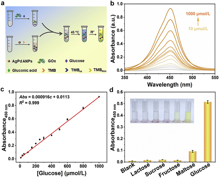
Colorimetry is the most commonly used strategy for biosensing [58]. In order to assess the excellent POD-like catalytic performance of AgPd ANPs, a colorimetric biosensor was constructed for the detection of H2O2 and glucose (Fig. 5a). We optimized the catalyst concentration, pH, temperature, and reaction time to improve the biosensor sensitivity (Fig. S5 in Supporting information). Using AgPd ANPs exhibiting superior POD-like activity under optimized conditions, different concentrations of H2O2 (10–700 µmol/L) display a good linear relationship with the absorbance of TMBdiox, with a limit of detection (LOD) of 0.44 µmol/L (Fig. S6 in Supporting information). Building on this, we determined the glucose concentrations. In a broad linear range (10–1000 µmol/L), the absorbance of TMBdiox increased proportionally with rising glucose concentrations (Figs. 5b and c). The linear regression equation, Abs = 0.000916c + 0.0113 (R2 = 0.999), results in a LOD of 1.56 µmol/L (S/N ≥ 3), outperforming most previous nanozymes for glucose detection (Table S3 in Supporting information). The selectivity of sensors to specific analytes is also critical [59]. To evaluate the selectivity of the constructed biosensing platform for glucose detection, various sugar analogues (maltose, fructose, sucrose, and lactose) were tested. Fig. 5d reveals a negligible interference from other analogs at the same concentration, highlighting the high selectivity of the proposed biosensor for glucose detection.
Figure 5
Figure 5.
(a) Schematic illustration of colorimetric detection of glucose with the POD-like of AgPd ANPs. (b) The UV–vis absorption spectra of TMBdiox with different glucose concentrations (10, 30, 50, 100, 150, 200, 250, 300, 400, 500, 600, 800, and 1000 µmol/L). (c) The linear calibration plots for glucose detection from 10 µmol/L to 1000 µmol/L. (d) Introduction of common sugar analogues to test the selectivity of the AgPd ANPs-based colorimetric sensor for the determination of glucose. Error bars refer to the standard deviation of three replicates.
To demonstrate the practical utility of the colorimetric analysis method, the AgPd ANPs-based biosensor was applied to human serum samples. Since the normal content of blood glucose in the human body is 3.9–6.1 mmol/L, samples underwent pretreatment to meet the requirements for analysis and detection. The spike-and-recovery experiment, detailed in Table 2 with various glucose solution samples, yielded recoveries between 99.2%−110.6%, indicating excellent accuracy in glucose detection. The relative standard deviation (RSD) of the three parallel tests was ≤ 3.37%, affirming the robust reproducibility and stability of the assay. These results underscore the feasibility, reliability, and accuracy of the proposed sensing platform is for glucose detection in serum samples.
Table 2
Table 2.
Detection of standard recovery glucose in serum (n = 3).
In summary, an alloying strategy was developed to achieve the reaction specificity of AgPd nanozymes, exclusively mimicking the catalytic activity of POD. The catalytic function of AgPd ANPs can be fine-tuned by appropriately adjusting the content of doped Ag. Particularly, increasing the Ag element enhances the POD-like activity and eliminates the OXD-like activity of Pd NPs, resulting in a nanozyme with specific POD-like activity. The valence-band hybridization between Ag and Pd hinders O2 adsorption and activation on the Pd surface. Meanwhile, H2O2 adsorbs onto AgPd nanozymes, decomposing into -OH, which desorbs and forms •OH, further oxidizing TMB. Utilizing this enzymatic activity, a highly sensitive cascade biosensor platform was constructed for the detection of H2O2 and glucose, demonstrating excellent selectivity against interfering sugar analogues. The practical application of this platform was further validated in human serum, indicating its potential for blood glucose-related real-time clinical diagnostics and routine care. This valence-band hybridization strategy in nanoalloy introduces a promising multi-noble metal nanostructure with exclusive POD-like activity, providing a new approach for controlling natural enzyme-like catalytic function specificity and opening avenues for colorimetric sensors based on nanozymes with singular catalytic activity.
Declaration of competing interest
The authors declare that they have no known competing financial interests or personal relationships that could have appeared to influence the work reported in this paper.
This research was financially supported by the National Natural Science Foundation of China (No. 22074038), the Luoyang Institute of Science and Technology Natural Science General Project (No. 21010905), and the Fundamental Research Funds for the Central Universities. This work was also sponsored by the Domestic Visiting Scholar Program of Shandong University of Science and Technology. The authors thank Mettler Toledo for the accurate weighing of all chemicals as well as the Analytical Instrumentation Center of Hunan University for TEM, XPS, and ESR measurements.
Supplementary materials
Supplementary material associated with this article can be found, in the online version, at doi:10.1016/j.cclet.2025.110916.
[1]
L.Z. Gao, J. Zhuang, L. Nie, et al., Nat. Nanotechnol. 2 (2007) 577–583. doi: 10.1038/nnano.2007.260
[2]
M. Zandieh, J.W. Liu, Adv. Mater. 36 (2024) 2211041.
[3]
G. Wei, S.J. Liu, Y.K. Peng, H. Wei, Chin. J. Chem. 42 (2024) 1515–1522. doi: 10.1002/cjoc.202300755
Figure 1
(a) Schematic diagram of the formation of TMBdiox (yellow) from TMB (transparent) in the presence of OXD or POD. (b) UV–vis absorption spectra of TMBdiox with pure ZrO2 or different AgPd ANPs loaded on ZrO2 in H2O2 solution (corresponding to POD-like activity). (c) UV–vis absorption spectra of TMBdiox with pure ZrO2 or different AgPd ANPs loaded on ZrO2 in aqueous solution (corresponding to OXD-like activity).
Figure 2
TEM (a), HRTEM (b), HAADF-STEM (c) and elemental mapping (d) images of AgPd ANPs loaded on ZrO2. Inset of panel (a) shows the size distribution of AgPd ANPs.
Figure 3
(a) UV–vis absorption spectra of TMBdiox of different reaction systems. (b) The absorbance of TMBdiox representing catalase-like activity of AgPd ANPs. (c) Steady-state kinetic assay of AgPd ANPs by varying the concentration of TMB. Inset: Lineweaver−Burk plot of photocatalytic activity of the AgPd ANPs with H2O2 as the substrate. (d) Steady-state kinetic assay of AgPd ANPs by varying the concentration of H2O2. Inset: Lineweaver−Burk plot of photocatalytic activity of the AgPd ANPs with TMB as the substrate. In panels b, c, and d, the reactions were conducted with 208 µmol/L TMB, 10 mmol/L H2O2, and 100 µg/mL AgPd ANPs in 0.2 mol/L acetate buffer (pH 4.0). Error bars refer to the standard deviation of three replicates.
Figure 4
(a) Fluorescence spectra of different reaction systems. (b) ESR spectra of •OH trapped by DMPO in different systems. (c) ESR spectra of •O2− trapped by DMPO in different systems. In panels a, b and c, the reactions were conducted with 10 mmol/L H2O2, 10 mmol/L TA, 10 mmol/L DMPO, and 100 µg/mL AgPd ANPs. (d) Mechanism for the POD-like nanozymatic reaction of AgPd ANPs.
Figure 5
(a) Schematic illustration of colorimetric detection of glucose with the POD-like of AgPd ANPs. (b) The UV–vis absorption spectra of TMBdiox with different glucose concentrations (10, 30, 50, 100, 150, 200, 250, 300, 400, 500, 600, 800, and 1000 µmol/L). (c) The linear calibration plots for glucose detection from 10 µmol/L to 1000 µmol/L. (d) Introduction of common sugar analogues to test the selectivity of the AgPd ANPs-based colorimetric sensor for the determination of glucose. Error bars refer to the standard deviation of three replicates.

 DownLoad:

 下载:
下载:


 下载:
下载:
