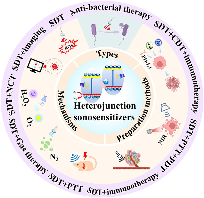
Figure 1.
Schematic illustration of heterojunction NSNs for synergistic SDT.
Ultrasound-responsive heterojunction sonosensitizers for multifunctional synergistic sonodynamic therapy
Li Qin , Wenjing Wei , Keqing Wang , Xianbao Shi , Guixia Ling , Peng Zhang

As an emerging non-invasive therapeutic method, sonodynamic therapy (SDT) has attracted extensive focus for its unparalleled therapeutic mechanisms and promising clinical applications. SDT utilizes ultrasound (US) to stimulate sonosensitizers (SNs), triggering the production of reactive oxygen species (ROS), which subsequently induces cell death [1,2]. This approach boasts deep tissue penetration capability, absence of phototoxicity, and high spatio-temporal controllability, offering novel strategies for disease treatment. SDT has been widely applied in areas such as antibacterial therapy and tumor treatment [3]. Compared to invasive treatments such as surgery, SDT does not require direct contact with the tumor, thereby reducing patient pain and risk. It can activate SNs at the tumor sites, directly killing tumor cells while avoiding damage to normal cells. SNs hold a crucial position in SDT, and the efficacy of SDT largely depends on the properties of the SNs, including their accumulation in local tissues, the efficiency of ROS generation, and biocompatibility [4-7]. However, traditional SNs possess certain drawbacks, such as the low efficiency in generating ROS under US, which limits their capabilities [8]. Furthermore, traditional SNs are often limited to a single treatment modality, making it difficult to integrate them with other therapeutic approaches to achieve multimodal therapy.
To overcome the limitations of traditional SNs, researchers have devoted themselves to developing novel SNs that can enhance therapeutic efficacy and reduce side effects. Based on this, heterojunction SNs have emerged as promising materials. Heterojunction SNs are a new class of SNs, which are composed of two or more different materials through a heterojunction structure, forming nanocomposites with unique electronic properties [9]. This structure typically encompasses the combination of p-type and n-type semiconductor materials [10], or other types of material such as the integration of metals and semiconductors [11], nanozymes [12], two-dimensional (2D) nanomaterials [13], and metal-organic frameworks (MOFs) [14]. The design of heterojunction SNs focuses on enhancing the efficiency of electron-hole pair separation and ROS generation between materials, thereby enhancing the efficiency of SDT [15]. Additionally, the design of heterojunction structures also offers the potential for multifunctionality of SNs, such as the integration of targeting ligands, drug carriers, and imaging capabilities, making SDT more precise and efficient [16].
This review systematically classified the types of heterojunction SNs, described the preparation methods and characterization methods, and then delved into the mechanisms by which they enhanced SDT. Following this, it focused on providing a comprehensive overview of the applications of heterojunction SNs in disease diagnosis and treatment, aiming to explore their design principles, synthesis strategies, and potential in this field. The review categorized heterojunction SNs based on their therapeutic areas and modalities. We primarily focused on the application of heterojunction SNs in bacterial infections and tumor treatment (Table S1 in Supporting information). In the context of bacterial infections, their applications primarily encompassed wound healing and bone regeneration. In terms of tumor treatment, the review systematically categorized them based on therapeutic modalities, including single-mode SDT, dual-mode therapies and triple-mode therapies that integrated SDT with multiple other modalities (Fig. 1). Finally, the review summarized the current challenges faced by heterojunction SNs and their future development directions. We anticipate that this review will contribute to the development of novel heterojunction SNs and facilitate the clinical translation of SDT.
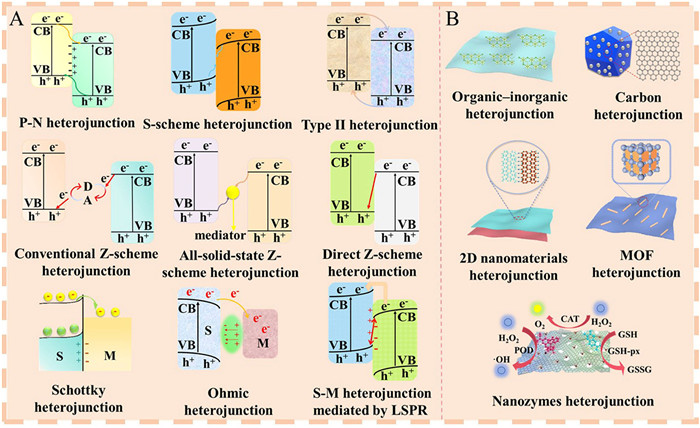
As promising candidates for enhancing SDT, heterojunction SNs are formed by coupling different functional materials. Based on conductivity of the materials, heterojunction can be divided into two types: semiconductor–semiconductor (S–S) heterojunction and semiconductor–metal (S–M) heterojunction. S–S heterojunction mainly includes P-N heterojunction, Z-scheme heterojunction, S-scheme heterojunction, and Type Ⅱ heterojunction (discussed in Section 2.1 in Supporting information). Based on the materials types to construct heterojunction, it can be divided into five types: organic–inorganic heterojunction, carbon-based heterojunction, 2D nanomaterials-based heterojunction, MOF-based heterojunction, and nanozymes-based heterojunction (discussed in Section 2.2 in Supporting information). The structures of heterojunction SNs were summarized in Fig. 2. Typical heterojunction SNs were summarized in Figs. S1 and S2 (in Supporting information).
Currently, several methods have been widely used to create heterojunction SNs, mainly including in situ growth, electrostatic attraction, atomic layer deposition (ALD), thermal injection, and in situ reduction or oxidation. In this section, the preparation methods applicable types of heterojunction SNs, and their advantages and disadvantages were introduced (discussed in Section 3.1 in Supporting information).
The techniques for characterizing heterojunction are also important. Therefore, the advanced methods for characterizing heterojunction SNs such as ultraviolet photoelectron spectroscopy, kelvin probe force microscopy, and DFT calculations were also summarized (discussed in Section 3.2 in Supporting information).
When US interacts with these SNs, they trigger a transition of electrons within the material, shifting them from conduction band (CB) to the valence band (VB), with holes being left in their original positions. This process results in the generation of electron-hole pairs, which serve as crucial intermediaries in the production of ROS, a vital component for the effectiveness of SDT (Fig. S3A in Supporting information) [17].
Heterojunctions can enhance the separation of electron-hole pairs via various mechanisms. For instance, in a type-Ⅱ heterojunction, both the CB and VB of the material with a wider bandgap are positioned above those of the material with a narrower bandgap. This arrangement facilitates the transfer of electrons and holes to different materials, thereby minimizing their recombination [18]. Furthermore, the Z-scheme heterojunction facilitates the efficient separation of electrons and holes through the establishment of an internal electric field, which directs their movement in mutually exclusive directions. Band structure engineering is a proven strategy where the heterojunction is composed of materials with differing bandgaps. The redistribution of charges at the heterojunction interface generates an internal electric field, which acts as a driving force, pushing photo-generated or sono-generated carriers in different directions and reducing the chances of electron-hole recombination [19]. Another approach involves interface engineering. The unique interface structure of the heterojunction provides additional active sites, serving as a platform for electron-hole pair separation and facilitating the generation of reactive oxygen under ultrasonic action [20]. Optimizing the quality and contact mode of the heterojunction interface can minimize defects and trap states, which might otherwise capture carriers and lead to recombination. Additionally, selecting materials with complementary characteristics is also crucial. For example, pairing a material that exhibits high electron mobility with one that demonstrates exceptional hole mobility can significantly enhance carriers transport efficiency [21,22].
One of the key factors affecting ROS formation is the hypoxic tumor microenvironment (TME) in SDT. SDT is an oxygen-consuming mechanism, and as a substrate for SDT to produce ROS, tumor hypoxia will inevitably affect the efficiency of ROS production and reduce the efficacy of SDT [23,24]. Therefore, increasing oxygen supply can improve this situation. Heterojunction can increase oxygen supply in different ways.
One is to promote oxygen supply. Some heterojunction materials have oxygen-carrying capacity, which can directly deliver oxygen to the tumor site or generate oxygen in situ in the TME through catalytic action, increasing local oxygen concentration. By constructing heterojunction with nanozymes with catalase (CAT) activity, nanozymes can decompose endogenous hydrogen peroxide (H2O2) into oxygen, subsequently mitigating hypoxia conditions and furnishing necessary reactants for SDT (Fig. S3A) [22].
Moreover, when constructing heterojunction with photothermal nanomaterials, photothermal effects can cause vascular dilation by locally heating tumor tissue, thereby increasing blood flow. This increased blood flow helps to improve the delivery of oxygen to the tumor sites [25]. In some cases, photothermal effects can promote the release of oxygen from the blood into tissues, as an increase in temperature can enhance the oxygen affinity of hemoglobin, thereby promoting the release of oxygen [26]. The photothermal effect can alter the TME, reduce hypoxic areas, and thus improve the accessibility of oxygen to the tumor tissue.
Heterojunction SNs greatly enhance the effectiveness of SDT. However, like traditional SNs, heterojunction SNs are distributed throughout the body after intravenous administration and can also induce sonodynamic effects in normal tissues, causing damage to normal tissues [27]. Therefore, constructing heterojunction SNs with active targeting ability can specifically target pathogenic sites, enhance therapeutic efficacy, and reduce damage to normal tissues (Fig. S3B in Supporting information). At present, different targeting ligands have been integrated into the surface of heterojunction to achieve active targeting. Hyaluronic acid (HA) can target the overexpressed CD44 receptor in tumor cells and coupling it to the surface of heterojunction can enhance the uptake of tumor cells. The Cu2O/Cu2-xS heterojunction achieved both photothermal therapy (PTT) and active targeting of tumor cells through surface modification of gambogic acid and HA [28]. Cyclic RGD peptide (cRGD) can target the integrin αvβ3 on the surface of tumor cells. By modifying cRGD onto heterojunction containing carbon nitride nanosheets (NSs) and copper-loaded molybdenum disulfide (MoS2) NSs, it was endowed with active targeting ability, achieving efficient delivery of heterojunction to tumor cells and enhancing the therapeutic effect of osteosarcoma (OS) [29]. In addition, heterojunction modified with homologous cell membranes can also enhance active targeting ability. For example, using 4T1 cell membrane to modify MoS2-Ti2C2 heterojunction enhanced its accumulation in 4T1 tumor cells [30]. Extracellular vesicles derived from CT26 cells were modified onto a heterojunction composed of transition metal carbides and nitrides (MXene) and liquid metal, enabling targeted delivery to CT26 tumor cells [31].
More importantly, different ligands are also used to modify heterojunction SNs to achieve their active targeting ability and improve the effectiveness of SDT. HA, as a targeting ligand, is also used to modify heterojunction SNs for active targeting. Fu et al. synthesized a HA-modified barium titanium (BTO)/black phosphorylation (BP) heterojunction, which first targeted tumor cells, and then induced tumor apoptosis under US stimulation. At the same time, PO43− produced by BP degradation interacted with hydrogen ions in lysosomes, thereby inhibiting tumor autophagy, disrupting tumor self-protection mechanisms, and enhancing the efficacy of immunotherapy [32]. Manganese porphyrin nanoparticles (NPs)/Au NPs heterojunction composed of Au@Cu2O nanocubes with surface modified titanium dioxide quantum dots also enhanced the targeting of tumor cells through modification of HA [33,34]. CD44 receptor is also highly expressed on the surface of proinflammatory macrophages. HA-modified heterojunction also targeted inflammatory macrophages to inhibit atherosclerosis [35]. Heterojunction SNs encapsulated by tumor cell membranes can also enhance their homologous targeting. For example, the tungsten disulfide/Pt heterojunction encapsulated by HeLa cell membranes achieved tumor localization through homologous targeting and effectively tumors inhibition through SDT [36].
In summary, heterojunction SNs connected by targeted ligands can accurately target disease tissues, thereby enhancing SDT and reducing damage to normal tissues.
Heterojunction not only enhances its catalytic efficiency, but also serves as a drug carrier for drug delivery to enhance synergistic therapeutic effects. Heterojunction has been used for the delivery of different chemotherapy drugs in tumor treatment. The heterojunction composed of Bi2Se3 with hollow mesoporous structure and Au NPs was used to load doxorubicin, thereby achieving the combination of PTT, photocatalytic therapy (PCT) and chemotherapy [37]. In the core-shell heterojunction composed of porphyrin based MOFs and upconversion NPs, due to the porous structure of MOFs, they were used to load tirapamine and programmed death-ligand 1 (PD-L1), thereby endowing the heterojunction with chemotherapy and immunotherapy functions [38].
Heterojunction SNs have also been constructed for drug delivery. The constructed Tungsten disulfide/Pt Schottky heterojunction was surface modified with polyethyleneimine to achieve effective loading of curcumin and further encapsulated by the tumor cell membrane for homologous targeting [36]. Curcumin inhibited the activity of tumor associated fibroblasts, and US triggered ROS generated by heterojunctions induced tumor cell apoptosis, thereby optimizing the therapeutic effect.
In short, heterojunction SNs with hollow or porous structures can also serve as carriers for drug delivery, further synergizing with SDT to enhance therapeutic efficacy.
Heterojunction integrated materials with different properties can be used in combination with other treatment modalities such as hyperthermia, chemodynamic therapy (CDT), and immunotherapy, to improve the therapeutic effect (Fig. S3B) [12,39]. For instance, by integrating photothermal materials into a heterojunction design, the SNs generate ROS in response to US. Concurrently, PTT converts light energy to heat energy. The combined application of these two modalities elicits both thermal and chemical effects, synergistically enhancing the efficacy of cell eradication [40]. Moreover, PTT can raise the temperature of local tissues, increase blood flow, and improve oxygen content, thus enhancing the activity and therapeutic effect of SNs [41]. Heterojunction SNs can also be combined with immunotherapy. TME usually has immunosuppressive properties, and heterojunction SNs can reduce the activity of immunosuppressive cells and enhance the infiltration and function of immune cells by altering the TME [42]. Heterojunction SNs contribute to the formation of immune memory cells and long-term anti-tumor immunity by enhancing the immunogenicity of tumor cells [43,44]. In addition to this, heterojunction SNs can also be combined with CDT, where heterojunction SNs produce ROS in response to US, while CDT produces ROS through a Fenton-like reaction [45]. The combination of the two can increase the local concentration of ROS in the tumor, thus improving the killing effect on tumor cells. In conclusion, constructing heterojunction SNs, on the one hand, can enhance the SDT therapy by itself, and on the other hand, it can be combined with other therapies to enhance the therapeutic effect of the disease.
Bacterial infection is a common problem in the wound healing process, which can cause inflammatory reactions, and delay the healing process [46]. Heterojunction has great potential in sonodynamic antibacterial for wound healing, providing effective alternative or complementary strategies for traditional antibiotic treatment, especially in the face of drug-resistant bacterial infections, demonstrating significant advantages.
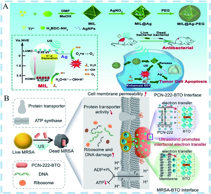
MOFs, being porous, exhibit promising potential in various biomedical applications, encompassing sensing, drug delivery systems, and enhancement of SDT [47]. Meng et al. developed polyethylene glycol (PEG)-functional Ti-based MOFs (MIL)@Ag heterostructures for treating deep-seated cancer and bacterial infections. Ag NPs were assembled in situ on the surface of MIL (Fig. 3A) [48]. In comparison to the pure MIL, the Ag NPs embedded within it significantly enhanced electron transfer, effectively separated electron-hole pairs, and improved O2 adsorption efficiency, thereby significantly boosting ROS generation efficiency and enhancing SDT. Furthermore, the Ag NPs within MIL@Ag-PEG were capable of generating ROS within the redox microenvironment, causing nonspecific cellular damage. Leveraging deep tissue penetration of US, MIL@Ag-PEG eliminated pathogens, enhancing wound healing.
Titanium oxide (TiO2), has garnered widespread attention across multiple fields for robust antibacterial ability, broad spectrum of effectiveness, long-lasting antibacterial effect, and non-toxic, safe characteristics [49]. For instance, a copper peroxide (CuO2)/TiO2 heterostructure was devised, consisting of CuO2 nanoclusters and ultrathin TiO2 NSs enriched with oxygen vacancies (OV-TiO2), which were then incorporated into microneedles (MNs) [50]. This innovative design aimed to achieve a dual enhancement of sono-chemodynamic and sonothermal antibacterial therapy, offering a bilateral therapeutic advantage. As confirmed through both in vitro and in vivo experiments, CuO2/TiO2 MNs (CTMN) demonstrated remarkable antibacterial efficacy. An exceptional killing rate surpassing 99.9999% was achieved against a diverse spectrum of multidrug-resistant (MDR) pathogens within 5 min, while simultaneously expediting the wound healing process.
Nanozymes, novel functional nanomaterials with enzyme-mimicking catalytic properties, have garnered significant attention in recent years and have been extensively applied in biosensor innovation, medical diagnostics, treatments, as well as tissue engineering [51,52]. Bai et al. reported a heterostructure skin patch comprising zinc oxide nanorod@graphdiyne NSs (ZnO@GDY NR), which exhibited enzyme-like activity, utilizing piezoelectric activation and nanozymes [53]. This patch effectively disrupted the proliferation of methicillin-resistant Staphylococcus aureus (MRSA) pathogens in infected skin wounds while simultaneously promoting rapid repair of the wound tissues.
While elevating local ROS levels can effectively eliminate bacterial infections, it poses a risk of inflicting oxidative damage to nearby cells and tissues, thereby causing adverse effects on the body [54]. Geng et al. crafted an intelligent nanocatalytic membrane, integrating poly(lactic-co-glycolic acid) (PLGA) with black phosphorus/V2C MXene bio-heterojunctions (2D2-bioHJs) for enhanced functionality [55]. The 2D2-bioHJs served as a precise "balancer", effectively striking a harmonious equilibrium between the generation and clearance of ROS, thereby imparting both antibacterial and anti-inflammatory attributed to the engineered membrane. Crucially, in vivo evaluation confirmed that the nanocatalytic membranes not only eradicated the bacterial population but also mitigated the nuclear factor kappa-B (NF-κB) inflammatory cascade and fostered angiogenesis, thereby transforming stagnant chronic wound conditions into a regenerative milieu.
Currently, a large number of antibiotics are clinically required in the treatment of osteomyelitis caused by bacterial infections, which lead to the emergence of antibiotic-resistant bacteria [56-58]. SDT proves to be an efficacious modality for addressing deep bone infections [59-61]. Employing heterostructure is an effective means of improving the electron transfer efficiency and enhance bone regeneration.
For the cause of ROS production, researchers usually focus on manipulating the generation, separation, and transport of electrons in the material [62,63], but seldom consider manipulating the spin-electron degrees of freedom for ROS generation. Ding et al. used porous TiO2 and Fe3O4 NPs to form a ferromagnetic heterojunction named CF(Fe3O4/TiO2) [64]. Fe3O4 affected the electronic spin state of TiO2, which endowed CF with an enhanced spin-polarized state and magnetic moment. When subjected to US, the built-in electric field formed by CF and the spin-polarized electrons synergistically inhibited US-activated charge complexation, and increased the ROS yield. The results suggested that modulation of spin polarization optimized US catalytic efficiency and showed great potential for bone regeneration in osteomyelitis in vivo.
Ti3C2 (TC) has been demonstrated to be an efficient catalyst [65-67]. In one study, an porphyrin metal-organic framework (HNTM)/TC hybrid nanomaterial (HN-Ti3C2) with Schottky heterojunction was synthesized [68]. Upon US excitation, HNTM produced an abundance of electron-hole pairs, while TC swiftly transferred the carriers generated by HNTM under US irradiation, effectively hindering the recombination of these electron-hole pairs. The results indicated that MXene-based Schottky heterojunction could successfully eliminate the infection and was prominent in the treatment of osteomyelitis.
Some studies have developed an interface engineering strategy, and this strategy accelerates electron transfer at the interface by constructing various heterostructures to achieve high efficiency in electron transfer [69-71]. BTO is a prototypical piezoelectric material that undergoes polarization when subjected to ultrasonic excitation [72]. Therefore, Yu et al. developed a new catalyst based on a zirconium-based porphyrin metal-organic skeleton (PCN-222) and BTO piezoresponsive heterojunction, PCN-222-BTO (Fig. 3B) [63]. Upon US irradiation, the polarization of BTO induced the formation of a built-in electric field at the interface between PCN-222 and BTO. This electric field facilitated the transfer of electrons generated by PCN-222 toward BTO, ultimately triggering the generation of ROS at the interface. Meanwhile, at the MRSA-BTO interface, the polarization of BTO prompted the migration of electrons from the bacterial membrane towards the BTO.
In addition, the heterojunction based on S-doped TiO2 NSs and CeO2 (S-TiO2-x/CeO2) heterojunction was prepared [73]. The CeO2 attached to S-TiO2-x not only enhanced the SDT but also exerted enzymatic effect to promote the osteogenic differentiation.
SDT has emerged as a novel and promising strategy in clinical cancer treatment, owing to its exceptional tissue penetration depth and non-toxic nature [74]. The electron-hole pair separation rate and bandgap width of the SNs are the keys to the SDT. The problem of fast electron-hole pair complexation rate and band gap width must be overcome to improve the efficiency of SDT [75].
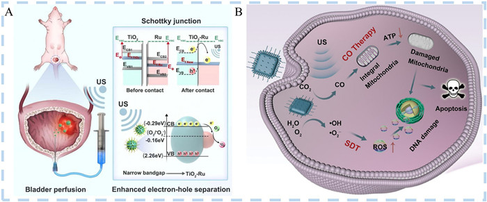
Schottky heterojunction SNs exhibit enormous potential. A TiO2-Ru-PEG Schottky heterojunction SNs with high electron-hole separation and narrow bandgap were synthesized by Li et al. (Fig. 4A) [9]. After the coordination of [Ru(phen)2dppz]2+ (Ru(Ⅱ) with TiO2 within the TiO2 shell), Ru precipitated to form Schottky heterojunction. Both experimental data and density functional theory (DFT) calculations showed that the formation of heterojunction decreased the band gap of TiO2-Ru-PEG. Under US irradiation, the effective separation rate of electron-hole pairs of TiO2-Ru-PEG was significantly enhanced, and the generation level of ROS was significantly increased. In addition, in vivo experimental findings underscored the remarkable ability of TiO2-Ru-PEG to suppress the proliferation of bladder tumor cells, while maintaining a high level of safety.
Hu et al. developed a biodegradable N-CD@LiFePO4 P-N heterojunction SNs, with efficient SDT and modulation of TME [10]. N-type conductive scavenge nitrogen-doped carbon dots (N-CDs) were arranged onto the surface of LiFePO4 nanorods. P-N heterojunction mediated a rapid charge transfer and production of 1O2 significantly increased, which resulted in an efficient separation of electron-hole pairs for SDT. N-CD@LiFePO4 P-N heterojunction improved the SDT efficiency. After US irradiation, N-CD@LiFePO4 P-N heterojunction eliminated tumors by a single drug injection.
Gas therapy is a novel treatment strategy. Gases such as hydrogen (H2), nitric oxide (NO) and carbon monoxide (CO) can induce mitochondrial damage to kill tumor cells [76-78]. Gas therapy also enhance the effectiveness of SDT [79].
Hydrogen therapy is a non-invasive treatment modality using molecular H2 as a therapeutic agent [80]. Yuan et al. used Schottky heterostructure nanocomposite (Pt-Bi2S3) to demonstrate the feasibility of the concept of sonodynamic hydrogen precipitation [81]. In this nanocatalytic system, electrons generated by US excitation were transferred from the surface of bismuth sulfide (Bi2S3) nanorods to platinum (Pt) NPs, and the transfer of electrons induced energy band bending to form a barrier, and this barrier could effectively prevent the electron backflow. The combined effect of the two aspects made electron-hole pair recombination less likely, improved the charge separation rate and promoted H2 generation. In vitro and in vivo experiments have shown that H2 led to mitochondrial dysfunction and interferes with the normal functioning of the intracellular respiratory system.
Yuan et al. also investigated the S-scheme heterojunction named BiOBr@Bi2S3 nano-heterojunction to combine gas therapy and SDT (Fig. 4B) [82]. The BiOBr@Bi2S3 S-scheme heterojunction consisted of a reducing catalyst (BiOBr) and Bi2S3. The contact between BiOBr NSs and Bi2S3 nanorods induced charge redistribution at the interface. The electrons of BiOBr were transferred to the holes of Bi2S3, leaving holes and electrons with strong redox ability in the positive VB of BiOBr and the negative CB of Bi2S3, respectively, which participated in the oxidation reaction and the reduction reaction, respectively. In vitro and in vivo experiments, the BiOBr@Bi2S3 nanocatalysts not only possessed the ability to control ROS and CO generation, but also the ability to enhance CO therapy. This experiment achieved the goal of combined SDT and CO therapy, which mechanistically and synergistically induced tumor cell apoptosis.
Based on the cost-effectiveness, robust stability, tunable catalytic activity, scalability for mass production, and exceptional reproducibility of nanozymes [83,84], it has also been used in combination with SDT [85].
Zhao et al. accomplished the fabrication of ternary Ru/TiO2-x@TiCN heterojunction through a one-step in situ synthesis process, involving the oxidation of TiCN NSs into TiO2-x NSs and the subsequent reductive deposition of Ru3+ ions into Ru NPs (Fig. S4A in Supporting information) [86]. Ru/TiO2-x@TiCN heterojunction exhibited peroxidase (POD), glutathione peroxidase (GSH-px), and CAT-like catalytic activity due to the presence of Ti3+ and Ti4+. Ru/TiO2-x@TiCN improved oxygen replenishment in the tumor and enhanced the SDT effect by rapidly converting intracellular H2O2 to O2 via its CAT-like catalytic activity.
Manganese oxide (MnOx) exhibited multiple enzyme-like activities. Therefore, different heterojunctions based on MnOx were constructed. A piezoelectric heterojunction nanoplatform (M-BOC@SP NSs) composed of bismuth chloride NSs (BiOCl NSs) and MnOx was synthesized by Zhao et al. [87]. BiOCl NSs exhibited piezoelectric effect and provided more active sites by virtue of their structure. Upon US irradiation, the piezoelectric effect significantly promoted carrier transport and enhanced ROS production. As MnOx had multiple enzyme-like activities, it could down-regulate GSH levels and also decompose endogenous H2O2 to generate O2 and •OH. M-BOC@SP NSs significantly enhanced ROS production and reversed the intra-TME. The in vitro and in vivo results demonstrated that M-BOC@SP NSs had significant anticancer effects.
It has been documented that the narrow bandgap of Pt facilitated the effective separation of electron-hole pairs and enhanced ROS generation, ultimately boosting the efficiency of SDT [88]. Li et al. designed Pt-ZnO SNs (Fig. S4B in Supporting information) [1]. Pt-ZnO, with a narrow bandgap and a moderate amount of oxygen defects, improved the separation efficiency of electron-hole pairs and the charge utilization efficiency under US, and promoted the generation of ROS. Meanwhile, Pt-ZnO exhibited both robust CAT-like and POD-like activities, significantly boosting the efficacy of SDT and catalytic treatment. In addition, Pt-ZnO significantly depleted GSH, further amplifying oxidative stress. Eventually, Pt-ZnO achieved a triple ROS amplification effect. In vivo evaluations demonstrated that Pt-ZnO cleared up to 98.1% of tumors.
Wang et al. synthesized novel SNs-hollow cobalt phosphides (CoP)@N-carbon@PEG nanospheres (CPCs@PEG) (Fig. S4C in Supporting information) [89]. Upon binding of the N-carbon component, the CPCs exhibited a Z-scheme mechanism that favored the separation and transferred of electron-hole pairs and enhanced the ROS level. In addition, CPCs@PEG utilized endogenous glucose as an in situ hole scavenger, which hindered electron-hole pair complexation and enhanced electron enrichment and ROS generation. Because of the higher oxidizing capacity of Co3+, CPCs@PEG was endowed with CAT activity, which expressed H2O2 as O2 and promoted ROS generation. The engineered CPCs@PEG heterojunctions exhibited extraordinary anticancer effects and significantly inhibited the proliferation and growth of tumor cells.
The combined use of PTT and SDT can produce synergistic effects and improve treatment efficacy [7,90]. On the one hand, the integration of PTT with SDT potentiates tumor eradication through the generation of ROS and thermal effects. On the other hand, PTT can augment blood flow to the tumor site, optimizing the hypoxic TME and thereby potentiating the efficacy of SDT [91,92].
As a new member of the 2D material family, MXenes have found applications in a broad spectrum of biomedical fields, spanning from nanomedicine to the regenerative biomaterials. This versatility is attributed to their extensive surface area, robust near-infrared (NIR) absorption capabilities, and customizable compositions [93,94]. Recently, the sonodynamic effect of MXenes has also explored. MXenes are used to construct heterojunction to combine PTT and SDT. Xu et al. engineered Nb2C MXenes as a superior SNs through gentle in-situ self-oxidation, calculating their work function values across varying oxidation degrees to pinpoint optimal conditions for constructing effective Schottky heterojunctions (Fig. S5A in Supporting information) [95]. The study revealed that the Schottky heterojunction effectively expedited the segregation of sonication-induced electron-hole pairs and hindered their recombination, significantly enhancing the ROS production efficiency of in-situ self-oxidized Nb2C MXenes (Nb2C-Ox) when exposed to US irradiation.
OS, the most prevalent malignant bone tumor particularly affecting children and adolescents, disrupts the homeostasis of the afflicted bone, resulting in osteolysis as it progresses [96]. Lv et al. devised an innovative switchable approach for achieving dynamic OS ablation and subsequent static bone regeneration. This was accomplished by integrating piezoelectric BaTiO3 (BTO) with atomically thin TC through a Schottky heterojunction, resulting in the synthesis of TC@BTO composites (Fig. S5B in Supporting information) [97]. The TC@BTO composites, fortified by the Schottky heterojunction, exhibited exceptional photothermal conversion and ROS generation capabilities under sequential US and NIR irradiation, synergistically leveraging the strengths of SDT and PTT.
Ti3C2 MXenes have garnered the much attention due to their ease of fabrication, cost-effectiveness, and superior performance characteristics. Geng et al. reported novel SNs based on 2D Ti3C2Tx NSs for high-efficiency SDT of deeply-seated tumors (Fig. S5C in Supporting information) [26]. By integrating NIR-Ⅱ-responsive CDs onto the surface of Ti3C2Tx NSs, the resulting 0D/2D hybrid CD@Ti3C2Tx heterojunctions (HJs) not only capitalized on the strengths of both components but also mitigated their limitations, thereby enhancing the efficacy of SDT and PTT. As a naturally abundant Ti source, Ti3C2 MXene could spontaneously transform into TiO2 in situ, forming a heterostructure with robust interactions. This process enhanced charge carrier transfer and facilitated efficient electron-hole separation [98]. In another study, Ti3C2-TiO2 MXenes were synthesized with enhanced sonosensitization properties through precise interface engineering (Fig. S5D in Supporting information) [99]. The Ar-Ti3C2-TiO2 composite demonstrated superior US-triggered ROS generation efficiency, attributed to its narrowed band gap (2.37 eV) and efficient interfacial electron transfer, which mitigated electron-hole recombination. Moreover, its preserved structural integrity contributed to high photothermal conversion efficiency, while laser irradiation further bolstered electron-hole pair separation, enhancing ROS production. Results validated its ability to accumulate efficiently at tumor sites, facilitating effective PTT-enhanced SDT for tumor eradication.
Apart from MXene-based heterojunction, Song et al. crafted a ternary heterojunction SNs, Bi@BiO(2-x)@Bi2S3-PEG (BOS), for tumors, enabling continuous SDT bolstered by thermal injury [100]. BOS not only preserved the individual functionalities of its constituent parts but also demonstrated superior catalytic efficacy in ROS generation. Concurrently, it continuously depleted GSH, fostering biodegradation and sustaining oxidative stress-induced injury. Furthermore, under NIR illumination, BOS's photothermal conversion facilitated thermal ablation of tumors while alleviating hypoxia, thereby enhancing the SDT.
CDT represents an efficacious approach for the elimination of excessively produced H2O2 within tumor cells [101]. CDT can catalyze H2O2 to generate •OH by Fenton reactions and reduce the production of GSH [102]. Different heterojunction SNs based on metallic oxide have been deported to combine SDT and CDT.
Cu2O emerges as a promising semiconductor candidate for inorganic SNs in SDT and CDT [103]. Feng et al. introduced novel Z-scheme heterojunction SNs, leveraging Fe-doped CDs as assistants to boost the sensitivity of cubic Cu2O, yielding Fe-CDs@Cu2O (Fig. S6A in Supporting information) [104]. Fe-CDs@Cu2O nanocomposites exhibited significantly enhanced SDT efficacy, stemming from their superior capability in facilitating electron-hole separation. Moreover, the incorporation of Fe ions into the CDs fostered a synergistic enhancement of Fenton-like reactions alongside the Cu2+ inherent in Cu2O, leading to a substantial augmentation in the efficiency of CDT.
Cu2-xO also exhibited the characteristics of narrow bandgap, which have been used to construct heterojunction [105]. For example, Geng et al. constructed cutting-edge Cu2-xO@TiO2-y heterojunction NSNs, harnessing the synergy of positively charged Cu2-xO nanodots and negatively charged TiO2-y NSs, integrated within a core-shell oxide semiconductor framework that boasted narrow bandgaps ranging from 2.1 eV to 2.2 eV (Fig. S6B in Supporting information) [106]. The nanocomposite possessed exceptional redox activities, enabling it to regulate the TME through Cu+/Ti3+ catalyzed •OH generation from endogenous H2O2 and Cu2+ mediated consumption of endogenous GSH to combine SCT/CDT.
CeO2, as a prototypical N-type semiconductor, possesses the inherent potential to serve as an effective SNs [107]. Zheng et al. introduced a groundbreaking formulation for the novel Pt/CeO2-xSx SNs, achieving oxygen defects by meticulously incorporating sulfur (S) doping and subsequently integrating Pt in situ [108]. Extensive oxygen defects were successfully introduced into Pt/CeO2-xSx through S doping, significantly enhancing its capacity to hinder electron-hole recombination, thereby fostering robust ROS generation. Notably, Pt/CeO2-xSx proficiently depleted overexpressed GSH through redox reactions, intensifying oxidative stress within the TME. Leveraging its exceptional POD-like activity, Pt/CeO2-xSx effectively orchestrated a highly efficient synergistic treatment strategy integrating SDT and CDT.
In addition to above metallic oxide, CoOx and MnO2-x were also used to construct heterojunction [109-112]. For instance, Liu et al. demonstrated that by utilizing ALD to construct a multifunctional TiO2@CoOx heterojunction (Fig. S6C in Supporting information) [109]. CoOx demonstrated a sequential generation of H2O2 → O2 → •O2− in response to the TME, effectively alleviating hypoxia during SDT and enhancing the efficacy of CDT. Zhou et al. fabricated a 2D TiO2@MnO2-x core/shell heterostructure by strategically depositing MnO2-x onto the surface of TiO2 NSs (Fig. S6D in Supporting information) [13]. The incorporation of MnO2-x imparted the heterostructure with multifaceted functionalities, including modulation of tumor hypoxia, GSH depletion, and induction of Fenton-like reactions. The combined effect of mitigating tumor-hypoxia and concurrently executing SDT/CDT therapy led to remarkable efficacies in tumor cell eradication and suppression.
The use of the immune system for tumor immunotherapy has shown synergistic effects with SDT [112]. Bismuth molybdate (Bi2MoO6, BMO) is a notable Aurivillius family member, renowned for its superior photocatalytic performance and unique layered configuration. For instance, Cheng et al. innovatively crafted an ultrasonically controllable 2D piezoelectric nanoagonist (Bi2MoO6−MXene) to effectively induce ferroptosis (Fig. S7A in Supporting information) [113]. The integration of BMO with MXene formed a Schottky heterojunction that significantly reduced the bandgap by 0.44 eV, enhancing carrier-separation efficiency and mitigating electron-hole pair recombination under US stimulation. Consequently, this enhancement facilitated a surge in the production of ROS. When subjected to precise spatiotemporal US excitation, the BMO-MXene system potently inhibited ovarian cancer (OC) proliferation by more than 90%, initiating lipid peroxidation and destabilizing the mitochondrial membrane potential. Furthermore, it inactivated crucial antioxidant systems, such as GSH-px and cystathionine transporter proteins, triggering ferroptosis in tumor cells. Notably, ferroptosis in OC cells ignited an immunogenic cell death (ICD) cascade, fostering dendritic cell maturation and amplifying antitumor immune responses.
Chemotherapy can damage the DNA of tumor cells, while SDT further damages tumor cells by generating ROS. The combination of the two may have a synergistic effect and improve treatment efficacy [114].
Since oxygen is required for ROS generation, and hypoxia-activated prodrugs (HAPs) based chemotherapeutic approaches require the creation of a hypoxic environment [115,116]. Chen et al. investigated the fabrication of BiOCl-Au-Ag2S Z-scheme heterostructure NPs, tirapazamine (TPZ) was loaded onto the NPs through non-covalent interactions, to prepare LPBAAS NPs [117]. Under US irradiation, UV light excited the BiOCl and Ag2S components, generating free electrons and holes. Depositing Au NPs on BiOCl NSs achieved an effective separation of electrons and holes and maintained its high redox potential. The subsequent reaction of the NSs followed the Z-scheme mechanism. Combining BiOCl with HAPs could realize the dual functions of HAPs activation and ROS generation in an anoxic environment. In vitro and in vivo analysis have conclusively shown that LPTBAAS NPs could efficiently eliminate tumor cells in a hypoxic environment, successfully achieving synergistic treatment with SDT and HAPs-based chemotherapy.
Nonmetallic graphitic carbon nitride quantum dots (g-C3N4 QDs) have been widely used in biological research. Numerous studies have also shown that g-C3N4 enhanced photocatalytic activity by constructing intimate heterojunctions [118]. He et al. synthesized nanostructured TiO2@g-C3N4 heterojunction by surface precipitating g-C3N4 quantum dots onto hollow mesoporous TiO2 nanorods (Fig. S7B in Supporting information) [119]. Stimuli-responsive release was achieved by loading TiO2@g-C3N4 with the prodrug romidepsin (RMD) under US irradiation. The disulfide bridge of the released RMD was readily reduced to monocyclic dithiols by highly reducing GSH, and the metabolites of monocyclic dithiols triggered apoptosis through the dual effects of blocking the G2/M phase of the cell cycle and enhancing histone acetylation. In brief, the successful experiments with TiO2@g-C3N4 have initiated a new research direction for the combined enhancement of cellular redox imbalance and tumor-specific chemotherapy.
RT and SDT have demonstrated a synergistic impact on tumor treatment, enhancing their effectiveness when combined [120,121]. Zhu et al. designed a bismuth-based heterostructure semiconductor, termed BFIP (BiF3: 10%Yb@BiOI-PEG), aimed at augmenting the effectiveness of RT and SDT in breast cancer treatment (Fig. S7C in Supporting information) [122]. This groundbreaking semiconductor was meticulously crafted via a two-stage method. Initially, a porous spherical structure of bismuth fluoride was crafted, which was then subjected to a partial transformation, resulting in the formation of bismuth oxyiodide. Subsequently, a pivotal surface modification phase was undertaken, involving the application of amphiphilic polyethylene glycol, culminating in the production of the BFIP semiconductor. Heavy atom integration in the BFIP boosted radiosensitivity, while its carrier separation efficiency surpassed that of bismuth fluoride, yielding abundant ROS upon US. BFIP effectively depleted glutathione through a coordination and oxidation mechanism, thereby disrupting the TME and eliciting oxidative stress responses. Both in vitro cellular and in vivo tumor models yielded encouraging outcomes.
US imaging is based on the principle of utilizing pulsed echoes of US, and gas-free systems with liquid-solid interfaces allow for high-intensity reflection and scattering, so loading NPs with high US impedance on the surface of the contrast agent can further enhance the reflective ability of the contrast agent [123,124]. Here is an example of the seamless integration of US imaging capabilities with SDT within unified nanoplatforms. Zhang et al. integrated r-SiO2 and MISO to synthesize SMISO composite NPs (Fig. S7D in Supporting information) [125]. Firstly, MISO was synthesized through a straightforward one-step hydrothermal process. Secondly, rattling SiO2 (r-SiO2) was synthesized by etching solid SiO2/hollow SiO2 (s/h SiO2). In this platform, r-SiO2 played an extraordinary role in providing multiple reflections/scattering for US imaging. And MISO improved the inherently limited spatial resolution of US, further enhancing the US signal. In addition, the innovative design of MISO was to tune the bandgap structure to improve the electron-hole separation performance, thus improving the SDT efficiency. After combining r-SiO2 and MISO, the combined effect of SMISO composite NPs provided enhanced contrast for US imaging.
α-Fe2O3 has a narrow band gap and excellent light absorption properties and has been widely used in different fields [126]. However, the application of α-Fe2O3 in SDT is still shallow. Zhang et al. synthesized fine α-Fe2O3 NPs coated with Pt nanocrystals (α-Fe2O3@Pt) by attempting to efficiently combine hematite with the noble metal Pt, which possessed different Fermi levels and specific nano-enzymatic activities, through a one-pot hydrothermal method [127]. In this system, the pairing of α-Fe2O3 with Pt formed an effective electron trapping trap, and the trapping trap effect induced the redistribution of electrons from the CB of α-Fe2O3 toward Pt. Then, the heterostructure formed at the interface built a Schottky barrier, which greatly facilitated the production of 1O2. Overall, the α-Fe2O3@Pt heterostructured particles, which achieved the dual effects of self-oxygenation and imaging-guided SDT therapy, were capable of effectively suppressing tumors in vitro and in vivo under imaging guidance.
MNs have been designed to penetrate skin and deliver drugs straight to tumor sites, viewed as a potential tool for tumor therapy [128-130]. For instance, Hu et al. introduced an innovative Bi-based soluble MNs, specifically designed for computed tomography (CT) imaging of breast tumors, while also enhancing phototherapy/SDT through the integration of starvation therapy and gas therapy (Fig. 5A) [131]. This heterojunction effectively prevented the swift recombination of electron-hole pairs, thereby amplifying photodynamic and sonodynamic therapeutic effects. The MNs were further endowed with glucose oxidase (GOx) and diallyl trisulfide, enabling tumor starvation and gas therapy, respectively. Under US or NIR stimulation, these agents were released in a controlled manner. Both in vitro and in vivo evaluations confirmed the remarkable therapeutic potential of this multifaceted MN, which achieved exceptional outcomes through the synergistic enhancement of starvation therapy, gas therapy, phototherapy, and SDT.
Semiconductors such as sonocatalysts play a significant role in enhancing ROS generation. However, rapid electron and hole complexation in semiconductors is a serious problem. Metal-sensitized photocatalyst technology plays an important role in optimizing the semiconductor performance level [132]. Chen et al. designed a metal-semiconductor Schottky heterostructure nanocomposite (ABO) [39]. Under ultrasonic irradiation, the excited electrons were transferred from the surface of semiconductor Bi2O3 to Au through a Schottky heterojunction, which triggered the energy band bending and formed a Schottky barrier, a barrier that effectively prevented the electrons from flowing back. At the same time, Bi2O3 left holes in the VB, and the holes were involved in the subsequent oxidation reaction. The synergistic effect of the two significantly increased the level of ROS generation. Under laser irradiation, Au exhibited excellent photothermal conversion efficiency. Importantly, under the combined effect of US and NIR, ABO nanocomposites activated ICD. At the same time, ABO depleted overexpressed GSH and generated a large amount of ROS. The combined effect of ICD and GSH depletion disrupted the redox state of the TME, promoted the activation of the immune system, and inhibited the development of tumor cells. The in vitro and in vivo results demonstrated that this is a new two-pronged approach for tumor treatment. The ABO system not only generated ROS through PTT/SDT therapy, but also linked with the activation of the immune system, which greatly improved the efficiency of tumor cell therapy.
ZnO NPs, have been developed for piezocatalysis due to their high piezoelectric coefficient, excellent biocompatibility, efficient charge transfer, and large specific surface area. Ma et al. constructed flying-saucer-shaped heterojunctions through the deposition of copper oxide (CuxO) onto the exterior of PEGylated ZnO NPs [133]. The presence of Cu+ in CuxO facilitated a Fenton-like reaction, harnessing abundant intracellular H2O2 to generate hydroxyl radicals. When combined with PD-L1, this integrated sonodynamic and chemodynamic therapeutic platform demonstrated remarkable efficacy in suppressing both orthotopic and distant colorectal tumors. This was achieved through the activation of systemic antitumor immunity, modulation of tumor cell glucose metabolism, and fostering a beneficial shift in intestinal microbiota composition.
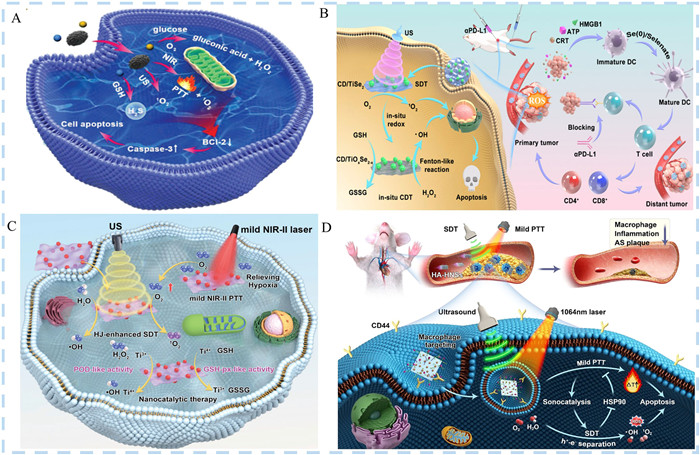
He et al. prepared multifunctional SNs of TiSe2 via a one-step facile oil phase process for in situ CDT-enhanced sono-immunotherapy (Fig. 5B) [45]. The acidic TME triggered the in situ redox transformation of TiSe2, resulting in the formation of TiOxSe2−x, a composite featuring Se(0), selenate, and a Ti3+/Ti4+ redox couple. This unique composition facilitated in situ CDT via a Ti3+-mediated Fenton-like reaction, while simultaneously depleting overexpressed GSH through a Ti4+-mediated mechanism. Furthermore, the in situ redox process generated Se ions, which accelerated the maturation of dendritic cells, thereby activating adaptive immune responses. Additionally, the integration of heterojunctions between CDs and TiSe2 enhanced the efficiency of ROS generation by TiSe2 in a cascaded fashion. This augmentation not only bolstered sonodynamic activity but also magnified the chemodynamic performance. More crucially, the ROS generated through the combined in situ CDT and SDT effectively induced ICD via a synergistic approach that harnessed antitumor immunity, further bolstered by the incorporation of an immune checkpoint blockade.
It is well known that combination therapy has the advantages of safety, high efficiency, and precision, and it is a trend to combine nanocatalytic therapy (NCT) with other therapeutic means (e.g., PTT, PDT, or SDT) for the treatment of tumors [134-136]. Therefore, Yan et al. assembled CDs onto TiCN NSs to make sono-responsive and NIR-Ⅱ-photoresponsive nanozymes (Fig. 5C) [12]. The presence of vacancies in the TiCN lattice structure and the narrow TiCN bandgap led to enhanced segregation of electron-hole pairs while inhibiting their re-combination into complexes. Meanwhile, TiCN was a multifunctional nanozyme with multiple enzyme-like catalytic activities. Under the excitation of Ti3+, TiCN produced POD catalytic activity, which facilitated the production of •OH via the decomposition of H2O2, and with Ti4+ serving as the mediator, TiCN possessed glutathione GSH-px-like catalytic activity, which depleted the overexpressed GSH. The combined effect exhibited a higher level of ROS. Of interest, CDs have excellent NIR-Ⅱ photothermal properties. The mild photothermal effect served to augment the catalytic prowess of the nanozymes, thereby enhancing both the NCT and SDT performance. Meanwhile, the mild PTT did not kill tumor cells directly through high temperature, so it will not cause damage to normal tissues. Experiments have shown that nanozyme has truly achieved the effective combination of NCT, SDT and PTT. The "three-in-one" mechanism completely inhibited the proliferation of tumor cells and cures the tumor.
When it comes to combination therapy, there is no shortage of tumor treatments mediated by combining all three SDT/PDT/PTT [137,138]. Xu et al. self-assembled ordered manganese porphyrin NPs under noncovalent interactions [34]. First, the metalloporphyrin network with a long-range ordered stacking structure provided a pathway for carrier separation and migration and shortened the migration distance, improving the PDT performance. In addition, the 1O2 generation capacity of the photosensitizer was observed to be significantly improved. Au in self-assembled ordered metalloporphyrin NPs (SM) surface was grown in situ to form a plasma SMA heterostructure. Subsequently, the color rendering range of the composite's absorption wavelength was shifted to the near-infrared region, the photon energy was converted to NIR-Ⅱ photoacoustic imaging, and the separation of electron-hole pairs was significantly increased, with both SDT and PTT improved. Finally, plasmonic heterostructured nanocomposites (SMAH) were constructed under the modification of HA. SMAH depleted endogenous GSH, disrupted the redox balance, and reversed the TME, further enhancing the efficacy of SDT and PDT. Animal experiments demonstrated that SMAH was safe and effective with excellent performance during tumor treatment under US and NIR-Ⅱ light irradiation.
SDT has been widely used to combine with other means for diseases such as inflammation and tumors [139]. At the present stage, Cao et al. used Cu sulfide/titanium oxide heterojunction NSs modified with HA and polyethylene glycol (Fig. 5D) [35]. Modified by Cu with high surface energy crystal faces, the electronic properties of TiO2 were improved. Therefore, Cu/TiO2 heterostructure NSs (HNSs) had high electron-hole separation efficiency and excellent sonodynamic properties. In addition, HNSs also exhibited PTT effect, which achieved PTT treatment of deep atherosclerotic plaques. Finally, HA was utilized to modify HNSs, and since HA can selectively bind CD44 receptor, HA-HNSs indirectly achieved the targeting of macrophages within the plaque. Subsequently, it was applied to imaging and drug delivery in atherosclerotic plaques. In vitro experiments showed that the photothermal effect promoted the HA-HNSs-mediated sonodynamic process and increased the ROS yield. SDT cleaved heat shock protein 90 to generate large amounts of ROS, resulting in greater efficacy of mild PTT. SDT and PTT achieved a mutually reinforcing, combined treatment of early atherosclerosis.
Heterojunction NSNs-driven synergistic SDT has emerged as an appealing option due to their enhanced ROS generation efficiency and the potential for combined multimodal treatments. This comprehensive review systematically summarized the research advancements in enhancing SDT through heterojunction NSNs, described the types of heterojunction SNs, preparation methods, characterization methods and the mechanisms underlying their enhancement of SDT. Furthermore, it highlighted the latest research on various synergistic therapies based on heterojunction SNs for the treatment of bacterial infections and tumors. Despite significant progress made, several crucial issues remain to be addressed.
(1) Material consistency and preparation difficulty: Heterojunction SNs are typically composed of different materials precisely combined at the nanoscale, which imposes stringent requirements on material purity and preparation techniques. The quality of the interfaces between materials directly influences their performance, yet maintaining consistency and high-quality interfaces during actual production poses a significant challenge.
(2) Stability and biocompatibility: Similar to other SNs, the stability and biocompatibility of heterojunction SNs within the biological environment are crucial issues. The low degradation of SNs raises concerns about their toxicity. SNs need to maintain the integrity of their structure and function without toxicity to achieve long-term therapeutic effects.
(3) ROS production efficiency: Although the heterojunction structure can enhance ROS production, further improving efficiency to enhance therapeutic effects remains a challenge. This may necessitate further optimization of the composition and structure of the SNs.
(4) TME adaptability: The complexity of the TME, including hypoxia and immune suppression, significantly impacts the therapeutic efficacy of SNs. Therefore, designing SNs that can adapt to and overcome these challenges is crucial for enhancing the effectiveness of SDT.
(5) Application scope limitations: Despite the high potential of heterojunction SNs, their application scope is still limited to fields such as bacterial infection and tumor treatment. Expanding their application scope requires further research and technological breakthroughs.
In summary, sonodynamic synergistic therapy supported by heterojunction NSNs has demonstrated promising therapeutic effects in disease treatment. If the aforementioned issues can be resolved, it will accelerate their clinical application, providing a more advanced non-invasive treatment strategy. Shortly, heterojunction NSNs may further expand from bacterial infection and tumor treatment to other disease treatment areas, making greater contributions to human health.
The authors declare that they have no known competing financial interests or personal relationships that could have appeared to influence the work reported in this paper.
Li Qin: Writing – original draft, Visualization, Conceptualization. Wenjing Wei: Writing – review & editing. Keqing Wang: Writing – review & editing. Xianbao Shi: Supervision, Project administration. Guixia Ling: Supervision, Project administration. Peng Zhang: Supervision, Project administration.
This work was supported by Key Research Project of the Educational Department of Liaoning Province, China (No. JYTZD2023139).
Supplementary material associated with this article can be found, in the online version, at doi:
Y. Li, W. Li, Y. Liu, et al., Acta Biomater. 171 (2023) 543–552.
M. Wu, J. Yong, H. Zhang, et al., Adv. Healthc. Mater. 12 (2023) e2301497. doi: 10.1002/adhm.202301497
Y. Zhang, J. Zhao, L. Zhang, et al., Nano Today 49 (2023) 101798.
J. Chen, Q. Zhou, W. Cao, Adv. Func. Mater. (2024), 2405844. doi: 10.1002/adfm.202405844
W. Qin, Q. Yang, C. Zhu, et al., Small 20 (2024) e2311228. doi: 10.1002/smll.202311228
H. Zhang, W. Zhu, W. Pan, et al., Asian J. Pharm. Sci. 19 (2024) 100954.
X. Zhu, S. Zhang, Y. Cao, et al., Chin. Chem. Lett. 34 (2023) 108234.
Z. Jiang, W. Xiao, Q. Fu, J. Control Release 361 (2023) 547–567. doi: 10.1016/j.jconrel.2023.08.003
G. Li, S. Wu, J. Liu, et al., Adv. Mater. 36 (2024) e2401252. doi: 10.1002/adma.202401252
J. Hu, B. Geng, J. Glowacki, et al., Chem. Eng. J. 446 (2022) 137320.
J. Jadwiszczak, J. Sherman, D. Lynall, et al., ACS Nano 16 (2022) 1639–1648. doi: 10.1021/acsnano.1c10524
L. Yan, Z. Cao, L. Ren, et al., Adv. Healthc. Mater. 13 (2024) e2302190. doi: 10.1002/adhm.202302190
Y. Zhou, L. Yu, C. Dong, et al., Chem. Eng. J. 431 (2022) 134017.
L. Liu, Z. Zhang, S. Gu, et al., J. Colloid. Interface Sci. 676 (2024) 52–60.
S. Wen, W. Zhang, J. Yang, et al., ACS Nano 18 (2024) 23672–23683. doi: 10.1021/acsnano.4c08236
S. Wu, G. Li, W. Ouyang, et al., Biomater. Res. 28 (2024) 0014.
L. Cai, J. Du, F. Han, et al., ACS Nano 17 (2023) 7901–7910. doi: 10.1021/acsnano.3c01856
M.Y. Zhang, J.K. Li, R. Wang, et al., Adv. Sci. 8 (2021) e2101884. doi: 10.1002/advs.202101884
J. Zhang, Y. Zhang, L. Li, et al., J. Mater. Chem. A 11 (2022) 434–446.
W. Liu, Y. Dong, J. Liu, et al., Chem. Eng. J. 451 (2023) 138666.
B. Chen, J. Xu, S. Shi, et al., ACS Appl. Mater. Interfaces 16 (2024) 28917–28927. doi: 10.1021/acsami.4c05896
J. Liu, S. Dong, S. Gai, et al., ACS Nano 18 (2024) 23579–23598. doi: 10.1021/acsnano.4c07904
D. Trachootham, J. Alexandre, P. Huang, Nat. Rev. Drug Discov. 8 (2009) 579–591. doi: 10.1038/nrd2803
F. Du, L. Liu, Z. Wu, et al., Adv. Mater. 33 (2021) e2101095. doi: 10.1002/adma.202101095
F. Gong, L. Cheng, N. Yang, et al., Nat. Commun. 11 (2020) 3712.
B. Geng, S. Xu, L. Shen, et al., Carbon 179 (2021) 493–504.
J. Hu, L. Yan, Z. Cao, et al., Adv. Sci. (2024), 2407196. doi: 10.1002/advs.202407196
W. Gao, J. Zhang, Y. Chang, et al., Adv. Func. Mater. (2024), 2408125. doi: 10.1002/adfm.202408125
J. Xia, C. Hu, Y. Ji, et al., ACS Nano 17 (2023) 21134–21152. doi: 10.1021/acsnano.3c04903
X. Zhang, X. Chen, P. Zhang, et al., Nano Res. 16 (2023) 7148–7163. doi: 10.1007/s12274-022-5313-3
Y. Li, L. Chen, Y. Chen, et al., Biomaterials 315 (2025) 122921.
Y. Fu, Y. He, X. Wei, et al., ACS Nano 18 (2024) 28793–28809. doi: 10.1021/acsnano.4c08468
Y. Huang, C. Chen, H. Tan, et al., Small 20 (2024) e2401147. doi: 10.1002/smll.202401147
P. Xu, C. Wen, C. Gao, et al., ACS Nano 18 (2024) 713–727. doi: 10.1021/acsnano.3c09011
Z. Cao, G. Yuan, L. Zeng, et al., ACS Nano 16 (2022) 10608–10622. doi: 10.1021/acsnano.2c02177
A. Li, T. Zhang, X. Zhang, et al., ACS Nano (2024) 5344–5357. doi: 10.1021/acsnano.3c09316
C. Yang, M. Chang, M. Yuan, et al., Small 17 (2021) e2100961. doi: 10.1002/smll.202100961
Y. Shao, B. Liu, Z. Di, et al., J. Am. Chem. Soc. 142 (2020) 3939–3946. doi: 10.1021/jacs.9b12788
G. Chen, J. Du, L. Gu, et al., Chem. Eng. J. 482 (2024) 148953.
J. Du, J. Ping, Q. Wang, et al., ACS Appl. Nano Mater. 7 (2024) 8151–8163. doi: 10.1021/acsanm.4c00746
X.D. Liu, B. Chen, G.G. Wang, et al., Adv. Func. Mater. 31 (2021) 2104424.
Z. Yang, Y. Luo, H. Yu, et al., Adv. Mater. 34 (2022) e2108908. doi: 10.1002/adma.202108908
M. Ou, C. Lin, Y. Wang, et al., J. Control Release 345 (2022) 755–769.
X. Song, X. Zhou, Y. Pan, et al., Adv. Func. Mater. 33 (2023) 2306734.
X. He, J. Cai, J. Hu, et al., Small Struct. 5 (2024) 2300558.
L. Zhou, H. Zheng, Z. Liu, et al., ACS Nano 15 (2021) 2468–2480. doi: 10.1021/acsnano.0c06287
Y. Li, X. Zhang, S. Liu, et al., Chin. Chem. Lett. 36 (2024) 110501. doi: 10.1088/1674-4926/24080031
X. Meng, S. Sun, C. Gong, et al., ACS Nano 17 (2023) 1174–1186. doi: 10.1021/acsnano.2c08687
J.R. Lex, R. Koucheki, N.A. Stavropoulos, et al., Acta Biomater. 140 (2022) 136–148. doi: 10.1016/j.actbio.2021.11.045
M. Liang, L. Shang, Y. Yu, et al., Acta Biomater. 158 (2023) 811–826. doi: 10.1016/j.actbio.2022.12.041
Z. Jia, X. Lv, Y. Hou, et al., Bioact. Mater. 6 (2021) 2676–2687. doi: 10.1016/j.bioactmat.2021.01.033
M.K. Sarangi, L. Patel, G. Rath, et al., Chin. Chem. Lett. 35 (2024) 109381.
Q. Bai, J. Zhang, Y. Yu, et al., ACS Appl. Mater. Interfaces 14 (2022) 26455–26468. doi: 10.1021/acsami.2c05114
Z. Lu, R. Zhao, Y. Li, et al., Bioact. Mater. 31 (2024) 509–524.
C. Geng, S. He, S. Yu, et al., Adv. Mater. 36 (2024) e2310599. doi: 10.1002/adma.202310599
L. Bernard, A. Dinh, I. Ghout, et al., Lancet 385 (2015) 875–882. doi: 10.1016/S0140-6736(14)61233-2
C. Hobson, A.N. Chan, G.D. Wright, Chem. Rev. 121 (2021) 3464–3494. doi: 10.1021/acs.chemrev.0c01214
P.D. Tamma, E. Avdic, D.X. Li, et al., JAMA Intern. Med. 177 (2017) 1308–1315. doi: 10.1001/jamainternmed.2017.1938
H. Chen, L. Liu, A. Ma, et al., Biomaterials 269 (2021) 120639. doi: 10.1016/j.biomaterials.2020.120639
Y. Qiao, X. Liu, B. Li, et al., Nat. Commun. 11 (2020) 4446. doi: 10.1038/s41467-020-18268-0
S. Son, J.H. Kim, X. Wang, et al., Chem. Soc. Rev. 49 (2020) 3244–3261. doi: 10.1039/c9cs00648f
Y. Xiang, J. Lu, C. Mao, et al., Sci. Adv. 9 (2023) eadf0854. doi: 10.1126/sciadv.adf0854
Y. Yu, Y. Zeng, Q. Ouyang, et al., ACS Nano 17 (2023) 21018–21029. doi: 10.1021/acsnano.3c03858
T. Ding, Y. Li, F. Liu, et al., Adv. Func. Mater. 34 (2024) 2401795.
M. Zhang, D. Yang, C. Dong, et al., ACS Nano 16 (2022) 9938–9952. doi: 10.1021/acsnano.2c04630
Y. Wu, X. Li, Q. Yang, et al., Chem. Eng. J. 390 (2020) 124519.
W. Yang, G. Ma, Y. Fu, et al., Chem. Eng. J. 429 (2022) 132381. doi: 10.1016/j.cej.2021.132381
H. Wang, N. Mu, Y. He, et al., Theranostics 13 (2023) 1669–1683. doi: 10.7150/thno.81511
Q.Q. Yan, D.X. Wu, S.Q. Chu, et al., Nat. Commun. 10 (2019) 4977. doi: 10.1038/s41467-019-12851-w
J. Li, Z. Li, X. Liu, et al., Nat. Commun. 12 (2021) 1224.
Y. Liu, J. Ding, F. Li, et al., Adv. Mater. 35 (2023) e2207114. doi: 10.1002/adma.202207114
Y. Wang, X. Wen, Y. Jia, et al., Nat. Commun. 11 (2020) 1328. doi: 10.1049/el.2020.2265
C. Wang, J. Lei, C. Mao, et al., Adv. Func. Mater. 33 (2023) 2306493.
G. Li, S. Wang, D. Deng, et al., ACS Nano 14 (2020) 1586–1599. doi: 10.1021/acsnano.9b06689
X. Cao, M. Li, Q. Liu, et al., Small 19 (2023) e2303195. doi: 10.1002/smll.202303195
Z. Yan, Z. Liu, H. Zhang, et al., Acta Biomater. 174 (2024) 1–25. doi: 10.56028/ijbm.2.4.1.2024
H. Cai, X. Wu, L. Jiang, et al., Chin. Chem. Lett. 35 (2024) 108946.
H. Tian, J. Lin, F. Zhu, et al., Chin. Chem. Lett. 34 (2023) 107577.
M. Pan, D. Hu, L. Yuan, et al., Acta Pharm. Sin. B 13 (2023) 2926–2954.
Y. Wu, M. Yuan, J. Song, et al., ACS Nano 13 (2019) 8505–8511. doi: 10.1021/acsnano.9b05124
M. Yuan, S. Liang, L. Yang, et al., Adv. Mater. 35 (2023) e2209589. doi: 10.1002/adma.202209589
M. Yuan, L. Yang, Z. Yang, et al., Adv. Sci. 11 (2024) e2308546. doi: 10.1002/advs.202308546
Y. Cheng, X. Kong, Y. Chang, et al., Adv. Mater. 32 (2020) e1908109. doi: 10.1002/adma.201908109
L. Chen, Z. Mao, Y. Wang, et al., Sci. Adv. 8 (2022) eabo7372. doi: 10.1126/sciadv.abo7372
X. Wang, X. Zhong, L. Bai, et al., J. Am. Chem. Soc. 142 (2020) 6527–6537. doi: 10.1021/jacs.9b10228
Y. Zhao, B. Yuan, L. Yan, et al., Adv. Sci. 11 (2024) e2307029. doi: 10.1002/advs.202307029
Y. Zhao, T. Huang, S. Wang, et al., J. Colloid. Interface Sci. 640 (2023) 839–850.
S. Liang, B. Liu, X. Xiao, et al., Adv. Mater. 33 (2021) e2101467. doi: 10.1002/advs.202101467
L. Wang, W. Song, S. Choi, et al., ACS Appl. Mater. Interfaces 15 (2023) 2552–2563. doi: 10.1021/acsami.2c15327
L. Jia, Y. Wang, T. Hu, et al., Chem. Eng. J. 469 (2023) 143969.
H. Hu, J. Zhao, K. Ma, et al., J. Controll Release 359 (2023) 188–205.
X. Wang, X. Wang, Q. Yue, et al., Nano Today 39 (2021) 101170.
Y. Wei, P. Zhang, R.A. Soomro, Q. Zhu, B. Xu, Adv. Mater. 33 (2021) e2103148. doi: 10.1002/adma.202103148
H. Huang, C. Dong, W. Feng, et al., Adv. Drug Deliv. Rev. 184 (2022) 114178. doi: 10.1016/j.addr.2022.114178
J. Xu, L. Chen, S. Ding, et al., Nano Today 48 (2023) 101750.
P.S. Meltzer, L.J. Helman, N. Engl. J. Med. 385 (2021) 2066–2076. doi: 10.1056/nejmra2103423
Z. Lv, Y. Wu, J. Lin, et al., Adv. Funct. Mater. 34 (2024) 2312032. doi: 10.1002/adfm.202312032
J. Heo, N. Her, M. Jang, et al. Crit. Rev. Env. Sci. Tec. 53 (2023) 987–1008. doi: 10.1080/10643389.2022.2101857
J. Xu, X. Wang, Y. Liu, et al., Acta Biomater. 175 (2024) 307–316.
K. Song, J. Du, X. Wang, et al., Adv. Healthc. Mater. 11 (2022) e2102503. doi: 10.1002/adhm.202102503
Z. Liu, S. Liu, B. Liu, et al., Chin. Chem. Lett. 35 (2024) 109626.
Q. Zhao, Y. Zhao, S. Zhong, et al., Chin. Chem. Lett. 35 (2024) 109644.
B. Zhang, Z. Zengcai, W. Lin, et al., Chem. Eng. J. 465 (2023) 142904.
C. Feng, L. Wang, D. Zhang, et al., J. Colloid. Interface Sci. 665 (2024) 681–692.
Y. Zhao, S. Wang, Y. Ding, et al., ACS Nano 16 (2022) 9304–9316. doi: 10.1021/acsnano.2c01968
B. Geng, S. Zhang, X. Yang, et al., Chem. Eng. J. 435 (2022) 134777.
S. Liu, L. Fang, H. Ding, et al., ACS Nano 16 (2022) 20805–20819. doi: 10.1021/acsnano.2c08047
H. Zheng, N. Yin, K. Lv, et al., J. Mater. Chem. B 12 (2024) 4162–4171. doi: 10.1039/d4tb00084f
W. Liu, R. Shao, L. Guo, et al., Adv. Sci. 11 (2024) 2304046.
B. Ding, P. Zheng, P. Ma, et al., Adv. Mater. 32 (2020) e1905823. doi: 10.1002/adma.201905823
X. Lin, S. Liu, X. Zhang, et al., Angew Chem. Int. Ed. Engl. 59 (2020) 1682–1688. doi: 10.1002/anie.201912768
Y. Li, W. Chen, Y. Kang, et al., Nat. Commun. 14 (2023) 6973. doi: 10.1038/s41467-023-42509-7
S. Cheng, T. Zhou, Y. Luo, et al., J. Nanobiotechnology 22 (2024) 408.
J. An, Y.G. Hu, K. Cheng, et al., Biomaterials 234 (2020) 119761. doi: 10.1016/j.biomaterials.2020.119761
K. Yang, L. Yue, G. Yu, et al., Biomaterials 275 (2021) 120822. doi: 10.1016/j.biomaterials.2021.120822
K. Yang, Z. Yang, G. Yu, et al., Adv. Mater. 34 (2022) e2107434. doi: 10.1002/adma.202107434
Y. Chen, T. Zou, G. Xin, et al., Adv. Mater. 36 (2024) e2307929. doi: 10.1002/adma.202307929
G. Xu, H. Zhang, J. Wei, et al., ACS Nano 12 (2018) 5333–5340. doi: 10.1021/acsnano.8b00110
M. He, H. Yu, Y. Zhao, et al., Small 19 (2023) e2300244. doi: 10.1002/smll.202300244
Y. Liao, D. Wang, S. Zhu, et al., Nano Today 44 (2022) 101510. doi: 10.1016/j.nantod.2022.101510
M. Zhang, L. Dong, D. Li, et al., Adv. Func. Mater. 33 (2023) 2303451.
L. Zhu, G. Chen, Q. Wang, et al., J. Colloid. Interface Sci. 662 (2024) 914–927.
J. Wu, G.R. Williams, S. Niu, et al., Adv. Sci. 6 (2019) 1802001. doi: 10.1002/advs.201802001
X. Meng, Y. Yi, Y. Meng, et al., ACS Nano 16 (2022) 4217–4227. doi: 10.1021/acsnano.1c10173
T. Zhang, Q. Zheng, C. Xie, et al., ACS Appl. Mater. Interfaces 15 (2023) 4883–4894. doi: 10.1021/acsami.2c18095
Y. Wei, S. Han, D.A. Walker, et al., Chem. Sci. 3 (2012) 1090–1094. doi: 10.1039/c2sc00673a
T. Zhang, Q. Zheng, Y. Fu, et al., J. Nanobiotechnology 19 (2021) 358.
Y. Shi, J. Zhao, H. Li, et al., Adv. Healthc. Mater. 11 (2022) e2200908. doi: 10.1002/adhm.202200908
Y. Li, G. He, L.H. Fu, et al., ACS Nano 16 (2022) 17298–17312. doi: 10.1021/acsnano.2c08098
Y. Chen, J. Zhu, J. Ding, et al., Chin. Chem. Lett. 35 (2024) 108706.
T. Hu, L. Jia, H. Li, et al., Adv. Healthc. Mater. 13 (2024) e2303147. doi: 10.1002/adhm.202303147
J. Tan, B. Ding, P. Zheng, et al., Small 18 (2022) e2202462. doi: 10.1002/smll.202202462
Y. Ma, Y. Cao, M. Zu, et al., Adv. Func. Mater. 34 (2024) 2402164.
L. Fan, X. Xu, C. Zhu, et al., ACS Appl. Mater. Interfaces 10 (2018) 4502–4511. doi: 10.1021/acsami.7b17916
M. Chang, Z. Hou, M. Wang, et al., Angew Chem. Int. Ed. 60 (2021) 12971–12979. doi: 10.1002/anie.202101924
M. Ge, H. Guo, M. Zong, et al., Angew Chem. Int. Ed. 62 (2023) e202215795. doi: 10.1002/anie.202215795
C. Zhou, L. Zhang, T. Sun, et al., Adv. Mater. 33 (2021) e2006532. doi: 10.1002/adma.202006532
Y. Zhu, Z. Wang, R. Zhao, et al., ACS Nano 16 (2022) 3105–3118. doi: 10.1021/acsnano.1c10732
M. Qian, L. Gong, T. Jia, et al., ACS Appl. Nano Mater. 7 (2024) 21072–21082. doi: 10.1021/acsanm.4c04184

Figure 2 Schematic illustration of types of heterojunction SNs. (A) Types of heterojunction SNs classified by the conductivity of the materials. (B) Types of heterojunction SNs classified by the materials types.

Figure 3 (A) Schematic illustration of MIL@Ag-PEG heterojunction for antibacterial application. Reproduced with permission [48]. Copyright 2023, American Chemical Society. (B) Schematic illustration of PCN-222-BTO heterojunction in osteomyelitis treatment. Reproduced with permission [63]. Copyright 2023, American Chemical Society.

Figure 4 (A) The mechanism of Schottky heterojunctions of TiO2-Ru-PEG for SDT. Reproduced with permission [9]. Copyright 2024, Wiley-Blackwell. (B) Schematic illustration and S-scheme transfer mechanism of BiOBr@Bi2S3 heterojunction for SDT and gas therapy. Reproduced with permission [82]. Copyright 2024, Wiley-VCH Verlag.

Figure 5 (A) Bi/BiVO4 Schottky heterojunction-loaded MNs for SDT/gas/photothermal therapy. Reproduced with permission [131]. Copyright 2024, John Wiley and Sons Ltd. (B) Schematic illustration of CD/TiSe2 heterojunctions for in situ CDT-enhanced sono-immunotherapy. Reproduced with permission [45]. Copyright 2024, Wiley-VCH. (C) CD/TiCN nanozymes heterojunction for "three-in-one" SDT/nanocatalysis/photothermal therapy. Reproduced with permission [12]. Copyright 2024, John Wiley and Sons Ltd. (D) CuS/TiO2 heterojunction NSs for atherosclerosis treatment. Reproduced with permission [35]. Copyright 2022, American Chemical Society.

 扫一扫看文章

扫一扫关注我们
 DownLoad:

 下载:
下载:
 下载:
下载: