Figure 1.
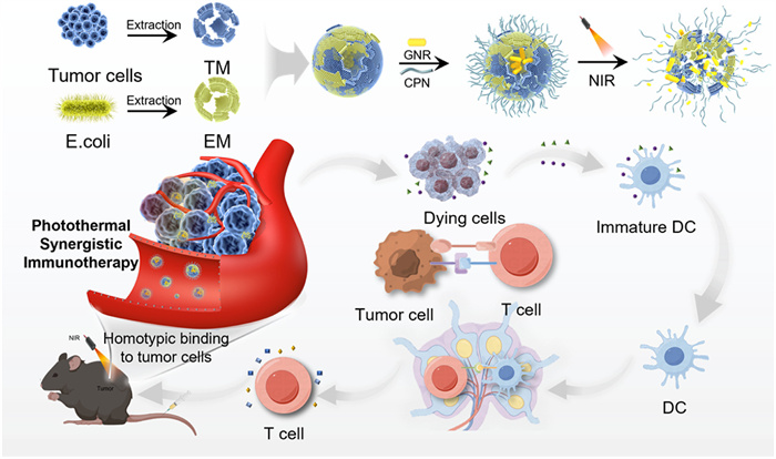
Schematic of the HM-NP combined with tumor targeting, controlled release, and photothermal functionalities for photothermal synergistic immunotherapy in vivo.
Intratumoral photo-controlled antigens burst release for synergistic immunotherapy by bio-membrane and organic membrane coated dual-functional nanoparticles
Chenkai Yang , Xiaoling Pan , Weiguang Zhao , Zhiwen Qiu , Lei He , Cong Wu , Ang Li , Zhengnan Huang , Yilin Yan , Shengzhou Li , Zhuofan Nan , Xiangqian Cao , Bing Shen , Wei Li

Nanomedicine, a submicron drug delivery system, has garnered significant attention in oncology research due to its ability to enhance tumor-specific drug delivery while minimizing systemic side effects [1]. Nanoparticle-based photothermal therapy (PTT) employs photothermal nanomaterials to transform light energy into heat, effectively eliminating tumors through direct heat-induced cell destruction [2]. Furthermore, PTT can induce a favorable tumor microenvironment conducive to immune response [3]. The release of tumor-associated antigens from heat-damaged tumor cells activates dendritic cells and attracts cytotoxic T lymphocytes (CTLs) [4-6]. Nevertheless, immune-mediated tumor eradication from PTT is often restricted, highlighting the importance of accurately identifying and processing released tumor antigens by the immune system for optimal immune stimulation following PTT. Additionally, co-administration of immunostimulatory agents can augment the antitumor immune response by innate immunity [7-9]. Recent research has presented compelling evidence endorsing the efficacy of photothermal combined immunotherapy for suppression of tumor growth [10,11].
The use of cell membranes as carriers for nanomaterials has attracted attention, offering functional modification approach for medication transport, immune regulation, and vaccine inoculation [12-16]. Recently, a technique involving the formulation of tumor cell membrane (TM) and Escherichia coli membrane (EM) through membrane fusion has been proposed to enhance antitumor effects by enhancing tumor targeting and increasing the immunogenicity of the two components while minimizing side effects [17-19]. Cancer cell membranes grant nanoparticles immune escape and targeted homing, enhancing the accuracy and effectiveness of treatment [20-23]. Moreover, isolated tumor membranes obtained from primary tumors are recognized for preserving membrane antigens, such as neoantigens, offering potential for personalized immunotherapy [24-26]. However, membrane antigens are frequently characterized by low immunogenicity, which limits the elicitation of an effective antitumor immune response [27]. Nanoparticles have the capacity to encapsulate a variety of antigens and adjuvants, enabling the co-delivery of crucial components to the same antigen-presenting cell (APC) to optimize the immune response [28]. Bacterial outer membrane vesicles (OMVs), containing diverse pathogen-associated molecular patterns (PAMPs), are recognized immunostimulatory agents [29-32], playing a crucial role in regulating antitumor immune response [33-37]. Nevertheless, formulations derived from bacteria may result in significant adverse effects, including cytokine storm, which impede their clinical utility [38]. Therefore, membrane-based biomimetic nanoplatforms, combining materials from different cells or polymers, show potential in enhancing tumor specificity and antitumor effects while minimizing side effects [39,40].
The precise release of therapeutic agents at the tumor site is crucial for enhancing anticancer efficacy while minimizing adverse effects. A self-adaptive drug delivery system typically comprises nanomaterials and encapsulated therapeutics, designed to transport agents to the tumor site and regulate drug release in response to endogenous or exogenous stimuli [41]. For instance, biologically responsive poly(amino acids) can facilitate the spatiotemporal selective delivery of various mechanistic drugs within tumor cells [42]. Poly(N-isopropylacrylamide) (PNIPAM) temperature sensitivity has been exploited to develop nanocarriers with exceptional drug loading capacity and temperature-controlled release characteristics [43-45]. PNIPAM demonstrates the ability to dynamically respond to fluctuations in the local microenvironment temperature, thereby inducing alterations in the carrier's structural conformation and cellular interactions, ultimately facilitating targeted delivery and controlled release [46-48]. And the dynamic response of PNIPAM to temperature fluctuations enables tailored drug release profiles, optimizing therapeutic efficacy. PNIPAM derivatization with natural polymers such as carboxymethyl chitosan (CMC) [49] and O-carboxymethyl chitosan (OCMC) [50] enhances biodegradability, biocompatibility, and controlled drug release capabilities [51]. Furthermore, the presence of chitosan can inhibit and evade immune clearance [52].
In this study, we synthesized dual-functional hybrid membrane nanoparticles (HM-NPs) incorporating gold nanorods (GNRs) and a thermally responsive polymer shell, demonstrating temperature and light dual-sensitive properties (Scheme 1). The shell, composed of HM and organic membrane OCMC-graft-PNIPAM (chitosan-graft-poly(N-isopropylacrylamide) (CPN)), provides homologous targeting, immune stimulation, and controlled release properties. Under in situ near-infrared (NIR) light stimulation, heat generated by the GNRs induces shrinkage of the CPN, promoting the disintegration of shell and the burst release of HM, enhancing local concentration of antigens at the tumor site. This photoimmunotherapy holds promise for safer and more effective anti-tumor efficacy, with reduced systemic toxicity, contributing to biomedical research and medicine.
In this research, GNR was fabricated using a previously established method [53]. TM and EM were isolated and utilized for further experimentation. Carboxymethyl CPN was synthesized through a polymerization reaction (Figs. S1 and S2 in Supporting information). Subsequently, HM-NP were successfully prepared by water bath sonication and a 400-nm cutoff extruder (Fig. 1). Dynamic light scattering (DLS) analysis revealed that the hydrodynamic size of HM was around 210 nm with a minimal polydispersity index (PDI) (Fig. 1C). Furthermore, the ζ potential of EM, TM, and HM samples was found to be negative (Fig. 1D). The dimension and surface charge of HM fell within the range of EM and TM, indicating successful combination of the two membranes into HM. Western blot analysis further validated the presence of proteins from the original EM and TM in HM, including the characteristic outer membrane protein-A (OmpA) band from EM and the ATPase band from TM (Fig. S3 in Supporting information). Subsequently, HM-CPN hybrids were prepared by repeated fusion of HM and CPN at mass ratios of 1:2, 1:1, 2:1, and 3:1 using water bath sonication and a 400-nm cutoff extruder. To identify the most effective ratio for inducing the disintegration of the shell and the release of HM, the hydrodynamic size and PDI of the different mass ratios of HM-CPN were measured. Notably, the group with a mass ratio of 3:1 between HM and CPN exhibited minimal changes in particle size and good dispersion between 27 ℃ and 37 ℃. However, a sudden increase in particle size and PDI at 47 ℃ indicated sufficient heat-induced rupture of the thermo-responsive polymer shell (Figs. 1A and B). Therefore, HM-CPN with a mass ratio of 3:1 (HM:CPN) was selected for subsequent experiments.
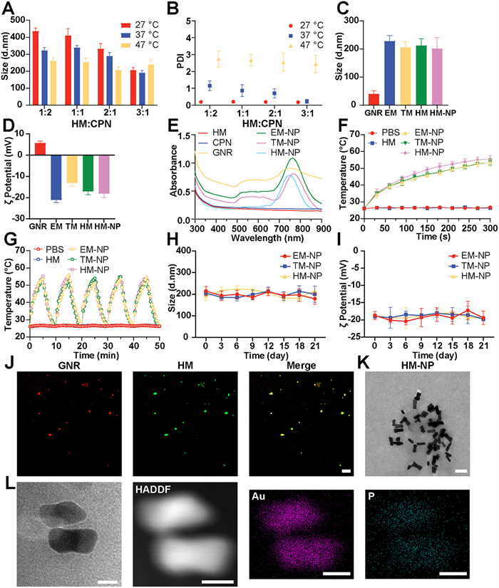
The ultraviolet-visible (UV–vis) absorption spectra demonstrated a clear absorption peak at 780 nm for GNR (Fig. 1E). The marginal displacement of the absorption peak towards longer wavelengths (Fig. 1E) and the analogous photothermal heating profile (Fig. 1F) for EM-NP, TM-NP, and HM-NP indicated the successful encapsulation of GNR, with a maximum encapsulation rate of approximately 70.8%. Following exposure to 780 nm laser (1.5 W/cm2), the photothermal conversion performance of HM-NP was investigated, and the HM-NP group swiftly rose to 55.5 ℃ after 5 min, demonstrating a superior photothermal performance of HM-NP (Fig. 1F and Fig. S4 in Supporting information). The photothermal conversion efficiency (η) of HM-NP was 28.0% (Fig. S5 in Supporting information). To assess thermal stability during laser exposure, sequential irradiation-cooling experiments were conducted (Fig. 1G). HM-NP sustained a consistent temperature over five cycles of irradiation, demonstrating the remarkable photothermal stability of HM-NP.
To confirm the fusion between GNR and HM, Cy5-labeled GNR and 3,3′-dioctadecyloxacarbocyanine perchlorate (DIO)-modified HM was sonicated and extruded repeatedly. The fusion was confirmed through confocal laser scanning microscopy (CLSM) imaging (Fig. 1J). To assess the stability of HM-NP, the nanoparticles were stored in phosphate buffered saline (PBS), and their hydrodynamic sizes and PDI were measured at various time points using DLS. The results, depicted in Figs. 1H and I, demonstrate the excellent stability of HM-NP over time. Additionally, the morphological analysis of HM-NP was conducted by transmission electron microscopy (TEM), revealing HM-NP approximately 250 nm in size, enveloped by a thin membrane (Figs. 1K and L).
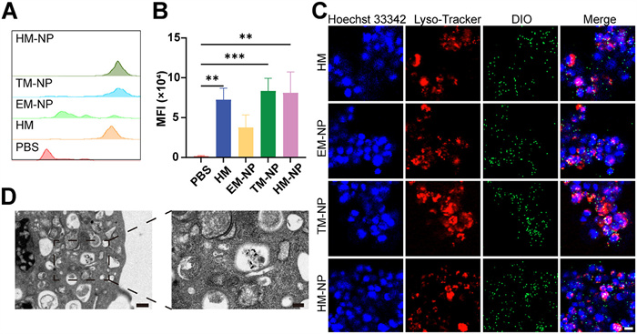
The internalization of HM-NP was first assessed using flow cytometry. The absorbance of the HM-NP group notably surpassed that of the other groups, indicating significantly enhanced uptake efficiency (Figs. 2A and B). Subsequently, colocalization analysis was further performed using CLSM (Fig. 2C). Samples labeled with DIO were co-incubated with MB49 cells, followed by lysosomal staining. The presence of both red and green fluorescence conclusively confirmed the successful internalization of HM-NP into the cells. The endocytic uptake of HM-NP was further illustrated using TEM in Fig. 2D, revealing that HM-NP was internalized and localized in lysosomes. This uptake pattern may potentially result from the targeting capability conferred by the tumor cell membrane [54].
The biocompatibility of HM, EM-NP, TM-NP, and HM-NP was then assessed using a cell counting kit-8 (CCK8) assay on MB49 tumor cells (Figs. 3A–D). Cell viability exceeded 80% after 24 h of incubation without irradiation at a dosage of 108 µg/mL. Nevertheless, upon laser irradiation, notable cell damage was detected, leading to around 50% cell viability at a dosage of 54 µg/mL of HM-NP. Notably, HM-NP exhibited enhanced phototoxicity compared to other groups at 1 equiv. concentration, potentially attributed to its superior cellular internalization or enhanced photothermal properties. Moreover, we evaluated the cytotoxic effects of HM-NP on cancer cells by cell dual staining with calcein AM and propidium iodide (PI), respectively (Fig. 3E). No significant cytotoxicity was observed in the PBS, HM, EM-NP, TM-NP, HM-NP, PBS/L, or HM/L groups. However, a substantial quantity of dead cells were observed in HM-NP/L group. The effectiveness of photothermal cell destruction was slightly diminished in the EM-NP/L and TM-NP/L groups, likely as a result of insufficient cellular internalization or suboptimal photothermal performance.
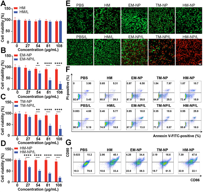
To further elucidate the cellular response to different therapies, flow cytometry analysis was performed (Fig. 3F). In the case of treatment with EM-NP/L, a late apoptotic percentage of 18.8% was observed. The late apoptotic rate increased to 19.6% upon treatment with TM-NP/L. Furthermore, administration of HM-NP/L further augmented the late apoptotic population, leading to a late apoptotic population of 25.9% at 1 equiv. concentration. Therefore, the findings suggested that HM-NP demonstrated promise in thermal ablation due to its advantageous targeting capabilities and photothermal characteristics.
To assess the immune activation induced by HM-NP, we conducted flow cytometry analysis to evaluate the maturation of dendritic cells (DCs). Bone marrow-derived dendritic cells (BMDCs) were cultured with various formulations in vitro for 24 h, followed by specific staining for CD11c (a DC marker), CD80, and CD86 (markers of mature DCs) using fluorescent antibodies. Subsequently, cellular fluorescence intensity was quantified by flow cytometry (Fig. 3G). The results demonstrated a significant increase in the population of mature BMDCs in the HM and HM-NP groups compared to the EM-NP and TM-NP groups. These findings indicated that HM-NP enhanced DC maturation, highlighting its potential for antitumor immunotherapy.
Prior to in vivo applications, it is essential to consider the biosecurity aspects. To assess potential adverse effects, a series of assessments was conducted, including hemolysis tests, blood biochemistry analysis, and blood routine assays. The hemolysis assay indicated that HM-NP exhibited minimal hemolysis, demonstrating excellent stability during blood circulation (Figs. 4A and B). Subsequently, blood samples were collected post intravenous administration of PBS, HM, EM-NP, TM-NP, and HM-NP for blood biochemistry and blood routine examinations to assess systemic toxicity. As depicted in Figs. 4C and D, levels of liver function-related enzymes, namely aspartate aminotransferase (AST) and alanine transaminase (ALT), remained within the normal range. The biomarkers of kidney function, such as blood urea nitrogen (BUN) and creatinine (CREA), exhibited stable levels (Figs. 4E and F), with minimal discernible distinctions observed between the HM-NP and PBS groups. Moreover, the HM group displayed a significantly reduced toxicity in comparison to the EM group (Fig. S6 in Supporting information). Additionally, the findings from the blood routine assay demonstrated that the levels of main blood cells remained relatively stable across all five groups, indicating no evident systemic toxicity of HM-NP (Fig. 4G).
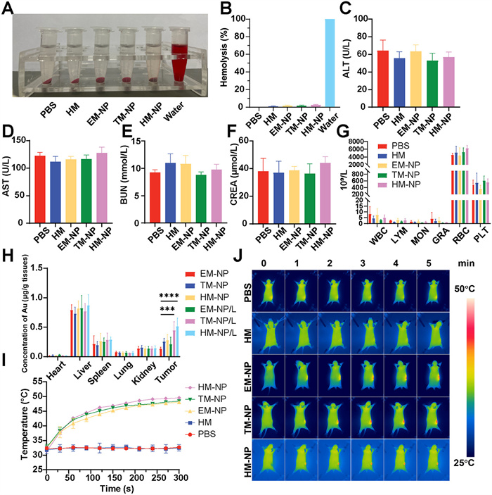
The biodistribution of nanomaterials in key organs was assessed using inductively coupled plasma mass spectrometry (ICP-MS) 24 h post-injection. The HM-NP/L group displayed a notably elevated gold content of tumors (0.51 µg/g) in comparison to the HM-NP group (0.28 µg/g) (Fig. 4H). The enhanced tumor retention in HM-NP/L may be attributed to the localized hyperthermia and release of tumor-targeted cancer cell membranes induced by NIR laser irradiation.
Following this, an in vivo investigation into the photothermal heating performance of HM-NPs was carried out. Animal welfare and experimental procedures were reviewed and approved by the Shanghai General Hospital Medical Ethics Committee (No. 2020SQ159–3). Infrared thermography was utilized to track the immediate temperature alterations in mice after intravenous administration of HM-NP. All groups exhibited a notable rise in local tumor temperature, with the exception of PBS and HM groups (Figs. 4I and J). Remarkably, the HM-NP group achieved a remarkable temperature increase of 49.57 ℃ in 5 min, showcasing its outstanding homologous homing capability and superior photothermal property, suggesting a promising application of HM-NP.
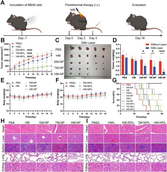
Considering the effective antitumor properties of HM-NP on cancer cells, the in vivo tumor-suppressing efficacy of the samples was assessed. C57 mice were chosen to establish MB49 tumor-carrying models, randomly divided into 10 groups. As shown in Fig. 5A, tumor dimensions and body weights were measured every other day throughout the course. The tumors exhibited rapid growth in all groups when not exposed to NIR irradiation (Fig. 5B; Figs. S7 and S8 in Supporting information). However, in the HM-NP/L group, tumor growth was efficiently suppressed, and even entirely eradicated with the assistance of NIR irradiation, demonstrating the positive treatment outcome of HM-NP. The images of mouse tumors taken 18 days post-treatment provided clear visual evidence of the varying therapeutic effects observed across the groups (Fig. 5C and Fig. S7). Notably, the HM-NP/L group demonstrated a substantial decrease in tumor volume by 95.43% and tumor mass by 85.22%, showcasing a remarkable inhibitory effect on tumor growth (Fig. 5D and Fig. S9 in Supporting information). Additionally, while the EM-NP/L and TM-NP/L groups exhibited some degree of tumor volume inhibition, their efficacy was inferior to that of the HM-NP/L group, indicating that the synergistic PTT and immunotherapy effect induced by HM-NP under NIR irradiation play a predominant role. Moreover, there was no noticeable decrease in body weight recorded across all the groups, demonstrating the excellent biocompatibility of HM-NP (Figs. 5E and F). The survival rates of tumor-bearing mice were assessed over a span of 60 days. There was a notable decline in the mouse population in the PBS, PBS/L, and HM groups before the 45th day, while the survival rate in HM-NP/L group was the highest (Fig. 5G). These observations underscore the effective tumor suppression achieved through HM-NP-mediated photoimmunotherapy. Additionally, histological sections stained with hematoxylin and eosin (H&E) were used to analyze the alterations in tumor tissue and major organs. As depicted in Figs. 5H and I, the H&E staining of major organs following various interventions demonstrated minimal tissue damage caused by HM-NP. The terminal-deoxynucleotidyl transferase-mediated nick end labeling (TUNEL) and H&E staining of tumors confirmed the significant pathological changes and excellent antitumor efficacy of HM-NP/L (Figs. 6A and B).
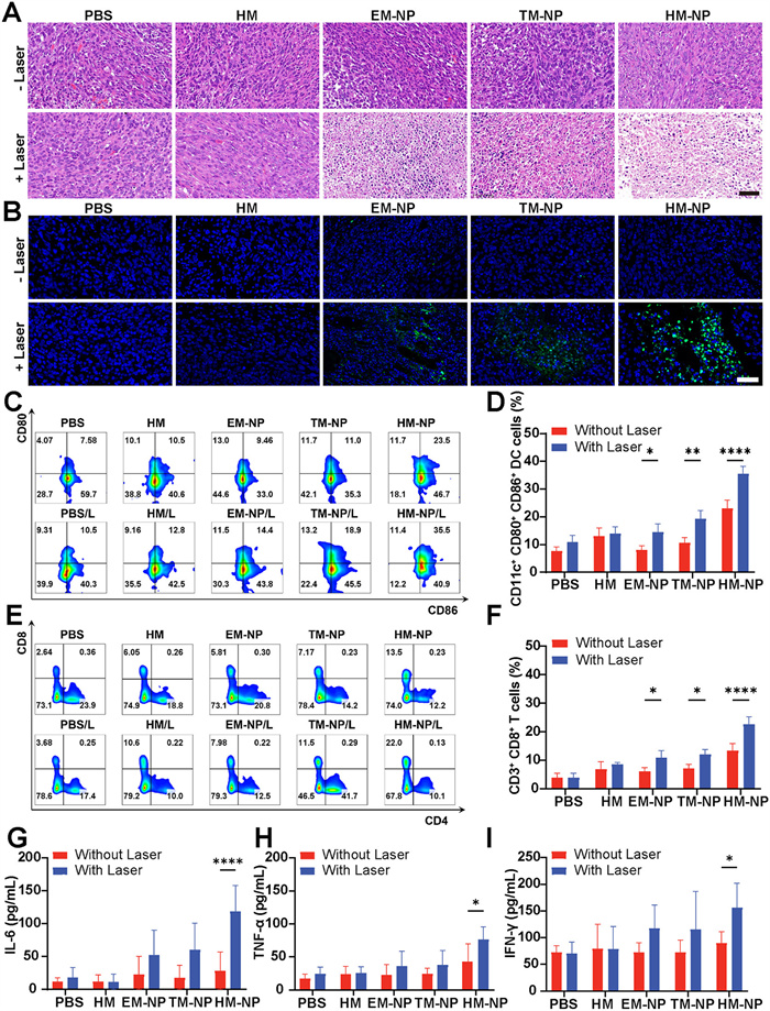
Following this, the immune activation at tumor sites was evaluated. As shown in Figs. 6C and D, HM-NP/L induced the highest increase in mature DCs (CD80+CD86+) compared to PBS. Percentages of mature DCs after EM-NP, EM-NP/L, TM-NP, TM-NP/L, and HM-NP were 8.14%, 14.57%, 10.64%, 19.30%, and 23%, respectively. These results suggest HM-NP under NIR irradiation activates APCs. Additionally, HM-NP/L treatment significantly increased CD3+CD8+ T cells in tumor tissues (22.59%), 5.73-fold higher than PBS (3.94%). Minor increases in CD3+CD8+ T cells were observed in the HM (6.82%), HM/L (8.6%), EM-NP (6.04%), and TM-NP (7.16%) groups. Besides, a substantial infiltration of activated CD3+CD8+ T cells was detected in the EM-NP/L (10.93%), TM-NP/L (12.02%), and HM-NP (13.33%) groups, indicating that EM and TM treatments induced the infiltration of T cells, with PTT further enhancing immune activation (Figs. 6E and F). Cytokine levels in blood serum were assessed for confirmation. The interleukin-6 (IL-6) levels induced by HM-NP/L were approximately 9.80 times higher compared to the PBS group and 4.22 times higher compared to the HM-NP group (Fig. 6G). Similarly, the concentrations of tumor necrosis factor-α (TNF-α) exhibited comparable outcomes, providing further evidence of the enhanced activation of DCs in vivo by HM-NP in conjunction with laser irradiation (Fig. 6H). Moreover, the tumoricidal cytokine interferon-γ (IFN-γ) produced by T cells showed a significant increase in the HM-NP/L group, surpassing levels in the HM-NP group by 1.73-fold (Fig. 6I). These findings validate the ability of HM-NP to enhance PTT and further improve the immunological effect against tumors, highlighting its synergistic effect in combining PTT and immunotherapy for effective tumor suppression.
In conclusion, we have developed a functional approach utilizing hybrid membrane nanoparticles with homologous targeting, immune stimulation and photothermal controlled release properties to enhance the synergistic immunotherapy following PTT. The encapsulation of CPN enables HM-NP to possess the ability to explosively release tumor antigens post PTT, which are then delivered to DCs and activate DC maturation, efficiently stimulating antigen-specific T lymphocytes. Additionally, loading the immune adjuvant EM can further enhance antitumor immune responses through innate immunity. Synergistic PTT and immunotherapy utilizing HM-NP for enhanced tumor suppression through amplified photothermal effects and immune stimulation have been demonstrated. Furthermore, biocompatibility studies have shown that HM-NP does not induce significant side effects. Therefore, this thermo-responsive nanoparticle system holds great promise as a rapid drug release system for photothermal synergistic immunotherapy.
The authors declare that they have no known competing financial interests or personal relationships that could have appeared to influence the work reported in this paper.
Chenkai Yang: Writing – original draft. Xiaoling Pan: Writing – review & editing. Weiguang Zhao: Data curation. Zhiwen Qiu: Project administration. Lei He: Methodology. Cong Wu: Resources. Ang Li: Software. Zhengnan Huang: Supervision. Yilin Yan: Validation. Shengzhou Li: Investigation. Zhuofan Nan: Visualization. Xiangqian Cao: Formal analysis. Bing Shen: Funding acquisition. Wei Li: Conceptualization.
This work was supported by the National Natural Science Foundation of China (Nos. 92059112, 82072821 and 31470964), University of Shanghai for Science and Technology (No. 10-21-302-405), the Program of Shanghai Academic/Technology Research Leader (No. 22XD1404700), and the Shanghai Songjiang Municipal Science and Technology Commission Natural Science Foundation (No. 20SJKJGG250).
Supplementary material associated with this article can be found, in the online version, at doi:
J.S. de Maar, A.M. Sofias, T.Porta Siegel, et al., Theranostics 10 (2020) 1884–1909. doi: 10.7150/thno.38625
X. Hou, Y. Tao, Y. Pang, et al., Int. J. Cancer 143 (2018) 3050–3060. doi: 10.1002/ijc.31717
L. Huang, Y. Li, Y. Du, et al., Nat. Commun. 10 (2019) 4871. doi: 10.1038/s41467-019-12771-9
Y. Ma, Y. Zhang, X. Li, et al., ACS Nano 13 (2019) 11967–11980. doi: 10.1021/acsnano.9b06040
Z. Zhou, H. Wu, R. Yang, et al., Sci. Adv. 6 (2020) eabc4373. doi: 10.1126/sciadv.abc4373
Q. Li, Q. Liu, H. Li, et al., Front. Bioeng. Biotechnol. 10 (2022) 1039154. doi: 10.3389/fbioe.2022.1039154
S. Qi, L. Lu, F. Zhou, et al., Theranostics 10 (2020) 1814–1832. doi: 10.7150/thno.38515
N.R.M. Reintjens, E. Tondini, A.R. de Jong, et al., J. Med. Chem. 63 (2020) 11691–11706. doi: 10.1021/acs.jmedchem.0c00851
H. Zhu, K. Ma, R. Ruan, et al., Chin. Chem. Lett. 35 (2024) 108536. doi: 10.1016/j.cclet.2023.108536
Y. Dai, Z. Guo, D. Leng, et al., Adv. Sci. 11 (2024) 2404886.
X. Pang, H. Xu, Q. Geng, et al., J. Nanobiotechnol. 21 (2023) 492. doi: 10.1186/s12951-023-02267-6
M. Xuan, J. Shao, J. Li, Natl. Sci. Rev. 6 (2019) 551–561. doi: 10.1093/nsr/nwz037
D. Patel, J. Solanki, M.M. Kher, A. Azagury, Small 20 (2024) 2401990. doi: 10.1002/smll.202401990
Y. Wang, Z. Zhao, C. Liu, et al., Int. J. Nanomed. 17 (2022) 855–868. doi: 10.2147/ijn.s338488
Y. Wang, D. Qian, X. Wang, et al., ACS Nano 18 (2024) 19283–19302. doi: 10.1021/acsnano.4c05369
G. Wang, Y. Qi, J. Xu, et al., ACS Appl. Nano Mater. 6 (2023) 2403–2412. doi: 10.1021/acsanm.2c04615
Q. Chen, G. Huang, W. Wu, et al., Adv. Mater. 32 (2020) 1908185. doi: 10.1002/adma.201908185
M. Li, H. Zhou, W. Jiang, et al., Nano Today 35 (2020) 101007. doi: 10.1016/j.nantod.2020.101007
R.B. Patel, M. Ye, P.M. Carlson, et al., Adv. Mater. 31 (2019) 1902626. doi: 10.1002/adma.201902626
J. Wang, J. Sun, W. Hu, et al., Adv. Mater. 32 (2020) 2001862. doi: 10.1002/adma.202001862
P. Lv, X. Liu, X. Chen, et al., Nano Lett. 19 (2019) 2993–3001. doi: 10.1021/acs.nanolett.9b00145
J. Zhou, S. Wan, Y. Wu, et al., Biomed. Pharmacother. 177 (2024) 117102. doi: 10.1016/j.biopha.2024.117102
Y. Gao, Y. Zhu, X. Xu, et al., Front. Cell Dev. Biol. 9 (2021) 688070. doi: 10.3389/fcell.2021.688070
X. Chen, B. Liu, R. Tong, et al., Biomater. Sci. 9 (2021) 590–625. doi: 10.1039/d0bm01617a
W. Liu, M. Zou, S. Qin, et al., Adv. Funct. Mater. 30 (2020) 2003559. doi: 10.1002/adfm.202003559
L. Chen, H. Qin, R. Zhao, et al., Sci. Transl. Med. 13 (2021) eabc2816. doi: 10.1126/scitranslmed.abc2816
B.T. Luk, Y. Jiang, J.A. Copp, et al., Mol. Pharm. 15 (2018) 3723–3728. doi: 10.1021/acs.molpharmaceut.8b00074
C. Feng, P. Tan, G. Nie, M. Zhu, Exploration 3 (2023) 20210263. doi: 10.1002/EXP.20210263
W.G. Land, Sultan Qaboos Univ. Med. J. 15 (2015) e157–e170. doi: 10.18295/2075-0528.1676
J. Fessler, V. Matson, T.F. Gajewski, J. Immunother. Cancer 7 (2019) 108. doi: 10.1186/s40425-019-0574-4
Y. Chew, H.Y. Chung, P.Y. Lin, et al., Int. J. Mol. Sci. 22 (2021) 3942. doi: 10.3390/ijms22083942
M. Berzosa, A. Nemeskalova, A. Calvo, et al., Pharmaceutics 14 (2022) 123. doi: 10.3390/pharmaceutics14010123
K.A. Fitzgerald, J.C. Kagan, Cell 180 (2020) 1044–1066. doi: 10.1016/j.cell.2020.02.041
M. Kaparakis-Liaskos, R.L. Ferrero, Nat. Rev. Immunol. 15 (2015) 375–387. doi: 10.1038/nri3837
O.Y. Kim, H.T. Park, N.T.H. Dinh, et al., Nat. Commun. 8 (2017) 626. doi: 10.1038/s41467-017-00729-8
C. Schwechheimer, M.J. Kuehn, Nat. Rev. Microbiol. 13 (2015) 605–619. doi: 10.1038/nrmicro3525
H. Dorner, I. Stolzer, J. Mattner, et al., Gastroenterology 167 (2024) 1183–1197 e16. doi: 10.1053/j.gastro.2024.06.032
C.A. Thaiss, N. Zmora, M. Levy, E. Elinav, Nature 535 (2016) 65–74. doi: 10.1038/nature18847
D. Dehaini, X. Wei, R.H. Fang, et al., Adv. Mater. 29 (2017) 1606209. doi: 10.1002/adma.201606209
Y. Liu, M. Li, J. Ding, X. Chen, Chin. Chem. Lett. 36 (2025) 110146. doi: 10.1016/j.cclet.2024.110146
G. Yang, Y. Liu, J. Chen, J. Ding, X. Chen, Acc. Mater. Res. 3 (2022) 1232–1247. doi: 10.1021/accountsmr.2c00147
G. Yang, J. Ding, X. Chen, WIREs Nanomed. Nanobiotechnol. 16 (2024) e1985. doi: 10.1002/wnan.1985
S.T. Jones, Z. Walsh-Korb, S.J. Barrow, et al., ACS Nano 10 (2016) 3158–3165. doi: 10.1021/acsnano.5b04083
Q. Zhang, C. Weber, U.S. Schubert, R. Hoogenboom, Mater Horiz. 4 (2017) 109– 116. doi: 10.1039/C7MH00016B
G. Cui, H. Wang, S. Long, et al., Front. Bioeng. Biotechnol. 10 (2022) 931830. doi: 10.3389/fbioe.2022.931830
M. Bathfield, J. Reboul, T. Cacciaguerra, P. Lacroix-Desmazes, C. Gérardin, Chem. Mater. 28 (2016) 3374–3384. doi: 10.1021/acs.chemmater.6b00595
H. Takebuchi, R. Jin, Macromol. Chem. Phys. 222 (2021) 2100174. doi: 10.1002/macp.202100174
D. Jara, N.H. Ávila, G.A.M. Medina, et al., ACS Appl. Nano Mater. 6 (2023) 16475–16485. doi: 10.1021/acsanm.3c02701
L. Upadhyaya, J. Singh, V. Agarwal, R.P. Tewari, J. Control. Release 186 (2014) 54–87. doi: 10.1016/j.jconrel.2014.04.043
A. Zhu, Mary.B. Chan-Park, S. Dai, L. Li, Colloids Surf. B: Biointerfaces 43 (2005) 143–149. doi: 10.1016/j.colsurfb.2005.04.009
C.H. Chen, C.Y. Kuo, S.H. Chen, et al., Int. J. Mol. Sci. 19 (2018) 1373. doi: 10.3390/ijms19051373
R. Upadhya, L.G. Baker, W.C. Lam, et al., MBio 9 (2018) e02087 -18.
A.M. Alkilany, L.B. Thompson, S.P. Boulos, P.N. Sisco, C.J. Murphy, Adv. Drug Deliv. Rev. 64 (2012) 190–199. doi: 10.1016/j.addr.2011.03.005
R.J. Bose, R. Paulmurugan, J. Moon, S.H. Lee, H. Park, Drug Discov. Today 23 (2018) 891–899. doi: 10.1016/j.drudis.2018.02.001

Figure 1 Schematic of the HM-NP combined with tumor targeting, controlled release, and photothermal functionalities for photothermal synergistic immunotherapy in vivo.

Figure 1 Preparation and characterization of HM-NP. (A, B) The size and PDI of HM-CPN prepared using different mass ratios of HM and CPN. (C, D) The size and ζ potential of GNR, EM, TM, HM and HM-NP. (E) UV absorption spectrum of HM, CPN, GNR, EM-NP, TM-NP and HM-NP. (F) Temperature changes of PBS, HM, EM-NP, TM-NP and HM-NP (all at a concentration of 200 µg/mL) under 780 nm laser at 1.5 W/cm2 for 5 min. (G) Temperature variations of PBS, HM, EM-NP, TM-NP and HM-NP throughout 5 cycles of laser irradiation on/off. (H, I) The size and ζ potential changes of EM-NP, TM-NP and HM-NP in PBS for 21 days. (J) Confocal microscopy images of HM-NP. Scale bar: 50 µm. (K) Exemplary images captured by TEM depicting HM-NP. Scale bar: 50 nm. (L) TEM and elemental mapping images of HM-NP. Scale bar: 20 nm. Data are presented as mean ± standard deviation (SD) (n = 3).

Figure 2 Internalization of HM-NP by cells in vitro. (A) Flow cytometry assessment of MB49 cell responses following intervention with PBS, HM, EM-NP, TM-NP and HM-NP and (B) associated mean fluorescence intensity (MFI) levels. Data are presented as mean ± SD (n = 3). **P < 0.01, ***P < 0.001. (C) Confocal microscopy was employed to analyze the colocalization of HM, EM-NP, TM-NP, and HM-NP (DIO; green) with lysosomal compartments (red) in MB49 cells. Scale bar: 10 µm. (D) TEM images displaying MB49 cells post 12-h treatment with HM-NP. Scale bar: 500 nm in the left image, 200 nm in the enlarged right image.

Figure 3 In vitro evaluation of the antitumor efficacy of HM-NP. (A–D) Cell survival rate of MB49 cells following HM, EM-NP, TM-NP, or HM-NP co-culturation with/without laser. Data are presented as mean ± SD (n = 3). P < 0.05, **P < 0.01, ****P < 0.0001. (E) The CLSM images depicting the staining of calcein AM (in green) and propidium iodide (in red) in MB49 cells post various interventions. Scale bar: 50 µm. (F) Flow cytometry-based apoptotic analysis of MB49 cells after different treatments. (G) Flow cytometric analysis of the expressed CD80 and CD86, which were the makers for activated BMDC cells, after in vitro incubation of BMDC cells with PBS, HM, EM-NP, TM-NP and HM-NP for 24 h.

Figure 4 Analysis of biosecurity, tissue distribution and photothermal performance. (A) Images and (B) quantitative assessment of the hemolysis assay following various interventions, with water serving as a positive control. (C–F) Biochemistry analysis of C57 mice following intravenous injection with PBS, HM, EM-NP, TM-NP and HM-NP. (G) Hematological analysis of C57 mice post administration with PBS, HM, EM-NP, TM-NP, and HM-NP. (H) Distribution of Au element 24 h following intravenous injection of EM-NP, TM-NP, and HM-NP with or without NIR irradiation, analyzed by ICP-MS. (I) Temperature fluctuations in tumor areas under laser exposure at 1.5 W/cm2 for 5 min. (J) Infrared thermal imaging of various groups following 780 nm laser at 1.5 W/cm2 for 5 min. Data are presented as mean ± SD (n = 3). ***P < 0.001, ****P < 0.0001.

Figure 5 In vivo assessment of the anti-tumor efficacy of HM-NP. (A) Illustration depicting the administration pathway for HM-NP in the tumor inhibition study. (B) Tumor volumes in each group following laser exposure. (C) Images showing excised tumors from various groups subjected to laser. (D) Weights of tumors in mice post different interventions. (E, F) The body weight changes in various groups over an 18-day period. (G) Mice survival rates across various experimental groups. (H, I) H&E staining of vital organs (spleen, kidney, lung, liver, and heart) post diverse interventions. Scale bar: 50 µm. Data are presented as mean ± SD (n = 5). ####P < 0.0001 vs. PBS group. P < 0.05, ***P < 0.001.

Figure 6 In vivo antitumor and immune activation effect of HM-NP. (A) H&E staining of tumor after different treatments. Scale bar: 50 µm. (B) Immunofluorescence analysis of TUNEL staining within the tumor regions following multiple interventions. Scale bar: 50 µm. (C) Flow cytometry analysis demonstrated the expression of CD80 and CD86 (gated by CD11c), and (D) the proportion of CD80+CD86+ DC cells in MB49 tumors. (E) Flow cytometry analysis revealed the expression of CD8 and CD4 (gated by CD3), and (F) the proportion of CD3+CD8+ T cells in MB49 tumors. (G–I) ELISA analysis of the levels of IL-6, TNF-α, and IFN-γ in serum after various interventions (n = 3). Data are presented as mean ± SD (n = 3). P < 0.05, **P < 0.01, ****P < 0.0001.

 扫一扫看文章

扫一扫关注我们
 DownLoad:
 下载:
下载:
 下载:
下载: