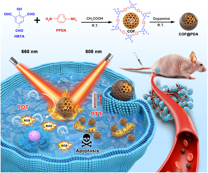
Figure 1.
Schematic illustration of the synthesis process of COF@PDA and its application in combined PDT/PTT therapy of breast cancer (R.T. = room temperature).
An imine-linked covalent organic framework with intrinsic photo-induced mitochondrial regulation for breast cancer therapy
Lulu He , Le Wang , Zhen He , Yiqian Yang , Cheng Heng Pang , Aiguo Wu , Bencan Tang , Juan Li

Instead of utilizing nanocarriers to deliver photosensitizers or photothermal agents for photodynamic therapy (PDT) or photothermal therapy (PTT), nanoagents with intrinsic photo-response show greater potential for PDT or PTT due to less uncertain leakage and serious nonspecific damage to normal tissues [1-4]. Besides, PDT or PTT monotherapy might cause normal tissue damage to some extent for over-lasting treatment duration and excessive laser power density [5,6]. Furthermore, the subcellular mitochondrial targeting ability of therapeutics may increase the production efficiency of reactive oxygen species (ROS), and further break redox balance and make mitochondrial membrane potential decrease rapidly causing apoptosis of cancer cells [7,8]. Thus, these agents which are not only provided with intrinsic photo-response ability but also have the mitochondrial regulation capacity will achieve a more accurate and efficiency therapy result [9-11].
Covalent organic frameworks (COFs) are one of newly developing porous crystalline materials, which have attracted significant attention for biomedical application owing to their various advantages [12-14]. Due to fascinating features of COFs including designable crystal structure, variable pores and high specific surface areas, and excellent chemical stability as well as biocompatibility, they show great potential in biomedical applications [15-17]. Interestingly, COFs with specific framework structures can afford to take charge of producing photodynamic effect themselves. For example, Tang's group fabricated three kinds of porphyrin-based COFs for inhibiting tumor growth through a type I PDT pathway [18]. In addition, COFs could also serve as an ideal substrate for integration of functional matrix in a moderate and controllable way, to achieve a synergistic PDT/PTT effect. For instance, Dong's group reported a CuS@COF-BDP dual-model therapeutic agent, which integrated an inorganic photothermal agent CuS and organic photosensitizer boron-dipyrromethene (BODIPY) into a nanoscale COF for combined PDT/PTT treatment [19]. Besides, COFs are supposed to regulate mitochondria by adjusting the surface property. Zhou's group developed a positive charged COF with subcellular mitochondria targeting ability for cancer cell suppression [20].
Only a few literatures have been reported for intrinsic photo-responsive COFs, let alone integrated with other substances for combined PDT/PTT treatment [21]. The introducing of polydopamine (PDA) out layer could work as one stone for killing three birds, which firstly considered as photothermal agent with excellent photothermal conversion efficiency, secondly improved water solubility, and thirdly enhanced in vivo stability and circulation time [22-24]. Herein, we designed a photo-response nanoplatform COF@PDA based on a novel imine-linked COF for PDT and integrating with PDA shell for PTT concurrently (Scheme 1). The COF took responsible for producing abundant ROS to break redox homeostasis under 660 nm laser, in the meanwhile, the PDA exhibited an excellent photothermal ability under 808 nm light, resulting in joint inhibiting of tumor growth through the apoptosis pathway owing to its function of mitochondrial regulation. These results demonstrate that COF@PDA is a promising candidate for the intrinsic PDT/PTT combined treatment, along with the photo-induced mitochondrial regulation ability towards breast cancer.
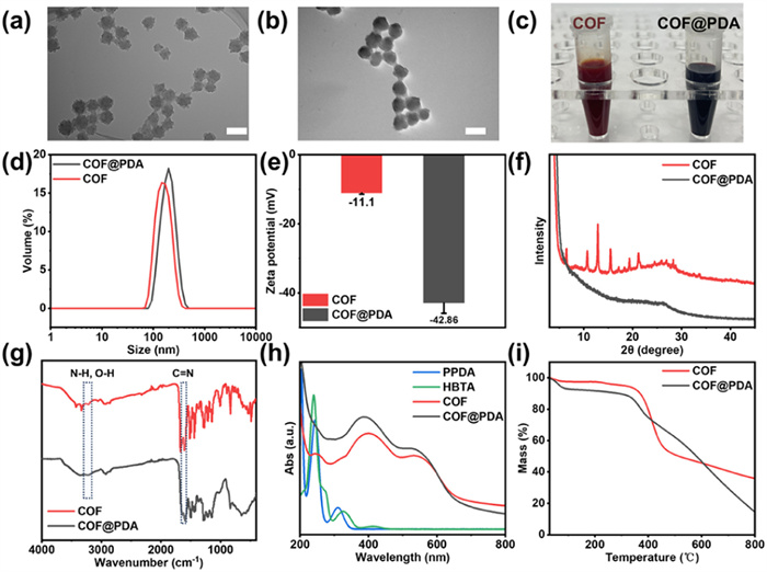
To achieve the photo-induced COF, a newly imine-linked HBTA-PPDA-COF (hereinafter referred to as the COF) generated via a Schiff-base condensation reaction between 2‑hydroxy-1,3,5-benzenetricarbaldehyde (HBTA) and para-phenylene-diamine (PPDA), was prepared according to the literature method with some improvements under ambient condition [25,26]. After that, the generated COF was then coated with photothermal and biocompatible PDA through the self-polymerization of dopamine monomer under the mixture solution of ethanol and ammonia (pH ≈ 11.5) at room temperature for 4 h, subsequently obtained the dark brown PDA-coated COF (termed as COF@PDA) [27]. The transmission electron microscope (TEM) image (Fig. 1a) and scanning electron microscope (SEM) image (Fig. S1a in Supporting information) of COF exhibited as a rough popcorn-like sphere, with an average diameter of 80 nm. Then the COF was modified with different ratio of dopamine and obtained diverse morphologies of PDA-coated COF, as shown in Fig. S2 (Supporting information). Fig. S2a showed when using 20 mg of dopamine, it was hard to observe an obvious core-shell structure. When the amount increased to 60 or 80 mg, the PDA cover was too thick, resulting in abnormal expansion in size (Figs. S2b and c). Only when the dopamine was at an appropriate dosage (40 mg), the COF@PDA nanoagent with perfect structure and size could be fabricated. Thus, under the optimal amount of dopamine, the obtained COF@PDA showed a slight size increase (∼90 nm), as shown in Fig. 1b of TEM image and Fig. S1b of SEM image. Also, a significant color change occurred during the modification, which changed from red brown to dark brown (Fig. 1c). Furthermore, the dynamic light scattering (DLS) results showed that the size distribution of the COF@PDA had a slight increase compare to COF alone (Fig. 1d), and the zeta potential value decreased from −11.1 ± 0.2 of COF to −42.86 ± 1.5 of COF@PDA (Fig. 1e). Notably, COF was highly crystalline and could be confirmed by its powder X-ray diffraction (PXRD) patterns. Compared with COF (2θ = 6.48°, 10.70°, 12.88°, 15.47°, 19.32°, 21.17°), no diffraction peak was observed for COF@PDA (Fig. 1f). The obtained COF and COF@PDA were further characterized by Fourier transform infrared (FT-IR) spectrometry. As shown in Fig. 1g, the characteristic peak at ∼1615 cm−1 for -C=N- stretching vibration indicated the present of imine linkages of COF, and a wide band ranging from 3000 cm−1 to 3500 cm−1 was observed, which indicated the successful introduction of PDA. Besides, both COF and COF@PDA exhibited a broad absorption band in the range of 200–800 nm of ultraviolet–visible (UV–vis) spectrum (Fig. 1h). In addition, thermogravimetric analysis (TGA) results proved that compared with only one weight loss for COF at around 400 ℃, COF@PDA possessed two obvious weight losses, as shown in Fig. 1i. All above observations confirmed the successful construction of COF and COF@PDA.
The photodynamic behavior of COF and COF@PDA were evaluated using 1,3-diphenylisobenzofuran (DPBF) as a chemical probe to detect the generation of singlet oxygen (1O2). Generally, essentially no decrease in DPBF aqueous solution was detected (Fig. 2a) under 660 nm irradiation with a power density of 0.5 W/cm2, indicating that this laser alone could not induce the generation of ROS. In contrast, when the COF was incubated with DPBF under the same condition, a rapid reduced UV–vis absorption at 420 nm could be found in Fig. 2b, suggesting the efficient generation of 1O2. When the surface of COF was covered by PDA, the absorption of 660 nm light might also be partially interrupted, which could be deduced by the result that the characteristic peak at 420 nm of DPBF mixed with COF@PDA showed weaker PDT effect compared with COF alone (Fig. 2c). Besides, Fig. 2d summarized the comparison of decay rates of different samples upon 660 nm irradiation within 10 min duration. In addition, the electron paramagnetic resonance (EPR) spectra for 2,2,6,6-tetramethylpiperidine (TEMP, a specific 1O2 trapping agent) in the presence of COF or COF@PDA kept silent before the irradiation, and an expected characteristic 1:1:1 signal appeared after 660 nm irradiation (Fig. 2e). Interestingly, due to the weak acidity of tumor microenvironment, the degradation of PDA shell could be occurred. Thus, the COF@PDA was dispersed in phosphate buffer solution (PBS) with different pH values (pH 7.4 or 6.4) to explore the stability of COF@PDA. Compare with pH 7.4 PBS that simulated regular physiological environment, COF@PDA showed a clear trend to degrade under pH 6.4 PBS which imitated the faintly acidity of tumor tissue (Fig. S3 in Supporting information).
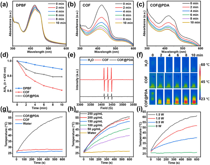
Further, the photothermal properties of COF@PDA were explored using NIR 808 nm light, as shown in Figs. 2f and g, compared with water or COF, COF@PDA showed a dramatic temperature increase when explored to 808 nm laser. The photothermal effect of COF@PDA under different concentration (0–250 µg/mL) and different power density (0–1.5 W/cm2) were given in Figs. 2h and i separately, and all results indicated COF@PDA displayed a concentration-dependent as well as laser density-dependent photothermal effect. For example, when COF@PDA (100 µg/mL) was irradiated with a 808 nm laser for 10 min under power of 1.0 W/cm2, the solution temperature increased from 25.69 ℃ to 46.07 ℃ (ΔT = 20.4 ℃), while only a 1.4 ℃ temperature increase for the deionized water. According to previous methods [16,28], the photothermal conversion efficiency of the COF@PDA was measured to be η = 18.42%, which was calculated based on the records of the temperature of COF@PDA solution during the cool-down process, as shown in Fig. S4 (Supporting information). Additionally, temperature curves had barely changed after five laser irradiation cycles, demonstrating the good photothermal stability of COF@PDA (Fig. S5 in Supporting information). Thereby, COF@PDA could be used as a controllable synergistic photodynamic/photothermal nanoagent in the following in vitro and in vivo experiments.
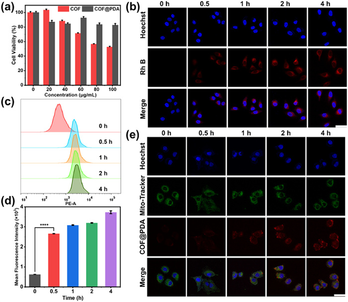
Following was the characterization of biosafety and therapeutic efficacy at cellular level. Firstly, the biosafety of COF@PDA was assessed by cell counting kit-8 (CCK-8) assay, and results showed that COF@PDA had no obvious cytotoxicity to MCF-7 cells (Fig. 3a) or MCF-10A cells (Fig. S6 in Supporting information) at concentrations up to 100 µg/mL after 24 h of co-incubation. Besides, it also indicated that compared to COF alone, COF@PDA showed a less toxicity under the same condition. Then the endocytosis of COF@PDA in cells was confirmed by confocal laser scanning microscopy (CLSM), in which the Rhodamine B (Rh B)-labeled COF@PDA absorbed by MCF-7 cells could be observed by the intracellular red fluorescence emitted by the Rh B (Fig. 3b). After that, the flow cytometry (FCM) was also used to further confirm and quantify this successful endocytosis (Fig. 3c), and it turned out that the COF@PDA could enter into cells successfully within half an hour according to mean fluorescence intensity statistics (Fig. 3d). Furthermore, as shown in Fig. 3e, the red fluorescence of COF@PDA displayed significant overlaps with the green fluorescence of Mito Tracker Green, which verified the colocalization of COF@PDA with mitochondrial.
In addition, to evaluate irradiation-induced intracellular ROS production ability of COF or COF@PDA, a specific green fluorescent probe for 1O2 within cells was introduced using 2,7-dichlorodihydrofluorescein diacetate (DCFH-DA) fluorescence probe. As shown in Fig. 4a, the control groups that without laser irradiation had hardly any fluorescence, however, after incubation with COF or COF@PDA (50 µg/mL) for 4 h and then irradiation with a 660 nm laser (0.5 W/cm2) for 10 min, a remarkable DCFH-derived fluorescence was recorded within MCF-7 cells by a CLSM, indicating an excellent intracellular photo-induced ROS generation ability. After that, the efficacy of monotherapy or combined therapy of COF@PDA was evaluated using MCF-7 cells via CCK-8 assay, JC-1 kit and calcein acetoxymethyl ester and propidium iodide (calcein-AM/PI) double staining analysis for assessing cell proliferation and cytotoxicity. As shown in Fig. 4b, the results showed that the cell survival rate remained as high as 76.75% ± 1.17% in the dark with a dosage of 150 µg/mL, in the meanwhile, neither 660 nm nor 808 nm irradiation would cause any damage for cell viability. In contrast, the PDT monotherapy (660 nm laser, 0.5 W/cm2, 10 min) and PTT monotherapy (808 nm laser, 1.0 W/cm2, 10 min) gave cell viabilities of 48.30% ± 1.96% and 43.56% ± 7.19%, respectively. However, the cell viability derived from the combined therapy sharply declined to 17.61% ± 0.47%, indicating the outstanding combined therapy effects of PDT/PTT.
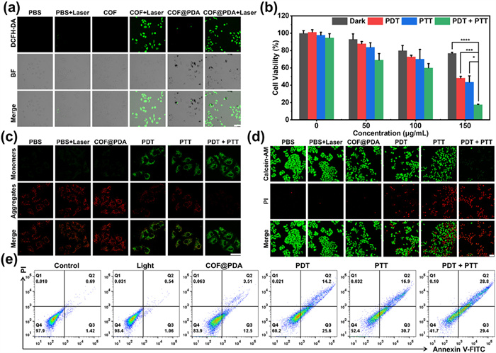
Furthermore, JC-1 kit was conducted to detect mitochondrial membrane potentials in MCF-7 cells. When incubated with COF@PDA alone, a strong red fluorescence was observed, which proved the normal mitochondrial membrane potential with JC-1 monomers. However, when exposed to COF@PDA as well as lasers, it could only find the green fluorescence which came from JC-1 monomers, indicating the collapse of mitochondrial membrane potential (Fig. 4c). After that, calcein-AM/PI live/dead double staining results showed that when incubated with COF@PDA followed by both 660 nm and 808 nm laser irradiation exposure, it displayed the highest cytotoxicity, for almost all cells had a red fluorescence compared with other groups (Fig. 4d). These experimental results were similar to those from the previous CCK-8 assay, demonstrating the superior combination PDT/PTT effect of prepared COF@PDA nanoagent towards breast cancer cells. Subsequently, the apoptosis of treated MCF-7 cells was quantitatively investigated by FCM, as exhibited in Fig. 4e. The irradiation alone resulted in negligible apoptosis. In contrast, the apoptosis rates of PDT and PTT-treated cells were 39.8% and 47.6%, respectively, and for the PDT/PTT-treated cells, it dramatically increased by 58.2%.
All animals were obtained from Hangzhou Ziyuan Laboratory Animal Technology Co., Ltd. (Hangzhou, China). All animal procedures were carried out by the Ningbo University Guidelines for the Care and Use of Laboratory Animals, and were approved by the Ningbo University Animal Ethics Committee (permit No. SYXK (Zhe) 2019-0005). To evaluate the biocompatibility and biosafety of COF@PDA, the hemolytic test was firstly conducted. As shown in Fig. S7 (Supporting information), the hemolytic rate was zero for the negative group and 100% for the positive control group separately, and all materials group were below 1%. To further monitor the in vivo safety of the COF@PDA, healthy mice were injected with PBS or COF@PDA at three dosages (5, 10, 15 mg/kg) through a single tail intravenous injection. After 21 days of observation, the mice were sacrificed, and the blood samples of mice were saved for blood routine analysis as well as blood biochemical examination, and both these two items showed no significant difference between the control group and administrated groups (Figs. S8 and S9 in Supporting information). Meanwhile, the major organs of mice were collected for haematoxylin and eosin (H&E) staining analysis, and microscopy images of H&E-stained organ slices collected from the administrated groups showed that there was no obvious histopathological abnormality compared with the control group (Fig. S10 in Supporting information). All experiment results verified COF@PDA possessed a good biosafety even at a high dosage of 15 mg/kg.
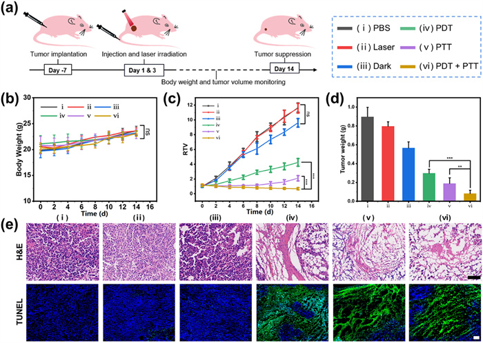
To evaluate the therapeutic efficiency in vivo, the tumor-bearing BALB/c nude mice were randomly divided into six groups (n = 5) with different treatments as follows: (ⅰ) PBS only; (ⅱ) PBS + 660 nm + 808 nm; (ⅲ) COF@PDA only; (ⅳ) COF@PDA + 660 nm; (ⅴ) COF@PDA + 808 nm; and (ⅵ) COF@PDA + 660 nm + 808 nm, respectively. The entire experimental protocol was presented in Fig. 5a and the irradiation was conducted at 12 h post injection of COF@PDA. The Figs. 5b and c showed the trend of body weight and relative tumor volume (RTV) within 14-day treatment respectively, which were recorded every two days. It was obvious that the body weight of mice in all groups showed no difference, indicating all treatments had no systematic toxicity. In addition, the RTV values of mice in groups (i–iii) increased rapidly, suggesting negligible therapeutic effects of the lasers and COF@PDA alone. However, no matter the monotherapy groups (ⅳ and ⅴ) or combined therapy group (ⅵ), the tumor volumes were significantly suppressed and reduced. Similarly, the results analysis of excised tumor weights (Fig. 5d) and tissue images (Fig. S11 in Supporting information) were consistent with that of tumor volume changes. The H&E and TdT-mediated dUTP nick end labeling (TUNEL) staining analysis were further carried out to investigate the tumor tissue damage degree and cancer cell apoptosis rate (Fig. 5e), and results showed that COF@PDA in the presence of 660 or 808 nm or both lasers significantly led to a high amount of tumor cell apoptosis or necrosis. Besides, Fig. S12 (Supporting information) presented the H&E results of major organs (heart, liver, spleen, lung, and kidney) of mice from all groups, and it confirmed no physiological damage to major organs, which further proved that COF@PDA had neglectable short-term toxicity towards mice.
In summary, we have successfully designed and fabricated a newly COF@PDA nanoagent through a facile stepwise modification under ambient condition. The obtained COF@PDA could serve as a PDT/PTT-in-one therapeutic agent with intrinsic photo-induced mitochondrial regulation for highly suppressing MCF-7 cancer cell proliferation due to its excellent ROS generation performance under 660 nm laser and efficient photothermal conversion ability within 808 nm laser. We believe that our strategy could provide ideas for further constructing more COF-based nanoplatforms for combined PDT/PTT treatment along with photo-induced mitochondrial modulation against breast cancer, and also promote the potential application of COFs in the biomedical field.
The authors declare that they have no known competing financial interests or personal relationships that could have appeared to influence the work reported in this paper.
Lulu He: Writing – review & editing, Writing – original draft, Validation, Methodology, Investigation, Conceptualization. Le Wang: Validation, Methodology, Data curation. Zhen He: Visualization, Investigation, Formal analysis. Yiqian Yang: Validation, Resources, Formal analysis. Cheng Heng Pang: Visualization, Supervision, Resources, Investigation. Aiguo Wu: Writing – review & editing, Supervision, Project administration, Funding acquisition, Conceptualization. Bencan Tang: Writing – review & editing, Supervision, Project administration, Funding acquisition, Conceptualization. Juan Li: Writing – review & editing, Writing – original draft, Supervision, Project administration, Investigation, Funding acquisition, Conceptualization.
This work was financially supported by the National Key R&D Program of China (No. 2023YFC2415700), the Natural Science Foundation of China (Nos. T2222021, 32025021, 22171153), Ningbo Science and Technology Bureau under CM2025 Program (No. 2020Z092), and Ningbo Natural Science Foundation Program (No. 2022J171).
Supplementary material associated with this article can be found, in the online version, at doi:
J. Hu, J. Hu, W. Wu, et al., Acta Biomater. 148 (2022) 206–217. doi: 10.1016/j.actbio.2022.06.012
S. Chen, T. Sun, M. Zheng, Z. Xie, Adv. Funct. Mater. 30 (2020) 2004680. doi: 10.1002/adfm.202004680
S. Gan, X. Tong, Y. Zhang, et al., Adv. Funct. Mater. 29 (2019) 1902757. doi: 10.1002/adfm.201902757
L. Zhang, Q.C. Yang, S. Wang, et al., Adv. Mater. 34 (2021) e2108174.
P. Gao, R. Wei, Y. Chen, et al., Biomaterials 297 (2023) 122109. doi: 10.1016/j.biomaterials.2023.122109
Y. Qin, M. Huang, C. Huang, et al., Chin. Chem. Lett. 35 (2024) 109171. doi: 10.1016/j.cclet.2023.109171
C. Huang, S. Zhou, C. Chen, et al., Small 18 (2022) e2205062. doi: 10.1002/smll.202205062
Y. Zhou, S. Jing, S. Liu, J. Nanobiotechnol. 20 (2022) 188. doi: 10.54691/bcpssh.v18i.974
H. Peng, F. Yao, J. Zhao, et al., Exploration 3 (2023) 20220115. doi: 10.1002/EXP.20220115
X. Yu, M. Lyu, X. Ou, et al., Adv. Healthc. Mater. 12 (2023) e2202907. doi: 10.1002/adhm.202202907
Z. Yu, X. Luo, C. Zhang, et al., Chin. Chem. Lett. 35 (2024) 109519. doi: 10.1016/j.cclet.2024.109519
N. Singh, J. Kim, J. Kim, et al., Bioact. Mater. 21 (2023) 358–380.
Y. Shi, J. Yang, F. Gao, Q. Zhang, ACS Nano 17 (2023) 1879–1905. doi: 10.1021/acsnano.2c11346
A. Mal, H. Ding, M. Li, W. Li, C. Wang, ACS Appl. Nano Mater. 5 (2022) 13972–13984. doi: 10.1021/acsanm.2c01517
L. Zhang, A. Song, Q.C. Yang, et al., Nat. Commun. 14 (2023) 5355. doi: 10.1038/s41467-023-41121-z
S. Wang, Y. Pang, S. Hu, et al., Chem. Eng. J. 451 (2023) 138864. doi: 10.1016/j.cej.2022.138864
L. Zhang, Y. Xiao, Q.C. Yang, et al., Adv. Funct. Mater. 32 (2022) 2201542. doi: 10.1002/adfm.202201542
P. Dong, H. Lv, R. Luo, Z. Li, X. Wu, J. Lei, Chem. Eng. J. 461 (2023) 141817. doi: 10.1016/j.cej.2023.141817
X.J. Dong, W.Y. Li, Q. Guan, Y.A. Li, Y.B. Dong, Chem. Commun. 58 (2022) 2387–2390. doi: 10.1039/d1cc06330h
L. Chen, J. Zhang, K. Cai, et al., Sens. Actuators B: Chem. 350 (2022) 130861. doi: 10.1016/j.snb.2021.130861
J. Chen, Y. Wang, Y. Yu, et al., Exploration 3 (2023) 20220144. doi: 10.1002/EXP.20220144
P. Gao, R. Wei, X. Liu, et al., Chem. Commun. 57 (2021) 5646–5649. doi: 10.1039/d1cc00314c
J. Feng, W.X. Ren, F. Kong, et al., Sci. China Mater. 65 (2021) 1122–1133.
D. Ren, G.R. Williams, Y. Zhang, et al., ACS Appl. Bio Mater. 5 (2022) 123–133. doi: 10.1021/acsabm.1c00926
C.L. Hu, L.H. Cai, S.N. Liu, et al., Chem. Commun. 55 (2019) 9164. doi: 10.1039/c9cc04668b
Q. Guan, L.L. Zhou, Y.A. Li, et al., ACS Nano 13 (2019) 13304–13316. doi: 10.1021/acsnano.9b06467
Y. Xiao, C. Ma, Z. Jin, et al., Chem. Eng. J. 421 (2021) 127837. doi: 10.1016/j.cej.2020.127837
D. Xi, M. Xiao, J. Cao, et al., Adv. Mater. 32 (2020) 1907855. doi: 10.1002/adma.201907855

Figure 1 Schematic illustration of the synthesis process of COF@PDA and its application in combined PDT/PTT therapy of breast cancer (R.T. = room temperature).

Figure 1 The physicochemical characterization of as-prepared COF and COF@PDA. TEM images of (a) COF and (b) COF@PDA. (c) The photograph of COF (left) and COF@PDA (right) solutions. (d) DLS curves, (e) zeta potential, (f) XRD patterns, and (g) FT-IR spectra of COF and COF@PDA. (h) UV–vis spectra of monomers, COF and COF@PDA. (i) TGA curves of COF and COF@PDA. Data are presented as the mean ± SD (n = 3). Scale bar: 100 nm.

Figure 2 UV–vis spectra of DPBF solutions containing (a) DPBF only, (b) COF (50 µg/mL) and (c) COF@PDA (50 µg/mL) when exposure to 660 nm irradiation (0.5 W/cm2) for different durations (0, 2, 4, 6, 8, and 10 min). (d) Comparison of the decay rate of DPBF induced by different samples. (e) Electron spin resonance spectra of different samples with TEMP. (f) Thermal images and (g) temperature variation curves of water, COF (250 µg/mL) and COF@PDA (250 µg/mL) under 808 nm laser (1.0 W/cm2, 10 min). Temperature variation curves of COF@PDA with (h) different concentrations and (i) different power density.

Figure 3 (a) Viabilities of MCF-7 cultured with COF or COF@PDA in different concentrations. (b) CLSM images of MCF-7 cells incubated with Rh B labelled COF@PDA (50 µg/mL) for 0, 0.5, 1, 2 and 4 h. (c) Histogram analysis and (d) Mean fluorescence intensity values of flow cytometry for MCF-7 cells incubated with Rh B labelled COF@PDA. (e) CLSM images of MCF-7 cells coincubated with COF@PDA and MitoTracker Green. Data are presented as mean ± SD (n = 3). Scale bar: 50 µm. ****P < 0.0001.

Figure 4 (a) Detection of ROS production in MCF-7 cells using DCFH-DA with or without 660 nm laser (0.5 W/cm2, 10 min). (b) Viabilities of MCF-7 treated with monotherapy or combination therapy. CLSM images of MCF-7 cells stained with (c) the mitochondrial membrane potential fluorescence dye (JC-1) and (d) cells live/dead fluorescence dyes (calcein-AM/PI) after various treatments. (e) Flow cytometry analysis of MCF-7 cells stained by Annexin V-fluorescein isothiocyanate (FITC) and PI after various treatments. PDT: 0.5 W/cm2, 10 min; PTT: 1.0 W/cm2, 10 min. Scale bar: 100 µm. P < 0.05, ***P < 0.001, ****P < 0.0001.

Figure 5 (a) Schematic diagram of the animal experiment. (b) Body weight variation curves, (c) relative tumor volume changes and (d) tumor weight statistics of mice in different groups during 14-day treatment. (e) H&E and TUNEL-stained histological section of tumor slices. Data are presented as mean ± SD (n = 5). Scale bar: 100 µm. ns, no significance. **P < 0.01, ***P < 0.001.

 扫一扫看文章

扫一扫关注我们
 DownLoad:
 下载:
下载:
 下载:
下载: