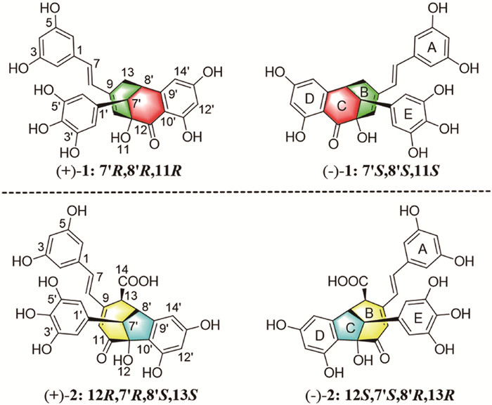
Figure 1.
Structures of (+)−1, (−)−1, (+)−2 and (−)−2.
Two racemic pairs of benzo bicyclo[3.3.1]/[4.2.1] nonene stilbenoid dimers from Heterosmilax yunnanensis and structural revision of syagrusin A
Ji-Ping Liao , Jiang Li , Wen-Jie Qin , Xiu-Mei Duan , Peng-Fei Wang , Jin-Ying Tian , Xiang Yuan , Pei-Cheng Zhang , Fei Ye , Ya-Nan Yang

Stilbenoids, an important class of naturally-occurring polyphenolic compounds already found in various plant families, are specialized secondary metabolites that act as phytoalexins in plants [1-3]. To date, approximately 459 natural stilbenoid compounds with diverse scaffolds have been identified and have been proven to exhibit cardioprotective, neuroprotective, anti-diabetes, antiobesity, depigmentation, anti-inflammatory and cancer prevention activities [4-8]. Among them, approximately 65 stilbenoid dimers with various structural patterns including cyclobutane, indane, dihydrofuran, dihydrobenzofuran, 1,4-benzodioxane ring, cyclohexene ring, cycloheptene-1-one, bicyclic [3.2.1], [3.2.2], [3.3.0] and [4.2.0] moieties, have been identified [9-17], with most of them based on resveratrol. The intriguing structural complexity and often relatively high bioactivities of dimeric stilbenoids have incentivized sustaining attentions from the scientific community [18-21].
Heterosmilax yunnanensis Gagnep. (Liliaceae), which is widely distributed in southern China, has been used in traditional Chinese medicine for clearing heat, dehumidifying, detoxifying and relieving joint pain, with specific applications in the treatment of muscle pain, scabies and gynecological disease [22,23]. Previously limited chemical investigations of H. yunnanensis revealed that in addition to phenolic acids, flavonoids, terpenoids as well as the recent disclosure of 2-benzylbenzofuran O-glycosides and alkaloids by our group [24,25], stilbenoids also exist in this herb: resveratrol and 3,3′,5,5′-tetrahydroxy-4′-methoxy stilbene [26]. In our continuous efforts to identify biologically active stilbenoids with unique structures, the phytochemical study of H. yunnanensis was performed, leading to the discovery of two racemic pairs of stilbenoid dimers, (±)-heterosmilaxones A (1) and B (2). The structure of previously reported syagrusin A was revised to (+)−1, which possesses a unique benzo bicyclo[3.3.1] nonene skeleton. Compound 2, bearing a novel 6/5/7 tricyclic core system, features an unprecedented benzo bicyclo[4.2.1] nonene scaffold with four continuous chiral centers. Compounds 1 and 2 could be generated through key inverse-electron-demand [4 + 2] and [5 + 2] cycloadditions, respectively. To the best of our knowledge, dimeric stilbenoids have never been discovered in the Lily family. Herein, we disclose the isolation, structural elucidation and biological evaluation of 1 and 2.
(±)-Heterosmilaxone A (1) was obtained as a white amorphous powder. Its molecular formula was assigned as C27H22O9 on the basis of its high resolution electrospray ionization mass spectroscopy (HRESIMS) data at m/z 491.1345 [M + H]+ (calcd. for C27H23O9, 491.1337), indicating seventeen degrees of unsaturation (DOUs). The infrared spectroscopy (IR) absorptions suggested the presence of hydroxyl (3376 cm−1), carbonyl (1624 cm−1), aromatic ring and olefin functionalities (1598, 1508 and 1458 cm−1). The 1H nuclear magnetic resonance (NMR) data (Table S1 in Supporting information) revealed signals of three aromatic protons assignable to 1,3,5-trisubstituted [δH 6.35 (2H, d, J = 2.0 Hz) and 6.14 (t, J = 2.0 Hz)], 1,3,4,5-tetrasubstituted [δH 6.51 (2H, s)] and 1,2,3,5-tetrasubstituted benzene rings [6.32 (d, J = 2.0 Hz) and 6.18 (d, J = 2.0 Hz)], two vinyls [δH 6.68 (d, J = 16.0 Hz), 6.38 (d, J = 16.0 Hz) and 5.81 (s)], two methines [δH 3.45 (brd) and 3.43 (m)], and a methylene [δH 2.57 (m) and 2.26 (brd, J = 16.5 Hz)]. Moreover, a total of twenty-seven carbons were observed in the 13C NMR spectrum. Combined analysis with the heteronuclear single quantum coherence (HSQC) spectrum revealed a keto-carbonyl (δC 202.9), thirteen sp2 quaternary carbons, twelve methines, and a methylene. The aforementioned structural units occupied fifteen of a total of seventeen DOUs, requiring 1 to be equipped with two additional cyclic fragments (Fig. 1).
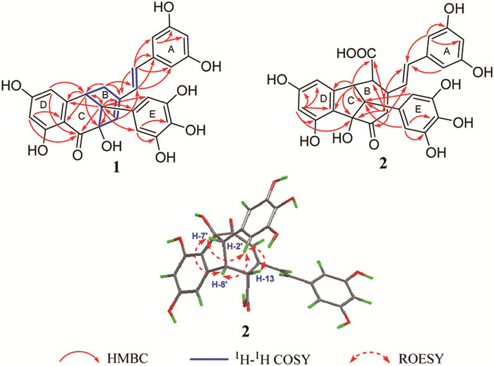
Interpretations of the 1H–1H correlation spectroscopy (COSY) spectrum revealed three spin-coupling systems: H-10/H-7′, H-10/H2–13 and H2–13/H-7′/H-8′, implying a C7′-C8′-C13 linkage (Fig. 2). The key heteronuclear multiple-bond correlations (HMBCs) from H-7′ to C-8′, C-9, C-11 and H-8′ to C-7′, C-9, C-11 permitted the deduction of a cyclohexene fragment. Further analysis of the HMBC spectrum allowed the assignment of the C11-C7′-C8′-C9′-C10′ linkage via correlations from H-7′ to C-9′, H-8′ to C-9′, C-10′, H-14′ to C-8′ and H-12′ to C-10′. Furthermore, a benzo cyclohexanone fragment could be built up according to the key HMBCs of H-7′ to C-12 and form a bicyclic bridge together with the cyclohexene fragment. Observations of correlations from H-7/H-8 to C-1, C-2/C-6 indicated a styrene fragment, which was positioned at C-9 evidenced by H-7 to C-9. The HMBC correlations from H-7′ to C-1′ and C-2′/C-6′ suggested that ring E was attached to C-7′. Therefore, the above evidence supported the formation of a cyclooctenone motif that is fused to ring D via C-9′ and C-10′. As a result, the planar structure of 1 with a rare benzo bicyclo[3.3.1] nonene skeleton was established (Fig. 1).
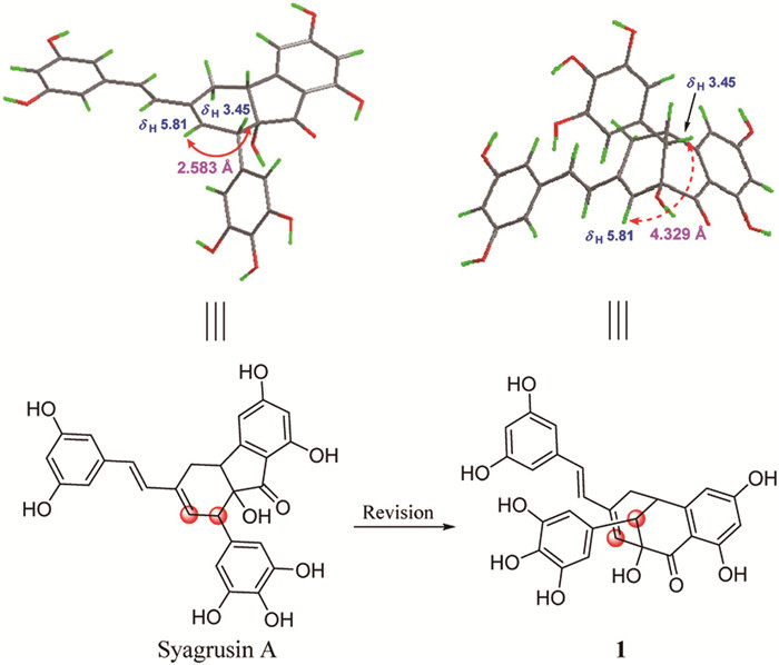
The lack of key rotating frame overhauser spectroscopy (ROESY) correlations of 1 prevented the assignment of its relative configuration (Fig. S18 in Supporting information). Based on a molecular model and an MM2 calculation, H-8′ and C-11 should be equatorially orientated because of the rigidity of the bridged ring (Fig. S3 in Supporting information), thus leaving the relative configuration of C-7′ to be determined. As a result, there are two possible relative configurations for 1, (7′S*,8′S*,11S*)−1 and (7′R*,8′S*,11S*)−1. The 13C NMR calculations following a reported sorted training sets protocol with improved accuracy by consideration of the hybridized types of carbon were conducted [27], which allowed the relative configuration of 1 to be assigned as 7′S*,8′S*,11S* (Figs. S5 and S6 in Supporting information).
When reviewing the reported literature, we found that 1 had the same molecular weight, 1H and 13C NMR data (detected in CD3OD) as those of syagrusin A
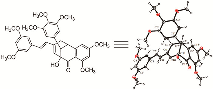
For definitive proof, we attempted to obtain single crystals of 1 in various solvent systems but did not succeed. Considering its polyphenolic features, the methylated derivatization of 1 in racemic form was conducted with dimethyl sulfate to yield an analogue (1a) (Supporting information) [33]. Luckily, we finally obtained a suitable crystal of 1a in an EtOH/H2O (65:35) mixture (Fig. 4, CCDC No. 2381496). Consistent with the structure deduced above, an X-ray diffraction experiment using Cu Kα radiation allowed the unambiguous determination of 1 with a relative configuration of 7′S*,8′S*,11S*, resulting in the structural revision of syagrusin A.
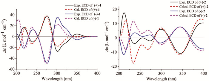
However, the optical inactivity of 1 and the presence of a centrosymmetric space group in the crystal structure suggested that it existed as racemates. A chiral resolution of 1 was subsequently performed to afford the anticipated enantiomers (+)−1 and (−)−1. The absolute configurations of (+)−1 and (−)−1 were determined using electronic circular dichroism (ECD) calculations. As shown in Fig. 5, the calculated ECD spectrum of (7′R,8′R,11R)−1 was in accordance with the experimental ECD spectrum of (+)−1, whereas the calculated (7′S,8′S,11S)−1 fitted well with the experimental one of (−)−1, allowing the determination of (+)−1 to be 7′R,8′R,11R and (−)−1 to be 7′S,8′S,11S. By measuring the optical rotatory values of
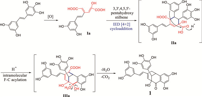
Biogenetically, Lee's original proposal of syagrusin A initiated by ortho-para oxidative coupling of resveratrol and gallic acid residues seemed unconvincing [28]. Considering both the structural features of 1 and the abundant existence of stilbenoid natural products in Syagrus romanzoffiana [34,35], we proposed a more reasonable biogenetic pathway for 1 (Scheme 1). First, the oxidation followed by ring-opening of ring A in 3,3′,4,5,5′-pentahydroxy stilbene results in 1,3-unsaturated hexanedioic acid (Ia). Then, an inverse-electron-demand (IED) [4 + 2] cycloaddition reaction occurs between Ia and another 3,3′,4,5,5′-pentahydroxy stilbene to yield the key intermediate IIa [36,37]. Finally, C-10′ attacks the electrophilic carboxyl C-12 via intramolecular Friedel-Crafts acylation followed by decarboxylation and dehydration, leading to the formation of 1.
(±)-Heterosmilaxone B (2), a yellowish amorphous powder, has a molecular formula of C28H22O11 as deduced from its HRESIMS ion peak at m/z 535.1234 [M + H]+ (calcd. for C28H23O11, 535.1235), indicative of eighteen degrees of unsaturation. The IR absorptions at 3338, 1664, 1618, 1555 and 1465 cm−1 suggested the presence of hydroxyl, carboxy, carbonyl and phenyl groups. Compound 2 displayed a similar 1H NMR spectrum to that of 1 other than the signals of three methine protons [δH 3.84 (d, J = 5.0 Hz), 3.75 (s) and 3.48 (d, J = 8.5 Hz)], instead of two methines and a methylene in 1 (Table S1). The 13C NMR spectrum associated with the HSQC spectrum attributed twenty-eight carbons to a keto-carbonyl group (δC 209.2), a carboxyl group (δC 176.1), ten methine carbons [δC 142.6, 123.7, 123.1, 109.1, 108.7, 107.5 (2C), 105.6, 105.4 and 103.6], thirteen sp2 quaternary carbons [δC 161.4, 159.8, 158.6 (2C), 157.1, 148.0, 147.2 (2C), 139.0, 133.5, 131.5, 118.4 and 90.0] and three methine carbons (δC 60.3, 59.7 and 52.5).
The HMBC correlations from H-2/H-6 to C-7 suggested the existence of a styrene group. The HMBCs from H-10 to C-7/C-8 positioned the styrene group at the olefinic quaternary carbon C-9. Further elucidation of the HMBCs established the C13-C8′-C9′-C10′-C12 linkage by observing correlations from H-8′ to C-9′, C-10′, C-12, C-13 and H-12′ to C-12, implying the attachment of C-8′ and C-12 to ring D through C-9′ and C-10′, respectively. Olefinic proton H-10 was correlated with the keto-carbonyl C-11, oxygenated aliphatic quaternary carbon C-12 and methine carbon C-7′, permitting the linkage of C9-C10-C11-C12-C7′. Moreover, the HMBC correlations from H-10 to C-13, C-14 and H-8′ to C-14 confirmed the existence of a benzo-octane fragment. So far, sixteen of the seventeen DOUs have been occupied and the necessity of another cycle rationalizes the connection of C-7′ to C-8′, as evidenced by the HMBC correlation from H-8′ to C-7′ and strong spatial correlations from H-7′ to H-8′, thus resulting in a 6/5/7 tricyclic core system. The key HMBC correlations (Fig. 2) from H-2′/H-6′ to C-7′ indicated the connection of ring E to the methine C-7′. Therefore, 2 was determined to have a benzo bicyclo[4.2.1] nonene skeleton.
The ROESY correlations from H-2′/H-6′ to H-13 indicated that ring E and H-13 were positioned on the same face of ring B. Moreover, the rigidity of the bridged ring forced H-8′ and OH-12 to stand in equatorial orientation. Hence the relative configuration of 2 was defined as 12S*,7′S*,8′R*,13R* (Fig. 2).
Similarly, 2 also existed as a racemic mixture due to its barely measurable optical rotation value and was then separated to provide two expected enantiomers. By comparing the experimental and calculated ECD spectra, where (12R,7′R,8′S,13S)−2 and (12S,7′S,8′R,13R)−2 agreed well with the measured ones of (+)−2 and (−)−2, respectively (Fig. 5), their absolute configurations were finally assigned.
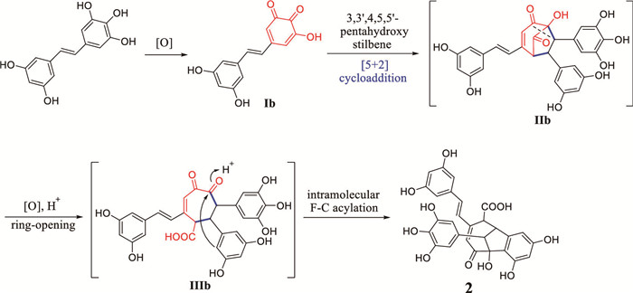
Biogenetically, we propose that 2 is also derived from the monomer 3,3′,4,5,5′-pentahydroxy stilbene. First, ring A in pentahydroxy stilbene is oxidated to obtain the electron-deficient o-quinone (Ib), which then undergoes a [5 + 2] cycloaddition reaction with another pentahydroxy stilbene to provide IIb with a [3.2.1] scaffold [38]. After that, ring-opening by oxidation followed by intramolecular Friedel-Crafts acylation finally led to the formation of 2 (Scheme 2) [39].
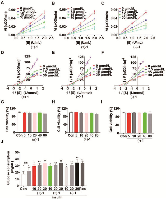
In the in vitro antidiabetic bioactivity assays, both (±)−1 and (±)−2 showed significant inhibition against α-glucosidase with half maximal inhibitory concentration (IC50) values of 1.56 and 7.71 µmol/L, respectively, which were superior to the positive control acarbose (IC50 = 228.77 µmol/L) (Table S2 in Supporting information). (±)−1 displayed a greater inhibition rate (96.6%) against the protein tyrosine phosphatase-1B (PTP1B) enzyme than that of (±)−2 (75.9%), each at a concentration of 10 µmol/L (CC06240 was used as a positive control, with an inhibition rate of 100.6%). Furthermore, the IC50 of compound (±)−1 and its pure enantiomers (+)−1/(−)−1 on the enzyme were determined to be 13.3, 13.7 and 14.6 µmol/L, respectively (Table S3 in Supporting information). The enzyme kinetics study at different PTP1B activity and substrate kinetics study indicated that their inhibition on PTP1B enzyme is reversible and exhibited a mixed-type inhibition (Fig. 6). In addition, the glucose consumption in HepG2 cells showed that, compared to the insulin group, treatment with (±)−1 and (+)−1/(−)−1 considerably increased glucose consumption without significant cytotoxicity, equal to the efficiency of rosiglitazone at the concentration of 30 µmol/L.
In summary, two racemic pairs of stilbenoid dimers, (±)-heterosmilaxones A (1) and B (2), possessing unprecedented benzo bicyclo[3.3.1] and [4.2.1] nonene scaffolds, respectively, were identified from the rhizomes of H. yunnanensis. The previously misassigned syagrusin A was revised to (+)−1 via 2D NMR analyses and X-ray diffraction. This study helps enrich the structural library of natural dimeric stilbenoids and the in vitro tests revealed that both (±)−1 and (±)−2 could significantly inhibit α-glucosidase activities. Besides, (±)−1, along with its pure enantiomers, modulated PTP1B enzyme activities and increased glucose consumption in HepG2 cells in a dose-dependent manner, providing a new structure motif for developing novel antidiabetic agent.
The authors declare that they have no known competing financial interests or personal relationships that could have appeared to influence the work reported in this paper.
Ji-Ping Liao: Writing – original draft, Visualization, Validation, Methodology, Investigation, Data curation, Conceptualization. Jiang Li: Writing – original draft, Visualization, Validation, Methodology, Investigation, Data curation, Conceptualization. Wen-Jie Qin: Validation, Investigation, Data curation. Xiu-Mei Duan: Validation. Peng-Fei Wang: Validation. Jin-Ying Tian: Validation. Xiang Yuan: Validation. Pei-Cheng Zhang: Writing – review & editing, Validation. Fei Ye: Writing – review & editing, Validation. Ya-Nan Yang: Writing – review & editing, Project administration, Funding acquisition, Conceptualization.
The conformational optimization, 13C NMR and ECD calculations were supported by Biomedical High Performance Computing Platform, Chinese Academy of Medical Sciences. This work was financially supported by the CAMS Innovation Fund for Medical Sciences (CIFMS, No. 2021-I2M-1-028).
Supplementary material associated with this article can be
found, in the online version, at doi:
P. Jeandet, B. Delaunois, A. Conreux, et al., Biofactors 36 (2010) 331–341. doi: 10.1002/biof.108
A. Valletta, Iozi L.M, F. Leonelli, Plants 10 (2021) 90. doi: 10.3390/plants10010090
C. Rivière, A.D. Pawlus, J.M. Mérillon, Nat. Prod. Rep. 29 (2012) 1317–1333. doi: 10.1039/c2np20049j
T. Teka, L.L. Zhang, X.Y. Ge, et al., Phytochemistry 197 (2022) 113128. doi: 10.1016/j.phytochem.2022.113128
B.C. Akinwumi, K.A.M. Bordun, H.D. Anderson, Int. J. Mol. Sci. 19 (2018) 792. doi: 10.3390/ijms19030792
H. Eräsalo, M. Hämäläinen, T. Leppänen, et al., J. Nat. Prod. 81 (2018) 1131–1142. doi: 10.1021/acs.jnatprod.7b00384
M.H. Pan, Y.C. Koh, T.L. Lee, et al., J. Agric. Food Chem. 67 (2019) 13605–13616. doi: 10.1021/acs.jafc.9b05963
M. Dvorakova, P. Landa, Pharmacol. Res. 124 (2017) 126–145. doi: 10.1016/j.phrs.2017.08.002
M.H. Keylor, B.S. Matsuur, C.R.J. Stephenson, Chem. Rev. 115 (2015) 8976–9027. doi: 10.1021/cr500689b
Z.W. Liu, J. Wu, D.J. Huang, J. Agric. Food Chem. 61 (2013) 4155–4161. doi: 10.1021/jf400144s
X.L. Li, B.X. Zhao, X.J. Huang, et al., Org. Lett. 16 (2014) 224–227. doi: 10.1021/ol403211a
C.Y. Wang, S.S. Lam, L.H. Tseng, et al., Phytochem. Anal. 22 (2011) 352–360. doi: 10.1002/pca.1286
Y.M. Zhai, K. Jiang, S.J. Qu, et al., RSC. Adv. 6 (2016) 50083. doi: 10.1039/C6RA08238F
S.G. Li, X.J. Huang, M.M. Li, et al., J. Nat. Prod. 81 (2018) 254–263. doi: 10.1021/acs.jnatprod.7b00540
P. Pel, H.S. Chae, P. Nhoek, et al., Bioorg. Chem. 99 (2020) 103869. doi: 10.1016/j.bioorg.2020.103869
D. Mittas, U. Spitaler, M. Bertagnoll, et al., Phytochemistry 200 (2022) 113241. doi: 10.1016/j.phytochem.2022.113241
Q.W. Yan, B.J. Su, S. He, et al., Bioorg. Chem. 143 (2024) 107060. doi: 10.1016/j.bioorg.2023.107060
K.J. Romero, M.H. Keylor, M. Griesser, et al., J. Am. Chem. Soc. 142 (2020) 6499–6504. doi: 10.1021/jacs.0c01714
J. Vinet, A.L. Flourat, C. Peyrot, et al., ACS Sustain. Chem. Eng. 10 (2022) 9166–9175. doi: 10.1021/acssuschemeng.2c02010
S. Nagasawa,Y. Itagaki, Y. Sasano, et al., Org. Lett. 26 (2024) 4178–4182. doi: 10.1021/acs.orglett.4c00839
Q.F. He, Z.L. Wu, X.J. Huang, et al., J. Org. Chem. 86 (2021) 5870–5882. doi: 10.1021/acs.joc.1c00295
H.Y. Shen, Z.P. Qu, Y.H. Lee, et al., Front. Oncol. 9 (2019) 632. doi: 10.3389/fonc.2019.00632
Y.H. Kuai, L.N. Hai, X.Y. Jian, et al., Chin. J. Experiment. Tradit. Med. Formulae 16 (2010) 207–209.
R.R. Du, J.C. Zhou, W.J. Qin, et al., Bioorg. Chem. 143 (2024) 107079. doi: 10.1016/j.bioorg.2023.107079
R.R. Du, R.Y. Wang, J.C. Zhou, et al., Bioorg. Chem. 151 (2024) 107618. doi: 10.1016/j.bioorg.2024.107618
L. Qiao, J.Z. Yuan, H.Y. Chen, et al., J. Chin. Chem. Mater. 10 (2007) 1242–1244.
H.W. Yan, R.R. Du, X. Zhang, et al., Chin. Chem. Lett. 33 (2022) 2555–2558. doi: 10.1016/j.cclet.2021.09.064
S.H. Lam, S.S. Lee, Phytochemistry 71 (2010) 792–797. doi: 10.1016/j.phytochem.2010.01.013
R. He, P. Liu, X.H. Huo, et al., Org. Lett. 19 (2017) 5513–5516. doi: 10.1021/acs.orglett.7b02577
T.D.S. Agostini-Costa, J. Ethnopharmacol. 224 (2018) 202–229. doi: 10.1016/j.jep.2018.05.035
B.M. Trost, C.J. Hung, G. Mata, Angew. Chem. Int. Ed. 59 (2020) 4240–4261. doi: 10.1002/anie.201909692
H. Kim, J. Rencoret, T.J. Elder, et al., Sci. Adv. 9 (2023) eade5519. doi: 10.1126/sciadv.ade5519
A. Cervi, P. Aillard, N. Hazeri, et al., J. Org. Chem. 78 (2013) 9876–9882. doi: 10.1021/jo401583q
S.H. Lam, J.M. Chen, C.J. Kang, et al., Phytochemistry 69 (2008) 1173–1178. doi: 10.1016/j.phytochem.2007.12.004
C.Y. Wang, S.H. Lam, et al., Phytochem. Anal. 22 (2011) 352–360. doi: 10.1002/pca.1286
A.T. Dang, D.O. Miller, L.N. Dawe, et al., Org. Lett. 10 (2008) 233–236. doi: 10.1021/ol702614b
Q. Li, M. Zhang, X.T. Zhang, et al., Chin. Chem. Lett. 35 (2024) 108193. doi: 10.1016/j.cclet.2023.108193
P. Ellerbrock, N. Armanino, M. K.Ilg, et al., Nat. Chem. 7 (2015) 879–882. doi: 10.1038/nchem.2336
J. Zhang, T. Zhang, Q. An, et al., Chin. Chem. Lett. 35 (2024) 108927. doi: 10.1016/j.cclet.2023.108927

Figure 5 Experimental and calculated ECD spectra of (+)−1 and (−)−1, (+)−2 and (−)−2, respectively.

Figure 6 Kinetic assessment of the effects of compounds (±)−1, (+)−1 and (−)−1 against PTP1B and their effects on glucose consumption in HepG2 cells. (A–C) Enzyme kinetics curve (n = 3). (D–F) Substrate kinetics study. Lineweaver-Burk plot of PTP1B activity at various substrate concentrations with various compounds (n = 3). (G–I) HepG2 cells were treated with 5, 10, 20, 40, and 80 µmol/L of compounds for 24 h. Cell viability was measured with a cell counting kit-8 (CCK-8) assay (n = 6). (J) Cellular glucose consumption was detected in insulin (10 nmol/L)-stimulated HepG2 cells after 24 h of treatment with 10, 20 and 30 µmol/L of compounds, and 10 µmol/L of rosiglitazone (ROS) was used as a positive control (n = 6). The data are expressed as the mean ± SD. P < 0.05, **P < 0.01 vs. the insulin treatment group. ns, no significance.

 扫一扫看文章

扫一扫关注我们
 DownLoad:

 下载:
下载:


 下载:
下载: