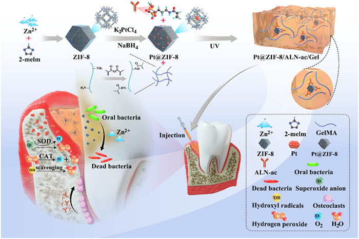
Scheme 1.
Schematic illustration of the synthesis of Pt@ZIF-8/ALN-ac/Gel Hydrogel and its antimicrobial, anti-inflammatory and osteogenic properties in periodontitis.
A Pt@ZIF-8/ALN-ac/GelMA composite hydrogel with antibacterial, antioxidant, and osteogenesis for periodontitis
Xicheng Li , Dong Mo , Shoushan Hu , Meng Pan , Meng Wang , Tingyu Yang , Changxing Qu , Yujia Wei , Jianan Li , Hanzhi Deng , Zhongwu Bei , Tianying Luo , Qingya Liu , Yun Yang , Jun Liu , Jun Wang , Zhiyong Qian

Periodontitis is a disease caused by pathogenic microflora in dental plaque that affects the periodontal tissues (including the gums, alveolar bone, and periodontal ligament), resulting in loosening of teeth, impaired mastication, and eventual tooth loss [1-3]. Epidemiologically, periodontitis affects 11% of the world population, and the burden of periodontitis has intensified due to population growth and aging, underscoring the enormous challenges to public health [4-6]. The activation of macrophages and neutrophils by bacterial deoxyribonucleic acid (DNA) and lipopolysaccharide (LPS) promotes the overproduction of reactive oxygen species (ROS), leading to the incidence of periodontitis and further exacerbating periodontal attachment loss [7-10]. Clinically, adopted strategies for periodontitis treatment (including periodontal curettage, local antimicrobial therapy, and regenerative periodontal surgery) have time-consuming, high-cost, and high-recurrence disadvantages [11-13]. Mechanical interventions are detrimental to the regeneration of periodontal tissues [14]. Frequent antibiotic therapy could lead to bacterial resistance and cause dose-related side effects [15,16]. Therefore, there is an urgent need for an intervention system capable of targeting multiple pathogenic factors to completely address the complex periodontal bone regeneration issues.
Platinum nanoparticles (Pt NPs) exhibited catalase (CAT)-like and superoxide dismutase (SOD)-like activities altogether, as well as tunable catalytic activity, broad pH and temperature adaptability and excellent stability [17-20]. The ultrasmall size of Pt NPs is crucial for their activity [21]. However, these ultrasmall-sized Pt NPs tend to aggregate to drastically reduce the enzyme activity [22]. Therefore, metal-organic frameworks (MOF) have been utilized for the encapsulation of catalytically active Pt NPs to modify the surface properties of Pt NPs and prevent the aggregation, thereby augmenting their stability under physiological conditions [23-25]. Zeolitic imidazolate framework-8 (ZIF-8) is a type of MOF constructed from zinc ions and 2-methylimidazolate (2-MI) [26-30]. Benefiting from its sustained release of Zn2+, which actively contributes to osteogenesis and antibacterial processes, ZIF-8 can be incorporated as an efficacious modification material for bone tissue engineering [31,32]. It is expected that ZIF-8 encapsulated Pt NPs (Pt@ZIF-8) will facilitate a synergistic therapy of mitigating inflammation and promoting local bone tissue repair.
In the treatment of periodontitis, single nanoparticles are difficult to stay at the site of inflammation due to salivary scour [13]. Thus, the gelatin methacryloyl (GelMA), with good flowability at body temperature and cross-linking properties after ultraviolet (UV) irradiation, is an ideal biomaterial to prolong the retention of Pt@ZIF-8 [33-35]. Furthermore, GelMA can be modified by functional molecule to better meet clinical needs [36]. Alendronate (ALN) not only inhibits osteoclast activity and proliferation, but also promotes the differentiation of stem cells to osteoblasts by activating the mitogen-activated protein kinase signaling pathway [37-39]. We therefore introduced modified ALN into GelMA backbone to enhance the retention of Pt@ZIF-8 at the site of inflammation [40]. Meanwhile, local and controlled release of ALN could increase the concentration of the drug at the defect site and avoid side effects caused by systemic administration at high doses, such as severe gastrointestinal reactions and jaw osteonecrosis [41,42].
In this study, we introduced acrylic-modified alendronate (ALN-ac) into the backbone of GelMA hydrogel and combined it with ZIF-8 encapsulating Pt NPs to form the Pt@ZIF-8/ALN-ac/Gel composite hydrogel system. As schematically illustrated in Scheme 1, the system achieves the following advantages: (1) The GelMA-based composite hydrogel system with injectability and photopolymerisability made Pt@ZIF-8/ALN-ac/Gel a convenient form of administration, effectively extending the residence time of the nano-enzymes in the periodontal pockets, thus reducing dose frequency. (2) The multifunctional Pt@ZIF-8/ALN-ac/Gel hydrogel significantly alleviated oxidative stress-induced damage, exhibited broad-spectrum antimicrobial activity against oral pathogens represented by Porphyromonas gingivalis (P. gingivalis) and promoted osteogenic differentiation of human periodontal ligament stem cells (hPDLSCs), thereby intervening in multiple pathogenic factors associated with periodontitis. (3) Sustained delivery of ALN could inhibit osteoclast activity and proliferation, while the bisphosphate group on ALN could stabilise Pt@ZIF-8 and prevent its aggregation. As a result, the Pt@ZIF-8/ALN-ac/Gel hydrogel had good biocompatibility and was able to effectively scavenge intracellular ROS and significantly promoted osteogenesis. In vivo, Pt@ZIF-8/ALN-ac/Gel could reduce bacterial load, mitigate inflammation and effectively guide alveolar periodontal regeneration. In a word, these advancements may provide new strategies for the clinical therapy of periodontitis.
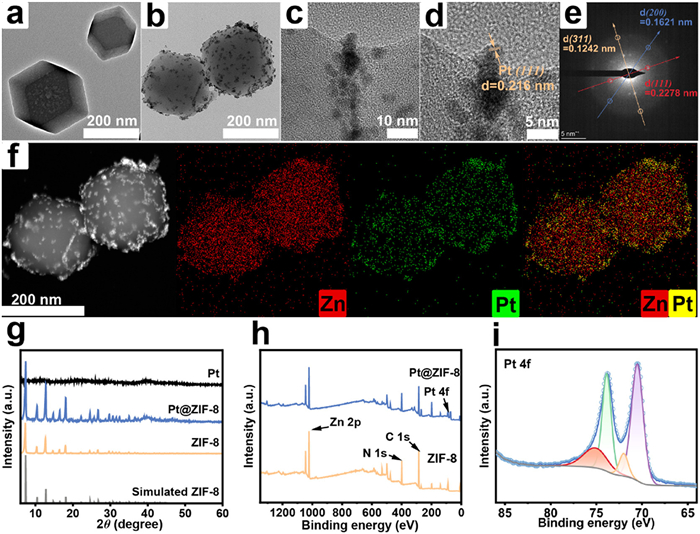
The Pt@ZIF-8 were prepared using a two-step method. Firstly, ZIF-8 was synthesized in methanol according to the previously reported method with appropriate modifications. Transport electron microscopy (TEM) characterization showed that the as-prepared ZIF-8 crystals had a dodecahedral structure with a mean size of 326.2 nm (Fig. 1a). Using the deposition–precipitation (DP) method, rapid reduction of metal cations in K2PtCl4 by NaBH4 within a ZIF-8 suspension was conducted to produce Pt@ZIF-8 (Figs. 1b and c). The change in zeta potential also demonstrated Pt NPs successfully loaded in ZIF-8 (Fig. S2 in Supporting information). Dynamic light scattering (DLS) measurements show that the average particle size of Pt@ZIF-8 is 396.1 ± 4.4 nm (Fig. S1 in Supporting information). The TEM images of Pt@ZIF-8 clearly show the crystal structure of Pt NPs, and regular alternating dark and bright spots (Figs. 1d and e). From the fast Fourier transform (FFT) pattern (Fig. 1e), (111), (200) and (311) planes of the pure Pt NPs were identified, where the d spacings corresponding to the (111) plane is 0.216 nm. These results indicate that encapsulation of ZIF-8 does not affect the crystal structure of Pt NPs. The elemental mapping showed that the Pt NPs were distributed evenly throughout the nanomaterial (Fig. 1f). The diffraction peaks of synthesised Pt@ZIF-8 were compared with those of ZIF-8 standard cards (2θ = 7.35°, 10.45°, 12.80°, 14.75°, 16.50°, 18.10°, 24.60°, 26.80°, 31.65°, 35.05°) using X-ray diffraction (XRD), confirming successful synthesis without alteration to the lattice structure of ZIF-8 due to Pt NP loading (Fig. 1g). Fourier transform infrared spectrometer (FTIR) spectra of Pt@ZIF-8 demonstrate the same result (Fig. S3 in Supporting information). X-ray photoelectron spectroscopy (XPS) revealed the presence of four elements (C, N, Zn, Pt) in Pt@ZIF-8, with Pt predominantly in the zero-valence state within the MOF, exhibiting peaks at 71.9 eV (4f7/2) and 75.1 eV (4f5/2), indicative of its effective catalytic ability (Figs. 1h and i). The Pt loading content in Pt@ZIF-8 was determined to be 1.6% (wt%) using Inductively coupled plasma optical emission spectroscopy (ICP-OES).
The ROS scavenging ability of Pt@ZIF-8 was assessed in this study (Fig. 2a). SOD is renowned for its capacity to catalyze the •O2− to generate oxygen (O2) and H2O2, and CAT is known to decompose H2O2 into O2 and H2O. Therefore, the enzyme-like catalytic activity of Pt@ZIF-8 was assessed by SOD activity kit and CAT activity kit respectively. The results showed that the elimination capacity of •O2− and H2O2 exhibited a positive correlation with increasing Pt concentrations (Figs. 2b and c). The SOD/CAT mimicry capability was further validated by monitoring the time course of O2 generation by dissolved oxygen probe. In the presence of Pt@ZIF-8, O2 levels gradually increased with time, and the increase of Pt@ZIF-8 concentration accelerated O2 generation (Fig. 2d). Additionally, based on the Michaelis-Menten curves and Lineweaver-Burk plot, the Michaelis-Menten constant (KM) and maximum velocity (Vmax) were determined to be 6.188 mmol/L and 1.018×10⁻⁶ mol L−1 s−1 (Figs. 2e and f). These findings collectively demonstrate that Pt@ZIF-8 exhibits excellent SOD and CAT-like activities, effectively scavenging •O2− and H2O2.
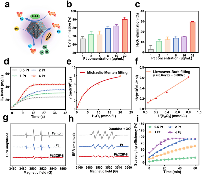
Electron paramagnetic resonance (EPR) spectra were utilized to investigate •OH scavenging capabilities of Pt NPs and Pt@ZIF-8. The •OH was generated through the Fenton reaction by Fe2+/H2O2 system and spin-trapped by 5,5-dimethyl-1-pyrroline-N-oxide (DMPO). The EPR spectra revealed distinctive signals corresponding to DMPO—OH adducts, indicating successful •OH generation. After adding Pt NPs and Pt@ZIF-8 in Fe2+/H2O2 system, the signal intensity decreased significantly, particularly for Pt@ZIF-8 (Fig. 2g). Furthermore, the •OH scavenging activity of Pt@ZIF-8 was also evaluated using UV–vis spectroscopy. The reaction between SA and •OH produced an absorption peak at 510 nm. As expected, the ability of Pt@ZIF-8 effectively scavenging •OH radicals exhibited a dose-dependent manner (Fig. S4 in Supporting information). Additionally, EPR spectra were conducted to assess •O2− scavenging ability of Pt NPs and Pt@ZIF-8. •O2− was generated by the reaction of xanthine and xanthine oxidase and spin-trapped by DMPO. Similarly, after adding Pt NPs and Pt@ZIF-8, the signal intensity sharply decreased, and Pt@ZIF-8 showed enhanced •O2− scavenging ability (Fig. 2h).
The overall antioxidant capacity of Pt@ZIF-8 was assessed using the 2,2′-azino-bis(3-ethylbenzothiazoline-6-sulfonic acid) (ABTS) assay. Significant reduction in absorbance of ABTS•+ radicals was observed in the presence of Pt@ZIF-8. The scavenging efficiency of radicals correlated positively with Pt concentration, which implies that Pt@ZIF-8 has good antioxidant capacity to scavenge ABTS•+ radicals (Fig. 2i).
Before fabrication of hydrogels, we first synthesised two monomers, GelMA and ALN-ac. The 1H NMR spectrum showed that the proton peaks of methyl (–CH═CH2) appeared at 5.43 and 5.67 ppm, indicating that the double bond was successfully introduced in gelatin and GelMA was synthesised (Fig. S5 in Supporting information). As for ALN-ac, the peak of the carbon double bond could be observed in the range of chemical shifts around 6.2 ppm (Fig. S6 in Supporting information). For hydrogel synthesis, pregel solutions were first prepared at 37 ℃ by dissolving lyophilised GelMA in an aqueous solution containing 0.05% lithium phenyl(2,4,6-trimethylbenzoyl)phosphinate (LAP). Afterwards, Pt@ZIF-8 or ALN-ac were added, such that the final concentrations of Pt@ZIF-8, ALN-ac, GelMA and LAP were fixed at 2%, 5%, 10% and 0.05%, respectively. All prepared pregel solutions were irradiated under 405 nm UV light to produce cross-linking (Fig. 3a). According to the components, we classified the hydrogels into the following four groups, Gel, Pt@ZIF-8/Gel, ALN-ac and Pt@ZIF-8/ALN-ac/Gel.
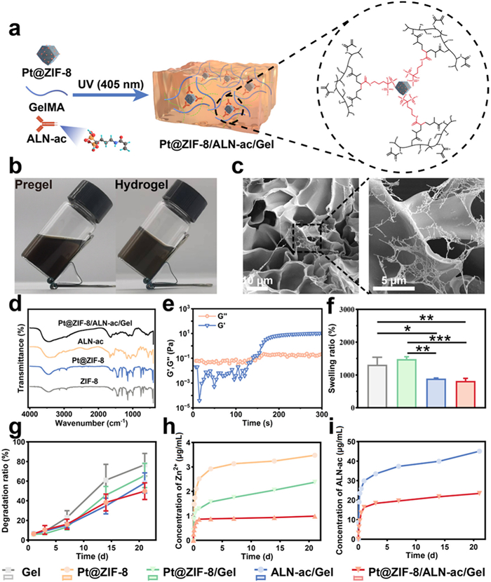
We observed that the Pt@ZIF-8/ALN-ac/Gel pregel solution formed a solid cross-linked hydrogel upon UV irradiation (Fig. 3b). Scanning electron microscopy (SEM) of the Pt@ZIF-8/ALN-ac/Gel hydrogel showed that the hydrogel exhibited a porous and homogeneous cross-linking structure, whereas Pt@ZIF-8 was uniformly distributed throughout the hydrogel (Fig. 3c). FTIR analysis confirmed that the Pt@ZIF-8/ALN-ac/Gel hydrogel exhibited characteristic peaks of Pt@ZIF-8 (Zn–N and C–N), ALN-ac (P=O and P–O), and GelMA (N–H and C=O), confirming successful incorporation of Pt@ZIF-8 and ALN-ac into GelMA (Fig. 3d).
To survey the hydrogel formation, the dynamic rheological properties of the Pt@ZIF-8/ALN-ac/Gel hydrogels were monitored by a rheometer. At the beginning, the loss modulus (G′′) was larger than storage modulus (G′), upon UV irradiation, G′ increased rapidly, and a crossover between G′ and G′′ indicated the sol-gel phase transition occurred (Fig. 3e). Swelling behavior of the hydrogels was also assessed. The results showed that the addition of Pt@ZIF-8 increased the swelling rate of the hydrogels, but the swelling rate decreased significantly after the addition of ALN-ac, which indicated that ALN-ac could increase the crosslink density of GelMA, which was beneficial to delay the degradation of the hydrogels (Fig. 3f). Further, we explored the degradation characteristics of the hydrogels, and the results showed that the addition of ALN-ac significantly reduced the degradation rate of the hydrogels, and it is noteworthy that the introduction of Pt@ZIF-8 could also reduce the degradation rate (Fig. 3g). The results of the swelling and degradation tests indicated that the Pt@ZIF-8/ALN-ac/Gel hydrogel exhibited a high degree of cross-linking and was able to sustain its integrity under the biological conditions.
The release of Zn2+ from the Pt@ZIF-8/ALN-ac/Gel was detected by ICP-OES, the release curve showed that the GelMA hydrogel was able to protect the nanoparticles and reduce the hydrolysis of Pt@ZIF-8, while introducing ALN-ac significantly reduced the release rate of Zn2+ and realised sustained release of Zn2+ (Fig. 3h). Similarly, we monitored the release characteristics of ALN-ac and found that the introduction of Pt@ZIF-8 retarded the release of ALN-ac, mainly possibly due to the interaction of the bisphosphate group with Pt@ZIF-8 (Fig. 3i). These results indicated that the Pt@ZIF-8/ALN-ac/Gel hydrogel could substantially release Zn2+ and ALN-ac, which was conducive to inhibiting the proliferation of oral pathogens and promoting the osteogenic differentiation of hPDLSCs.
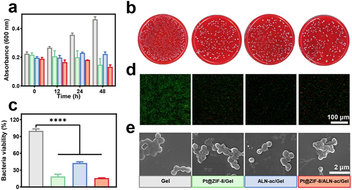
We chose anaerobic P. gingivalis as the typical bacteria of periodontitis. The hydrogel was cocultured with bacteria to test its antibacterial effect. By monitoring the absorbance of the bacteria at 600 nm, we found that the bacteria in the Gel group showed good growth within 48 h, while the Pt@ZIF-8/ALN-ac/Gel group had a significant inhibition of bacterial growth (Fig. 4a). It is worth mentioning that the ALN-ac/Gel group also showed the ability to inhibit the growth of bacteria. Plate colony images showed that compared with the Gel group, the colonies on plates of the Pt@ZIF-8/Gel, ALN-ac/Gel and Pt@ZIF-8/ALN-ac/Gel groups were all decreased in different degrees, with the Pt@ZIF-8/ALN-ac/Gel group having the best antibacterial effect (Fig. 4b). By evaluating the bacterial viability, the Pt@ZIF-8/ALN-ac/Gel group was also demonstrated to have good antibacterial performance (Fig. 4c). Live-dead staining (Fig. 4d) and relative fluorescence intensity (Fig. S7 in Supporting information) indicated significantly higher antibacterial activity in the Pt@ZIF-8/ALN-ac/Gel group compared to the Gel group. Compared with Gel group, P. gingivalis in group Pt@ZIF-8/ALN-ac/Gel exhibited severe shrinkage and deformation, indicating that the bacteria had lost their activity. These results indicate that the Pt@ZIF-8/ALN-ac/Gel hydrogel has excellent antibacterial properties.
The Pt@ZIF-8/ALN-ac/Gel hydrogel was further analyzed for antioxidation capacity and inducing osteogenic differentiation capacity by hPDLSCs in the cellular microenvironment. Firstly, hPDLSCs were cocultured with Pt@ZIF-8/ALN-ac/Gel hydrogel and after incubation for 1, 3, 7 days, cell activity was detected by cell counting kit-8 (CCK-8). No significant inhibitory effect on cell proliferation was observed, indicating that the Pt@ZIF-8/ALN-ac/Gel hydrogel has excellent cytocompatibility (Fig. S8 in Supporting information). Live/dead staining assay was performed after seeding cells on the surface of hydrogel for 24 h. The results showed that most of cells in each group survived after treating with different hydrogels, and cell proliferation was not inhibited (Fig. 5a). Furthermore, 4′,6-diamidino-2-phenylindole (DAPI)/tetramethylrhodamine isothiocyanate (TRITC)-phalloidin fluorescence staining showed that hPDLCs displayed well-stretched structures and favourable viability in all groups, indicating that the Pt@ZIF-8/ALN-ac/Gel hydrogel was able to support cell adhesion well (Fig. 5b). These findings underscore that the Pt@ZIF-8/ALN-ac/Gel hydrogel has good biocompatibility and can be suitable for further practical applications.
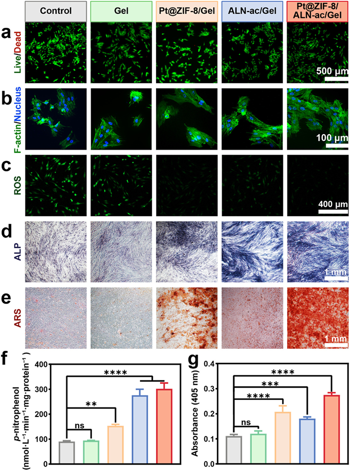
Oxidative stress occurs when excess ROS cannot be removed quickly enough and leads to apoptosis. To explore the scavenging capacity of Pt@ZIF-8/ALN-ac/Gel on intracellular ROS, H2O2 was used to stimulate intracellular oxidative stress. Subsequently, we monitored intracellular ROS levels using 2′,7′-dichlorofluorescin diacetate (DCFH-DA) as a fluorescent probe following treatment with various hydrogels. The results showed that the control group had a strong fluorescence signal and the addition of GelMA had no significant antioxidant effect. The introduction of ALN-ac decreased the intracellular fluorescence intensity a bit, while with the addition of Pt@ZIF-8, the intracellular fluorescence intensity decreased significantly, and the Pt@ZIF-8/ALN-ac/Gel group had the lowest fluorescence intensity (Fig. 5c). This demonstrated that Pt@ZIF-8/ALN-ac/Gel hydrogel could effectively scavenge ROS and could protect cells from oxidative stress damage.
Given the importance of osteogenesis in treating periodontitis defects, osteogenic capacity was assessed using alizarin red S (ARS) and alkaline phosphatase (ALP) staining. First, we evaluated ALP activity, an early osteogenesis marker. Staining was performed after 14 days of culture, the results indicated that the ALN-ac/Gel and Pt@ZIF-8/ALN-ac/Gel groups stained significantly stronger than the other groups (Fig. 5d). The quantitative results further demonstrated that ALN-ac was able to significantly facilitate ALP production, with the highest activity in the Pt@ZIF-8/ALN-ac/Gel group (Fig. 5f). The ARS staining results showed that compared with the control group, the hydrogels of the Pt@ZIF-8/Gel, ALN-ac/Gel and Pt@ZIF-8/ALN-ac/Gel groups generated different degrees of mineralisation, with the Pt@ZIF-8/ALN-ac/Gel group exhibiting the greatest degree of mineralisation, and the quantitative results showed the same tendency (Figs. 5e and g). In conclusion, Pt@ZIF-8/ALN-ac/Gel hydrogels effectively enhanced osteogenic differentiation of hPDLSCs in vitro.
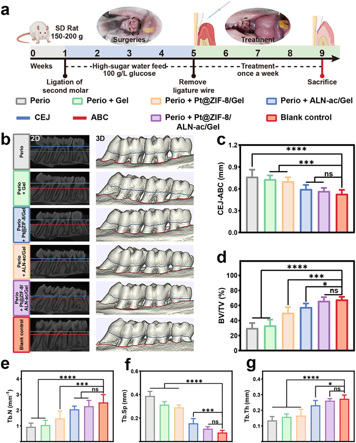
The aforementioned in vitro experiments collectively demonstrated that the Pt@ZIF-8/ALN-ac/Gel hydrogel not only effectively inhibited the proliferation of oral pathogens, but also exhibited excellent antioxidant and osteogenic differentiation abilities. However, given the variability and complexity of the oral microenvironment, the ability of anti-inflammatory and osteogenic regeneration in vivo needs further research. Thus, the therapeutic efficacy of Pt@ZIF-8/ALN-ac/Gel on periodontitis was assessed using the typical ligature-induced periodontitis model of rats. All animal experiments and procedures adhered to institutional guidelines on animal welfare and were approved by the Institutional Laboratory Animal Care and Use Committee of Sichuan University (permit No. 20230307030). The ligation was performed around the maxillary second molar in rats, and high glucose diet was given simultaneously to establish ligature-induced periodontitis model in rat. After four weeks, different hydrogels were injected into the periodontal pocket once a week for four weeks to observe the therapeutic effect of Pt@ZIF-8/ALN-ac/Gel on the periodontal tissues of rats (Fig. 6a).
In vivo antimicrobial effect was assessed using the spread plate method. The results showed that the number of colonies was significantly lower in the Pt@ZIF-8/ALN-ac/Gel group than in the periodontitis group and the Gel group, indicating that the Pt@ZIF-8/ALN-ac/Gel hydrogel has excellent in vivo antibacterial activity (Fig. S9 in Supporting information).
The therapeutic effect was assessed by micro-computed tomography (Micro-CT) analysis. The vertical distance between the alveolar bone crest (ABC) and the cementoenamel junction (CEJ) was assessed as the therapeutic index using 3D reconstruction. Micro-CT imaging clearly showed a significant loss of alveolar bone in the periodontitis group, whereas the Pt@ZIF-8/ALN-ac/Gel group significantly reduced bone loss (Fig. 6b). After quantitative analysis, bone loss was found to be most pronounced in the periodontitis group compared to the control group. After treatment with Pt@ZIF-8/ALN-ac/Gel hydrogel, CEJ-ABC was significantly reduced, showed no statistically significant difference compared to the control group (Fig. 6c). These results suggest that Pt@ZIF-8/ALN-ac/Gel hydrogel has a positive effect on the therapy of alveolar bone loss. Upon further assessment of bone volume in the vicinity of the second molar, it was observed augmentation in bone tissue (bone volume/total volume, BV/TV), elevation in trabecular thickness (Tb.Th) and trabecular number (Tb.N), and reduction in trabecular separation (Tb.Sp) after treatment with Pt@ZIF-8/ALN-ac/Gel, demonstrating the favourable therapeutic effect of Pt@ZIF-8/ALN-ac/Gel (Figs. 6d–g).
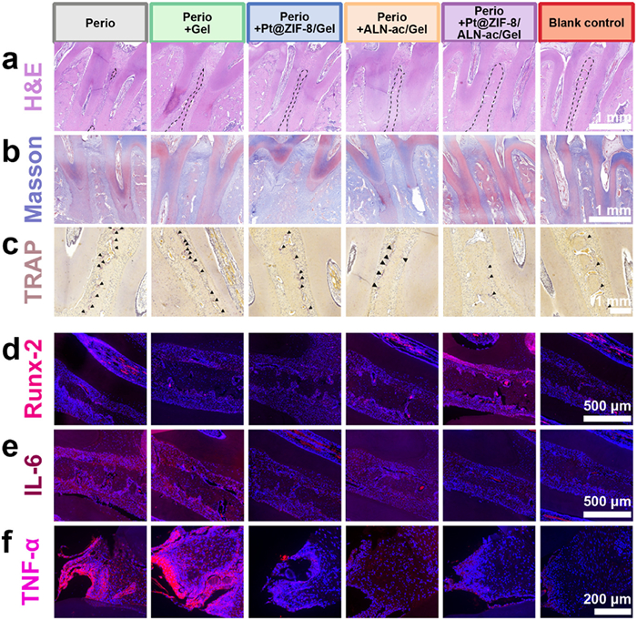
After four weeks of therapy, periodontal bone regeneration was further assessed using hematoxylin-eosin (H&E) staining and Masson trichrome staining. H&E staining showed that the substantial presence of inflammatory cells was observed accompanied by a decrease in alveolar ridge height and bone mass in the periodontitis group, which were significantly reversed in the Pt@ZIF-8/ALN-ac/Gel group (Fig. 7a). Masson trichrome staining showed that collagen fiber in the periodontitis group exhibited a disordered and irregular structure, and Pt@ZIF-8/ALN-ac/Gel significantly ameliorated these abnormalities and enhanced collagen deposition (Fig. 7b). We then used tartrate-resistant acid phosphatase (TRAP) staining to detect osteoclast activity. Compared with the periodontitis group, both the ALN-ac/Gel and Pt@ZIF-8/ALN-ac/Gel groups led to a decline in the number of osteoclasts, demonstrating that ALN-ac was able to significantly reduce osteoclast activity (Fig. 7c). To further investigate the mechanism of bone regeneration, immunofluorescence staining was conducted. Runt-related transcription factor 2 (RUNX-2) is known to be a factor involved in the regulation of mineralisation and bone tissue maturation [43]. Immunofluorescence images of RUNX-2 showed that Pt@ZIF-8/ALN-ac/Gel had significant osteogenic properties in inflammatory environment (red fluorescence, Fig. 7d). Tumor necrosis factor-α (TNF-α) and inflammatory cytokine interleukin-6 (IL-6) involved in a variety of pathological responses and are important inflammatory markers [44]. Immunofluorescence images showed that IL-6 expression was the highest in the periodontitis group, suggesting inflammation. significantly decreased after treatment with Pt@ZIF-8/ALN-ac/Gel hydrogel, which indicated that Pt@ZIF-8/ALN-ac/Gel had an obvious capacity to alleviate inflammation in vivo (red fluorescence, Fig. 7e). The immunofluorescence images of TNF-α also proved the above results (red fluorescence, Fig. 7f). These results collectively indicate that Pt@ZIF-8/ALN-ac/Gel hydrogel have good therapeutic effects, reduce inflammatory cell infiltration, decrease collagen destruction, inhibit osteoclast formation, and accelerate the restoration of periodontitis.
The treatment of periodontitis has been a persistent and pressing clinical challenge. Therefore, we synthesized injectable Pt@ZIF-8/ALN-ac/Gel hydrogels with anti-inflammatory, antibacterial and osteogenic functions to accelerate periodontal bone regeneration in patients. Pt@ZIF-8 exhibited excellent ROS scavenging ability due to ZIF-8 encapsulation modifying the surface properties of Pt NPs. Also, Pt@ZIF-8/ALN-ac/Gel hydrogel sustained the release of Zn2+ exhibiting excellent antibacterial activity against oral pathogens. In vitro, Pt@ZIF-8/ALN-ac/Gel hydrogel effectively scavenged intracellular ROS and increased ALP activity to promote extracellular matrix mineralisation. We further revealed that Pt@ZIF-8/ALN-ac/Gel hydrogel could significantly reduce the bacterial load, alleviate inflammation, inhibit osteoclast activity, and promote alveolar bone regeneration using a rat model of periodontitis. In summary, Pt@ZIF-8/ALN-ac/Gel hydrogel not only exhibits great promise as a potential therapeutic strategy for periodontitis, but also for other bone defects and inflammatory diseases.
The authors declare that they have no known competing financial interests or personal relationships that could have appeared to influence the work reported in this paper.
Xicheng Li: Writing – review & editing, Writing – original draft, Visualization, Validation, Software, Methodology, Investigation, Formal analysis, Data curation, Conceptualization. Dong Mo: Writing – review & editing, Validation, Software, Project administration, Formal analysis, Conceptualization. Shoushan Hu: Visualization, Resources, Methodology. Meng Pan: Software, Methodology. Meng Wang: Methodology. Tingyu Yang: Software, Methodology. Changxing Qu: Resources, Methodology. Yujia Wei: Methodology. Jianan Li: Methodology. Hanzhi Deng: Methodology. Zhongwu Bei: Methodology. Tianying Luo: Methodology. Qingya Liu: Methodology. Yun Yang: Methodology. Jun Liu: Resources. Jun Wang: Resources. Zhiyong Qian: Resources, Project administration, Methodology, Funding acquisition.
This work was financially supported by the National Natural Science Funds (NSFC, Nos. U21A20417 and 31930067) and 1·3·5 project for disciplines of excellence, West China Hospital, Sichuan University (No. ZYGD18002).
Supplementary material associated with this article can be found, in the online version, at doi:
B. Pihlstrom, B. Michalowicz, N. Johnson, Lancet 366 (2005) 1809–1820. doi: 10.1016/S0140-6736(05)67728-8
C. Ren, X. Hao, L. Wang, et al., Adv. Healthc. Mater. 10 (2021) 2100196. doi: 10.1002/adhm.202100196
B. Zeng, Z. Mu, T. Shen, et al., Chin. Chem. Lett. 36 (2025) 110350. doi: 10.1016/j.cclet.2024.110350
D. Richards, Evid. Based Dent. 15 (2014) 70–71. doi: 10.1038/sj.ebd.6401037
J. Li, S. Song, J. Meng, et al., J. Am. Chem. Soc. 143 (2021) 15427–15439. doi: 10.1021/jacs.1c07875
L. Wu, S. Zhang, L. Zhao, Z. Ren, C. Hu, J. Periodontol. 93 (2022) 1445–1454. doi: 10.1002/JPER.21-0469
Y. Xu, Y. Luo, Z. Weng, et al., ACS Nano 17 (2023) 18732–18746. doi: 10.1021/acsnano.3c01940
J. Gong, C. Ye, J. Ran, et al., ACS Nano 17 (2023) 16573–16586. doi: 10.1021/acsnano.3c02407
G. Hajishengallis, Nat. Rev. Immunol. 15 (2015) 30–44. doi: 10.1038/nri3785
L.M. Silva, A.D. Doyle, T. Greenwell-Wild, et al., Science 374 (2021) eabl5450. doi: 10.1126/science.abl5450
D. Herrera, P. Matesanz, C. Martín, et al., J. Clin. Periodontol. 47 (2020) 239–256. doi: 10.1111/jcpe.13230
P. Angst, A. Stadler, M. Mendez, R. Oppermann, U. Velden, S. Gomes, J. Clin. Periodontol. 46 (2019) 1083–1093. doi: 10.1111/jcpe.13178
F. Graziani, D. Karapetsa, B. Alonso, D. Herrera, Periodontology 2000 75 (2017) 152–188. doi: 10.1111/prd.12201
F. Graziani, L. Music, D. Bozic, G. Tsakos, Br. Dent. J. 227 (2019) 621–625. doi: 10.1038/s41415-019-0735-3
D. Mo, M. Pan, W. Chen, et al., Adv. Funct. Mater. 34 (2024) 2313569. doi: 10.1002/adfm.202313569
M. Zhang, X. Peng, H. Xu, et al., Adv. Sci. 11 (2024) 2404143. doi: 10.1002/advs.202404143
H. Wang, K. Wan, X. Shi, Adv. Mater. 31 (2019) 1805368. doi: 10.1002/adma.201805368
G. Tarricone, V. Castagnola, V. Mastronardi, et al., Nano Lett. 23 (2023) 4660–4668. doi: 10.1021/acs.nanolett.3c01479
J. Mu, C. Li, Y. Shi, et al., Nat. Commun. 13 (2022) 2513. doi: 10.1038/s41467-022-29772-w
D. Pedone, M. Moglianetti, E. De Luca, G. Bardi, P.P. Pompa, Chem. Soc. Rev. 46 (2017) 4951–4975. doi: 10.1039/C7CS00152E
F. Xia, X. Hu, B. Zhang, et al., Small 18 (2022) 2201558. doi: 10.1002/smll.202201558
X. Ji, Q. Li, H. Song, C. Fan, Adv. Mater. 34 (2022) 2201562. doi: 10.1002/adma.202201562
P. Horcajada, R. Gref, T. Baati, et al., Chem. Rev. 112 (2012) 1232–1268. doi: 10.1021/cr200256v
Y. Guo, S. Ding, C. Shang, et al., Adv. Mater. 36 (2024) 2306292. doi: 10.1002/adma.202306292
Y. Wan, J. Fang, Y. Wang, et al., Adv. Healthc. Mater. 10 (2021) 2101515. doi: 10.1002/adhm.202101515
L. He, G. Huang, H. Liu, et al., Sci. Adv. 6 (2020) eaay9751. doi: 10.1126/sciadv.aay9751
Y. Liu, Z. Zhu, X. Pei, et al., ACS Appl. Mater. Interfaces 12 (2020) 36978–36995. doi: 10.1021/acsami.0c12090
D. Mo, Z. Wang, K. Sun, et al., J. Mater. Chem. C 8 (2020) 11110–11118. doi: 10.1039/D0TC00908C
W. Wang, S. Chen, E. Guisasola Cal, et al., Inorg. Chem. Front. 7 (2020) 3945–3952. doi: 10.1039/D0QI00831A
T. Luo, H. Yang, R. Wang, et al., ACS Nano 17 (2023) 16715–16730. doi: 10.1021/acsnano.3c03169
K. Zhou, Z. Shi, X. Yi, et al., Chin. Chem. Lett. 36 (2025) 110226. doi: 10.1016/j.cclet.2024.110226
H. Wang, S. Chen, Z. He, et al., Chin. Chem. Lett. 35 (2024) 108597. doi: 10.1016/j.cclet.2023.108597
D. Steinberg, M. Friedman, Periodontology 2000 84 (2020) 176–187. doi: 10.1111/prd.12341
Y. Liu, T. Li, M. Sun, et al., Acta Biomater 146 (2022) 37–48. doi: 10.1016/j.actbio.2022.03.046
B. Maharjan, D. Kumar, G.P. Awasthi, et al., Composites Part B 177 (2019) 107415. doi: 10.1016/j.compositesb.2019.107415
T. Yu, Y. Hu, W. He, et al., Materials Today Bio. 19 (2023) 100558. doi: 10.1016/j.mtbio.2023.100558
W. Shi, X. Zhang, L. Bian, et al., Int. J. Biol. Macromol. 204 (2022) 441–456. doi: 10.1016/j.ijbiomac.2022.02.007
R. Wang, L. Che, Q. Feng, K. Cai, ACS Appl. Mater. Interfaces 14 (2022) 12038–12049. doi: 10.1021/acsami.1c23017
L. Xue, N. Gong, S.J. Shepherd, et al., J. Am. Chem. Soc. 144 (2022) 9926–9937. doi: 10.1021/jacs.2c02706
Q. Yao, Y. Liu, Y. Pan, et al., J. Colloid Interface Sci. 607 (2022) 1500–1515. doi: 10.1016/j.jcis.2021.09.089
L. Yang, X. Chen, L. Chen, et al., Macromol. Biosci. 24 (2024) 2300416. doi: 10.1002/mabi.202300416
J. Abtahi, F. Agholme, O. Sandberg, P. Aspenberg, J. Dent. Res. 92 (2013) 279–283. doi: 10.1177/0022034512472335
J. Li, L. Li, T. Wu, et al., Small Methods 8 (2024) e2300843. doi: 10.1002/smtd.202300843
Y. Qu, B. Chu, J. Li, et al., Small Methods 8 (2024) e2301178. doi: 10.1002/smtd.202301178

Scheme 1 Schematic illustration of the synthesis of Pt@ZIF-8/ALN-ac/Gel Hydrogel and its antimicrobial, anti-inflammatory and osteogenic properties in periodontitis.

Figure 1 Typical TEM image of (a) ZIF-8, and (b) Pt@ZIF-8. (c) High-resolution TEM image of Pt@ZIF-8 and (d) Pt NPs on ZIF-8. (e) FFT pattern obtained from Pt@ZIF-8. (f) EDS element mapping of Pt@ZIF-8. (g) XRD patterns of Pt@ZIF-8. (h) Survey and (i) Pt 4f of XPS spectra of Pt@ZIF-8.

Figure 2 (a) Schematic representation of ROS scavenging by Pt@ZIF-8 in vitro. (b, c) SOD and CAT activity of Pt@ZIF-8. (d) Concentration-dependent generation of O2 in the presence of Pt@ZIF-8 at different concentrations (0.5, 1.0, 2.0 and 4.0 µg/mL). (e, f) Michaelis-Menten steady-state kinetics of O2 generation from the decomposition of H2O2 by Pt@ZIF-8. (g) EPR spectra analysis of •OH scavenging by Pt@ZIF-8. (h) EPR spectra analysis of •O2− scavenging with Pt@ZIF-8. (i) Assessment of ABTS radical scavenging efficiency by different concentrations of Pt@ZIF-8. All data were shown as means ± SD (n = 3).

Figure 3 (a) Schematic demonstration of Pt@ZIF-8/ALN-ac/Gel. (b) Photographs of the pregel solution before and after hydrogel transition by UV irradiation. (c) SEM images of Pt@ZIF-8/ALN-ac/Gel. (d) FTIR spectrums of Pt@ZIF-8/ALN-ac/Gel. (e) Rheological testing of Pt@ZIF-8/ALN-ac/Gel. (f) Swelling ratio of Pt@ZIF-8/ALN-ac/Gel. (g) Degradation rates of Pt@ZIF-8/ALN-ac/Gel. (h) Cumulative release of Zn2+ from Pt@ZIF-8/ALN-ac/Gel. (i) Cumulative release of ALN-ac from Pt@ZIF-8/ALN-ac/Gel. All data were shown as means ± SD (n = 3). P < 0.05, **P < 0.01, ***P < 0.001.

Figure 4 (a) Absorbance of P. gingivalis with 48 h. (b) Images of the P. gingivalis colonies on blood agar plates. (c) Bacterial viability. (d) Live/dead staining (live bacteria were green, dead bacteria were red). (e) Morphology of P. gingivalis at 12 h. All data were shown as means ± SD (n = 3). ****P < 0.0001.

Figure 5 (a) Live/dead staining of hPDLSCs on 1 d (living cells were green and dead cells were red). (b) Morphology of hPDLSCs on 1 d (cytoskeleton were green and nuclei were blue). (c) Intracellular ROS-scavenging performance of Pt@ZIF-8/ALN-ac/Gel. (d) ALP staining of hPDLSCs on 14 d. (e) ARS staining of hPDLSCs on 21 d. (f) ALP activity of hPDLSCs on 14 d. (g) Quantification of extracellular matrix mineralization on 21 d. All data were shown as means ± SD (n = 3). **P < 0.01, ***P < 0.001, ****P < 0.0001. ns, no significant.

Figure 6 (a) Schematic experimental schedule of ligature-induced periodontitis model in rat and treatment with Pt@ZIF-8/ALN-ac/Gel. (b) Micro-CT scanning and 3D reconstructed image of the maxillary molars with different treatments by Micro-CT (blue lines represent the CEJ and red lines represent the ABC). (c) Quantitative analysis of the distance between CEJ and ABC. The quantitative analysis of bone-related parameters: (d) BV/TV, (e) Tb.N, (f) Tb.Sp and (g) Tb.Th from the Micro-CT images. All data were shown as means ± SD (n = 3). P < 0.05, ***P < 0.001, ****P < 0.0001.

Figure 7 (a) H&E staining images of the periodontal tissue. The area within black dotted line represents the alveolar bone. (b) Masson staining illustrates the maturing collagen fibers (blue). (c) The osteoclasts in periodontal tissue using TRAP staining (black arrowheads). (d) Immunofluorescence staining of RUNX-2 (red) and DAPI (blue). (e) Immunofluorescence staining of IL-6 (red) and DAPI (blue). (f) Immunofluorescence staining of TNF-α (red) and DAPI (blue).

 扫一扫看文章

扫一扫关注我们
 DownLoad:
 下载:
下载:
 下载:
下载: