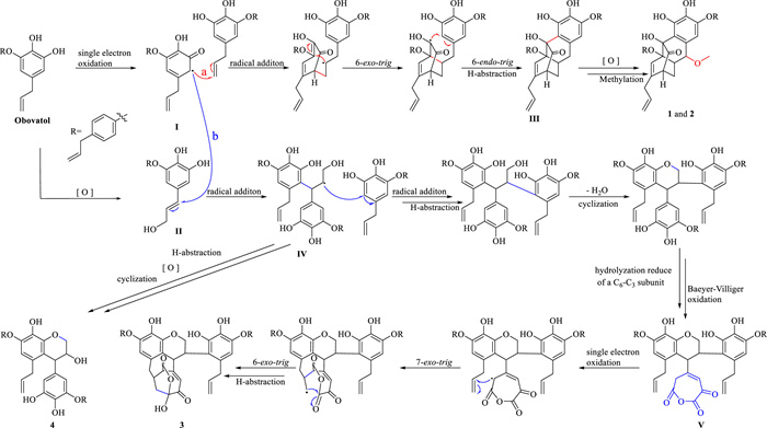
Scheme 1.
Possible biosynthetic pathways of compounds 1–4.
Houpolignols A–D, enantiomeric obovatol oligomeric neolignans with anti-NASH activities from Magnolia officinalis var. biloba
Wei-Ming Huang , Yue-You Yang , Ping Ying , Yu-Qian Cai , Tian-Jie Cao , Chuan-Lu Fu , Ling-Yi Kong , Wen-Jun Xu

Magnolia officinalis Rehd. et Wils var. biloba, named "Aoye Houpo" in Chinese, is a deciduous perennial tree belonging to the genus Magnolia (family Magnoliaceae), is native to southern regions of China. The cortex of the titled plant is one of the sources for the traditional Chinese medicine Magnolia officinalis Cortex [1]. For millennia, Magnolia officinalis Cortex has been regarded as an essential traditional medicine for preventing and treating various ailments, such as vomiting, phlegm, diarrhea, and phlegm [1]. In recent decades, botanists have extensively explored the secondary metabolites of M. officinalis var. biloba, resulting in the identification of a diverse array of compounds including neolignans, meroterpenoids, and alkaloids [2-8]. These compounds have demonstrated to exhibit various biological activities, such as anti-inflammatory, antibacterial, antioxidant, anti-nonalcoholic steatohepatitis (NASH), and protein tyrosine phosphatase 1B inhibitory activities, among which neolignans are the most significant bioactive components [9-21]. Obovatol, magnolol, and honokiol, typical neolignans of M. officinalis, can further form structurally attractive oligomeric neolignans through carbon-carbon or ether bonds. In an effort to search for oligomeric neolignans, four pairs of novel enantiomeric neolignanes, (+)/(−)-houpolignols A–D (1–4), were identified from the cortex of M. officinalis var. biloba. The unprecedented tetracyclo[9.3.1.02, 7.09, 14]pentadecane (1 and 2) and 8, 18-dioxapentacyclo[13.3.1.15, 9.04, 16.013, 20]icosane (3) core structures of 1–3 were confirmed by their 1D and 2D nuclear magnetic resonance (NMR) spectroscopic data and single crystal X-ray crystallography data of (±)-1 and (+)-3. The radical cascade cyclizations of obovatol and its dearomatized derivatives were considered as key steps in the proposed biogenetic pathway of 1–4 (Scheme 1). Herein, the isolation, structural elucidation, and anti-NASH activities of these compounds (Fig. 1) are detailed.
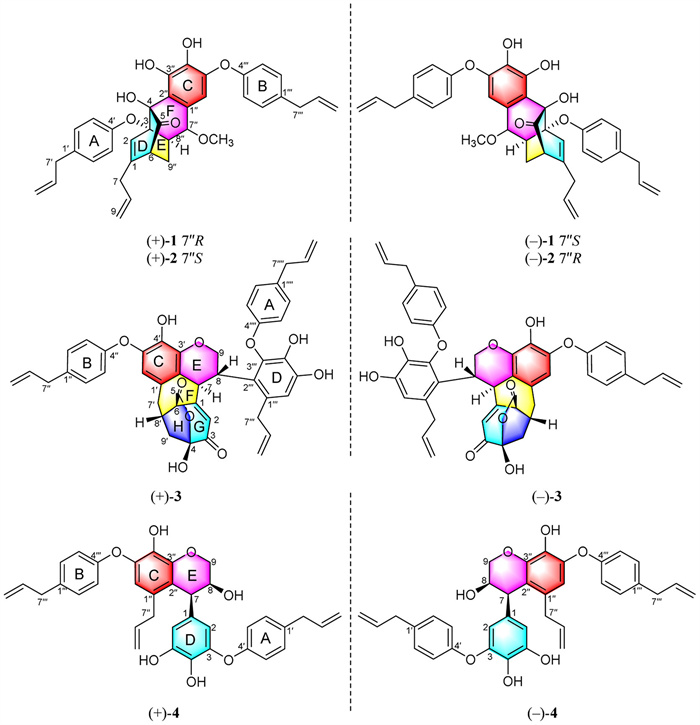
Houpolignol A (1) was obtained as a light yellow oil with a molecular formula of C37H36O7 on the basis of the positive ion high resolution electrospray ionization mass spectroscopy (HRESIMS) data (m/z 615.2354 [M+Na]+, calcd. for C37H36O7Na, 615.2353), corresponding to 20 indices of hydrogen deficiency (IHDs). The infrared spectroscopy (IR) absorptions of 1 indicated the presence of C═O (1734 cm−1), C═C (1637 cm−1), and benzene (1650, 1505 cm−1) groups. The 1H NMR spectrum of 1 (Table S1 in Supporting information) displayed two sets of signals for a 1, 4-disubstituted benzene rings, three groups of allyl proton signals, three phenol hydroxyl hydrogen signals, and one methoxy. In conjunction with the analyses of its heteronuclear single quantum correlation (HSQC), heteronuclear multiple-bond correlation (HMBC), and 1H—1H correlation spectroscopy (1H—1H COSY) spectra (Fig. 2), the 13C NMR data (Table S1 in Supporting information) of compound 1 revealed 18 aromatic carbons, 8 olefin carbons, and one carbonyl carbon, which accounted for 17 IHDs. The remaining 3 IHDs suggested that 1 had a tricyclic structure. The HMBC correlations from H-7′ to C-1′ and from H-7′′′ to C-1′′′, combined with the 1H—1H COSY correlations (Fig. 2) of H-7′/H-8′/H-9′, H-2′/H-3′, H-5′/H-6′, H-7′′′/H-8′′′/H-9′′′, H-2′′′/H-3′′′, and H-5′′′/H-6′′′ revealed the presence of two 1-allylphenol subunits (subunits A and B). In addition, the HMBC correlations from H-7′′ to C-1′′, C-2′′, C-6′′, C-8′′, and C-9′′, from H-6′′ to C-4′′ and C-5′′, and from 7′′-OCH3 to C-7′′, in conjunction with the 1H—1H COSY correlations of H-7′′/H-8′′/H-9′′, revealed the presence of a 1′′-(1-methoxypropyl)-3′′, 4′′, 5′′-trihydroxyphenyl (subunit C). The presence of a dearomatized 1-allyl-trihydroxyphenyl subunit (subunit D) was revealed via the HMBC correlations from H-7 to C-1, C-2, C-6, C-8, and C-9, and from H-6 to C-1, C-2, C-4, C-5, and C-7. Based on the HMBC correlations from 4-OH to C-3, C-4, C-5, and C-2′′, from H-8′′ to C-3, C-4, C-6, C-1′′, C-7′′, and C-9′′, and from H-6 to C-8′′, and C-9′′, as well as the 1H—1H COSY correlations of H-6/H-9′′/H-8′′/H-7′′, the connection patterns of C-2′′-C-4, C-6-C-9′′, and C-3-C-8′′ were identified. The rings C, D, E, and F in 1, fused with a tetracyclo[9.3.1.02, 7.09, 14]pentadecane core structure, were thus constructed. The rotating-frame overhauser effect spectroscopy (ROESY) cross-peaks of H-2 (subunit D) with H-3′/H-5′ (subunit A), and of H-6′′ (subunit B) with H-3′′′/H-5′′′ (subunit C) evidenced that subunits A and B were attached to the core structure via C-4′-O-C-3 and C-4′′′-O-C-5′′ ether linkages, respectively. The planar structure of 1 was resolved as shown (Fig. 1), which represented the first dearomatized obovatol dimer with a highly rigid cage-like 6/6/6/6 tetracyclic system based on the tetracyclo[9.3.1.02, 7.09, 14]pentadecane core structure.
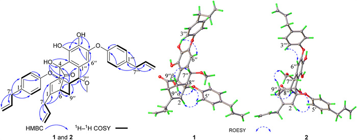
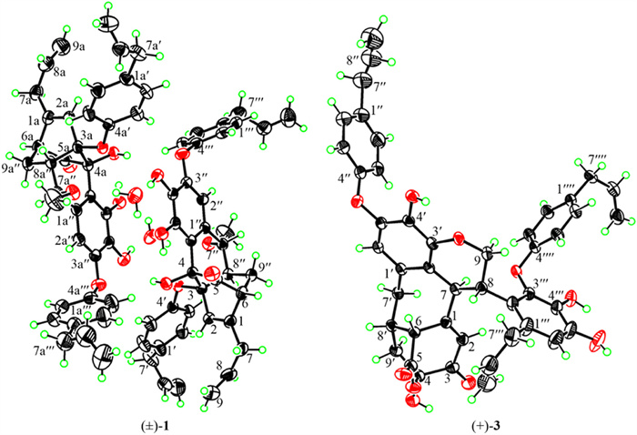
The relative configurations of C-3, C-4, C-6, and C-8′′ were fixed as a result of the highly rigid cage-like core structure of 1 [22], and was confirmed by the ROESY cross-peaks of H-9′′α/H-8′′, H-8′′/H-5′, and H-2/H-5′. The ROESY cross-peaks of H-9′′β/H-7′′ enabled the orientation of H-7′′ as β. The needle-like single crystals of 1 were obtained through slow evaporation in a 50% methanol-water solution at 4 ℃. The X-ray crystallographic data (CCDC: 2339297) confirmed the structure of 1, and indicated that compound 1 existed in a racemic form (Fig. 3). Compounds (±)-1 was then subjected to preparative high performance liquid chromatography (HPLC) separation on a Phenomenex Cellulose-3 chiral column (MeOH/H2O = 80:20, v/v) to yield two optically pure enantiomers, (+)-1 and (−)-1. The absolute configurations of (+)-1 and (−)-1 were determined by electronic circular dichroism (ECD) calculations using time-dependent density functional theory (TDDFT) at B3LYP/6–311+G(d, p) level. The calculated ECD spectra of (3S, 4R, 6R, 7′′R, 8′′R)-1 and (3R, 4S, 6S, 7′′S, 8′′S)-1 were in good agreements with the experimental ECD spectra of (+)-1 and (−)-1 (Fig. S1 in Supporting information), respectively. This indicated that the absolute structures of (+)-1 and (−)-1 were 3S, 4R, 6R, 7′′R, 8′′R and 3R, 4S, 6S, 7′′S, 8′′S.
Houpolignol B (2) was obtained as an optically inactive light yellow oil with the same molecular formula of C37H36O7 as that of 1, assigned by its 13C NMR and positive ion HRESIMS data (m/z 615.2354 [M+Na]+, calcd. for C37H36O7Na, 615.2353). The 1D NMR spectroscopic data of 2 and 1 (Table S1) were similar, except for the resonances around C-7′′, indicating that 2 was the C-7′′ epimer of 1. This assignment was further supported by the ROESY correlation of 7′′-OCH3 and H2-9′′ in 2, instead of the ROESY correlation of H-7′′ and H-9′′β in 1. In addition, DP4+ probabilistic analyses based on the computed 1H and 13C NMR chemical shifts of 1 and the C-7′′ epimer of 1 supported the latter as the correct structure of 2 with a probability of 100% (Fig. S3 in Supporting information). The DP4+ probability analysis for compound 1 was also conducted to confirmed the reliability of the NMR calculations of 2. The results showed that isomer 1 (3S*, 4R*, 6R*, 7′′R*, 8′′R*−1a) was the correct structure of 1 with a probability of 100%, which was consistent with the structure defined by the X-ray crystallographic data of (±)-1 (Fig. S2 in Supporting information). Identically, 2 was obtained as a racemic mixture, and was further subjected to chiral HPLC resolution to yield (+)-2 and (−)-2. The calculated ECD spectra indicated (Fig. S1) that the absolute structures of (+)-2 and (−)-2 were 3S, 4R, 6R, 7′′S, 8′′R and 3R, 4S, 6S, 7′′R, 8′′S.
Houpolignol C (3) was obtained as a white powder with a molecular formula of C45H40O10 on the basis of the negative-ion HRESIMS data (m/z 739.2550 [M−H]−, calcd. for C45H39O10, 739.2549), corresponding to 26 IHDs. The 1H NMR spectroscopic data of 3 (Table S2 in Supporting information) displayed two sets of signals for para-substituted benzene rings, two singlet signals of penta-substituted benzene ring proton, three phenol hydroxyl hydrogen signals, and three allyl proton signals. The 13C NMR, distortionless enhancement by polarization transfer (DEPT), and HSQC spectroscopic data of 3 showed 45 carbon resonances, which were classified to four benzene rings carbon signals, three sets of allyl carbon signals, one α, β-unsaturated carbonyl groups [δ 160.8 (C-1), 127.2 (C-2), and 188.9 (C-3)], one ester carbonyl [δ 169.4 (C-5)], one hemiacetal carbon [δ 102.4 (C-4)], 3 methylene carbons, and 4 methine carbons. The aforementioned functional groups accounted for 22 IHDs. The remaining 4 IHDs suggested that 3 had an additional tetracyclic structure. The presence of two C6C3 subunits (subunits A and B) was defined by the HMBC correlations from H-7′′ to C-1′′ and from H-7′′′′ to C-1′′′′, and by the 1H—1H COSY correlations (Fig. 4) of H-7′′/H-8′′/H-9′′, and H-7′′′′/H-8′′′′/H-9′′′′. Additionally, the HMBC correlations from H-7′ to C-1′ and from H-7′′′ to C-1′′′, in conjunction with the 1H—1H COSY correlations of H-7′/H-8′/H-9′ and of H-7′′′/H-8′′′/H-9′′′ revealed the presence of a 1-propyl-3′, 4′, 5′-trihydroxyphenyl (subunit C) and a 1-allyl-3′′′, 4′′′, 5′′′-trihydroxyphenyl (subunit D), respectively. The 1H—1H COSY correlations elucidated the connectivity of the H-7/H-8/H-9 segment, which in conjunction with the HMBC correlations from H-7 to C-1, C-2, C-6, C-8, C-9, C-2′, and C-3′, and from H-9 to C-3′, from H-7′ to C-6 and C-8′ established the presence of rings E and F in 3. Concurrently, it was ascertained that subunit D was connected to the ring E through C-2′′′-C-8 linkage on the basis of the HMBC correlations from H-7/H-8 to C-2′′′. The unassigned ester carbonyl, hemiacetal carbon, and α, β-unsaturated carbonyl moiety indicated that a C6 benzene unit in 3 was oxidized to an Oza-hepta-unsaturated lactone moiety (ring G). This deduction and the HMBC correlations from H-2 to C-1, C-3 C-4, and C-6, from H-6 to C-5, from 4-OH to C-3, C-4 and C-9′, from H-9′ to C-3, C-6 and C-8′, and from H-7′ to C-6, C-8′ and C-9′ confirmed the presence of rings G and H, which were deduced to be formed between a C-1′ allyl of subunit C and the Oza-hepta-unsaturated lactone moiety in 3. This result was consistent with the molecular formula of 3 with 2 remaining IHDs besides of rings E and F. Moreover, the HMBC correlations were observed from 4′′′-OH to C-3′′′, C-4′′′, and C-5′′′, from 5′′′-OH to C-4′′′, C-5′′′, and C-6′′′, and from 4′-OH to C-3′, C-4′, and C-5′. These correlations, combined with ROESY cross-peaks between H-6′ and H-3′′/H-5′′, evidenced that subunits A and B were attached to C-3′′′ and C-5′, respectively, through ether bonds. Accordingly, the planar structure of compound 3 was resolved as shown (Fig. 1), which resented the first dearomatized obovatol oligomeric with 6/6/7/6/7 pentacyclic system based on a 8, 18-dioxapentacyclo[13.3.1.15, 9.04, 16.013, 20]icosane core structure.
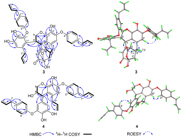
The relative configuration of 3 was determined by the analyses of its ROESY data and coupling constant. In the ROESY spectrum of 3, the key correlations of H-6/H-8′, H-8′/H-7′β, and H-7/H-7′α (Fig. 4) were observed. Additionally, the coupling constant (JH-7/H-8 = 11.7 Hz) between H-7 and H-8 indicated their trans orientations [23-25]. These data determined that H-6/H-8/H-8′ were β-oriented, and that H-7 was α-oriented. Compound (±)-3 was obtained as a racemic mixture as well. Chiral HPLC resolution on a Daicel Chiralpak AD-H column (n-hexane: isopropanol 3:2, v/v) afforded enantiomers (+)-3 and (−)-3. The absolute configurations of (+)-3 and (−)-3 were determined as 4S, 6R, 7S, 8S, 8′S and 4R, 6S, 7R, 8R, 8′R, respectively, through ECD calculation (Fig. S1). Finally, a needle-like single crystal of (+)-houpolignol C [(+)-3] was obtained from a n-hexane-CH2Cl2 (1:2) solution; and then the absolute configuration of compound (+)-3 (Fig. 3) was further verified as 4S, 6R, 7S, 8S, 8′S by its X-ray crystallographic data [CCDC: 2355403, Flack = 0.1 (7)].
Houpolignol D (4) was obtained as a light yellow oil with a molecular formula of C36H34O7 on the basis of the positive-ion HRESIMS data (m/z 601.2199 [M+Na]+, calcd. for C36H34O7Na, 601.2197), indicative of four sets of C6C3 subunits in the structure of 4. The 1H and 13C NMR spectroscopic data of 4 (Table S2) displayed characteristic signals for an obovatol dimer, including of two para-substituted benzene rings, a 1, 3, 4, 5-substituted benzene ring, a penta-substituted benzene ring, and three allyl moieties. The presences of two obovatol-like units were further elucidated by HMBC, 1H—1H COSY, and ROESY spectra. The HMBC correlations (Fig. 4) from H-7 to C-1, C-2, C-6, C-8, C-9, C-2′′, and C-3′′, and from H-9 to C-7, C-8, and C-3′′, in conjunction with the 1H−1H COSY correlations (Fig. 4) of H-7/H-8/H-9, indicated that an allyl moiety of subunit D was oxidized to a 1, 2-diol-propyl moiety in 4, and then formed ring E with C-7-C-2′′ and C-9-O—C-3′′ linkages. The broad singlet 1H NMR resonances of H-7 and H-8 and the ROESY cross-peak of H-7/H-8 indicated their cis orientations [23-25]. The near-zero specific rotation value suggested that 4 might be obtained as a racemate. Likewise, compounds (±)-4 was further subjected to chiral HPLC separation to yield two optically pure enantiomers, (+)-4 and (−)-4. The calculated ECD curves for (7S, 8S)-4 and (7R, 8R)-4 corresponded closely with the experimental ones of (+)-4 and (−)-4, respectively (Fig. S1). This correlation allowed the determination that the absolute structures of (+)-4 and (−)-4 were 7S, 8S and 7R, 8R.
Houpolignols A–C (1–3) were unusual oligomers of dearomatized obovatol tetracyclo[9.3.1.02, 7.09, 14]pentadecane (1 and 2) and 8, 18-dioxapentacyclo[13.3.1.15, 9.04, 16.013, 20]icosane (3) core structures; and compound 4 was a obovatol dimer with similar subunits of 1–3, especially for 3. Considering the racemic properties of 1–4, a plausible biogenetic pathway of 1–4, with radical cascade cyclization as key steps, was put forward and illustrated in Scheme 1. Initially, obovatol underwent oxidation steps to produced key intermediates I and II. Subsequently, intermediate I underwent a radical [4 + 2] cycloaddition, 6-endo-trig cyclization, and H-abstraction (path a) to produce intermediate III, which then underwent oxidation and methylation to yield compounds 1 and 2 [26-29]. Next, a radical addition of intermediates I and II formed a key intermediate IV (path b). Compound 4 was then formed through H-abstraction, oxidation, and cyclization steps starting from IV. The key intermediate IV and one more obovatol unit gothrough radical addition, H-abstraction, cyclization, hydrolysis (reduce one C6C3 subunit), and Baeyer-Villiger oxidation to yield intermediate V, which further underwent single electron oxidation, radical cyclization, and H-abstraction to obtain compound 3 [26-31].
Eight optically pure compounds [(+)/(−)-1–4] were evaluated for their anti-NASH activity induced by free fatty acid (FFA) in L02 cells in vitro. At safe concentrations, compounds [(+)/(−)-1–3] could significantly reduce intracellular lipid accumulation (Fig. S4A and D in Supporting information). As shown in Fig. S5 (Supporting information), following FFA administration, the model group cells showed a significant increase in triglyceride (TG) and malondialdehyde (MDA) levels compared to the control group, while compound (±)-1 dose-dependently reduced the levels of TG and MDA at 2.5, 5, and 10 µmol/L (Figs. S5A and B). These results indicated that compound (±)-1 effectively ameliorated FFA-induced lipid peroxidation damage. Furthermore, (±)-1 significantly increased protein expression levels of peroxisome proliferator activated receptor α (PPARα), acyl-CoA oxidase 1 (ACOX1), acyl-CoA synthetase long chain family member 1 (ACSL1), carnitine palmitoyltransferase 1A (CPT1A), and carnitine palmitoyltransferase 2 (CPT2) (Fig. S5C). Real-time quantitative reverse transcription PCR (RT-qPCR) experiment suggested that the expressions of fatty acid synthesis genes PPARα, ACOX1, ACSL1, CPT1A, and CPT2 were notably upregulated after treatment with (±)-1 of 2.5, 5, and 10 µmol/L, compared with that of the FFA-treated group (Fig. S5D). The research results showed that compound (±)-1 exhibited beneficial effects anti-lipid accumulation by promoting fatty acid beta-oxidation in FFA-induced L02 cells.
In conclusion, this investigation of M. officinalis var. biloba unveiled three unprecedented obovatol oligomers with tetracyclo[9.3.1.02, 7.09, 14]pentadecane (1 and 2) and 8, 18-dioxapentacyclo[13.3.1.15, 9.04, 16.013, 20]icosane (3) core structures. The dearomatization of obovatol via radical or oxidative steps played key roles in the formations of compounds 1–3. The anti-NASH capabilities by promoting fatty acids beta-oxidation of these structures were explored.
The authors declare that they have no known competing financial interests or personal relationships that could have appeared to influence the work reported in this paper.
Wei-Ming Huang: Writing – original draft, Validation, Supervision, Software, Formal analysis, Data curation, Conceptualization. Yue-You Yang: Software, Data curation. Ping Ying: Formal analysis, Data curation. Yu-Qian Cai: Software, Data curation. Tian-Jie Cao: Formal analysis, Data curation. Chuan-Lu Fu: Software, Formal analysis, Data curation. Ling-Yi Kong: Writing – review & editing, Supervision, Resources, Project administration, Investigation, Data curation, Conceptualization. Wen-Jun Xu: Writing – review & editing, Writing – original draft, Project administration, Funding acquisition, Formal analysis, Data curation, Conceptualization.
Financial support for this study is from the National Natural Science Foundation of China (No. 32170406), the Social Development Special Projects-Key Research and Development Plan of Science and Technology Department of Yunnan Province (No. 202303AC100025), and the 111 Project from Ministry of Education of China and the State Administration of Foreign Export Affairs of China (No. B18056).
Supplementary material associated with this article can be found, in the online version, at doi:
National Pharmacopoeia CommitteePharmacopoeia of Peoples Republic of China, Part 1, China Medical Science and Technology Publishing House, Beijing, 2020, pp. 263.
H. f. Ni, X. Cai, X. Qiu, et al., Fitoterapia 147 (2020) 104769.
V.T. Vu, X.Q. Liu, M.T. Nguyen, et al., Bioorg. Chem. 96 (2020) 103586.
C. Li, K. Xu, C. Li, et al., Chin. Chem. Lett. 31 (2020) 1248–1250.
C. Li, C.J. Li, J. Ma, et al., Bioorg. Chem. 88 (2019) 102948.
C. Li, C.J. Li, J. Ma, et al., Org. Lett. 20 (2018) 3682–3686. doi: 10.1021/acs.orglett.8b01476
Z.F. Guo, X.B. Wang, J.G. Luo, et al., Fitoterapia 82 (2011) 637–641.
L. Ge, W. Zhang, G. Zhou, et al., Sci. Rep. 7 (2017) 45342.
H. Luo, H. Wu, X. Yu, et al., J. Ethnopharmacol. 236 (2019) 412–442.
Z. Luo, F. Yin, X. Wang, et al., Chin. J. Nat. Med. 22 (2024) 195–211.
H.C. Shih, P.C. Kuo, S.J. Wu, et al., Bioorg. Med. Chem. 24 (2016) 1439–1445.
V.T. Vu, X.J. Xu, K. Chen, et al., Chin. J. Nat. Med. 19 (2021) 491–499.
N.H. Choi, G.J. Choi, B.S. Min, et al., J. Appl. Microbiol. 106 (2009) 2057–2063. doi: 10.1111/j.1365-2672.2009.04175.x
C. Yang, T. Li, L. Jiang, et al., Bioorg. Chem. 94 (2020) 103469.
R. Amorati, J. Zotova, A. Baschieri, et al., J. Org. Chem. 80 (2015) 10651–10659. doi: 10.1021/acs.joc.5b01772
Y.D. Zang, C.X. Zang, J.Y. Tian, et al., Phytochemistry 219 (2024) 113964.
A. Woodbury, S.P. Yu, D. Chen, et al., J. Nat. Prod. 78 (2015) 2531–2536. doi: 10.1021/acs.jnatprod.5b00225
K.L. Xu, J. Ma, C. Li, et al., Tetrahedron 123 (2022) 132964.
D.Y. Choi, J.W. Lee, J. Peng, et al., J. Neurochem. 120 (2012) 1048–1059.
Y. Chu, S. Gui, Y. Zheng, et al., Eur. J. Pharmacol. 969 (2024) 176438.
U.J. Youn, N. Fatima, Q.C. Chen, et al., Phytother. Res. 27 (2012) 1419–1422.
M. Jiang, Q. Wu, H. Guo, et al., J. Nat. Prod. 86 (2023) 2651–2660. doi: 10.1021/acs.jnatprod.3c00664
E. Subbotina, M.V. Galkin, J.S.M. Samec, ACS Sustain. Chem. Eng. 5 (2017) 3726–3731. doi: 10.1021/acssuschemeng.7b00428
L.J. Meng, G.L. Guo, C.G. Zhu, et al., Chin. Chem. Lett. 29 (2018) 119–122.
Y. X, L.J. Hu, J.G. Song, et al., Chin. Chem. Lett. 36 (2025) 110149. doi: 10.1016/j.cclet.2024.110149
Y. Chen, Q. Xue, H. Teng, et al., J. Org. Chem. 85 (2020) 6620–6625. doi: 10.1021/acs.joc.0c00637
H.P. Pepper, S.J. Tulip, Y. Nakano, et al., J. Org. Chem. 79 (2014) 2564–2573. doi: 10.1021/jo500027k
H.P. Pepper, H.C. Lam, W.M. Bloch, et al., Org. Lett. 14 (2012) 5162–5164. doi: 10.1021/ol302524q
Y.H. Liu, Y.T. Liao, X.D. Shao, et al., Org. Lett. 26 (2024) 2376–2380. doi: 10.1021/acs.orglett.4c00378
R.E. Arnold, J. Saska, R. Mesquita-Ribeiro, et al., Chem. Sci. 15 (2024) 11783–11793. doi: 10.1039/d4sc03232b
C.M. Yuan, Y.R. Zeng, L. Huang, et al., Chin. Chem. Lett. 36 (2025) 109859. doi: 10.1016/j.cclet.2024.109859

 扫一扫看文章

扫一扫关注我们
 DownLoad:

 下载:
下载:


 下载:
下载: