Department of Surgical Oncology and General Surgery, The First Hospital of China Medical University, Shenyang 110001, China
b.
Department of Pharmaceutics, School of Pharmacy, Fudan University and Key Laboratory of Smart Drug Delivery, Ministry of Education, Shanghai 201203, China
c.
Key Laboratory of Precision Diagnosis and Treatment of Gastrointestinal Tumors, China Medical University, Ministry of Education, Shenyang 110001, China
d.
Department of Obstetrics and Gynecology, Shengjing Hospital of China Medical University, Shenyang 110004, China
e.
Department of Gynecology, The Fourth Affiliated Hospital of China Medical University, Shenyang 110031, China
f.
Koç University Iş Bank Center for Infectious Diseases (KUISCID), Koç University School of Medicine and American Hospital, Istanbul 34450, Turkey
g.
Phase I Clinical Trials Center, The First Hospital, China Medical University, Shenyang 110102, China
* Corresponding authors. E-mail addresses: fnliu@cmu.edu.cn (F. Liu)
Received Date:
20 June 2024 Accepted Date:
23 September 2024 Revised Date:
19 September 2024 Available Online:
15 July 2025
Abstract:
Though oncolytic viruses (OVs) hold significant potential for comprehensive treatment of malignant tumors, their systemic administration faces substantial challenges such as insufficient circulation time, inadequate tumor targeting, and spontaneous antiviral immune response of the body, which seriously limits the clinical application of OVs. Herein, we proposed a tumor targeting strategy of tumor cell membrane biomimetic liposomes to encapsulate OVs for intravenous delivery, which enables OVs to target the homotypic tumor lesions and exert their oncolytic effect. On the one hand, this cell membrane biomimetic carrier enhanced the encapsulation of OVs by the hybrid lipid membranes, concealed the viral capsid proteins, and diminished the neutralization and clearance of the virions from the bloodstream. On the other hand, enhanced tumor targeted delivery can be achieved through the utilization of homologous adhesion molecules on the surface of tumor cell membrane. In addition, this strategy also promoted the tumor infiltration of CD4+, CD8+ T cells mediated by the oncolytic effect of OVs and increased the levels of inflammatory factors such as tumor necrosis factor-α (TNF-α) and interleukin-6 (IL-6) in the tumor, thereby effectively enhancing the anti-tumor effect of intravenous administration of OVs. The findings of our study demonstrate that T-L@Ad11 offers a handy and efficient approach for targeting tumors, thereby enhancing the antitumor efficacy of intravenous administration of OVs.
Oncolytic viruses (OVs), as a novel class of immunotherapy agents utilized in clinical practice for the treatment of malignant tumors, are primarily genetically engineered from naturally less pathogenic viruses, such as herpes virus, poxvirus, adenovirus, among others [1-5]. Under the condition of ensuring safety, OVs can selectively infect and eliminate tumor cells, while also activating an anti-tumor immune response to achieve therapeutic efficacy [1, 6, 7]. Currently, four OV-related products including Delytact, Lmlygic, Oncorine and Rigivir have been approved for cancer treatment via intratumoral injection [8]. However, this local drug delivery method is confronted with challenges such as limited diffusion of viruses within tumor tissues post-administration, potential leakage of viruses and suboptimal therapeutic outcomes, resulting in impeding the widespread adoption of OVs [9-11]. Theoretically, systemic administration of OVs facilitates their penetration into deep tumor lesions [12-14]. In contrast, viral particles exposed in the circulating blood will be readily neutralized by antibodies and eliminated by the mononuclear phagocyte system (MPS) [15]. And unmodified oncolytic adenoviruses exhibited an extremely short blood circulation time, resulting in their infrequent delivery to the target lesions [16].
In this study, we presented a novel approach to encapsulating OVs utilizing tumor cell membrane (TCM) biomimetic liposomes. This method enabled active targeting of oncolytic adenovirus type 11 (Ad11) to tumors following intravenous administration, and effectively suppressing tumor growth (Fig. 1A). Through complete encapsulation of OVs by the hybrid membranes composed of TCMs and synthetic lipid membranes, the viral capsid proteins were effectively sequestered within the biomimetic lipid membranes. After intravenous injection, the biomimetic lipid membranes can confer protection to OVs against immune cell clearance, such as macrophages, and the neutralizing antibody binding, thereby significantly prolonging the circulation time of OVs in the bloodstream. Moreover, the surface ligands and cognate adhesion molecules (such as CD44, galectin-3, and E-cadherin) derived from the TCMs facilitated the active targeting of OVs to the homotypic tumor in vivo and enhanced the aggregation of T-L@Ad11 at the tumor site [17-21]. The prolonged circulation time and the homologous targeting of the biomimetic membranes to tumors enhanced the accumulation of Ad11 at the tumor site, resulting in a significant inhibition of tumor growth. Furthermore, there was a substantial increase in infiltration of CD4+ and CD8+ T cells, leading to excellent anti-tumor efficacy exhibited by T-L@Ad11.
Figure 1
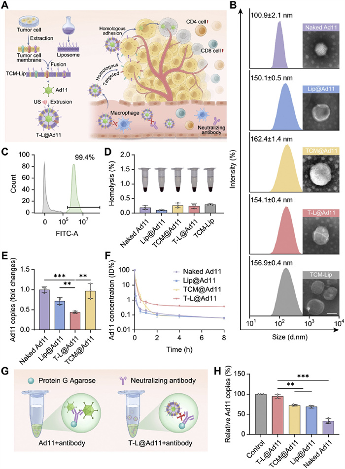
Figure 1.
Preparation, characterization, and functional verification of T-L@Ad11. (A) Schematic illustration of T-L@Ad11 preparation and the mechanism of T-L@Ad11 to enhance the anti-tumor efficacy of Ad11. (B) Particle size intensity and TEM images of naked Ad11, Lip@Ad11, TCM@Ad11, T-L@Ad11 and TCM-Lip. Scale bar: 50 nm. (C) The encapsulation efficiency of T-L@Ad11 evaluated by nanoparticle flow analysis. Ad11 was labeled with Cy5.5 (APC-A700 channel) while TCM-Lip was labeled with coumarin 6 (fluorescein isothiocyanate (FITC) channel). (D) Centrifuged erythrocytes after incubation mixed with naked Ad11, Lip@Ad11, TCM@Ad11, T-L@Ad11 and TCM-Lip and the corresponding relative percent hemolysis, n = 3. PBS and Triton X-100 were used as the negative and positive controls, respectively. (E) The amount of naked Ad11, Lip@Ad11, TCM@Ad11 and T-L@Ad11 endocytosed by macrophages at the MOI of 20, n = 3. (F) The circulation profiles of naked Ad11, Lip@Ad11, TCM@Ad11 and T-L@Ad11 after intravenous administration, n = 3. (G) Schematic diagram of the anti-neutralizing ability of T-L@Ad11 and other Ad11 formulations. (H) The amount of T-L@Ad11, TCM@Ad11, Lip@Ad11 and naked Ad11 in the supernatant that evaded binding to neutralizing antibodies, n = 3. Data are displayed as mean ± SD. The data in (F) are presented as the percentage of the initial dose (%ID). Statistical significance was analyzed by one-way ANOVA with a Tukey post hoc test. **P < 0.01, ***P < 0.001. "ns" indicates no significant difference.
In this study, Ad11 [22] was used as the OVs model. Transmission electron microscopy (TEM) images showed that the Lip-coated Ad11 (termed Lip@Ad11), TCM-coated Ad11 (termed TCM@Ad11) and TCM-Lip-coated Ad11 (termed T-L@Ad11) were spherical shell-core structures, distinguished from the regular icosahedral shape of naked Ad11. In contrast, TCM-Lip, the hybrid liposomes that were not loaded with Ad11, appeared as round-like vesicles (Fig. 1B). The hydrodynamic diameter was then evaluated by dynamic laser scattering. Results showed that the particle size of naked Ad11 was 100.90 ± 2.14 nm, and that of the empty TCM-Lip was 156.87 ± 0.40 nm. The particle sizes of Lip@Ad11, TCM@Ad11 and T-L@Ad11 after encapsulation of Ad11 were 150.10 ± 0.46 nm, 162.40 ± 1.39 nm and 154.13 ± 0.35 nm, respectively (Fig. 1B and Fig. S1A in Supporting information). The polydispersity indexes (PDI) of Lip@Ad11, TCM@Ad11, T-L@Ad11 and TCM-Lip were 0.18 ± 0.02, 0.20 ± 0.01, 0.15 ± 0.01 and 0.16 ± 0.02, respectively. All these results indicated that the formulations prepared by the ultrasound combined with mechanical extrusion method were uniformly dispersed (Fig. S1B in Supporting information). As shown in Fig. S2 (Supporting information), the zeta potential of naked Ad11 was −28.29 ± 0.85 mV. After the coating of the bionic membrane, the zeta potential of T-L@Ad11 was −21.80 ± 0.43 mV, which closely resembled the zeta potential of TCM-Lip (−21.91 ± 0.41 mV). To evaluate the stability of the formulation, the particle size and PDI of T-L@Ad11 stored in phosphate saline buffer (PBS) buffer was detected. The results demonstrated that the particle size and PDI of T-L@Ad11 remained stable at about 160 nm and 0.2, respectively, over a period of 7 days (Fig. S3 in Supporting information). To verify the coating efficiency of the bionic hybrid membrane on Ad11, nanoparticle flow assay was performed. The result showed that 99.4% of Ad11 was encapsulated with TCM-Lip (Fig. 1C). The result of SDS-PAGE demonstrated that the membrane protein components of TCM-Lip and T-L@Ad11 were similar to those of TCM proteins and retained the homologous adhesion molecules from TCM, which provided a basis for the functionalization of Ad11 coated with biomimetic liposomes (Fig. S4 in Supporting information). The retention of E-Cadherin, an adhesion molecule located on the surface of TCMs, was also confirmed in T-L@Ad11 through Western blot analysis (Fig. S5 in Supporting information). The blood compatibility of each Ad11 formulation was assessed through a hemolysis assay, with PBS and Triton X-100 serving as negative and positive control groups respectively. The supernatant of each group, as depicted in Fig. 1D, exhibited no significant hemolysis, with the relative quantitative percentage of hemolysis being below 0.5%. These results indicated that all three kinds of membranes could form a membrane coating on Ad11, which laid a foundation for evaluating their shielding and protection effects on Ad11 antigens.
According to our previous study [23], the circulation profile of OVs in vivo after intravenous administration is contingent upon the integrity of their surface membrane coating, specifically referring to their antigen shielding capacity against Ad11. Here, we evaluated the antigen shielding ability of the membrane encapsulation by examining its ability to protect Ad11 from the binding of neutralizing antibodies and uptake by macrophages, and then performed in vivo pharmacokinetic studies of various Ad11 formulations.
Firstly, the uptake of T-L@Ad11 with different mass ratios of TCM protein to lipid (TCM/Lip) by macrophages was evaluated. As shown in Fig. S6A (Supporting information), the amount of Lip@Ad11 without TCM components endocytosed by macrophages was reduced compared with naked Ad11, while other three formulations (TCM/Lip ratio of 1:4, 1:2 and 1:1) further reduced the uptake by macrophages, which was only about 40%–50% of that of naked Ad11. This indicated that the protective effect of the hybrid membrane on Ad11 was better than that of the lipid membrane alone. To further investigate the effect of in vivo circulation of Ad11 formulations with different TCM/Lip ratios, the pharmacokinetics of T-L@Ad11 were performed. Compared with Lip@Ad11, more Ad11 particles were retained in the circulating blood within 1 h after intravenous administration of T-L@Ad11 containing TCM components. Compared with T-L@Ad11 with other TCM/Lip ratios, more virions were retained in the circulating blood 2 h after intravenous administration of T-L@Ad11 with the TCM/Lip ratio of 1:2. Eight hours after intravenous administration, T-L@Ad11 with the TCM/Lip ratio of 1:2 remained above 0.3%ID (%ID, percentage of initial dose) in the bloodstream, while all other groups had fallen below 0.05%ID (Fig. S6B in Supporting information). Based on the pharmacokinetic results, T-L@Ad11 with the TCM/Lip ratio of 1:2 was selected for further research. The effect of multiplicity of infection (MOI) on macrophage uptake was investigated (Fig. 1E and Fig. S7 in Supporting information). The results demonstrated that T-L@Ad11 exhibited better resistance to macrophage phagocytosis at the MOI of 20 compared to the MOI of 10 and 40, while Lip@Ad11 showed a slight resistance to macrophage phagocytosis only at the MOI of 20 and 40. The macrophage uptake assay and pharmacokinetics study showed that PEG-liposomes marginally attenuated uptake by macrophages and failed to improve pharmacokinetic results. In contrast, coating Ad11 with TCMs increased the concentration of Ad11 in the circulating blood within the first hour after the intravenous administration but failed to neither improve its circulation profile nor reduce the endocytosis by macrophages. Notably, T-L@Ad11 had remarkable resistance to the endocytosis by macrophages and better circulation profile compared with other Ad11 formulations (Figs. 1E and F and Fig. S8 in Supporting information). The uptake of T-L@Ad11 in dendritic cells (DCs) was additionally investigated. As shown in Fig. S9 (Supporting information), the amount of T-L@Ad11 endocytosed by DCs was approximately 50% of that of naked Ad11, similar to the uptake by macrophages. Notably, there was a significant increase in the uptake of TCM@Ad11 by DCs, which was about 9-fold higher compared to the uptake of naked Ad11. We speculated that this phenomenon could be attributed to the excessive presence of tumor-associated antigens in TCM@Ad11, leading to its phagocytosis by DCs. Following previously reported methods, we evaluated the binding of Ad11 formulations to neutralizing antibodies, obtained from the serum of mice preimmunized with Ad11, by an immunoprecipitation assay (IPA, Fig. 1G) [22]. Most of the naked Ad11 particles were bound with neutralizing antibodies and pulled down by protein G-coated agarose beads, only 30% of free Ad11 remained in the supernatant. Lip@Ad11 and TCM@Ad11 reduced the binding of Ad11 to neutralizing antibodies, about 70% of the formulations were retained in the supernatant. T-L@Ad11 was minimally bound to neutralizing antibodies compared with other Ad11 formulations, with > 90% of the free particles in the supernatant (Fig. 1H). The above results further indicated that the hybrid liposomes had a more complete encapsulation of Ad11 and a stronger shielding ability to Ad11 antigens, which was beneficial for Ad11 to escape from the binding with neutralizing antibodies and the clearance of MPS in vivo.
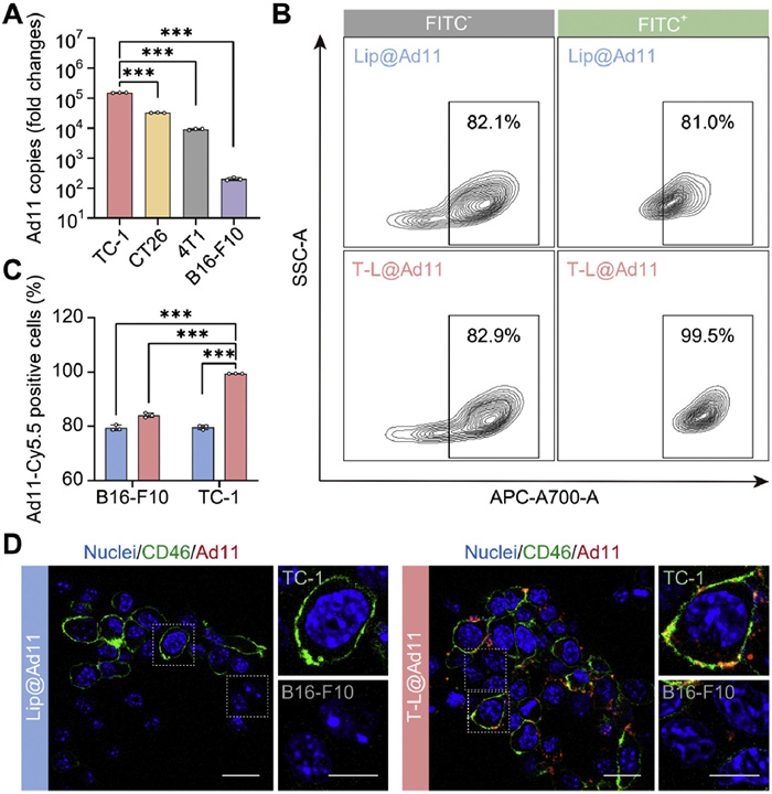
The homologous targeting ability of T-L@Ad11 was initially validated through quantitative real-time polymerase chain reaction (qPCR) assay. The results showed that the uptake of T-L@Ad11 by TC-1-hCD46 cells exhibited a significantly higher level, with 4.68-fold, 16.69-fold, and 751.25-fold increases compared to CT26, 4T1 and B16-F10 cells, respectively (Fig. 2A). To further confirm the target ability of T-L@Ad11 to its cognate tumor cells, TC-1-hCD46 and B16-F10 cells were co-cultured in vitro for uptake assay, and Lip@Ad11 was used as the control group. As depicted in Figs. 2B and C, Cy5.5-labeled Lip@Ad11 was endocytosed by 81.0% TC-1-hCD46 cells and 82.1% B16-F10 cells in the Lip@Ad11-treated group. In the T-L@Ad11-treated group, the proportion of Cy5.5 positive cells in B16-F10 cells was 82.9% and 99.5% in TC-1-hCD46 cell population. This demonstrated that the uptake of T-L@Ad11 by TC-1-hCD46 cells in the co-culture system was specific and that TCM components enabled our biomimetic hybrid membrane-coated Ad11 to actively target to the cognate tumor cells. To visually depict the variation in cellular uptake of different Ad11 formulations, confocal laser scanning microscopy (CLSM) imaging of co-cultured tumor cells was performed. In the T-L@Ad11-treated group, the red fluorescence of Cy5.5-Ad11 and the green fluorescence of TC-1-hCD46 cell membranes showed apparent colocalization, but no obvious red fluorescence was observed in B16-F10 cells with only the nuclei visualized. In the Lip@Ad11-treated group, neither TC-1-hCD46 nor B16-F10 cells exhibited obvious uptake behavior of Cy5.5-labeled Ad11 (Fig. 2D).
Figure 2
Figure 2.
T-L@Ad11 exhibited conspicuous active targeting ability to homotypic tumor cells in vitro. (A) The amount of T-L@Ad11 endocytosed by TC-1-hCD46, CT26, 4T1, and B16-F10 cells, n = 3. (B) Flow cytometry analysis and (C) quantitative results of T-L@Ad11 uptake experiments, n = 3. (D) CLSM images of Cy5.5-labeled T-L@Ad11 and Lip@Ad11 endocytosed by the co-cultured tumor cells. Scale bars: 20 µm (low magnification) and 10 µm (high magnification). Data are displayed as mean ± SD. Statistical significance was analyzed by one-way ANOVA with a Tukey post hoc test. ***P < 0.001.
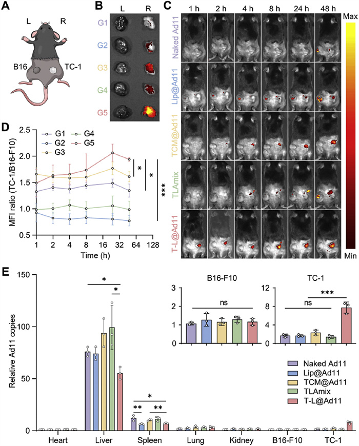
The bilateral subcutaneous tumor model in mice simultaneously bearing TC-1-hCD46 and B16-F10 tumors was constructed to explore the accumulation of Cy5.5-labeled T-L@Ad11 in tumors (Fig. 3A). All animal protocols were approved by the Animal Ethics Committee of China Medical University. In vivo fluorescence imaging was performed by the in vivo imaging system (IVIS) at prespecified time points after tail vein injection of Cy5.5-Ad11 formulations. As illustrated in Figs. 3C and D and Fig. S10 (Supporting information), T-L@Ad11 exhibited preferential accumulation at the TC-1-hCD46 tumor sites compared to Lip@Ad11, TCM@Ad11, and TLAmix (a simple mixture of TCM, Lip, and Ad11) at each time point. After 48 h of intravenous injection, TCM@Ad11 and T-L@Ad11 were sustainably concentrated only in the TC-1-hCD46 tumors but not in the B16-F10 tumors, whereas only a small amount of other Ad11 formulations accumulated in the TC-1-hCD46 tumors. This outcome could be better visually demonstrated through the fluorescence imaging of ex vivo tumors after 48 h of intravenous administration (Fig. 3B). This suggested that T-L@Ad11 possesses a strong homing ability towards the corresponding tumors in vivo due to the presence of TCM components, including homologous adhesion molecules. Subsequently, the biodistribution of T-L@Ad11 was evaluated by qPCR assay. Naked Ad11, T-L@Ad11, TCM@Ad11, TLAmix, and Lip@Ad11 were predominantly distributed in the liver and spleen (Fig. 3E). However, the amount of T-L@Ad11 in the liver was significantly lower compared with naked Ad11, TCM@Ad11 and TLAmix, accounting for 72.83%, 58.96%, and 55.66% of the liver distribution of the other Ad11 formulations, respectively. The distribution of T-L@Ad11 and Lip@Ad11 in the spleen was reduced compared with naked Ad11, accounting for 58.01% and 51.88% of the spleen distribution of naked Ad11, respectively. The distribution of T-L@Ad11 in TC-1-hCD46 tumors was notably higher compared with other Ad11 formulations, while no significant enrichment of Ad11 formulations was observed in B16-F10 tumors. These results suggest that T-L@Ad11 can both protect Ad11 from capture by MPS-related organs (e.g., livers and spleens) and enhance the targeted delivery of Ad11 to tumors, providing support for enhancing the antitumor efficacy of Ad11 intravenous administration.
Figure 3
Figure 3.
T-L@Ad11 increased the accumulation of Ad11 in homotypic tumors in vivo. (A) Schematic illustration of the bilateral subcutaneous tumor model in mice simultaneously bearing B16-F10 (on the left) and TC-1-hCD46 (on the right) tumors. (B) Ex vivo fluorescence imaging of tumor tissues after various treatment by IVIS, n = 3. G1: naked Ad11; G2: Lip@Ad11; G3: TCM@Ad11; G4: TLAmix; G5: T-L@Ad11. (C) Fluorescence imaging of mice bearing B16-F10 and TC-1-hCD46 tumors after being injected (i.v.) with Cy5.5-labeled Ad11 formulations, n = 3. (D) The mean fluorescence intensity (MFI) ratio of TC-1-hCD46 to B16-F10 tumors in vivo, n = 3. G1: naked Ad11; G2: Lip@Ad11; G3: TCM@Ad11; G4: TLAmix; G5: T-L@Ad11. Data in (D) are displayed as mean ± SEM. (E) The quantitative distribution of naked Ad11, Lip@Ad11, TCM@Ad11, TLAmix and T-L@Ad11 in organs and tumors determined by qPCR assay at 2 h after injection, n = 3. Data in (E) are displayed as mean ± SD and presented as relative copies compared to the PBS group. Statistical significance was analyzed by one-way ANOVA with a Tukey post hoc test. *P < 0.05, **P < 0.01, ***P < 0.001.
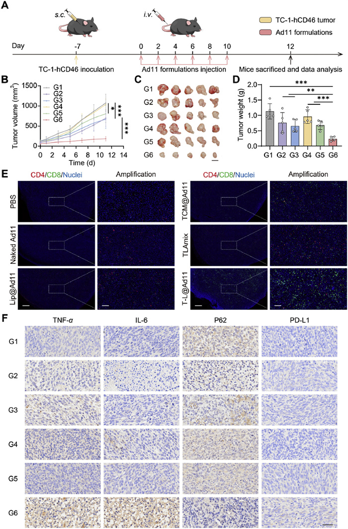
To evaluate the antitumor efficacy of each Ad11 formulation, the subcutaneous tumor model was constructed, and the treatment schedule was presented in Fig. 4A. As shown in Figs. 4B–D and Fig. S11 (Supporting information), the tumor growth in mice treated with TCM@Ad11 was found to be comparable to that observed in mice treated with PBS, indicating that TCM presented negligible effect on promoting the antitumor efficacy of Ad11. The administration of Lip@Ad11 partially attenuated tumor growth, demonstrating potential anti-tumor efficacy. Among all treatment groups, T-L@Ad11 exhibited the most potent anti-tumor effect with the smallest tumor volume and weight. On day 11, the tumor volume of T-L@Ad11-treated group accounted for 17.31%, 17.90%, 23.09%, 26.70% and 27.81% of that in the groups treated with PBS, TCM@Ad11, TLAmix, naked Ad11 and Lip@Ad11, respectively. To investigate the mechanism underlying the antitumor effect of T-L@Ad11, immunohistochemical staining for inflammatory factors and programmed cell death 1 ligand 1 (PD-L1) in the tumors was conducted (Fig. 4F). In comparison to other treatments, T-L@Ad11 treatment exhibited an upregulation of tumor necrosis factor-α (TNF-α) and interleukin-6 (IL-6) levels while downregulation of P62 in tumors, confirming that T-L@Ad11 enhances the anti-tumor response and Ad11-induced autophagy in tumor cells (Fig. S12 in Supporting information). The administration of T-L@Ad11 also resulted in an upregulation of PD-L1 expression within tumors, indicating that T-L@Ad11 therapy may not only elicit an anti-tumor immune response but also induce immunosuppression and exhaustion [24, 25]. Similarly, immunofluorescence images also demonstrated a significant increase in the infiltration of CD4+ and CD8+ T cells within tumors treated with T-L@Ad11 compared to other treatment groups, suggesting that T-L@Ad11 treatment enhanced cellular immune responses within tumors (Fig. 4E and Fig. S13 in Supporting information).
Figure 4
Figure 4.
T-L@Ad11 enhanced the antitumor efficacy of Ad11 in the subcutaneous tumor model. (A) The treatment schedule of the antitumor efficacy study. (B) The tumor growth curves of mice in different treatment groups, n = 5. (C) The photograph of tumors dissected from mice at day 12, n = 5, scale bar: 10 mm. (D) The weight of tumors at day 12, n = 5. G1: PBS; G2: naked Ad11; G3: Lip@Ad11; G4: TCM@Ad11; G5: TLAmix; G6: T-L@Ad11. (E) Immunofluorescence staining of CD4+/CD8+ T cells of TC-1-hCD46 tumor sections after different treatments. Scale bars: 500 µm (low magnification) and 100 µm (high magnification). (F) The immune-histochemical staining images of TNF-α, IL-6, P62 and PD-L1 in subcutaneous tumor sections, scale bar: 40 µm. Data are displayed as mean ± SD. Statistical significance was analyzed by two-way ANOVA or one-way ANOVA with a Tukey post hoc test. *P < 0.05, **P < 0.01, ***P < 0.001.
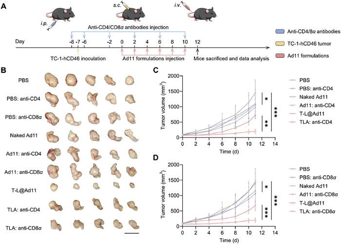
The antitumor efficacy study in the subcutaneous tumor model revealed a significant infiltration of CD4+ and CD8+ T cells in tumor sections following T-L@Ad11 treatment. To further validate the role of CD4+ and CD8+ T lymphocytes in vivo, the antitumor efficacy of T-L@Ad11 was investigated in the CD4+/CD8+ T lymphocyte-depletion mouse models bearing TC-1-hCD46 subcutaneous tumors based on the methods described in the literatures (Fig. 5A) [26, 27]. As shown in Figs. 5B–D and Fig. S14 (Supporting information), the intravenous administration of naked Ad11 exhibited only a marginal antitumor effect, and the depletion of CD4+/CD8+ T cells did not significantly impact its antitumor efficacy. The T-L@Ad11 treatment exhibited conspicuous antitumor effect, resulting in significantly reduced tumor volume and tumor weight. The antitumor efficacy of T-L@Ad11 was considerably attenuated when we selectively depleted CD4+/CD8+ T cells in vivo. However, it still outperformed naked Ad11, which due to the enhanced enrichment of Ad11 at the tumor site following biomimetic membrane coating. The findings demonstrated that CD4+/CD8+ T cells played a crucial role in the antitumor efficacy of T-L@Ad11, which was closely associated with the induction of antitumor immune responses by T-L@Ad11.
Figure 5
Figure 5.
The assessment of the roles of CD4+/CD8+ T lymphocytes in the antitumor efficacy of T-L@Ad11. (A) The treatment schedule of the antitumor efficacy study in the CD4+/CD8+ T lymphocyte-depletion mouse models bearing TC-1-hCD46 subcutaneous tumors. (B) The photograph of tumors dissected from mice on day 12, n = 5. Scale bar: 20 mm. (C) The tumor growth curves of mice in the CD4+ T lymphocyte depletion mice and normal mice, n = 5. (D) The tumor growth curves of mice in the CD8+ T lymphocyte depletion mice and normal mice, n = 5. Data are displayed as mean ± SD. Statistical significance was analyzed by two-way ANOVA with a Tukey post hoc test. *P < 0.05, **P < 0.01, ***P < 0.001.
The safety of the Ad11 formulations was then evaluated. As illustrated in Figs. S15 and S16 (Supporting information), no significant weight loss or changes of hepatic and renal function indicators were observed among mice from any treatment group (Fig. S17 in Supporting information). Moreover, the safety of the Ad11 formulations in vivo was also supported by the results of hematoxylin and eosin (H & E) staining of vital organs (Fig. S18 in Supporting information).
Recently, cell membrane biomimetic drug delivery systems have been widely investigated in the treatment of malignant tumors and have shown promising therapeutic potential in the preclinical stage [20, 21, 28-34]. These cell-derived biomaterials offer the benefits of excellent biocompatibility, prolonged circulation time and appropriate biodegradability [35]. The TCM serves as an ideal carrier for the delivery of anti-tumor drugs due to its surface composition, which includes the immunosuppressive signal molecule CD47, a variety of adhesion molecules and specific tumor antigens [28, 30, 36]. Compared with erythrocyte membranes, the coating of TCM on nanoparticles not only extends their circulation time in vivo, but also facilitates their targeted delivery to specific tumor tissues through the cognate adhesion molecules (such as N-cadherin, galectin-3 and EpCAM) on the membrane surface. TCM can be extracted from patient-derived tissues or cells and can be repeatedly obtained by culturing tumor cells in vitro. The ease of TCM fabrication facilitates the production of biomimetic nano-coating that exhibits comparable properties to original tumor cells. Previous studies on the delivery of OVs using biofilm vesicles have mainly focused on cell membranes [19]. However, the findings from our previous studies have demonstrated that the utilization of only cell membranes coating OVs is insufficient to fully conceal viral capsid proteins [23], as supported by the results of in vitro cellular uptakes and in vivo pharmacokinetic studies. By contrast, the incorporation of synthetic lipid membranes into native cell membranes enhances the encapsulation of OVs by biofilm vesicles. Additionally, the surface of tumor cells is enriched with tumor-associated antigens and adhesion molecules, which also enable the biomimetic membrane carriers to possess practical biological functions [20].
In this study, we have developed a cell membrane biomimetic lipid membrane carrier for targeted systemic delivery of OVs. Oncolytic Ad11 particles were encapsulated in the TCM biomimetic liposome (TCM-Lip). TCM-Lip is composed of synthetic long-circulating liposomes hybridized with TCM. When administered intravenously, OVs are susceptible to capture by the MPS and binding with neutralizing antibodies. However, TCM-Lip can effectively shield OVs from rapid clearance within the bloodstream. Simultaneously, the presence of cognate adhesion molecules on the surface of tumor cells facilitates specific targeting of T-L@Ad11 towards its corresponding tumors in vivo, thereby enhancing the accumulation of OVs at tumor sites. In the bilateral subcutaneous tumor model, T-L@Ad11 effectively enhanced the accumulation of OVs in tumors homologous to the biomimetic membrane. Meanwhile, adequate circulation time and abundant accumulation in the tumor guarantee the antitumor efficacy of T-L@Ad11 in vivo. Relying on the advantage of OVs in modulating the tumor immune microenvironment, we observed enhanced infiltration of immune cells and elevated levels of inflammatory factors in tumor sections by immunofluorescence and immunohistochemical staining.
The previous studies on drug delivery using TCMs have demonstrated the applicability of this technology to various types of tumor cells, indicating the broad applicability of our T-L@Ad11 strategy across diverse tumor types [19, 37]. In future clinical applications, individualized tumor cells can be obtained through surgical procedures or needle biopsies, and subsequently sorted, purified, and massively expanded in vitro using well-established experimental techniques. The utilization of tumor cell components (such as cell lysates) in the formulation of tumor vaccines has been supported by several recent studies [38-46]. This indicates that the combination of TCM constituents with OVs holds promise for personalized tumor vaccines. The preclinical studies have demonstrated the favorable safety profiles of various cell-membrane carriers for drug delivery, thereby alleviating concerns regarding the pathogenicity of biologic components derived from tumor cells. Further explorations are needed to enhance the intravenous delivery strategy of OVs in the design of the hybrid membrane carriers.
Declaration of competing interest
The authors declare that they have no known competing financial interests or personal relationships that could have appeared to influence the work reported in this paper.
We thank the staff members of the Beijing Bio-Targeting Therapeutics Technology Co., Ltd. (China) for providing technical support. This work was supported by the National Key R & D Program of China (No. 2022YFC2403401), the National Natural Science Foundation of China (nos. 82073368, 82303766), and the Liaoning Revitalization Talents Program (No. XLYC2007071), the China Postdoctoral Science Foundation (No. 2023M743908) and the Joint Program of Science and Technology Program of Liaoning Province (No. 2023JH2/101700094).
Supplementary materials
Supplementary material associated with this article can be found, in the online version, at doi:10.1016/j.cclet.2024.110493.
[1]
H.L. Kaufman, F.J. Kohlhapp, A. Zloza, Nat. Rev. Drug Discov. 15 (2016) 660. doi: 10.1038/nrd.2016.178
K.L. Oxley, B.M. Hanson, A.N. Zani, G.A. Bishop, Cancer Immunol. Immunother. 70 (2021) 3093–3103. doi: 10.1007/s00262-021-02914-7
[44]
H. Ogino, J.W. Taylor, T. Nejo, et al., J. Clin. Invest. 132 (2022) e151239.
[45]
A. Harari, M. Graciotti, M. Bassani-Sternberg, L.E. Kandalaft, Nat. Rev. Drug Discov. 19 (2020) 635–652. doi: 10.1038/s41573-020-0074-8
[46]
C.C. Feng, P. Tan, G.J. Nie, M.T. Zhu, Exploration 3 (2023) 20210263.
Figure 1
Preparation, characterization, and functional verification of T-L@Ad11. (A) Schematic illustration of T-L@Ad11 preparation and the mechanism of T-L@Ad11 to enhance the anti-tumor efficacy of Ad11. (B) Particle size intensity and TEM images of naked Ad11, Lip@Ad11, TCM@Ad11, T-L@Ad11 and TCM-Lip. Scale bar: 50 nm. (C) The encapsulation efficiency of T-L@Ad11 evaluated by nanoparticle flow analysis. Ad11 was labeled with Cy5.5 (APC-A700 channel) while TCM-Lip was labeled with coumarin 6 (fluorescein isothiocyanate (FITC) channel). (D) Centrifuged erythrocytes after incubation mixed with naked Ad11, Lip@Ad11, TCM@Ad11, T-L@Ad11 and TCM-Lip and the corresponding relative percent hemolysis, n = 3. PBS and Triton X-100 were used as the negative and positive controls, respectively. (E) The amount of naked Ad11, Lip@Ad11, TCM@Ad11 and T-L@Ad11 endocytosed by macrophages at the MOI of 20, n = 3. (F) The circulation profiles of naked Ad11, Lip@Ad11, TCM@Ad11 and T-L@Ad11 after intravenous administration, n = 3. (G) Schematic diagram of the anti-neutralizing ability of T-L@Ad11 and other Ad11 formulations. (H) The amount of T-L@Ad11, TCM@Ad11, Lip@Ad11 and naked Ad11 in the supernatant that evaded binding to neutralizing antibodies, n = 3. Data are displayed as mean ± SD. The data in (F) are presented as the percentage of the initial dose (%ID). Statistical significance was analyzed by one-way ANOVA with a Tukey post hoc test. **P < 0.01, ***P < 0.001. "ns" indicates no significant difference.
Figure 2
T-L@Ad11 exhibited conspicuous active targeting ability to homotypic tumor cells in vitro. (A) The amount of T-L@Ad11 endocytosed by TC-1-hCD46, CT26, 4T1, and B16-F10 cells, n = 3. (B) Flow cytometry analysis and (C) quantitative results of T-L@Ad11 uptake experiments, n = 3. (D) CLSM images of Cy5.5-labeled T-L@Ad11 and Lip@Ad11 endocytosed by the co-cultured tumor cells. Scale bars: 20 µm (low magnification) and 10 µm (high magnification). Data are displayed as mean ± SD. Statistical significance was analyzed by one-way ANOVA with a Tukey post hoc test. ***P < 0.001.
Figure 3
T-L@Ad11 increased the accumulation of Ad11 in homotypic tumors in vivo. (A) Schematic illustration of the bilateral subcutaneous tumor model in mice simultaneously bearing B16-F10 (on the left) and TC-1-hCD46 (on the right) tumors. (B) Ex vivo fluorescence imaging of tumor tissues after various treatment by IVIS, n = 3. G1: naked Ad11; G2: Lip@Ad11; G3: TCM@Ad11; G4: TLAmix; G5: T-L@Ad11. (C) Fluorescence imaging of mice bearing B16-F10 and TC-1-hCD46 tumors after being injected (i.v.) with Cy5.5-labeled Ad11 formulations, n = 3. (D) The mean fluorescence intensity (MFI) ratio of TC-1-hCD46 to B16-F10 tumors in vivo, n = 3. G1: naked Ad11; G2: Lip@Ad11; G3: TCM@Ad11; G4: TLAmix; G5: T-L@Ad11. Data in (D) are displayed as mean ± SEM. (E) The quantitative distribution of naked Ad11, Lip@Ad11, TCM@Ad11, TLAmix and T-L@Ad11 in organs and tumors determined by qPCR assay at 2 h after injection, n = 3. Data in (E) are displayed as mean ± SD and presented as relative copies compared to the PBS group. Statistical significance was analyzed by one-way ANOVA with a Tukey post hoc test. *P < 0.05, **P < 0.01, ***P < 0.001.
Figure 4
T-L@Ad11 enhanced the antitumor efficacy of Ad11 in the subcutaneous tumor model. (A) The treatment schedule of the antitumor efficacy study. (B) The tumor growth curves of mice in different treatment groups, n = 5. (C) The photograph of tumors dissected from mice at day 12, n = 5, scale bar: 10 mm. (D) The weight of tumors at day 12, n = 5. G1: PBS; G2: naked Ad11; G3: Lip@Ad11; G4: TCM@Ad11; G5: TLAmix; G6: T-L@Ad11. (E) Immunofluorescence staining of CD4+/CD8+ T cells of TC-1-hCD46 tumor sections after different treatments. Scale bars: 500 µm (low magnification) and 100 µm (high magnification). (F) The immune-histochemical staining images of TNF-α, IL-6, P62 and PD-L1 in subcutaneous tumor sections, scale bar: 40 µm. Data are displayed as mean ± SD. Statistical significance was analyzed by two-way ANOVA or one-way ANOVA with a Tukey post hoc test. *P < 0.05, **P < 0.01, ***P < 0.001.
Figure 5
The assessment of the roles of CD4+/CD8+ T lymphocytes in the antitumor efficacy of T-L@Ad11. (A) The treatment schedule of the antitumor efficacy study in the CD4+/CD8+ T lymphocyte-depletion mouse models bearing TC-1-hCD46 subcutaneous tumors. (B) The photograph of tumors dissected from mice on day 12, n = 5. Scale bar: 20 mm. (C) The tumor growth curves of mice in the CD4+ T lymphocyte depletion mice and normal mice, n = 5. (D) The tumor growth curves of mice in the CD8+ T lymphocyte depletion mice and normal mice, n = 5. Data are displayed as mean ± SD. Statistical significance was analyzed by two-way ANOVA with a Tukey post hoc test. *P < 0.05, **P < 0.01, ***P < 0.001.

 DownLoad:

 下载:
下载:


 下载:
下载:
