Citation:
Shupeng Han, Caiting Deng, Meichen Zheng, Linwei Yang, Hancun Kong, Yongchao He, Yinuo Zheng, Guowei Deng, Yu Ren, Feifei An. A GSH-responsive NIR-BODIPY fluorophore with large Stokes-shift for tumor specific fluorescence imaging and surgical guidance[J]. Chinese Chemical Letters,
2025, 36(7): 110459.
doi:
10.1016/j.cclet.2024.110459
A GSH-responsive NIR-BODIPY fluorophore with large Stokes-shift for tumor specific fluorescence imaging and surgical guidance
English
A GSH-responsive NIR-BODIPY fluorophore with large Stokes-shift for tumor specific fluorescence imaging and surgical guidance
Department of Breast Surgery, the First Affiliated Hospital of Xi'an Jiaotong University, Xi'an 710061, China
b.
School of Public Health, Health Science Center, Xi'an Jiaotong University, Xi'an 710061, China
c.
Institute of Medical Engineering, Department of Biophysics, School of Basic Medical Science, Health Science Center, Xi'an Jiaotong University, Xi'an 710061, China
d.
College of Chemistry and Life Science, Sichuan Provincial Key Laboratory for Structural Optimization and Application of Functional Molecules, Chengdu Normal University, Chengdu 611130, China
Received Date:
30 June 2024 Accepted Date:
13 September 2024 Revised Date:
11 September 2024 Available Online:
15 July 2025
Abstract:
The tumor microenvironment (TME)-activatable probes have proven effective in enhancing the signal-to-background ratio (SBR) for precise fluorescence imaging in tumor diagnosis. However, many fluorophores have suboptimal emission spectra and a short Stokes shift, which may lead to overlap with bio-autofluorescence, excitation, and emission spectra, limiting their use in intraoperative guidance. Herein, a γ-glutathione (GSH) responsive near-infrared (NIR) BODIPY probe, named "Pro-Dye" was synthesized with a large Stokes shift of 91 nm. The Pro-Dye can be rapidly and specifically activated by high concentrations of GSH both in solution and inside cancer cells, while remaining inactive in normal cells (Human umbilical vein endothelial cells, HUVECs). The Pro-Dye was further encapsulated by 1, 2-distearoyl-sn‑glycero-3-phosphoethanolamine-N-(polyethylene glycol)-5000 (DSPE-PEG5000) to form Pro-Dye nanoparticles (NPs), making it water-dispersible for in vivo application. In vivo fluorescence imaging demonstrated that Pro-Dye NPs can accumulate at the tumor and exhibit an improved SBR compared to the "always-on" probe (Dye NPs). Moreover, the tumor can be precisely resected under the real-time guidance of fluorescence imaging of Pro-Dye NPs, showing a well-defined tumor margin.
Among the various modalities for tumor therapy, surgery remains critical treatment of solid tumors. Identification and removal of solid tumors are typically carried out by surgeons, heavily relying on their training, experience, and somatosensory information gathered during surgery [1]. However, accurately delineating the tumor margins presents a challenge for surgeons based on subjective experience, potentially resulting in incomplete resection and recurrence of disease [2]. Accordingly, a significant number of cases have reported tumor-positive resection margins [3], which significantly impacts the prognosis of patients [4-6]. Fluorescence imaging can provide real-time surgical guidance and estimate tumor margins, enabling surgeons to adjust the surgical strategy rapidly [1, 7, 8]. Therefore, a fluorophore with good optical properties is desired for efficient tumor identification.
Within the near-infrared (NIR, 700–900 nm) wavelength range, most in vivo biomolecules possess relatively low scattering and autofluorescence, resulting in less attenuation of excitation and emission light, which enhances imaging depth [9, 10]. Therefore, NIR fluorophores are ideal for providing real-time guidance for surgeons during operations [11, 12]. Additionally, fluorophores with a large Stokes shift are preferred because they enable the separation of excitation and emission signals, leading to improved sensitivity and accuracy in fluorescence imaging [13].
Boron-dipyrromethene (BODIPY) fluorophore shows great potential in clinical applications because it possesses high emission quantum yield, large molar absorption coefficient, chemical structure stability, highly tunable structure, and photophysical properties [14, 15]. However, the absorption and emission peaks of most BODIPY dyes do not fall within the "NIR window", limiting their potential for imaging deep tissue [16, 17]. In addition, most BODIPY dyes typically have a small Stokes shift, which hinders their application in real-time in vivo fluorescence imaging [18]. Therefore, a BODIPY derivative with absorption and emission within the "NIR window" and a large Stokes shift is anticipated for in vivo fluorescence imaging.
The tumor microenvironment (TME) differs significantly from the normal tissue environment in various physicochemical aspects, such as the high expression of enzymes [19, 20], elevated levels of reactive oxygen species (ROS), increased γ-glutathione (GSH) concentration [21], and low pH value [22]. These characteristics of the TME have been investigated as specific factors that activate activable fluorophores for precise cancer theranostics. The dinitrobenzene sulfonate (DNBS) group has been widely used in developing GSH-responsive prodrugs [23, 24]. Moreover, the DNBS group can act as a potent quencher when linked to a fluorophore, which can be selectively activated by the high GSH concentration present in the TME [25]. BODIPY-based fluorophores have been created for detecting bio-thiols and are commonly employed as chemical probes in vitro [26]. Nevertheless, a GSH-responsive NIR BODIPY dye with a large Stokes shift has not yet been described for in vivo fluorescence imaging and surgical navigation.
Herein, a GSH responsive NIR BODIPY dye prodrug (Pro-Dye) with a large Stokes shift of 91 nm and satisfactory biocompatibility was synthesized for precise fluorescence imaging. The Pro-Dye possesses inhibited fluorescence emission and can be activated to recover strong fluorescence emission (×33 folds) upon incubation with GSH (Fig. 1a). The encapsulation of Pro-Dye by 1, 2-distearoyl-sn‑glycero-3-phosphoethanolamine-N-(polyethylene glycol)−5000 (DSPE-PEG5000) resulted in Pro-Dye nanoparticles (Pro-Dye NPs) with a diameter of approximately 100 nm. Pro-Dye NPs exhibited stronger fluorescence emission in 4T1 cancer cells compared to that in the human umbilical vein endothelial cells (HUVECs) and 4T1 cancer cells pretreated with l-buthionine-sulfoximine (BSO), a GSH synthetase inhibitor. In the in vivo fluorescence imaging, Pro-Dye NPs showed good tumor accumulation and increasing fluorescence intensity at the tumor site over time, resulting in a higher tumor/normal tissues ratio compared to the Dye NPs. Under real-time fluorescence guidance, the tumor was completely resected from the mouse, confirming the effective use of the designed fluorophore in diagnostic imaging and surgical navigation.
Figure 1
Figure 1.
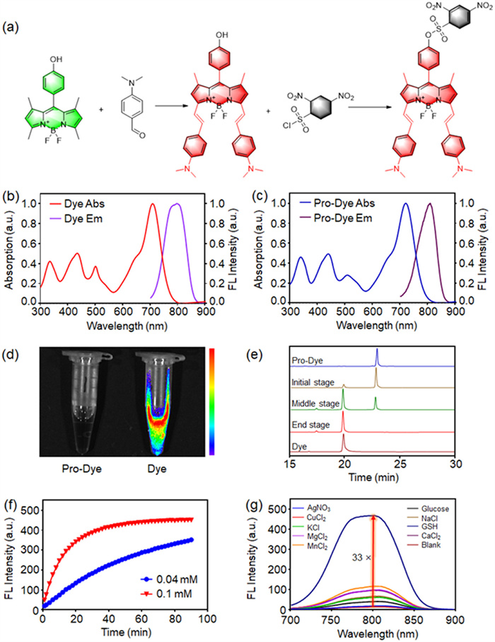
(a) Structural activation process of Pro-Dye by reacting with GSH. (b) UV–vis absorption spectra and normalized fluorescence emission spectrum (Ex = 680 nm) of Dye in DMSO. (c) UV–vis absorption spectra and normalized fluorescence emission spectrum (Ex = 680 nm) of Pro-Dye in DMSO. (d) Fluorescence image of Dye and Pro-Dye in DMSO (10 µmol/L). (e) HPLC analysis of Pro-Dye during incubation with GSH. (f) Kinetics curves of Pro-Dye activated by GSH at different concentrations (mM = mmol/L). (g) Fluorescence spectra of Pro-Dye at 1 h after incubation with different reagents (NaCl, 50 mmol/L; KCl, 50 mmol/L; CaCl2, 4 mmol/L; MgCl2, 1 mmol/L; MnCl2, 50 mmol/L; AgNO3, 2 mmol/L; CuCl2, 1 mmol/L; glucose, 10 mmol/L; GSH, 10 mmol/L).
As depicted in Fig. S1 (Supporting information), the GSH-responsive NIR fluorescent probe Pro-Dye was designed and synthesized from the green light-emitting BODIPY core (OH-Bo) through Knoevenagel reaction. p-Dimethylaminobenzaldehyde was used as the aromatic aldehyde to react with the 3, 5-methyl BODIPY substituent, resulting in a red-shifted styryl-substituted NIR fluorophore (Dye) with an extended π conjugation system. Subsequently, the hydroxyl group of the Dye was substituted with the DNBS group, serving as both a quenching moiety and a GSH recognition moiety, leading to the quenched NIR fluorophore named "Pro-Dye". The Pro-Dye exhibited quenched fluorescence compared to Dye. The quenching mechanism was attributed to photoinduced electron transfer (PET) caused by an electrophilic substituent group, as reported in various literature sources [27-29].
The photophysical properties of Pro-Dye and Dye were investigated using an ultraviolet–visible (UV–vis) spectrometer and a fluorescence spectrometer. As shown in Figs. 1b and c, compared to the absorbance peak of Dye at 709 nm, the absorbance peak of Pro-Dye slightly red-shifted to 721 nm due to the substitution of DNBS. The fluorescence emission peak of Dye was located at 800 nm, exhibiting a large Stokes shift of 91 nm. Both absorption and emission peaks were within the NIR wavelength range, allowing for deep biological tissue penetration. The large Stokes shift can efficiently reduce interference between excitation and emission channels during fluorescence imaging. The fluorescence emission study revealed that the fluorescence intensity of Pro-Dye was significantly inhibited compared to the unquenched Dye, confirming the successful quenching strategy of employing DNBS as the quencher and all the photophysical parameters were listed in Table S1 (Supporting information). Additionally, the fluorescence image of the Dye and Pro-Dye solution was captured using a fluorescence imaging system with 680 nm excitation and 820 nm emission filters. The fluorescence intensity of the Pro-Dye solution was much weaker than that of the Dye solution, further confirming the efficacy of the quenching strategy (Fig. 1d). As designed with possible activation mechanism by GSH (Fig. S2 in Supporting information), the Pro-Dye activation by GSH was further evaluated by using a series of Pro-Dye solutions (DMSO/H2O = 9/1) to incubate with GSH and analyzed with high-performance liquid chromatography (HPLC). As shown in Fig. 1e, the retention times of Dye and Pro-Dye were 19.9 min and 22.0 min, respectively. The HPLC profiles exhibited a gradual increment in the Dye peaks following the incubation of Pro-Dye with GSH over varying durations, indicating a progressive conversion from Pro-Dye to Dye. After a 40-min reaction period, only the peak of Dye remained in the chromatogram, indicating the complete conversion of Pro-Dye to Dye in response to GSH. Then, the fluorescence spectra of the Pro-Dye solution containing GSH (0.04 and 0.1 mmol/L, respectively) were measured at 2-min intervals (Figs. S9 and S10 in Supporting information). It was observed that the fluorescence intensity of Pro-Dye at 800 nm wavelength increased steadily. The activation kinetics curve of Pro-Dye fluorescence intensity showed that it took approximately 90 min to reach a plateau when incubated with GSH at a concentration of 0.1 mmol/L. In contrast, it took a longer time to reach the plateau when incubated with GSH at a lower concentration (0.04 mmol/L) (Fig. 1f). The Pro-Dye solution containing GSH exhibited an increased fluorescence intensity up to 33-fold compared to that without GSH (Fig. 1g). To verify the selectivity of Pro-Dye's response to GSH, incubations were performed with Pro-Dye and a series of solutions containing a panel of inorganic ions and organic molecules (NaCl, 50 mmol/L; KCl, 50 mmol/L; CaCl2, 4 mmol/L; MgCl2, 1 mmol/L; MnCl2, 50 mmol/L; AgNO3, 2 mmol/L; CuCl2, 1 mmol/L; glucose, 10 mmol/L; GSH, 10 mmol/L). Subsequent analysis using a fluorescence spectrometer revealed a significantly smaller increase in the fluorescence intensity of the Pro-Dye solution, except for the one incubated with GSH. Due to the structural similarities of common biothiols in vivo, some cross-reactivity is possible, implying that cysteine (Cys) or homocysteine (Hcy) might also activate the designed fluorescent molecules to some extent. Therefore, activation experiments of Pro-Dye by Cys and Hcy were conducted at their physiological concentrations (Fig. S11 in Supporting information). The results showed that both Cys and Hcy at their physiological concentrations had little activation on the Pro-Dye molecule, while GSH can rapidly activate the Pro-Dye molecule (Fig. S11). This indicated that the activation of Pro-Dye molecule in physiological environments has good selectivity and will not be influenced by other structurally similar thiols present in serum or tissue. All the tested compounds are typical biochemical components or oxidizing ions in vivo, suggesting that Pro-Dye will be specifically activated by GSH in the complicated in vivo environment, showing promising potential for fluorescence bioimaging in vivo.
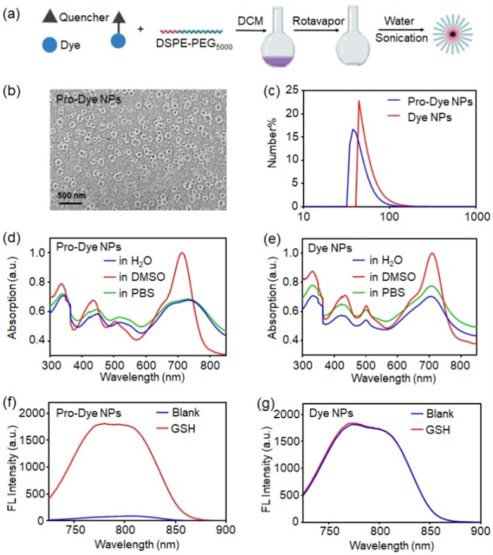
NIR BODIPY derivatives generally feature substantial aromatic structures, rendering them hydrophobic and challenging for direct use in vivo. To improve the water dispersity of Pro-Dye, DSPE-PEG5000, a frequently employed amphiphilic compound in liposome and micelle formulations, was utilized to disperse the Dye and Pro-Dye by forming NPs. As depicted in Fig. 2a, the Pro-Dye NPs were prepared via the membrane hydration method. Both the fluorophore and DSPE-PEG5000 were dissolved in dichloromethane (DCM) at a ratio of 1:9 (w/w). Subsequently, the solvent was dried in a flask using a rotavapor, resulting in a film on the inner wall of the flask. Finally, the flask was added with water and subjected to sonication, yielding a homogeneous NPs solution. Transmission electron microscopy (TEM) was employed to characterize the morphology of formed Pro-Dye NPs, which exhibited a predominantly spherical shape with a diameter < 100 nm (Fig. 2b). Furthermore, dynamic light scattering (DLS) analysis was applied and demonstrated that the particle size of Pro-Dye NPs was about 40 nm with a polydispersity index (PDI) of 0.25 (Fig. 2c), while the size of Dye NPs was about 43 nm with a polydispersity index of 0.27. Even more, quantitative analysis of particle size in TEM imaging was done, and the results were depicted in Figs. S13 and S14 (Supporting information). TEM image statistics revealed that the particle sizes of both Pro-Dye NPs and Dye NPs are within the range of 10–100 nm, which was agree with the statistics of DLS, benefiting tumor accumulation via the enhanced permeation and retention (EPR) effect [21, 30]. Furthermore, the particle sizes of Pro-Dye NPs remained below 100 nm in water, phosphate buffer saline (PBS), and DMEM medium over a period of 72 consecutive hours, suggesting efficient maintenance of the EPR effect during in vivo blood circulation (Figs. S15–S21 in Supporting information). Then, the absorption and emission spectra of Pro-Dye NPs and Dye NPs were measured, respectively. Both Pro-Dye NPs and Dye NPs exhibited absorption peaks at wavelengths beyond 700 nm, facilitating the use of NIR light as an excitation source for illuminating deep tissues (Figs. 2d and e). Furthermore, the GSH activation experiment was repeated to verify that the Pro-Dye NPs retained their GSH-activatable property. The activation experiments of fluorescence spectra demonstrated a notable increase in the fluorescence intensity of the Pro-Dye NPs solution (10 µmol/L) following incubation with GSH (10 mmol/L), while the fluorescence intensity of Dye NPs showed negligible enhancement (Figs. 2f and g), suggesting the retained GSH-responsive nature of the Pro-Dye NPs. The remarkable GSH-responsive properties of Pro-Dye NPs guarantee great promise for applications in vitro and in vivo imaging.
Figure 2
Figure 2.
(a) Schematic procedure for preparing Dye NPs and Pro-Dye NPs. (b) TEM characterization of Pro-Dye NPs. (c) Diameters and PDIs of Pro-Dye and Dye NPs in H2O as characterized by DLS. Pro-Dye NPs PDI = 0.25, Dye NPs PDI = 0.27. (d) Absorption spectra of Pro-Dye NPs in H2O, DMSO, and PBS, respectively. (e) Absorption spectra of Dye NPs in H2O, DMSO, and PBS, respectively. (f) Fluorescence spectra of Pro-Dye NPs (10 µmol/L). Blank: in DMSO (containing 10% H2O); GSH: incubated with 10 mmol/L GSH for 1 h. (g) Fluorescence spectra of Dye NPs (10 µmol/L). Blank: in DMSO (containing 10% H2O); GSH: incubated with 10 mmol/L GSH for 1 h.
MTT assay was conducted to assess the cytotoxicity of the Pro-Dye NPs. 4T1 breast cancer cells and HUVECs normal cells were incubated with Dye and Pro-Dye NPs at various concentrations for 24 h, following by cytoviability assessment. The results showed that the Pro-Dye NPs exhibited negligible cytotoxicity even at a concentration up to 50 µmol/L (Fig. S22 in Supporting information). Furthermore, the hemolytic test of Pro-Dye NPs was conducted to determine their in vivo safety. All the animal experiments in this research were approved by the Biomedical Ethics Committee of Xi'an Jiaotong University Health Science Center (No. 2021–1612). The results of hemolytic test demonstrated that Pro-Dye NPs did not induce hemolysis even at a concentration of 500 µg/mL (Figs. S24 and S25 in Supporting information), suggesting high safety via intravenous injection in further experiments. 4T1 and HUVEC cells were incubated with Pro-Dye NPs and imaged using a fluorescence microscope. As shown in Fig. S26 (Supporting information), the fluorescence intensity of 4T1 cells exhibited a notable enhancement as the incubation time increased from 3 h to 6 h (Figs. S26a and d). In contrast, the fluorescence images of HUVEC showed weak fluorescence even with increased incubation time (Fig. S26b). Semi-quantitative fluorescence analysis for fluorescence imaging of 4T1 cells indicated almost three-fold higher intensity than that of HUVEC cells (Fig. S26d). The results suggested that Pro-Dye NPs can be specifically activated by cancer cells but remain quenched when exposed to normal cells. To further demonstrate the intracellular activation of Pro-Dye NPs induced by GSH, BSO was used as a GSH synthetase inhibitor and pre-incubated with 4T1 cells to reduce the intracellular GSH concentration. Subsequently, BSO-pretreated 4T1 cells were incubated with Pro-Dye NPs for 6 h, followed by fluorescence imaging. The fluorescence intensity of BSO-pretreated 4T1 cells was significantly lower than that of the normal 4T1 cells, indicating a strong relationship between the intracellular activation process of Pro-Dye and high GSH concentration within cancer cells (Figs. S26c and e). All the results preliminarily demonstrated the effectiveness of the GSH activation strategy in cancer cell imaging. Additionally, it confirmed the potential of the designed fluorophore for tumor-selective in vivo imaging and surgical guidance.
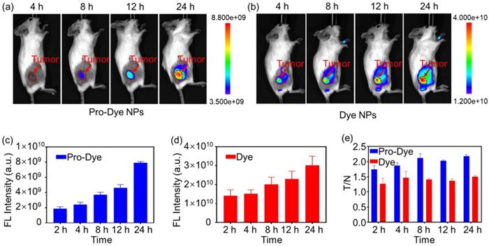
Subsequently, in vivo fluorescence imaging was conducted in 4T1 tumor-bearing mice after intravenous injection of Pro-Dye NPs and Dye NPs (control group), respectively. The fluorescence signals at tumor sites in mice injected with Pro-Dye NPs and Dye NPs enhanced over time, suggesting efficient accumulation of both Pro-Dye NPs and Dye NPs at their respective tumor sites (Figs. 3a–d). Due to the fluorescence quenching effect in Pro-Dye, the fluorescence signal at the tumor site in the Dye NPs group was stronger than that in the Pro-Dye NPs group. However, the fluorescence intensity of the Pro-Dye group at 24 h post-injection increased almost 4-fold compared to that at the 2-h time point post-injection, whereas the fluorescence intensity in the Dye group increased only about 2-fold. The enhanced fluorescence intensity in the Pro-Dye NPs group is not only contributed by the increased accumulation of the Pro-Dye NPs but also by the gradual activation of Pro-Dye at the tumor site. The Pro-Dye NPs accumulated at the tumor via the EPR effect and subsequently released the quenched Pro-Dye molecule, which was gradually activated by the intratumoral GSH over time. Compared with the strong fluorescence signal at the tumor-adjacent area in the Dye NPs group, the fluorescence signal of the tumor-adjacent area in the Pro-Dye NPs group was much weaker due to the lack of activation (Figs. 3a and b). Therefore, the tumor/normal tissue (T/N) fluorescence signal ratio in the Pro-Dye NPs group was significantly higher than that in the Dye NPs group, resulting in a better tumor edge for further surgical guidance (Fig. 3e).
Figure 3
Figure 3.
(a) In vivo fluorescence images of tumor-bearing mice injected with Pro-Dye NPs (0.83 mmol/L, 200 µL) at 4, 8, 12, and 24 h post-injection. (b) In vivo fluorescence images of tumor-bearing mice injected with Dye NPs (0.83 mmol/L, 200 µL) at 4, 8, 12, and 24 h post-injection. (c) Average fluorescence intensity of the tumor area of the Pro-Dye NPs injected group at 2, 4, 8, 12, and 24 h post-injection. (d) Average fluorescence intensity of the tumor area of the Dye NPs injected group at 2, 4, 8, 12, and 24 h post-injection. (e) Ratios of the average fluorescence intensity between the tumor area and tumor-adjacent normal tissue (T/N) at 2, 4, 8, 12, and 24 h post-injection. Data are presented as mean ± standard deviation (SD) (n = 4).
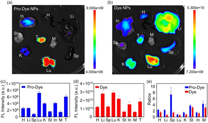
At 24 h after injection, the tumor-bearing mice were sacrificed and dissected to collect major organs (heart, liver, spleen, lung, kidney, stomach, intestine), as well as the tumor and tumor-adjacent tissue. The collected tissues were placed in a petri dish to assess the biodistribution of Dye NPs and Pro-Dye NPs through fluorescence imaging. As depicted in Figs. 4a and c, the fluorescence intensity of the resected tumor in the Pro-Dye group was notably higher than that of all other organs, except for the lung. Conversely, the fluorescence intensity of the tumor in the Dye NPs group was not prominent compared to that of normal organs due to the distributed Dye with "always on" fluorescence properties (Figs. 4b and d). The significant difference between the two groups demonstrated the advantage of Pro-Dye activation in tumors but not in normal tissues. In the Dye NPs group, the tumor fluorescence intensity was considerably weaker than that of the liver, kidney, and lung, indicating lower specificity for tumor accumulation. The fluorescence intensity of the lung was notably high in both groups, suggesting the accumulation of nanoparticles in the lung and the activation of Pro-Dye within the lung tissue. This phenomenon can be attributed to the high concentration of GSH in the lung epithelial lining fluid [31]. The fluorescence intensity ratios of tumor to organ/muscle were further quantified and calculated (Fig. 4e). The increased ratio in the Pro-Dye NPs group compared to the Dye NPs group indicated enhanced selectivity in tumor imaging and promising potential for surgical guidance.
Figure 4
Figure 4.
Biodistribution analysis of the Pro-Dye NPs group and Dye NPs group. (H: heart; Li: liver; Sp: spleen; Lu: lung; K: kidney; St: stomach; In: intestine; T: tumor; M: muscle). (a) Fluorescence image of major organs, tumor, and tumor-adjacent normal muscle from mice of the Pro-Dye NPs group at 24 h post-injection. (b) Fluorescence image of major organs, tumor, and tumor-adjacent normal muscle from mice of Pro-Dye NPs group at 24 h post injection. (c) Average fluorescence intensities of organs and tissues in (a). (d) Average fluorescence intensities of organs and tissues in (b). (e) Fluorescence intensity ratios of tumor to normal organs/muscle in (a) and (b). Data are presented as mean ± SD (n = 4).
To perform NIR fluorescence imaging-guided surgery, the tumor-bearing mice were intravenously injected with Pro-Dye NPs (0.83 mmol/L, 200 µL) and then subjected to preoperative fluorescence imaging to delineate the surgical area of the tumor. Complete resection of the tumor tissue was confirmed when the fluorescence signal disappeared completely within the surgical area. After tumor resection, the isolated tumor tissue was set aside and subjected to fluorescence imaging again (Fig. S27 in Supporting information). The image clearly showed that there was no fluorescence signal at the tumor site anymore after the surgery, while the isolated tumor tissue exhibited strong fluorescence intensity. This result suggested complete resection of the tumor tissue, confirming the success of the operation.
To demonstrate the biocompatibility of the prepared fluorescence probe, a series of pathological and toxicological experiments were conducted on Pro-Dye. The organs collected post fluorescence imaging were sectioned and stained with hematoxylin and eosin (H & E) for microscope imaging to evaluate their cellular morphology [32, 33]. These images revealed the absence of inflammation or mutation in major organs (heart, liver, spleen, lung, kidney, stomach, intestine), indicating good biocompatibility of Pro-Dye NPs and Dye NPs for in vivo applications (Fig. S28 in Supporting information). A routine blood test and blood biochemistry analysis were further conducted at 1 and 7 days after injecting Pro-Dye [34] NPs to verify the hematological safety. The results showed that all parameters in the blood routine test (WBC: white blood cell; RBC: red blood cell; HGB: hemoglobin; PLT: platelets; HCT: hematocrit; MCHC: mean corpuscular hemoglobin concentration; MCV: mean corpuscular volume) fell within the normal ranges (Fig. S29 in Supporting information), demonstrating the absence of acute hematotoxicity caused by Pro-Dye NPs. These results demonstrated that Pro-Dye NPs possessed good biocompatibility and safety for in vivo bioapplications.
In summary, this study has developed a novel GSH responsive BODIPY-based NIR fluorophore (Pro-Dye) with a large Stokes shift. The Pro-Dye was specifically designed and synthesized to be activated by high concentrations of GSH at the tumor site while remaining quenched in normal tissue. The in vitro activation test of the fluorophore demonstrated high efficiency and specificity in response to GSH, indicating potential applications in bioimaging. DSPE-PEG5000 was utilized to create Pro-Dye NPs for the efficient dispersion of the hydrophobic Pro-Dye, with particle sizes ranging from 40 nm to 70 nm. Pro-Dye NPs can accumulate at the tumor site and be further activated by GSH, thereby improving the SBR from 1.5 to 2 and enhancing the contrast of in vivo fluorescence imaging. The T/N ratio can facilitate precise intraoperative tumor resection, potentially reducing the occurrence of positive tumor margins and tumor recurrence. The GSH-responsive NIR fluorophore exhibits promising potential for application in precise tumor resection, with the capability to significantly improve the prognosis of clinical patients.
Declaration of competing interest
The authors declare that they have no known competing financial interests or personal relationships that could have appeared to influence the work reported in this paper.
This work was supported by the Natural Science Foundation of Shaanxi Province (Nos. 2023-YBSF-270, 2024SF-ZDCYL-02–08), Fundamental Research Funds for the Central Universities (No. xzy022024033), and Horizontal Project of the First Affiliated Hospital of Xi'an Jiaotong University (No. 202304174). This research was also supported by the Opening Project of Structural Optimization and Application of Functional Molecules Key Laboratory of Sichuan Province (No. 2023GNFZ-03), and The Key Laboratory for Screening and Diagnosis of Maternal and Child Genetic Disease of Health Commission of Jiangxi Province. The characterization assistance from the Instrument Analysis Center of Xi'an Jiaotong University is also acknowledged.
Supplementary materials
Supplementary material associated with this article can be found, in the online version, at doi:10.1016/j.cclet.2024.110459.
J. Xin, S. Han, M. Zheng, et al., Chin. Chem. Lett. 35 (2024) 109165.
[31]
J.S. Armstrong, K.K. Steinauer, B. Hornung, et al., Cell Death Differ. 9 (2002) 252–263. doi: 10.1038/sj.cdd.4400959
[32]
H. Chen, S. Yan, L. Zhang, et al., Sens. Actuators B: Chem. 405 (2024) 135346.
[33]
Y. Zhang, H. Zhao, J. Tang, et al., Bioorg. Chem. 140 (2023) 106800.
[34]
B. Chen, S. Mao, Y. Sun, et al., Chem. Commun. 57 (2021) 4376–4379. doi: 10.1039/d1cc01104a
Figure 1
(a) Structural activation process of Pro-Dye by reacting with GSH. (b) UV–vis absorption spectra and normalized fluorescence emission spectrum (Ex = 680 nm) of Dye in DMSO. (c) UV–vis absorption spectra and normalized fluorescence emission spectrum (Ex = 680 nm) of Pro-Dye in DMSO. (d) Fluorescence image of Dye and Pro-Dye in DMSO (10 µmol/L). (e) HPLC analysis of Pro-Dye during incubation with GSH. (f) Kinetics curves of Pro-Dye activated by GSH at different concentrations (mM = mmol/L). (g) Fluorescence spectra of Pro-Dye at 1 h after incubation with different reagents (NaCl, 50 mmol/L; KCl, 50 mmol/L; CaCl2, 4 mmol/L; MgCl2, 1 mmol/L; MnCl2, 50 mmol/L; AgNO3, 2 mmol/L; CuCl2, 1 mmol/L; glucose, 10 mmol/L; GSH, 10 mmol/L).
Figure 2
(a) Schematic procedure for preparing Dye NPs and Pro-Dye NPs. (b) TEM characterization of Pro-Dye NPs. (c) Diameters and PDIs of Pro-Dye and Dye NPs in H2O as characterized by DLS. Pro-Dye NPs PDI = 0.25, Dye NPs PDI = 0.27. (d) Absorption spectra of Pro-Dye NPs in H2O, DMSO, and PBS, respectively. (e) Absorption spectra of Dye NPs in H2O, DMSO, and PBS, respectively. (f) Fluorescence spectra of Pro-Dye NPs (10 µmol/L). Blank: in DMSO (containing 10% H2O); GSH: incubated with 10 mmol/L GSH for 1 h. (g) Fluorescence spectra of Dye NPs (10 µmol/L). Blank: in DMSO (containing 10% H2O); GSH: incubated with 10 mmol/L GSH for 1 h.
Figure 3
(a) In vivo fluorescence images of tumor-bearing mice injected with Pro-Dye NPs (0.83 mmol/L, 200 µL) at 4, 8, 12, and 24 h post-injection. (b) In vivo fluorescence images of tumor-bearing mice injected with Dye NPs (0.83 mmol/L, 200 µL) at 4, 8, 12, and 24 h post-injection. (c) Average fluorescence intensity of the tumor area of the Pro-Dye NPs injected group at 2, 4, 8, 12, and 24 h post-injection. (d) Average fluorescence intensity of the tumor area of the Dye NPs injected group at 2, 4, 8, 12, and 24 h post-injection. (e) Ratios of the average fluorescence intensity between the tumor area and tumor-adjacent normal tissue (T/N) at 2, 4, 8, 12, and 24 h post-injection. Data are presented as mean ± standard deviation (SD) (n = 4).
Figure 4
Biodistribution analysis of the Pro-Dye NPs group and Dye NPs group. (H: heart; Li: liver; Sp: spleen; Lu: lung; K: kidney; St: stomach; In: intestine; T: tumor; M: muscle). (a) Fluorescence image of major organs, tumor, and tumor-adjacent normal muscle from mice of the Pro-Dye NPs group at 24 h post-injection. (b) Fluorescence image of major organs, tumor, and tumor-adjacent normal muscle from mice of Pro-Dye NPs group at 24 h post injection. (c) Average fluorescence intensities of organs and tissues in (a). (d) Average fluorescence intensities of organs and tissues in (b). (e) Fluorescence intensity ratios of tumor to normal organs/muscle in (a) and (b). Data are presented as mean ± SD (n = 4).

 DownLoad:

 下载:
下载:


 下载:
下载:
