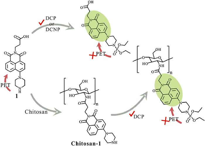
Scheme 1.
Schematic illustration of chitosan-1 used for specific detection of chemical nerve agent simulants (DCP).
Ultrasensitive and selective detection of chemical nerve agent simulants based on naphthalimide functionalized chitosan as fluorescent nanofibers
Qian Chen , Anyang Shen , Taotao Huang , Xinya Han , Jian Zhang , Hui Jiang , Renyong Liu , Yong Pan , Kui Zhang

Chemical nerve agents, known for their extreme toxicity, are subject to strict regulations regarding storage and preparation [1-3]. Consequently, chemical nerve agents are subsequently used in terrorist attacks, posing a threat to homeland security and terrorist threats. Due to the lack of protective measures and reliable detection methods, exposure to chemical nerve agents can irreversibly damage acetylcholinesterase resulting in lethal injury [4,5]. Therefore, there is a growing interest in the development of simple and highly sensitive detection methods for nerve agents, particularly those that enable visual detection. Traditionally, various methods such as gas chromatography, electrochemical sensors, and mass spectrometry have been employed to detect chemical nerve agents [6-10]. However, these methods have limitations in terms of portability, on-site detection capabilities, and equipment costs, which restrict further application. In recent years, colorimetric and fluorescent methods have emerged as promising alternatives for on-site detection of chemical nerve agents [11-15]. However, most of the organic fluorescent probes are afflicted by photobleaching, limited detection capability, and long-time response [16-20]. The designed structures of the probe allow for the improvement of sensing performance through the nucleophilic reaction of oxime groups [21-23], active oxygen [24-26], and nitrogen groups [27,28]. Additionally, the acid is also an interference effect to practical detection [29]. Therefore, the design of a fluorescent probe with high sensitivity, fast detection, and flexible modification remains highly desirable.
Small organic fluorescent probes modified with polymers have been widely explored for fluorescent sensing applications due to their high porosity, tailorable functionalization, and flexible modification [30-34]. The organic molecular fluorescent probe was fabricated with polymer to form nanofiber through the electrospinning technique which serves as a promising strategy to improve detection performance [35-38]. For example, the Yoon et al. group has developed a fluorescent sensor embedded in a nanofiber with o-phenylenediamine units to discriminate between nerve agent mimic vapor and phosgene [39]. Zeng et al. have developed a fluorescent sensor based on anthracene carboxyimide with polystyrene film for sensing DCP in solution and gas phases [40]. Chitosan has attracted significant attention because it has a rigid chain and biodegradable, and has low toxicity and chemical stability. The main chain of chitosan has numerous hydroxyl and amino groups which are suitable for functional modification. On the other hand, 1,8-naphthalimide is the fluorescent segment that has excellent photostability and unique spectral characteristics [41-44]. Inspired by the above studies, the chitosan was connected with fluorescent sensors may provide multiple detection units to improve the sensing performance. Especially, compared with the simple organic fluorescent probes, the conjugated fluorescent nanofiber significantly increased sensing sensitivity, flexibility, and response signal amplification. Introducing fluorescent probes to chitosan is considered a promising method to overcome the challenges bothered by small organic fluorescent probes.
In this study, we have developed a chitosan-based fluorescent nanofiber that provides ultrasensitive and highly selective detection of diethyl chlorophosphate (DCP) as shown in Scheme 1. The fluorescent detection unit was synthesized through the combination of 1,8-naphthalene and piperazine. The nanofiber chitosan-1 exhibits ultrasensitive and rapid response to DCP solution with the limitation of detection 2.2 nmol/L, which is significantly lower than compound 1. Moreover, the sensing mechanism was demonstrated by NMR spectra, mass spectrum, and density functional theory calculation. Therefore, the prepared sensing nanofiber not only shows impressive sensing performance but also specific detection of chemical nerve agent simulants DCP in both solution and vapor phases. The modification of chitosan may provide a feasible strategy to improve sensitivity, reduce response time, and enhance fluorescent signal accumulation.
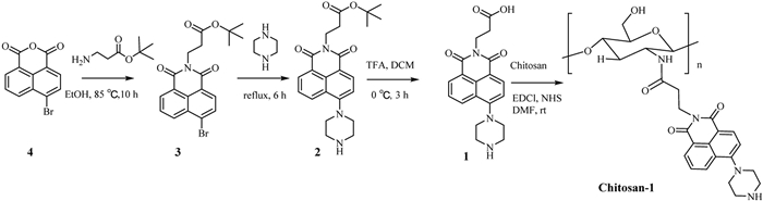
The naphthalimide is frequently introduced in organic molecule fluorescent probes due to its high quantum yield and convenient modification. Compound 1 was synthesized from 4‑bromo-1,8-naphthalic anhydride with a satisfactory overall yield. The synthetic routine of chitosan-1 as depicted in Scheme 2. The structure of compound 1 was verified through NMR and HRMS characterization. The IR spectra of chitosan, compound 1, and chitosan-1 were collected respectively as shown in Fig. S1 (Supporting information). By analyzing the IR spectrum, we confirmed the successful attachment of compound 1 to chitosan, as evidenced by the main peak occurring at 1650 cm-1, which are the characteristic peaks demonstrate the successful connection between compound 1 and the amine group of chitosan [45].
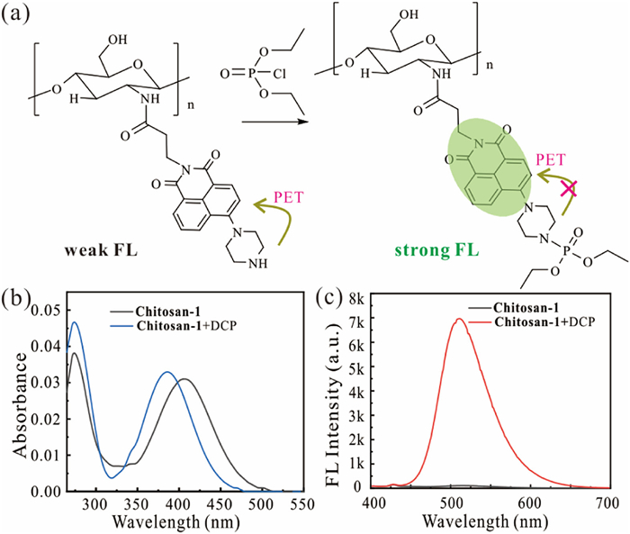
To evaluate the fluorescent detection performance of chitosan-1, the absorption and fluorescence spectra of chitosan-1 were explored in DMF solution. The photoinduced electron transfer (PET) process between the naphthalimide and piperazine resulted in a negative fluorescent response, as shown in Fig. 1a. In the presence of DCP, the fluorescence intensity sharply increased owing to the generation of a phosphorous ester. The maximum absorption wavelength exhibited a slight blue shift from 407 nm to 386 nm in the DMF solution, which can be attributed to the reduced electron donor ability of the phosphate ester compared to piperazine. The strong fluorescence intensity of chitosan-1 increased about 60-fold which is caused by the inhibition of the PET process, as shown in Figs. 1b and c. Thus, the rapid fluorescent response and photostability indicated that chitosan-1 is suitable for visual detecting DCP in solution.
To explore the sensitivity, we conducted titration fluorescent experiments. Firstly, we investigated the fluorescent response of compound 1 with various phosphorus analysts. We found that the fluorescent intensity was gradually increased with various concentrations of DCP and DCNP. The fluorescent intensity was increased at 510 nm and exhibited a satisfactory linear correlation with DCP and DCNP as shown in Figs. S4 and S5 (Supporting information), respectively. Under the same conditions, the fluorescent intensity of chitosan-1 was sharply increased and stayed at a plateau period with the gradual addition of DCP, as shown in Figs. 2a and b. The limitation of detection was calculated as low as 2.2 nmol/L, which is satisfied compared to the related literature in Table S1 (Supporting information). These results have demonstrated that the modification of compound 1 to chitosan remarkably decreases the detection limitation and amplifies fluorescent intensity. Considering the excellent specific detection of DCP, chitosan-1 was a promising candidate probe for sensitive and selective detection of DCP.
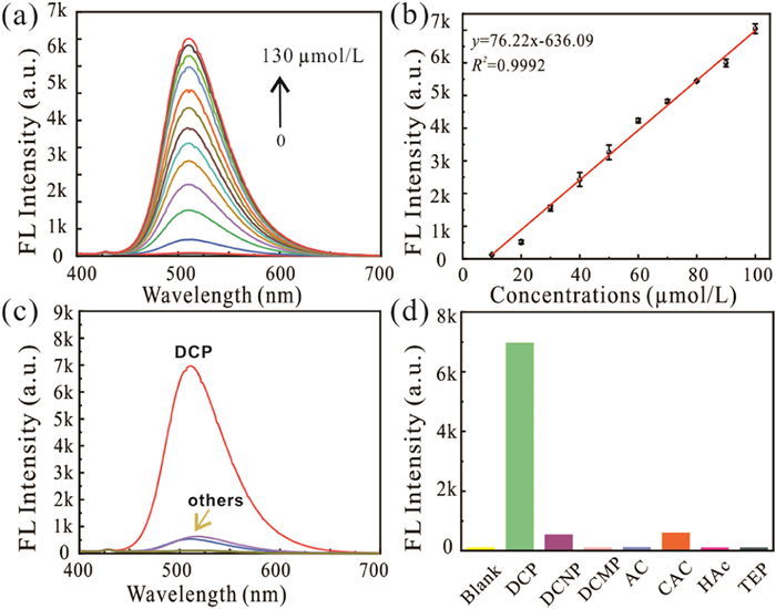
To explore the sensing performance, the selectivity of chitosan-1 was also investigated with various analytes including triethyl phosphate (TEP), acetic acid (HAc), acetyl chloride (AC), chloroacetyl chloride (CAC), diethyl cyanophosphonate (DCNP), and diethyl cyanomethyl phosphate (DCMP). Additionally, we conducted fluorescent detection experiments of DCP with compound 1 as a comparison. Under the optimal conditions, compound 1 could not discriminate the difference between DCP and DCNP due to the similar alkalinity of piperazine as shown in Fig. S4. Additionally, in the presence of DCP, the fluorescent intensity of chitosan-1 was dramatically increased about 60-fold as illustrated in Fig. 2c. The addition of DCNP and chloroacetyl chloride (CAC) resulted in a slight increase in fluorescent signal. Furthermore, the addition of other relative interfering analytes exhibited a negative fluorescent response as shown in Fig. 2d. These results demonstrated that the connection of compound 1 and chitosan not only provided multiple detection segments but also amplified the fluorescent response compared to compound 1.
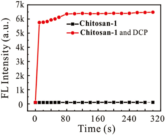
With the excellent sensing performance achieved, we were encouraged to detect DCP in solution and vapor. The reaction time was measured upon the addition of DCP in DMF solution, as shown in Fig. 3. The fluorescence enhancement increased within a few seconds and exhibited good photostability. Furthermore, we also evaluated the detection of DCP in the presence of relevant interferents, including DCNP, DCMP, acetyl chloride (AC), chloroacetyl chloride (CAC), acetic acid (HAc), and triethyl phosphate (TEP) as shown in Fig. 4a. It clearly shows that only DCP exhibited a distinct green color under 365 nm excitation. The detection of DCP vapor at concentrations of 0, 30, 60, and 90 ppm was conducted using chitosan-1 nanofibers as shown in Fig. 4b. These data demonstrated that chitosan-1 nanofibers can serve as an ultra-sensitive and selective probe for the detection of DCP vapor.

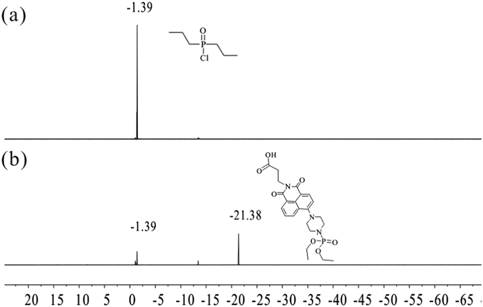
To explore the sensing mechanism, the 1H and 31P NMR spectra of only DCP and the mixture of DCP and compound 1 as illustrated in Figs. S10 and S11 (Supporting information), respectively. The peak of −1.39 was ascribed in the neat DCP as shown in Fig. 5a. In addition, after the addition of DCP in compound 1, the peak at −21.38 appeared which represented the formation of phosphate ester (Fig. 5b). To investigate the origin of sensing performance, the mass spectrum was also conducted. The main peak has appeared at 354.14401 which can be assigned to compound 1. Meanwhile, the mixture of compound 1 with DCP was measured to find the main peak shown in 489.07785 (Fig. S3 in Supporting information). With the method of density function theory (DFT) calculation, we try to illustrate the detection performance and origin mechanism as illustrated in Fig. S7 (Supporting information). DFT calculations have been carried out at the B3LYP/6–31G computational level, within the Gaussian-09 package. The obtained geometries were confirmed to be energetic minima through frequency calculations in all cases. The energy and molecular distribution of the frontier orbitals HOMO and LUMO have been obtained with the Gaussian-09 facilities and displayed with the GaussView-6 program. The calculation of compound 1 and the reaction of DCP were consistent with the sensing behavior.
In summary, we prepared a fluorescent nanofiber using fluorescent compound 1 and chitosan. The nanofiber based on chitosan-1 was confirmed by FT-IR and NMR spectrum which showed ultrasensitive and selective detection of DCP in solution and vapor. The fluorescent sensing unit was covalently bonded with the chitosan amine resulting in the abundance of repetitive units and the fluorescent signal accumulation compared with compound 1. The fluorescent response was significantly increased about 60-fold < 1 min and the limitation of detection was measured as low as 2.2 nmol/L. The fluorescent nanofiber was explored to detect the concentration of DCP vapor in the range from 0 ppm to 90 ppm with obvious color change. The facile strategy may provide a promising method for rapid visual detection of chemical nerve agents.
The authors declare that they have no known competing financial interests or personal relationships that could have appeared to influence the work reported in this article.
Qian Chen: Writing – original draft, Project administration, Conceptualization. Anyang Shen: Validation, Formal analysis. Taotao Huang: Software, Methodology. Xinya Han: Supervision, Funding acquisition. Jian Zhang: Methodology, Investigation. Hui Jiang: Resources, Methodology. Renyong Liu: Validation, Resources. Yong Pan: Supervision, Software. Kui Zhang: Writing – review & editing, Funding acquisition.
We are grateful for financial support from the National Natural Science Foundation of China (Nos. 82104065, 32061143045, 22276142, 22474003), the National Key Research & Development Program (Nos. 2019YFE0123100, 2022YFE0199800), Anhui Provincial Natural Science Foundation (No. 2208085MB38), Anhui Provincial Natural Science Foundation for Distinguished Young Scholars (No. 2008085J11) and Foundation of Education Department of Anhui Province (No. 2022AH010023).
Supplementary material associated with this article can be found, in the online version, at doi:
L. Gorecki, O. Soukup, J. Korabecny, Trends Pharmacol. Sci. 43 (2022) 593–606.
M. Schwenk, Toxicol. Lett. 293 (2018) 253–263.
K. Kim, O.G. Tsay, D.A. Atwood, D.G. Churchill, Chem. Rev. 111 (2011) 5345–5403. doi: 10.1021/cr100193y
V. Aroniadou-Anderjaska, J.P. Apland, T.H. Figueiredo, M. De Araujo Furtado, M.F. Braga, Neuropharmacology 181 (2020) 108298.
Q. Zhuang, A.J. Franjesevic, T.S. Corrigan, et al., J. Med. Chem. 61 (2018) 7034–7042. doi: 10.1021/acs.jmedchem.7b01620
R.L. Webster, S.P.B. Ovenden, L.J. McDowall, et al., Anal. Bioanal. Chem. 414 (2022) 3863–3873. doi: 10.1007/s00216-022-04027-1
D.T. Snyder, P.S. Demond, L.J. Szalwinski, et al., Int. J. Mass Spectrom. 444 (2019) 116171.
K.M. Rubin, B.A. Goldberger, T.J. Garrett, J. Anal. Toxicol. 44 (2020) 391–401. doi: 10.1093/jat/bkz118
G. Manco, E. Porzio, Y. Suzumoto, J. Chem. Technol. Biotechnol. 93 (2018) 2064–2082. doi: 10.1002/jctb.5603
J. Nawała, K. Czupryński, S. Popiel, D. Dziedzic, J. Bełdowski, Anal. Chim. Acta. 933 (2016) 103–116.
X. Wang, X. Wang, R. Feng, et al., Chem. Asian. J. 17 (2022) e202200284.
W.Q. Meng, A.C. Sedgwick, N. Kwon, et al., Chem. Soc. Rev. 52 (2022) 601–662. doi: 10.2991/978-94-6463-052-7_70
G. Wang, Y. Li, Z. Cai, X. Dou, Adv. Mater. 32 (2020) 1907043.
H. Ma, F. Li, P. Li, et al., Adv. Funct. Mater. 26 (2016) 2025–2031. doi: 10.1002/adfm.201504692
Y. Gong, Y. Guo, C. Qiu, et al., Sci. China Mater. 64 (2021) 1189–1196. doi: 10.1007/s40843-020-1517-8
B. Zhu, R. Sheng, T. Chen, et al., Coord. Chem. Rev. 463 (2022) 214527.
M.H. Lee, J.S. Kim, J.L. Sessler, Chem. Soc. Rev. 44 (2015) 4185–4191.
S.S. Ali, A. Gangopadhyay, A.K. Pramanik, et al., Dyes Pigm. 170 (2019) 107585.
J. Tan, Z. Li, Z. Lu, et al., Dyes Pigm. 193 (2021) 109540.
V. Kumar, Chem. Commun. 57 (2021) 3430–3444. doi: 10.1039/d1cc00132a
M.C. de Koning, G. Horn, F. Worek, M. van Grol, Eur. J. Med. Chem. 157 (2018) 151–160.
Y.C. Cai, C. Li, Q.H. Song, J. Mater. Chem. C 5 (2017) 7337–7343.
S. Zhang, B. Yang, B. Yuan, et al., ACS Sens. 8 (2023) 1220–1229. doi: 10.1021/acssensors.2c02611
Z. Lu, W. Fan, X. Shi, et al., Sens. Actuators B: Chem. 255 (2018) 176–182.
K.C. Behera, B. Bag, Chem. Commun. 56 (2020) 9308–9311. doi: 10.1039/d0cc03985c
F.W. Dagnaw, Y.P. Cai, Q.H. Song, Dyes Pigm. 189 (2021) 109257.
S. Gharami, K. Aich, S. Das, L. Patra, T.K. Mondal, New J. Chem. 43 (2019) 8627–8633. doi: 10.1039/c9nj02218j
N. Dey, J. Kulhánek, F. Bureš, S. Bhattacharya, J. Org. Chem. 86 (2021) 14663–14671. doi: 10.1021/acs.joc.1c01488
S. Fan, G.H. Dennison, N. FitzGerald, et al., Commun. Chem. 4 (2021) 45.
M.S.J. Khan, Y.W. Wang, M.O. Senge, Y. Peng, J. Hazard. Mater. 342 (2018) 10–19.
Y.C. Cai, C. Li, Q.H. Song, ACS Sens. 2 (2017) 834–841. doi: 10.1021/acssensors.7b00205
X. Yan, H. Li, X. Su, TrAC, Trends Anal. Chem. 103 (2018) 1–20. doi: 10.1117/1.oe.57.11.117114
P.P. Jia, Y.X. Hu, Z.Y. Zeng, et al., Chin. Chem. Lett. 34 (2023) 107511.
B.X. Huang, X. Liu, G.L. Yang, et al., CCS Chem. 4 (2022) 2090–2101. doi: 10.31635/ccschem.021.202100950
Y. Zhang, H. Mu, P. Zheng, Y. Zhao, M. Zhang, Sens. Actuators B: Chem. 343 (2021) 130140.
L. Chen, H. Oh, D. Wu, M.H. Kim, J. Yoon, Chem. Commun. 54 (2018) 2276–2279. doi: 10.1039/c7cc09901k
E. Jeong, J.K. Kim, J. Jin, H.I. Lee, Carbohydr. Polym. 295 (2022) 119845.
W. Mo, Z. Zhu, F. Kong, et al., Nat. Commun. 13 (2022) 5189.
Y. Hu, X. Zhou, H. Jung, et al., Anal. Chem. 90 (2018) 3382–3386. doi: 10.1021/acs.analchem.7b05011
L. Zeng, H. Zeng, L. Jiang, et al., Anal. Chem. 91 (2019) 12070–12076. doi: 10.1021/acs.analchem.9b03230
L. Gopala, Y. Cha, M.H. Lee, Dyes Pigm. 201 (2022) 110195.
A.S. Oshchepkov, M.S. Oshchepkov, M.V. Oshchepkova, et al., Adv. Opt. Mater. 9 (2021) 2001913.
H. Xu, H. Zhang, L. Zhao, et al., New J. Chem. 44 (2020) 10713–10718. doi: 10.1039/d0nj00416b
Y.T. Wang, Y. Qin, X.L. Zhao, et al., Chin. Chem. Lett. 34 (2023) 107576.
G.Q. Ying, W.Y. Xiong, H. Wang, Y. Sun, H.Z. Liu, Carbohydr. Polym. 83 (2011) 1787–1796.

Scheme 1 Schematic illustration of chitosan-1 used for specific detection of chemical nerve agent simulants (DCP).

Figure 1 (a) The illustration of the sensing mechanism between chitosan-1 and DCP, (b) UV–vis spectrum and (c) fluorescence spectra of chitosan-1 in the presence and absence of DCP in DMF solution.

Figure 2 (a) The fluorescence titration of chitosan-1. (b) The linear relationship between fluorescent intensity and variety concentration of DCP (0–100 µmol/L). (c, d) Fluorescence spectra of chitosan-1 on addition of relevant interferes.

Figure 4 (a) The detection images of chitosan-1 and DCP with various organophosphorus analytes. (b) The exposure of nanofiber to different amounts of DCP vapor range from 0 ppm to 90 ppm.

 扫一扫看文章

扫一扫关注我们
 DownLoad:
 下载:
下载:


 下载:
下载: