College of Chemistry, Fuzhou University, Fuzhou 350108, China
b.
State Key Laboratory of Structural Chemistry, Fujian Institute of Research on the Structure of Matter, Chinese Academy of Sciences, Fuzhou 350002, China
c.
Fujian Science & Technology Innovation Laboratory for Optoelectronic Information of China, Fuzhou 350108, China
Received Date:
03 April 2024 Accepted Date:
04 June 2024 Revised Date:
09 May 2024 Available Online:
15 July 2025
Abstract:
2D Ruddlesden-Popper (RP) polar perovskite, displaying the intrinsic optical anisotropy and structural polarity, has a fantastic application perspective in self-powered polarized light detection. However, the weak van der Waals interaction between the organic spacing bilayers is insufficient to preserve the stability of RP-type materials. Hence, it is of great significance to explore new stable 2D RP-phase candidates. In this work, we have successfully constructed a highly-stable polar 2D perovskite, (t-ACH)2PbI4 (1, where t-ACH+ is HOOC8H12NH3+), by adopting a hydrophobic carboxylate trans-isomer of tranexamic acid as the spacing component. Strikingly, strong O-H···O hydrogen bonds between t-ACH+ organic bilayers compose the dimer, thus decreasing van der Waals gap and enhancing structural stability. Besides, such orientational hydrogen bonds contribute to the formation of structural polarity and generate an obvious bulk photovoltaic effect in 1, which facilitates its self-powered photodetection. As predicted, the combination of inherent anisotropy and polarity leads to self-powered polarized-light detection with a high ratio of around ~5.3, superior to those of inorganic 2D counterparts. This work paves a potential way to design highly-stable 2D perovskites for high-performance optoelectronic devices.
2D hybrid perovskites have lately emerged as a prospective candidate for diverse optoelectronic applications, owing to their unique structural flexibility [1,2] and physical properties [3-10]. Among them, polarization-sensitive detectors benefit from the unique anisotropic properties from their crystal structures and have been extensively used in the fields of optoelectronic imaging, military, and wireless environmental sensing [11-14]. In addition, semiconductor materials with the polar structures are capable of inducing the bulk photovoltaic effect (BPVE) and generating a built-in electric field under light irradiation due to the intrinsic spontaneous polarizations (Ps), which promotes the separation of photogenerated carriers and future enables high-performance self-powered photodetection without an external power source [15,16]. However, for most 2D RP-type materials, the adjacent organic spacers are connected by relatively weak van der Waals interactions that cannot preserve structural stability, thus leading to the degradation of detection performance in harsh environments. Therefore, strengthening the interlayer connection between the neighboring organic spacer moieties and reducing the van der Waals energy gap in 2D perovskite topology is imperative to improve phase stability. From the perspective of structure, Dion-Jacobson (DJ) perovskites with single-layer organic cations have shorter interlayer distances without van der Waals gap [17], so the structural stability is stronger than that of RP-type. However, it should not be ignored that the symmetry of most diammonium spacer cations tends to induce non-polarized characteristics of DJ-type perovskites [18]. In other word, while reducing the energy gap of the RP-type lattice, delicate consideration should be given to preserving the lattice polarity. Therefore, the elimination or decreasing of van der Waals gaps is urgent to develop stable 2D RP-type perovskites and extend feasible device applications.
A hint is to combine the respective structural advantages of 2D RP and DJ phases, which might facilitate the assembly of new perovskite material with the target physical properties. In previous studies, it is known that bi-functional amino acids with both hydrogen donor and acceptor parts can be used as organic spacing bilayers, of which the adjacent organic cations are linked together via strong hydrogen-bonding interactions [19]. The formation of intermolecular H-bonds reduces or even eliminates the van der Waals gap to strengthen the stability of 2D perovskites. For example, studies of some groups [20,21] have demonstrated that H-bonding interactions can enhance structural stability and heat resistance. Meanwhile, the directed orientation of hydrogen bonds may favor the creation of polar structures or polarization [22], as demonstrated by a substantial spontaneous polarization observed in the pure organic ferroelectric of 1,4-diazabicyclo[2.2.2]octane N,N′-dioxide [23]. Inspired by the above results, we have selected trans-aminomethylcyclic acid (t-ACH+) as an organic spacer, of which terminal amino and carboxylic groups allow abundant hydrogen-bonding interactions toward the conception of new stable 2D RP-type perovskites.
In this work, we have obtained 2D RP-type hybrid perovskite of (t-ACH)2PbI4 (1) by adopting the bi-functional organic spacer with amino and carboxyl functional groups. O-H···O hydrogen bonding holds the interspaced organic t-ACH+ bilayers together, which not only inhibits the van der Waals gap to improve stability but also facilitates the formation of polar structure. Due to the distinct quantum-well structure causing significant anisotropy, 1 has the potential for polarization-sensitive light detection. The combination of bulk photovoltaic effect based on polarity allows self-powered polarized-light detection with a high ratio (ω = Imax/Imin) up to ~5.3 at 520 nm, superior to those of inorganic 2D counterparts. This work offers a new perspective for the conception of highly-stable layered perovskites and opens up opportunities for future applications of self-powered polarized-light detection.
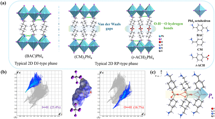
Single crystal X-ray diffraction (XRD) measurements show that 1 adopts a typical layered RP-type structure and crystallizes in the asymmetric space group Pca21 (Table S1 in Supporting information). Initially, we compare the crystal structure of 1 with those of typical 2D DJ-phase and RP-phase with different organic spacers (Fig. 1a). For all 2D perovskite frameworks, the octahedra PbI6 are interconnected by iodine atoms at shared corners to form inorganic sheets that are stacked alternately with organic layers in the b-axis direction. At the same time, N-H···I hydrogen bonds interlink the organic cations spacing layers with the inorganic slabs together (Fig. 1a, Fig. S3 and Table S5 in Supporting information). However, the most evident difference compared to the CM+ cation is an additional carboxyl group in t-ACH+. The carboxyl group in 1 acts as a donor and acceptor, providing an additional intermolecular hydrogen bond between adjacent bilayer organic amino acid molecules (O3-H3···O4, ~2.06 Å), eliminating the van der Waals gap (Fig. S4 and Table S4 in Supporting information) [19]. This hydrogen bond network differs from the one-dimensional hydrogen bond chain in (HO2C(CH2)3NH3)2(CH3NH3)Pb2I7 [24]. As we all know, the O-H···O bond is generally recognized as a strong hydrogen bond [25,26]. The intermolecular O-H···O hydrogen bonds (between alkyl ammonium cation bilayer) can cause electrostatic connections between alkyl ammonium cation bilayer, thereby enhancing the cohesion of perovskite and coupling between inorganic [PbX6] plates and improving the structural stability.
Figure 1
Figure 1.
(a) Layered perovskite structure of (BAC)PbI4 (left), (CM)2PbI4 (middle) and 1 (right). (b) Hirshfeld surfaces of t-ACH+ cations. Red regions represent N-H···I and O-H···O contacts. Inset: 2D fingerprint plots for t-ACH+ cations. (c) Oriented arrangement of O-H···O hydrogen bonding between nearby organic layers in 1.
Further analyses of the Hirshfeld surface and the 2D fingerprint show that t-ACH+ cations are tightly connected to inorganic skeleton through N-H···I hydrogen bond, and the N-H···I hydrogen bond interaction strength is up to 25.4%. Notably, the adjacent t-ACH+ cation layers also exist in addition to the O-H···O hydrogen bond, with an interaction strength is ~16.7% (Fig. 1b). The increased number of hydrogen bonds and stronger hydrogen bonds can enhance the lattice stiffness and structural stability. Compared to the RP-type perovskite, the DJ-type perovskite (BAC)PbI4 (BAC2+ is 1,4-bis(aminomethyl) cyclohexane) consists of the two terminal amino groups of BAC2+ cation connected to the inorganic layer by hydrogen bonding, eliminating the van der Waals gap (Fig. 1a). However, the structure of (BAC)PbI4 only has a single layer of cations with high symmetry. As a result, its dipole moment is generally extremely small or even zero. Contrarily, the carboxyl groups in 1 are almost aligned towards the c-axis (Fig. 1c). Such long-range ordered O-H···O hydrogen bonding arrangement could facilitate produce electric polarization or composition of polar structure [22]. In the structure of 1, the O-H···O hydrogen bonds between the t-ACH+ spacer cation not only effectively suppress the van der Waals gap to improve phase stability, but their oriented arrangement also facilitates the generation of polarization.
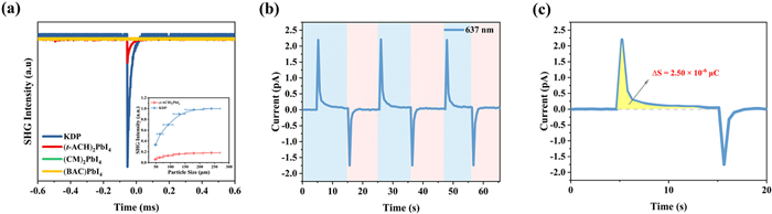
Generally, structural polarity confers the second harmonic generation (SHG) effect, where only 1 exhibits SHG response about 0.18 times that of KH2PO4 (KDP) at room temperature (Fig. 2a). This finding corresponds well to the single crystal structure analysis of 1. In addition, we acquired the SHG signal of 1 at room temperature using the Kurtz-Perry method. The SHG signal strength of 1 gradually increases with the increase of particle size and almost reaches saturation, indicating its phase-matching property (inset of Fig. 2a). Pyroelectric effect is also prevalent in 2D polar dielectric materials [27], which can directly convert thermal energy to electric current in response to external thermal stimuli, resulting in the photoinduced pyroelectric effect [28,29]. Experiments of photoinduced pyroelectric effect were carried out on a single crystal 1 approximately parallel to the polar axis at room temperature. As expected, a significant photoexcited pyroelectric current was observed in the I-t curve of compound 1 under 637 nm with a power density of 85.6 mW/cm2 (Fig. 2b). In addition, we measured the photo-pyroelectric switching cycle at different power densities (Fig. S5 in Supporting information). With the increase of laser power density, the current peak increases significantly and shows an obvious intensity dependent behavior. We integrated 6.82 × 10⁻⁴ µC/cm2 for its yellow region over one switching response cycle (Fig. 2c), and thermal imaging identified a 0.2 K temperature change on the 1 surface (Fig. S6 in Supporting information). The pyroelectric coefficient (pe) was estimated to be 3.41 × 10⁻³ µC cm−2 K−1 at 85.60 mW/cm2. The above results show that the strategy of introducing carboxyl groups in 2D hybridized perovskites is feasible to design polar structures.
Figure 2
Figure 2.
(a) SHG intensities of 1, (CM)2PbI4 and (BAC)PbI4 compared to the conventional KDP reference. Inset: SHG signal intensities variations as a function of particle size. (b) Photoresponse behavior of 1 under 637 nm laser illumination under zero bias. (c) Diagram depicting the heating portion throughout a single on-off response cycle.
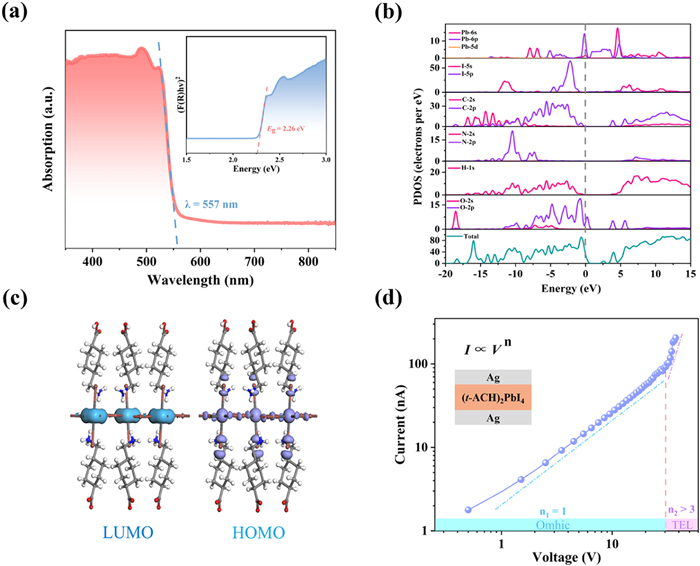
Electric polarization in polar semiconductors can interact with light to induce rich physical properties, including anomalous photovoltaic effects, fast photoelectric response, and polarized-light photoelectric dependence [30-32], which are conducive to achieving high-performance self-powered photodetection. The optical and semiconductor properties of materials are the basis for exploring potential optoelectronic applications. According to Fig. 3a, the ultraviolet-visible (UV–vis) absorption spectrum of 1 reveals its optical cutoff at ≈ 557 nm, giving an estimated optical band gap (Eg) of ≈ 2.26 eV using the Tauc equation (inset of Fig. 3a). The first-principle density functional theory (DFT) calculation [33] show that the minimum conduction band (CBM) and maximum valence band (VBM) of 1 are located at the G-point, indicating it should be a direct bandgap semiconductor. The simulated bandgap value of 2.07 eV is a bit lower than the obtained experimental value (Fig. S7 in Supporting information). Moreover, according to the partial density of states (PDOS), study along with LUMO and HOMO, the band gap is mainly caused by I-p and Pb-p orbitals (Figs. 3b and c). In addition, the electrical conductivity of 1 at different temperatures was also studied. As the temperature increases, its conductivity is positively correlated with temperature in the range of 315–375 K (Fig. S8 in Supporting information), further confirming its typical semiconductor properties. Furthermore, we measure the charge trap density on the crystal-based device (Ag/(t-ACH)2PbI4/Ag) the using space charge-limited current (SCLC) technique. Fig. 3d demonstrates two distinct situations of logarithmic I-V trace: an ohmic region (blue line, n = 1) and a trap-filled region (purple line, n > 3). The charge trap density can be estimated by the limiting voltage (VTFL) with the following formula [34]:
where ε is the dielectric constant, ε0 is the dielectric constant in vacuum, L is the thickness of the sample, and q is the fundamental charge. The ntrap of 1 is calculated to be ≈ 3.6 × 1010 cm−3, surpassing that of inorganic semiconductors like Si (1013–1014 cm−3) [35] and comparable with other 2D hybrid perovskites (C4H9NH3)2(NH2CHNH2)Pb2Br7 (3.86 × 1011 cm−3) [36].
Figure 3
Figure 3.
(a) UV–vis absorption spectra of 1. Inset: The bandgap calculated from the Tauc equation. (b) PDOS of 1 near the Fermi level. (c) LUMO-HOMO orbitals of 1. (d) Dark I-V curves were measured by using the SCLC method.
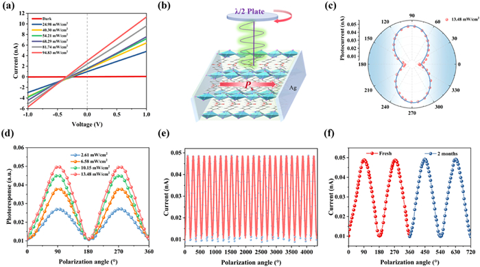
The wavelength-dependent optical response of our crystal-based device was measured in the wavelength range of 377–785 nm (Fig. S9 in Supporting information). The result indicates that photocurrent was pronounced near 520 nm but significantly weak at other wavelengths. The rapid decay of photocurrent signal above 520 nm may be attributed to weak light absorption. In addition, the polarity-induced BPVE of 1 serves as a power supply, driving the separation as well as conveyance of photoexcited carriers during self-powered photodetection, which also shows the angular dependence of polarized light [37-39], as verified in (3-bromopropylammonium)2PbBr4 and (isoamylammonium)2Cs3Pb4Br13 [40,41]. The bias dependency of BPVE-based photocurrent along the c-axis of 1 was tested at ambient temperature. The short-circuit photocurrent (Isc = 3.10 nA) and open-circuit photovoltage (Voc = 0.36 V) were obtained under 520 nm, 71.03 mW/cm2 (Fig. 4a), indicating the potential of 1 for the self-powered photodetection of polarized light.
Figure 4
Figure 4.
(a) An obvious BPVE of 1 under zero bias observed from the I-V curves at 520 nm. (b) Diagram of crystal-based photodetector for polarized-light detection. (c) Angle-resolved photocurrents collected in the polar coordinate (0 V, 520 nm). (d) The normalized polarization-dependent photocurrent at the different intensities of 520 nm polarized-light. (e) Cycles of polarization-dependent photocurrents (520 nm, 13.48 mW/cm2). (f) Polarization-dependent photocurrents for fresh sample versus air-exposed 2-month sample under the same illumination.
For 1, the alternating arrangement of organic (barrier) and inorganic (well) components generates a 2D quantum well structure [42] (Fig. S10 in Supporting information), which leads to obvious anisotropy of physical properties. Among them, the anisotropy of light absorption is essential for polarized-light responses. Here, the optical absorption coefficients of 1 are calculated with the following formula [43]:
where ω is the light frequency, ε1 and ε2 denote the real part and imaginary part of the dielectric function, and α is the absorption coefficient. The predicted optical absorption of 1 along the different axes displays an obvious anisotropy in the range of 365–900 nm (Fig. S11 in Supporting information). At 520 nm, the absorption coefficients of the a-axis and c-axis are significantly greater than that of the b-axis, and the dichroic ratio (α/α) reaches ~7.3. This value exceeds that of other classic 2D substances, such as TlSe2 (≈1.4) [44]. Combining the semiconductor properties, anisotropy structure, and BPVE, 1 is expected to achieve self-powered polarized-light photodetection. We fabricated a self-powered polarized detector based on a single crystal of 1 (Fig. 4b). The 520 nm laser passed through the polarizer and half-wave plate to generate polarized light. Fig. 4d shows the photocurrent variation obtained in the range of 0° to 360° polarization angles under zero bias, following a sinusoidal form of a 180° period. When the polarized light is perpendicular to the polar c-axis direction the minimal photocurrent (Imin) appears at θ = 0° and 180°. The maximum photocurrent (Imax) occurs at θ = 90° and 270° parallel to the polar c-axis direction. Repeating this process, we can obtain the Imax and Imin through a continuous rotation of the polarized light. When the incident optical power is 13.48 mW/cm2, 1 has a strong polarization ratio (ω = Imax/Imin = 5.3) (Fig. 4c), which is superior to detectors of other 2D materials such as ReS2 (~0.5) [45] and [CH3(CH2)3NH3]2CsPb2Br7 (~1.5) [46]. Moreover, fast and reproducible response times to illumination are essential for photodetectors. The rise time (τrise) and decay time (τdecay) of the response time represent an increase in photocurrent from 10% to 90% and decay from 90% to 10%, respectively. The τrise and τdecay were respectively measured to be 1.12 and 1.2 ms of single-crystal-based photodetectors of 1 under 520 nm (Fig. S12 in Supporting information). This polarization-sensitive behavior suggests that 1 is a potential candidate for polarized light detection in self-powered mode.
The thermal and phase stability of single crystal and crystal-based detector of 1 was evaluated. Thermogravimetric analysis shows that 1 has excellent phase stability up to 488 K in the atmosphere (Fig. S13 in Supporting information). The X-ray diffraction (PXRD) patterns of 1 after two months of exposure to air with a relative humidity of 70% ± 10% were consistent with PXRD patterns of fresh samples, indicating that the sample remained stable (Fig. S2 in Supporting information). The angularly resolved reproducible Isc of 1 remains almost constant after several cycles (Fig. 4e). Comparing its polarization sensitivity before and after two months in air, the curves do not change significantly under the same condition (520 nm, 13.48 mW/cm2), indicating the remarkable stability of 1 (Fig. 4f). These findings indicate that the photodetector of 1 has excellent anti-fatigue stability and long-term environmental stability.
In summary, we have obtained a 2D hybrid perovskite, (t-ACH)2PbI4, which contains the unique carboxyl acid dimer as the interlayered spacer. It is notable that the directional arrangements of O-H···O hydrogen bonds between adjacent layers of t-ACH+ cations not only facilitate its polar structure, but also enhance the phase stability of 2D perovskite motif. In addition, stemming from the BPVE effect generated by spontaneous polarization, crystal-based photodetector of 1 realizes self-powered polarized-light detection with a high sensitivity and large polarization-sensitive ratio of ~5.3 at 520 nm. This work has inspired the design of new phase stable 2D perovskites by utilization of hydrogen bonding interactions, and also provides the prospects for the assembly of high-performance optoelectronic devices.
Declaration of competing interest
The authors declare that they have no known competing financial interests or personal relationships that could have appeared to influence the work reported in this paper.
This work was supported by the National Natural Science Foundation of China (NSFC, Nos. 22125110, U23A2094, 22205233, 22193042, 21921001, 22305248 and U21A2069), the Natural Science Foundation of Fujian Province (No. 2023J02028), the Key Research Program of Frontier Sciences of Chinese Academy of Sciences (No. ZDBS-LY-SLH024), Fujian Science & Technology Innovation Laboratory for Optoelectronic Information of China (No. 2021ZR126), the National Key Research and Development Program of China (No. 2019YFA0210402), and the China Postdoctoral Science Foundation (Nos. 2022TQ0337 and 2023M733497).
Supplementary materials
Supplementary material associated with this article can be found, in the online version, at doi:10.1016/j.cclet.2024.110092.
[1]
Y. Gao, Z. Wei, S.N. Hsu, B.W. Boudouris, L. Dou, Mater. Chem. Front. 4 (2020) 3400–3418. doi: 10.1039/d0qm00233j
[2]
M. Yang, H. Cheng, Y. Xu, M. Li, Y. Ai, Chin. Chem. Lett. 33 (2022) 2143–2146.
[3]
Y. Fu, X. Jiang, X. Li, et al., J. Am. Chem. Soc. 142 (2020) 4008–4021. doi: 10.1021/jacs.9b13587
Y. Niu, R. Frisenda, E. Flores, et al., Adv. Opt. Mater. 6 (2018) 7. doi: 10.31438/trf.hh2018.3
[46]
J.Q. Wang, Y. Liu, S.G. Han, et al., Sci. Bull. 66 (2021) 158–163.
Figure 1
(a) Layered perovskite structure of (BAC)PbI4 (left), (CM)2PbI4 (middle) and 1 (right). (b) Hirshfeld surfaces of t-ACH+ cations. Red regions represent N-H···I and O-H···O contacts. Inset: 2D fingerprint plots for t-ACH+ cations. (c) Oriented arrangement of O-H···O hydrogen bonding between nearby organic layers in 1.
Figure 2
(a) SHG intensities of 1, (CM)2PbI4 and (BAC)PbI4 compared to the conventional KDP reference. Inset: SHG signal intensities variations as a function of particle size. (b) Photoresponse behavior of 1 under 637 nm laser illumination under zero bias. (c) Diagram depicting the heating portion throughout a single on-off response cycle.
Figure 3
(a) UV–vis absorption spectra of 1. Inset: The bandgap calculated from the Tauc equation. (b) PDOS of 1 near the Fermi level. (c) LUMO-HOMO orbitals of 1. (d) Dark I-V curves were measured by using the SCLC method.
Figure 4
(a) An obvious BPVE of 1 under zero bias observed from the I-V curves at 520 nm. (b) Diagram of crystal-based photodetector for polarized-light detection. (c) Angle-resolved photocurrents collected in the polar coordinate (0 V, 520 nm). (d) The normalized polarization-dependent photocurrent at the different intensities of 520 nm polarized-light. (e) Cycles of polarization-dependent photocurrents (520 nm, 13.48 mW/cm2). (f) Polarization-dependent photocurrents for fresh sample versus air-exposed 2-month sample under the same illumination.

 DownLoad:

 下载:
下载:


 下载:
下载:
