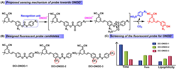
Scheme 1.
Design strategy of the fluorescent probes for ONOO−.
“Three-in-one” strategy of trifluoromethyl regulated blood-brain barrier permeable fluorescent probe for peroxynitrite and antiepileptic evaluation of edaravone
Lei Shen , Hongmei Liu , Ming Jin , Jinchao Zhang , Caixia Yin , Shuxiang Wang , Yutao Yang

Epilepsy is a neurodegenerative disease occurring at all ages with high disability and fatality rate. It is a kind of serious disease which threatens human’s health and life [1-3]. Rapid and accurate diagnosis of epilepsy and effective treatment are essential to control seizures. Recently, as numerous studies have shown, oxidizing stress is one of the precipitating factors, and a large number of reactive oxygen species (ROS) are produced during epileptic seizure [4-7]. The superoxide anion (•O2−) produced in the body rapidly combines with nitric oxide (NO) to generate excessive ONOO−, which causes irreversible damage to biomacromolecules for example DNA, protein and so on, leading in turn to neuron cells death [8-15]. Therefore, oxidative stress should be considered in the treatment of patients with epilepsy [16,17]. Abnormal levels of ONOO− lead to the redox imbalance in cells, and it may serve as the potential diagnostic biomarker and therapeutic target for epilepsy.
Since 2019, due to the impact of corona virus disease 2019 (COVID-19), the strategy of “drug repurposing” is attracting a growing interest recently, especially for the research of marketed drugs for new indications. As we know, edaravone (EDA) is a first-line drug for the clinical treatment of cerebral ischemia. It was first developed by Mitsubishi Tanabe Pharma Corporation and approved for market in Japan in 2001 [18]. EDA has good effects in preventing and treating cerebral ischemia, and the mechanism of treatment may be related to protect the oxidative injury of nerve cell through antioxidative action of scavenging free radicals and inhibiting lipid peroxidation [19]. Hence, we hypothesize that EDA might alleviate epilepsy, since the onset of epilepsy is closely related to oxidative nerve damage. Thus, it is imperative to develop novel methods for monitoring the ONOO− fluctuations during seizures for evaluating the efficacy of EDA in the brains of epilepsy.
Fluorescent probes, as the ideal tool for mapping bioactive molecules in biological systems because of the superior performance of simple operation, high sensitivity, high selectivity, and non-invasive visualization, have shown excellent effects in diagnosing diseases and evaluating pharmacodynamics in recent years [20-25]. To date, several fluorescent probes have been developed for real-time detection of enzymes, ROS, active nitrogen (RNS), and other active molecules for pharmacodynamic evaluation in mice [26-28]. However, evaluation of EDA for the antiepileptic pharmacodynamics using fluorescent probes has not been reported. Therefore, it is necessary to develop a novel near-infrared (NIR) fluorescent probe to monitor ONOO− fluctuations in order to evaluate the pharmacodynamics of EDA in epileptic mice brain. However, few probes have been able to detect ONOO− in the brain, which limit the efficacy evaluation of antiepileptic drugs in vivo. Thus, development of fluorescent probes that can effectively track endogenous ONOO− fluctuations in kainic acid (KA) induced seizures is necessary. There are several challenges of probes to achieve the goal, possessing the characteristic feature of crossing the BBB, high sensitivity, high selectivity and fast response for ONOO− in NIR region.
In view of the above considerations, we are committed to developing fluorescent probes to visualize ONOO− fluctuations in the epileptic brain. Hence, we proposed a promising recognition unit for ONOO−, thiocarbonate derivatives, due to the susceptibility to oxidation and decomposition under mild conditions [29]. ONOO− induced probes to remove thiocarbonate derivatives, and significantly restored the fluorescence emission of fluorophores (Scheme 1A). In this study, we firstly designed the probe DCI-ONOO-1 for detection of ONOO−. However, ONOO− induced DCI-ONOO-1 to release DCI-OH with emission wavelength at 598 nm, which was not suitable for in vivo imaging. As we know, to construct an ideal fluorescent probe for brain imaging, several factors must be considered, such as emission wavelength, molecular weight, signal intensity, stability, lipophilicity. Therefore, the DCI-CF3-OH was developed by introduction of trifluoromethyl into DCI-OH to adjust the acidity of the phenol hydroxyl group, prolonging the emission wavelength at 652 nm (Figs. S1 and S2 in Supporting information). Then, DCI-ONOO-2 and DCI-ONOO-3 were following closely synthesized by introducing trifluoromethyl due to its high lipidsolubility, good metabolic stability, high electronegativity and bioavailability (Scheme 1B and Scheme S1 in Supporting information). Subsequently, the physicochemical properties of the DCI-ONOO-1, DCI-ONOO-2 and DCI-ONOO-3 such as the oil-water partition coefficient (LogP) and BBB permeability (Pe) of were investigated, and the corresponding results were 0.71, 1.03, 1.27 for LogP and 1.22, 2.36, 3.27 for Pe, respectively (Figs. S3 and S4, Table S1 in Supporting information). Also, the kinetics of DCI-ONOO-1, DCI-ONOO-2 and DCI-ONOO-3 with ONOO− were also studied with response times of 730, 520 and 210 s, respectively (Fig. S5 and Table S1 in Supporting information).
To sum up, by the “three-in-one” strategy, the introduction of trifluoromethyl in the DCI-ONOO-3 can (1) improve the reaction activity and shorten the reaction time of DCI-ONOO-3 with ONOO− by facilitating the hydrolysis of oxidation products, (2) extend the emission wavelength of the fluorophore into the NIR region by adjusting the acidity of the phenol hydroxyl group, (3) increase lipophilicity (Scheme 1C). Taking of these advantages, using the probe DCI-ONOO-3, we found that the concentration of ONOO− in the epileptic mice was increased. Importantly, the antagonism of EDA on epilepsy at different concentrations was explored by observing ONOO− levels in the brains of epileptic mice treated with EDA. This provides a compelling method for the accurate diagnosis of epilepsy and the antiepileptic evaluation of EDA.
Dicyanoisophorone derivatives are used extensively in the design of fluorescence probes on account of the advantages of typical D-π-A structure, easy synthesis modification. Thus, we used dicyanoisophorone as a fluorophore to synthesize the probe DCI-ONOO-1 for the detection of ONOO− (Scheme S1 in Supporting information). The presence of ONOO− induced DCI-ONOO-1 to remove (4-nitrophenylsulfide)carbonyl and restored the fluorescence emission of fluorophore (Scheme 1A). By introducing a strong electron-withdrawing group (trifluoromethyl) at the site of the phenol hydroxyl group, a new fluorophore is expected to emit NIR fluorescence (Scheme 1C and Fig. S2 in Supporting information). As far as we know, trifluoromethyl is widely used in the design of drugs for the treatment of brain diseases to improve lipophilicity and biocompatibility [30,31]. Therefore, we then developed the probes DCI-ONOO-2 and DCI-ONOO-3 for ONOO− (Scheme S1). By screening, DCI-ONOO-3 with the outstanding physicochemical and optical properties was used as the target probe to detect ONOO−. The sensing mechanism of the DCI-ONOO-3 to ONOO− was confirmed by HRMS and HPLC analysis. As shown in Fig. S6B (Supporting information), the peaks of 208.9831 ([M-H]−), 357.1221 ([M-H]−), 561.1082 ([M-H]−) were to be the compound A, DCI-CF3-OH, and the probe DCI-ONOO-3, respectively, demonstrating that successful release of fluorophore DCI-CF3-OH and compound A caused by the reaction of DCI-ONOO-3 and ONOO−. Moreover, the results of HPLC analysis showed that the retention times of DCI-ONOO-3, DCI-CF3-OH and compound A were 49.5, 11.6 and 34.4 min, respectively. Then, with addition of ONOO− (0.5 equiv.) to the solution of DCI-ONOO-3, the peak at 49.5 min decreased, the peaks at 11.6 and 34.4 min were increased, corresponding to DCI-ONOO-3, DCI-CF3-OH and compound A. Upon adding excessive ONOO−, the peak at 49.5 min (DCI-ONOO-3) disappeared, meanwhile, the peaks at 11.6 min (DCI-CF3-OH) and 34.4 min (A) increased. The above results indicated that DCI-ONOO-3 was triggered by ONOO−, restoring the fluorescence of DCI-CF3-OH (Fig. S6C in Supporting information).
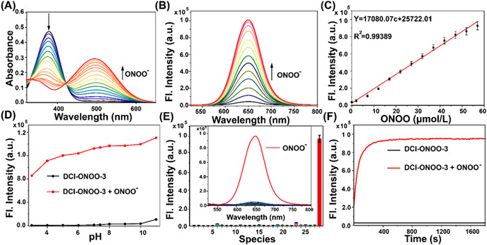
The spectral properties of the DCI-ONOO-3 for ONOO− were systematically performed in the CH3CN/PBS buffer (10 mmol/L, pH 7.4, 2/5, v/v). The optical titration experiments of DCI-ONOO-3 (10 µmol/L) toward ONOO− (0−60 µmol/L) were carried out. As shown in Fig. 1A, the absorption peak decreased at 380 nm and increased at 500 nm after adding ONOO−. Correspondingly, 61-fold fluorescence enhancement at 652 nm can be observed with an increase of ONOO− concentration (Fig. 1B), and a good linearity was obtained between the fluorescence intensity and the ONOO− concentration (0−60 µmol/L) with the limit of detection calculated to be around 73 nmol/L (Fig. 1C), illustrating the high sensitivity of DCI-ONOO-3 for ONOO−. Besides, The DCI-ONOO-3 can detect ONOO− over a wide range of pH 5–10 (Fig. 1D), indicating that DCI-ONOO-3 was suitable for ONOO− detection under complex physiological and biological condition. Moreover, the selectivity of DCI-ONOO-3 for ONOO− was investigated, as shown in Fig. 1E, only addition with ONOO− generated obvious fluorescence enhancement, whereas addition of other species (Na+, K+, Fe2+, Cu2+, Ca2+, Fe3+, Mg2+, Zn2+, Cl−, NO3−, CO32−, SO42−, HSO3−, Na2S, SO32−, NO, NO2−, H2O2, ClO−, TBHP, 1O2, •OH, Cys, GSH, Hcy, NE) showed a negligible response. Finally, as shown in Fig. 1F, the time-dependent was performed, indicating fast response of DCI-ONOO-3 to ONOO− in 210 s. All the above results illustrated that DCI-ONOO-3 has high selectivity, sensitivity and fast response of ONOO−, showing the possibility of DCI-ONOO-3 for monitoring ONOO− in biosystem.
Encouraged by the excellent performance of DCI-ONOO-3 for the detection of exogenous ONOO−, we further explored the application of DCI-ONOO-3 in the detection of intracellular ONOO−. The cytotoxicity of DCI-ONOO-3 for PC12 cells, SH-SY5Y cells and HT-22 cells was firstly performed by cell counting kit-8 (CCK8) method. As shown in Fig. S7 (Supporting information), the cells survival rate remained at about 82%, indicating that the DCI-ONOO-3 presented low cytotoxicity when the concentration of DCI-ONOO-3 up to 20 µmol/L. Then, DCI-ONOO-3 was used to fluorescence image of ONOO− in HT22 cells. As shown in Figs. S8A and D (Supporting information), the fluorescence intensity was concentration-dependent with DCI-ONOO-3. Meanwhile, Fig. S8C (Supporting information) indicated the high sensitivity of DCI-ONOO-3 for endogenous ONOO− by flow cytometry. Subsequently, we conducted the kinetic study of DCI-ONOO-3 towards ONOO− within 300 s in HT22 cells, and the fluorescence intensity gradually enhanced and stabilized in 200 s, indicating a fast response of DCI-ONOO-3 for ONOO− (Figs. S8B and E in Supporting information).
Furthermore, the levels of endogenous ONOO− under oxidative stress mediated by lipopolysaccharide (LPS) and 3-morpholino-sydnonimine (SIN-1) were investigated, and the ONOO− fluxes were also visualized by adding N-acetylcysteine (NAC, a ROS inhibitor), Fe(Ⅲ)tetrakis(1-methyl-4-pyridyl) (FeTMpyP, peroxynitrite deconstructor) and EDA, respectively. As shown in Fig. 2A, in contrast with the DCI-ONOO-3 (10 µmol/L) incubated group, HT22 cells were pretreated with LPS (1 µg/mL) or SIN-1 (100 µmol/L), followed by incubation of DCI-ONOO-3 (10 µmol/L), the increased intensity was observed. However, the cells were pretreated with LPS or SIN-1, followed by incubation of NAC (1 mmol/L), FeTMPyP (50 µmol/L) or EDA (150 µmol/L), the fluorescence intensity decreased, indicating that EDA has the potential of scavenging ONOO− in cells and alleviating oxidative stress.
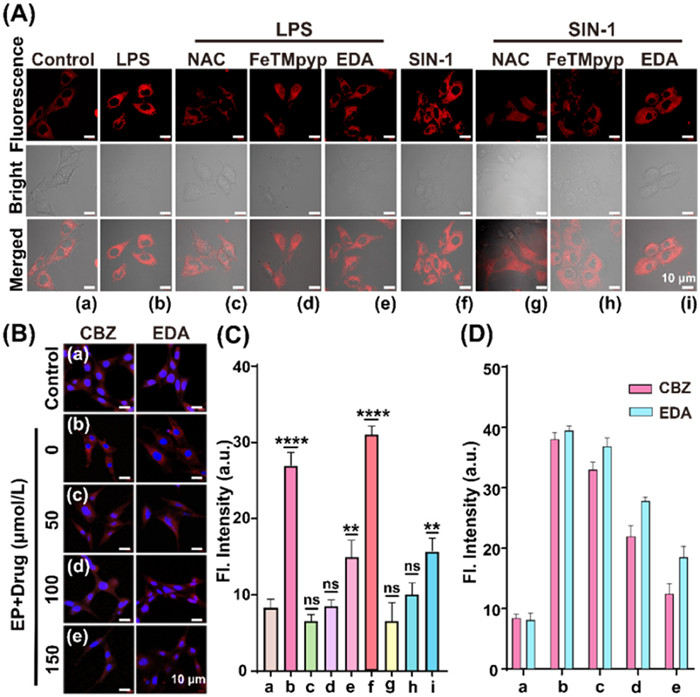
Moreover, we established the cell model of epilepsy by incubating HT22 cells in Mg2+-free media (ACSF, Artificial cerebro-spinal fluid (-Mg) [32] for studying antiepileptic efficacy of EDA. The HT22 cells of epilepsy were incubated with carbamazepine (CBZ, a clinical drug for epilepsy) and EDA respectively. Fig. 2B showed that the fluorescence intensity of the HT22 cells of epilepsy incubated with DCI-ONOO-3 increased compared to the control group. Whereas, after pretreating the epilepsy model groups with CAZ and EDA, the fluorescence increased compared to the epilepsy model groups. The above results proved that EDA has the potential of anti-epilepsy.
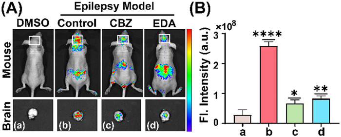
Having verified the selectivity and sensitivity of the DCI-ONOO-3 for ONOO− in living cells, as well as the good BBB permeability of the DCI-ONOO-3 by establishing a PAMPA model [33], we aimed to establish a mouse model of epilepsy [34] suitable for the study of antiepileptic activity of EDA. In this study, all animal experiments were conducted in accordance with the protocol approved by the Declaration of Animal Welfare and Ethical Approval of Hebei University, using 5-week-old BALB/c purchased from the SPF (Beijing) BIO-TECHNOLOGY Co., Ltd. The appropriate time for KA (20 mg/kg) induction to establish epileptic mice was firstly selected as 0.5 h by Racine grading and fluorescence imaging of ONOO− levels in the mouse brain (Fig. S9 in Supporting information). Immediately, the mice were divided into four groups: (a) Control group (injection of DCI-ONOO-3 for 0.5 h), (b) Epileptic group (KA-induced for 0.5 h, following injection of DCI-ONOO-3 for 0.5 h), (c, d) CBZ or EDA treated epileptic mice groups (KA-induced for 0.5 h, then treated with CBZ or EDA for 0.5 h, following injection of DCI-ONOO-3 for 0.5 h). As shown in Fig. 3, the fluorescence intensity of epileptic group increased in comparison to control group. However, compared to epileptic group, the fluorescence intensity of CBZ or EDA treated epileptic mice groups decreased to some extent, indicating the good antiepileptic activity of EDA. This provides a promising method for the clinical treatment of epilepsy.
In summary, we focused on developing a novel fluorescent probe passing through the BBB to monitor ONOO− in the brain. The rational strategy for designing probe DCI-ONOO-3 is proposed by introducing trifluoromethyl groups, solving the requirements of NIR emission and lipophilicity of probe for brain imaging. The DCI-ONOO-3 has high signal-to-noise ratio and BBB penetration, achieving real-time monitoring of ONOO− fluctuations in the epileptic mice brains and evaluating antiepileptic effect of EDA. This study provides a new strategy for exploring the pathogenesis of epilepsy, which helps to better understand the pathology of epilepsy. It also provides a new reference for clinical treatment of epilepsy, and accelerates the discovery of potential anti-epileptic drugs.
The authors declare that they have no known competing financial interests or personal relationships that could have appeared to influence the work reported in this paper.
The work supported by the National Natural Science Foundation of China (Nos. 22177025, 21807021, 22325703, 22177026), Science Fund for Creative Research Groups of Nature Science Foundation of Hebei Province (No. B2021201038), the Interdisciplinary Research Program of Natural Science of Hebei University (No. DXK202113), the Central Government Guided Local Science, Technology Development Fund (Hebei Province, No. 216Z2603G), National High-End Foreign Expert Recruitment Plan (No. G2022003007L).
Supplementary material associated with this article can be found, in the online version, at doi:
P. Kwan, M.J. Brodie, N. Engl. J. Med. 342 (2000) 314–319. doi: 10.1056/NEJM200002033420503
R.D. Thijs, R. Surges, T.J. O’Brien, J.W. Sander, Lancet 393 (2019) 689–701. doi: 10.1016/S0140-6736(18)32596-0
E. Perucca, Epilepsia 55 (2014) 473–474. doi: 10.1111/epi.12584
N.T. El Tantawi, D.S. Abd Elmegid, E. Atef, Seizure 65 (2019) 166–171. doi: 10.1016/j.seizure.2019.01.003
J.K. Knowles, H. Xu, C. Soane, et al., Nat. Neurosci. 25 (2022) 596–606. doi: 10.1038/s41593-022-01052-2
J. Goldstein, C.S. Kwon, M. Harmon, et al., Epilepsy Behav. 125 (2021) 108383. doi: 10.1016/j.yebeh.2021.108383
P.G. Winyard, C.J. Moody, C. Jacob, Trends Biochem. Sci. 30 (2005) 453–461. doi: 10.1016/j.tibs.2005.06.001
M.R. Ashrafi, S. Shams, M. Nouri, et al., Epilepsia 48 (2007) 1750–1755. doi: 10.1111/j.1528-1167.2007.01143.x
H. Li, X. Li, X. Wu, W. Shi, H. Ma, Anal. Chem. 89 (2017) 5519–5525. doi: 10.1021/acs.analchem.7b00503
N. Ma, M. Sasoh, S. Kawanishi, H. Sugiura, F.J. Piao, Biomed. Sci. 17 (2010) S7. doi: 10.1186/1423-0127-17-S1-S7
N. Ahmed, W. Zareen, Y. Ye, Chin. Chem. Lett. 33 (2022) 2765–2772. doi: 10.1016/j.cclet.2021.12.092
D.H. Ma, S.M. Hou, C. Bae, et al., Chin. Chem. Lett. 32 (2021) 3886–3889. doi: 10.1016/j.cclet.2021.05.048
T. Obata, Neural. Transm. 113 (2006) 1131–1144. doi: 10.1007/s00702-005-0415-0
N.N. Wang, H. Wang, J. Zhang, et al., Chin. Chem. Lett. 33 (2022) 1584–1588. doi: 10.1016/j.cclet.2021.09.046
Y.T. Yang, Y.Y. Zhang, M. Ma, et al., Anal. Chem. 94 (2022) 14443–14452. doi: 10.1021/acs.analchem.2c03390
R.S. Fisher, C. Acevedo, A. Arzimanoglou, et al., Epilepsia 55 (2014) 475–482. doi: 10.1111/epi.12550
K. Takei, K. Watanabe, S. Yuki, et al., Amyotroph. Lateral Scler. Frontotemporal Degener. 18 (2017) 5–10. doi: 10.1080/21678421.2017.1353101
N. Tanahashi, Y. Fukuuchi, Intern. Med. J. 41 (2002) 337–344. doi: 10.2169/internalmedicine.41.337
B. Daly, J. Ling, A.P. De Silva, Chem. Soc. Rev. 44 (2015) 4203–4211. doi: 10.1039/C4CS00334A
Y. Fu, N.S. Finney, RSC Adv. 8 (2018) 29051–29061. doi: 10.1039/c8ra02297f
J.V. Jun, D.M. Chenoweth, E.J. Petersson, Org. Biomol. Chem. 18 (2020) 5747–5763. doi: 10.1039/d0ob01131b
D. Wu, A.C. Sedgwick, T. Gunnlaugsson, et al., Chem. Soc. Rev. 46 (2017) 7105–7123. doi: 10.1039/C7CS00240H
M. Yang, J. Fan, J. Du, X. Peng, Chem. Sci. 11 (2020) 5127–5141. doi: 10.1039/d0sc01482f
H.H. Han, H. Tian, Y. Zang, et al., Chem. Soc. Rev. 50 (2021) 9391–9429. doi: 10.1039/d0cs01183e
N. Karton-Lifshin, E. Segal, L. Omer, et al., J. Am. Chem. Soc. 133 (2011) 10960–10965. doi: 10.1021/ja203145v
P. Li, J. Wang, X. Wang, et al., Chem. Sci. 10 (2019) 2805–2810. doi: 10.1039/c8sc04891f
S. Siriwibool, N. Kaekratoke, K. Chansaenpak, et al., Sci. Rep. 10 (2020) 1283. doi: 10.1038/s41598-020-58239-5
W. Liu, C. Fan, R. Sun, Y. Xu, J. Ge, Org. Biomol. Chem. 13 (2015) 4532–4538. doi: 10.1039/C5OB00042D
J.J. Willard, E. Pacsu, J. Am. Chem. Soc. 82 (1960) 4347–4350. doi: 10.1021/ja01501a056
O.A. Tomashenko, V.V. Grushin, Chem. Rev. 111 (2011) 4475–4521. doi: 10.1021/cr1004293
X. Wang, D. Su, C. Liu, et al., Anal. Chem. 94 (2022) 14965–14973. doi: 10.1021/acs.analchem.2c02805
S. Sombati, R.J.J. Delorenzo, J. Neurophysiol. 73 (1995) 1706–1711. doi: 10.1152/jn.1995.73.4.1706
X. Zhang, T. Liu, X. Fan, N. Ai, Mol. Graph. Model. 75 (2017) 347–354. doi: 10.1016/j.jmgm.2017.05.021
G. Marsicano, S. Goodenough, K. Monory, et al., Science 302 (2003) 84–88. doi: 10.1126/science.1088208

Figure 1 (A) Absorption and (B) fluorescence titration of DCI-ONOO-3 (10 µmol/L) to ONOO− (60 µmol/L) in phosphate buffered saline (PBS) (10 mmol/L, pH 7.4, containing 40% MeCN) solution. (C) Fluorescence intensity versus ONOO− concentration at 652 nm. (D) Fluorescence spectral response of DCI-ONOO-3 (10 µmol/L) to ONOO− (10 equiv.) in pH 3–11. (E) Fluorescence response of DCI-ONOO-3 (10 µmol/L) with addition of ONOO− (60 µmol/L) and other species (1 mmol/L). (F) The effect of reaction time between DCI-ONOO-3 (10 µmol/L) and ONOO− (100 µmol/L). λex = 500 nm, slit/slit = 2/2 nm.

Figure 2 (A) Fluorescence images of DCI-ONOO-3 in HT22 cells. (a) HT22 cells were incubated with DCI-ONOO-3 (10 µmol/L) for 20 min. (b–e) HT22 cells were pretreated with LPS (1 µg/mL) for 30 min, following incubation of none, or NAC (1 mmol/L), or FeTMpyp (50 µmol/L), or EDA (150 µmol/L) for 30 min, then incubated with DCI-ONOO-3 (10 µmol/L) for 20 min. (f–i) HT22 cells were pretreated with SIN-1 (100 µmol/L) for 30 min, following incubation of none, or NAC (1 mmol/L), or FeTMpyp (50 µmol/L), or EDA (150 µmol/L) for 30 min, then incubated with DCI-ONOO-3 (10 µmol/L) for 20 min, respectively. (B) Fluorescence images of HT22 cells of epilepsy. (a) Control group, HT22 cells were treated with DCI-ONOO-3 (10 µmol/L). (b–e) The HT22 cells of epilepsy pretreated with EDA or CBZ (0, 50, 100, 150 µmol/L), followed by incubation of DCI-ONOO-3 (10 µmol/L). (C) The fluorescence intensity of (A). (D) The relative fluorescence intensity of (C). λex = 561 nm, λem = 640–745 nm. Scale bar: 10 µm. Data are presented as the mean value ± standard deviation (SD) (n = 3). ****P ≤ 0.0001, **P < 0.01. ns, no significance.

Figure 3 (A) Top: fluorescence imaging of mice; Bottom: fluorescence imaging of corresponding cerebrum of mice. (a) Control group, only treated with DCI-ONOO-3 (200 µmol/L) for 0.5 h. (b–d) The mice treated with none, or CBZ (50 mg/kg), or EDA (50 mg/kg) for 0.5 h after injection with KA (20 mg/kg) for 0.5 h, then injection with DCI-ONOO-3 (200 µmol/L) for 0.5 h, respectively. λex = 510 nm, λem = 600–700 nm. (B) The relative fluorescence intensity of (A). λex = 510 nm, λem = 600–700 nm. Data are presented as the mean value ± SD (n = 3). ****P ≤ 0.0001, **P < 0.01, *P < 0.05.

 扫一扫看文章

扫一扫关注我们
 DownLoad:

 下载:
下载:
 下载:
下载: