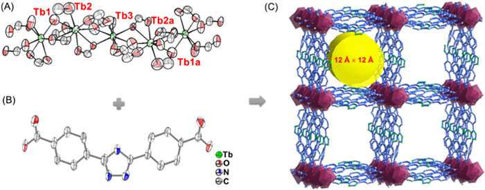
Figure 1.
The coordination environment of (A) [Tb5] unit, the structure of (B) H2TDA and (C) TDA-Tb.
Visual and portable detection of metronidazole realized by metal-organic framework flexible sensor and smartphone scanning
Xiangshuai Li , Jian Zhao , Li Luo , Zhuohao Jiao , Ying Shi , Shengli Hou , Bin Zhao

Metronidazole (MNZ) is a type of synthetic nitroimidazole antibiotic which is capable of helping people and animals resist the threaten of various bacterial infections, such as parasitic infections, trichomoniasis, whipworm infection, and amebiasis [1-3]. However, the abuse of MNZ have brought lots of troubles, for example, the accumulation of MNZ in human body will lead to a series of poisoning reactions including spasms, peripheral neuropathy, and ataxia [4]. Besides, MNZ also has genotoxic and carcinogenic effects on animals, which greatly threats the safety of food production, and has been listed into the National Toxicology Program. In these cases, developing an efficient pathway for detecting MNZ is important and necessary for human health [5]. In fact, MNZ lacks strong UV-absorbing chromophores and has a high boiling point (405 ± 25 ℃), which make it hardly be detected by conventional UV–vis spectrophotometry, and gas chromatography. Although some methods including high-performance liquid chromatography [6], electro-chemical method [7-9], and supercritical fluid chromatography [10] have been employed for MNZ sensing, most of these techniques always require expensive instruments and complicated processes, which greatly limit their wide application ranges [11-13]. Therefore, flexible sensors that can realize portable and visual detection of MNZ are desirable and important, but the corresponding explorations have not been reported hitherto.
Actually, luminescent metal-organic frameworks (MOFs) are promising materials to construct flexible sensors in detecting MNZ, because their advantages of sensitivity, selectivity, recyclability, and convenience [14,15]. The past few decades have witnessed the fast development of MOFs owing to their regular topology structure [16-19], easily modifiable channel [20-22], and high porosity [23-27]. And utilizing MOFs as luminescent sensors have attracted wide attention in detecting antibiotics [28,29], metal ions [30,31], pesticides [32], bio-molecules [33,34], and explosives [35]. Among them, lanthanide MOFs, such as Eu and Tb ions-based MOFs, are ideal platforms for constructing luminescent sensors [36]. Because they have characteristic luminescent signals, which endows them good anti-interference ability in luminescence detection. As a typical lanthanide ion, Tb ion has four luminescence signal peaks at 492, 545, 585, and 622 nm in the luminescence spectrum owing to the 5D4 → 7FJ (J = 6, 5, 4, 3) transitions. And constructing Tb-based MOFs can enhance the intensity of these peaks due to the antenna effect, the ligand absorbs light and transfers the absorbed energy to the luminescent Tb centers. Hence, designing a Tb-based MOF to construct flexible sensor may have an excellent performance in portable and visual detection of MNZ.
With this consideration, a new three-dimensional (3D) MOF {(CH3)2NH2·[Tb5(TDA)8(H2O)2]·6DMF·2C2H5OH}n (TDA-Tb) was fabricated through solvothermal reaction between 4,4′-(1H-1,2,4-triazole-3,5-diyl)dibenzoic acid (H2TDA) and Tb(NO3)3. The obtained TDA-Tb exhibits good luminescence properties owing to the conjugated molecular configuration of the ligands, which is helpful to the energy transfer process from itself to Tb3+. Experiment results indicate that TDA-Tb as a luminescent sensor has a good sensitivity and selectivity in detecting MNZ, and the LOD value can reach 4.1 × 10⁻⁷ mol/L. This sensor can be reused at least five times without obvious luminescence and structure change. Moreover, to realize portable and visual sensing, TDA-Tb was processed as flexible sensor without compromising its sensitivity and selectivity, which greatly facilitate its practical application.
Synthesis of TDA-Tb: Tb(NO3)3·6H2O (40 mg), H2TDA (15 mg) and benzoic acid (70 mg) were dissolved in mixed solvent of DMF (3 mL) and C2H5OH (1 mL), and diethylamine (100 µL) was added into the solution. Then, the obtained solution was transferred into a glass bottle and heated at 120 ℃ for 24 h. After that, transparent block crystals were obtained.
Preparation of TDA-Tb contained membrane (TDA-Tb-M): Firstly, PVDF (200 mg) was dissolved in 2 mL DMF, after that, thoroughly grinded TDA-Tb (10 mg) powder were added into the solution and sonicated for 10 min to give MOF contained PVDF solution. On another hand, PVA (30 mg) was added into 1 mL H2O and heated at 100 ℃ for 30 min to give PVA solution. And then, the PVA solution was uniformly injected on the surface of PVDF solution producing a solid gel. The gel was heated at 80 ℃ for 4 h for slowly evaporation of the solvent, and then the TDA-Tb-M was obtained after absolutely removal of the solvent.
MNZ sensing experiment: TDA-Tb (3 mg) was dispersed in 10 mL H2O and sonicated for 10 min. Next, 2.5 mL of the suspension and MNZ or other interfering substances was mixed and put into a quartz cuvette, and the performance of TDA-Tb was explored by recording the change of luminescence intensity at 545 nm using a luminescence spectrometer. The instrument parameter was set as follows: λex = 320 nm. All the experiments were carried out at room temperature.
Phone scanning method for MNZ sensing: TDA-Tb-M with size of 3 mm × 3 mm was insert into a plastic box with exposed sample hole. Then, the MNZ solution was drop into the sample hole to make the solution fully coating on the surface of the membrane, and using UV light to irradiate the MNZ solution coated membrane to active its luminescence. After that, using the smart phone APP "Color Collect" to scan the luminescence from the membrane and resulted "R" "G" "B" will be given. Calculating the "G/B" value tendency influenced by the concentration of MNZ solution will give their corresponding relationship.
Structure analysis reveals that the TDA-Tb crystallizes in the P-1 space group. Each asymmetric unit is composed of four independent ligands, one coordinated water, and 2.5 of Tb ions. There are three kinds of Tb ions, Tb1 is coordinated with nine oxygen atoms from six ligands and one H2O (Fig. S1A in Supporting information), and Tb2 is coordinated with nine oxygen atoms from six ligands (Fig. S1B in Supporting information); And Tb3 locates on the axis of symmetry, which connects with eight oxygen atoms from six ligand (Fig. S1C in Supporting information). Five adjacent Tb ions are linked with oxygen atoms, forming a penta-nuclear secondary building unit [Tb5] (Fig. 1A). The adjacent [Tb5] unit is bridged with two carboxyls from two TDA ligands, and further extends into a chain (Fig. S1D). The Tb based chains are supported by TDA ligands (Fig. 1B), leading to the formation of a 3D framework (Fig. 1C). In addition, TDA-Tb possesses one-dimensional channels with diameter of 12 Å (atom to atom) along the c axis in the porous framework. The porous structure of TDA-Tb was identified by N2 sorption isotherms measured at 77 K. Its saturated uptake is 110.6 cm3/g and the pore size is 1.0 nm as calculated from the N2 sorption study (Fig. S2 in Supporting information). Accessible Connolly surface was also used to help us observe the channel of TDA-Tb more unambiguously, and the pore size was calculated to be 10 Å × 9.4 Å by using a probe with the radius of 1.0 Å, which is similar with the result from N2 sorption result (Fig. S3 in Supporting information) [37]. Based on the calculation result from PLATON software, the total void volume of TDA-Tb is 59%.
Powder X-ray diffraction (PXRD) patterns were conducted to ascertain the phase purity of synthesized TDA-Tb. The PXRD pattern of synthesized samples was in good accordance with the simulated pattern, suggesting the obtained samples have high phase purity (Fig. S4 in Supporting information). According to the Fourier transform infrared (FT-IR) spectra, the peak belongs to C=O in the ligand was found at 1680 cm−1, and it was bathochromic-shifted from 1680 cm−1 to 1645 cm−1 in the TDA-Tb, which was attributed to the coordination interaction between the carboxyl groups and Tb ions (Fig. S5A in Supporting information). The thermal stability of TDA-Tb was tested by Thermogravimetric analysis (TGA), and the results were shown in Fig. S5B (Supporting information). Two stages of weight loss were found from 0 to 110 ℃ (2.4%) and 110–300 ℃ (13.7%), which were ascribed to the loss of C2H5OH (theoretical value: 2.4%), DMF and coordinated H2O (theoretical value: 13.4%), respectively. TDA-Tb can remain stable until 300 ℃, and the framework begins to collapse by further increasing the temperature (Fig. S5B). In addition, the acid, base and solvent-resistant abilities of TDA-Tb were also investigated. As-synthesized TDA-Tb crystals were immersed in various solvents (including CH3CN, C2H5OH, H2O, DMF) and water solution with different pH values from 2 to 12 for 24 h, and the resulting samples were tested by PXRD. As shown in Fig. S6 (Supporting information), their patterns could still match well with the simulated one, indicates that TDA-Tb has high solvent and pH stabilities.
The luminescence properties of TDA-Tb were also explored. Upon excitation with 320 nm light, TDA-Tb displays a broad emission peak at 374 nm ascribed to the ligand emission (Fig. S7 in Supporting information), and four characteristic peaks at 489, 545, 586 and 620 nm derived from 5D4 → 7FJ (J = 6, 5, 4, 3) transitions of Tb3+, respectively (Fig. 2A). The solid-state fluorescent spectrum is also similar with its emission behaviors in aqueous dispersion state (Fig. S8 in Supporting information).
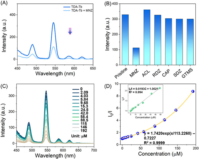
Owing to the good luminescence property and solvent stabilities, TDA-Tb was applied for MNZ sensing. Interestingly, the luminescence intensity of TDA-Tb decreased with increasing the amount of MNZ, and an exhaustive quenching phenomenon was observed after addition of 0.05 mg MNZ (Fig. 2A), suggesting that TDA-Tb can serve as a luminescence sensor to detect MNZ in water. To investigate its anti-interference performance, various cations and anions (including K+, Na+, Li+, Mg2+, Cd2+, Al3+, Cl−, SO42−, PO43−) and other antibiotics include ronidazole (RDZ), amoxicillin (ACL), chloramphenicol (CAP), gentamicin sulfate (GTMS), and sulfadiazine (SDZ) were also tested. As shown in Fig. 2B and Figs. S9 and S10 (Supporting information), other analytes have negligible influence on this sensor, revealing that TDA-Tb has a good selectivity in detecting MNZ. Moreover, to explore the sensitivity of TDA-Tb as the MNZ probe, TDA-Tb was treated with a serial of MNZ solution with gradient concentration (2.09 × 10⁻⁶ mol/L to 1.92 × 10⁻⁴ mol/L). As shown in Fig. 2C, the luminescence intensity of TDA-Tb gradually decreased with increasing the amount of MNZ. There maintains a linear relationship between the concentration of MNZ (C) and I0/I (where I0 represents the luminescence intensity of original TDA-Tb and I represents the luminescence intensity of TDA-Tb after adding MNZ) in the MNZ concentration range from 0 to 58.4 µmol/L (Fig. 2D). This linear relationship can be expressed in a mathematical formula I0/I = 0.0193C + 1.0021, which is in good agreement with the Stern−Volmer (S−V) equation. The LOD for TDA-Tb sensing MNZ was calculated to be 4.1 × 10⁻⁷ mol/L based on the 3σ method, implying the high sensitivity of TDA-Tb in detecting MNZ.
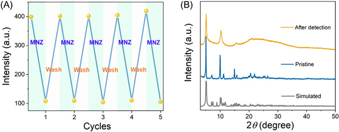
Recyclability is also an important property for luminescence probe in practical application. Hence, the luminescence intensity of TDA-Tb was monitored after 5 cycles. As shown in Fig. 3A, the luminescence of TDA-Tb can be quenched by addition of MNZ, and it can be recovered by water washing. In addition, the PXRD pattern of recycled TDA-Tb is well agree with the original one (Fig. 3B). Furthermore, the TGA curve of TDA-Tb after detection is also obtained, and the temperature at which the frame collapses (450 ℃) is consistent with the original one, revealing the framework of TDA-Tb is not destroyed during detection process (Fig. S11 in Supporting information). These demonstrate that the quenching phenomenon is not derived from the collapse of TDA-Tb. The TDA-Tb also possesses good long-term sensing performance. As exhibited from Fig. S12 (Supporting information), the TDA-Tb, which has been soaked in water for 1 day, still shows sensitive luminescence quenching responding ability to MNZ, and the quenching efficiency was nearly unchanged compare with pristine TDA-Tb.
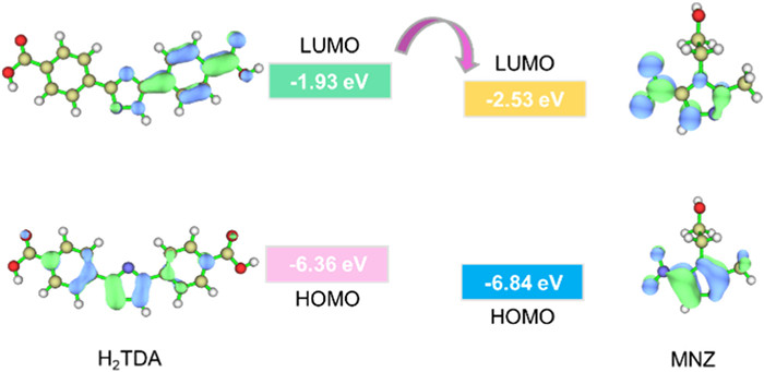
To understand the detailed mechanism that MNZ quenching TDA-Tb, density functional theory (DFT) calculations were performed at the B3LYP/6–31G* level. As shown in Fig. 4, the lowest unoccupied molecular orbitals (LUMO) of ligand lies above the LUMO of MNZ, suggesting that there exists photoinduced electron transfer (PET) from TDA-Tb to MNZ, leading to luminescence quenching [38,39]. In addition, UV–vis absorbance spectra of TDA-Tb and MNZ treated TDA-Tb were recorded (Fig. S13 in Supporting information), the absorbance of TDA-Tb shows highly overlap with MNZ at around 320 nm, illustrating that the existence of photo competing absorbance between TDA-Tb and MNZ which is also responsible for the luminescence quenching [40]. To understand this process, further experiments were conducted. As shown in Fig. S14A (Supporting information), after addition of MNZ into the TDA-Tb dispersion, the absorbance in the range of 220–370 nm of the system was enhanced, indicating MNZ consumes a part of energy while UV light irradiation, resulting in absorbing less energy by TDA-Tb. Meanwhile, MNZ shows no signals in the emission spectra while 320 nm light irradiation, implying the energy grabbed by MNZ can hardly influence the emission spectrum of TDA-Tb (Fig. S14B in Supporting information). However, the interference antibiotics have little or no absorbance at 320 nm, indicating that there is little or no light competing absorbance among TDA-Tb and other antibiotics (Fig. S13). Therefore, the luminescence quenching effect of MNZ on TDA-Tb is attributed to the combined influences of PET and photo competing absorbance effect between MOF host and MNZ. Besides, the interaction between MNZ and ligand was analyzed by the 1H NMR spectra. As illustrated in the 1H NMR spectra of mixed MNZ and H2TDA (Fig. S15 in Supporting information), the proton in the triazole group of H2TDA shifted to the high field, and the Hb hydrogen atom adjacent with N in the H2TDA was accordingly downshifted, which can be ascribed to the decrease of deshielding effect induced by the N—H…N hydrogen bond. This suggests that hydrogen-bond interaction exists between MNZ and H2TDA, and a part of absorbed energy was transferred to MNZ, leading to the luminescence quenching in TDA-Tb.
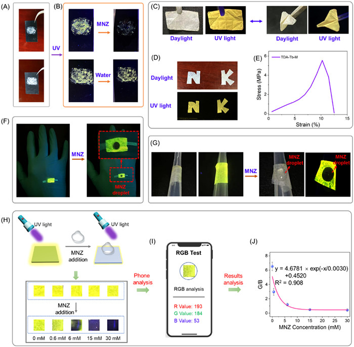
To pursuit the fast-visualization and portability of this luminescent probe, we further prepared a test paper by cohering the TDA-Tb powder with carbon paper (Fig. 5A). The test paper exhibits strong green-yellow color luminescence, and the luminescence is quenched while encountered with MNZ, realizing a naked eye detectable sensing process. For comparation, the luminescence shows no change while treated with water, further confirms the MNZ sensing ability of the test paper (Fig. 5B). Supporting the luminescence sensors onto membrane materials will endow them with good machinability and wide application range. Herein, flexible sensor TDA-Tb-M was prepared by incorporating TDA-Tb into a membrane material, which containing 1 mg of TDA-Tb, and 10 mg polymer (the polymer was consists of 90% mass ratio PVDF and 10% mass ratio PVA). The mechanical property of TDA-Tb-M is firstly tested because it is crucial for their practical application. As shown in Fig. 5C, the TDA-Tb-M can be folded into "airplane" shape and recovered to its pristine shape without any damage on the membrane, and it can also be tailored into desired shapes such as "N K" (Fig. 5D). The stress-strain test was further tested, and the maximum tensile stress of TDA-Tb-M can reach 5.6 MPa and its elongation is 12.5% (Fig. 5E). These results suggest that the flexible sensor TDA-Tb-M has a good mechanical property. The flexible TDA-Tb-M can be fabricated to be "fluorescent skin" or portable device by pasting it onto human's body or easy-achieved carrier for realizing convenient and real-time MNZ detection. As shown in Figs. 5F and G, the "fluorescent skin" and portable device possess good MNZ detection ability that obvious luminescence quenching can be observed after MNZ droplet was added onto their surfaces. Based on these results, the performance of TDA-Tb-M in luminescence detection of MNZ is further investigated. As shown in Fig. 5H, the intense yellow light of TDA-Tb-M gradually turn to weak blue color and finally quenched with increasing the concentration (0–0.03 mmol/L) of MNZ. The change of luminescence color can be recognized by the color recognizer application of mobile phone, and the corresponding RGB values can be obtained (Fig. 5I) [41-43]. As exhibited in the results, the ratio of G value to B value (G/B) of TDA-Tb-M gradually decreased with increasement of MNZ concentration, and the relationship between obtained G/B value versus MNZ concentration follows the formula of y = 4.6781 × exp(-x/0.0030) + 0.4520 (Fig. 5J). According to this method, the concentration of MNZ can be easily and sensitively recognized by phone scanning. All in all, the TDA-Tb-M realized the convenient and visual detection of MNZ, which exhibits broad application prospects in the fields of luminescence detection. And this work firstly realized luminescent sensing method for MNZ detection without usage of luminescence spectrometer (Table S2 in Supporting information).
In conclusion, a porous framework TDA-Tb with one-dimensional channel was prepared, which can serve as a luminescent sensor for MNZ detection. This sensor exhibits good anti-interference performance and high sensitivity, and the LOD can reach 4.1 × 10⁻⁷ mol/L. Moreover, the framework possesses good recyclability, and it still exhibits sensitive detection ability after at least five cycles. In addition, TDA-Tb can be fabricated to be a flexible sensor TDA-Tb-M, and the obtained flexible sensor shows good mechanical property and detection ability on MNZ. Importantly, the concentration of MNZ can be easily and sensitively recognized by smartphone scanning the TDA-Tb-M. This work not only provides a pathway for designing sensitive and recyclable MNZ-detecting sensor based on MOFs, but also realizes the portable and visual detection of MNZ by MOF based flexible sensor.
The authors declare that they have no known competing financial interests or personal relationships that could have appeared to influence the work reported in this paper.
This work was supported by the National Natural Science Foundation of China (Nos. 92161202, 21971125, 22101138, 21625103, and 22121005). The Fundamental Research Funds for the Central Universities (No. D5000230371).
Supplementary material associated with this article can be found, in the online version, at doi:
E. Itala, J. Niskanen, L. Pihlava, E. Kukk, J. Phys. Chem. A 124 (2020) 5555–5562. doi: 10.1021/acs.jpca.0c03045
L. Kumar, A. Jain, N. Lal, et al., ACS Med. Chem. Lett. 3 (2012) 83–87. doi: 10.1021/ml200161t
M.E. Falagas, A.P. Grammatikos, A. Michalopoulos, Expert Rev. Anti. Infect. Ther. 6 (2008) 593–600. doi: 10.1586/14787210.6.5.593
Y. Liu, J. Liu, H. Tang, et al., Sensor. Actuat. B 206 (2015) 647–652. doi: 10.1016/j.snb.2014.10.019
Y. Zhu, J. Li, Z. Yan, N. Zhao, X. Yang, Langmuir 38 (2022) 15442–15450. doi: 10.1021/acs.langmuir.2c02886
H.M. Maher, R.M. Youssef, R.H. Khalil, S.M. El-Bahr, J. Chromatogr. B 876 (2008) 175–181. doi: 10.1016/j.jchromb.2008.10.033
S. Bai, Q. Hu, Q. Zeng, M. Wang, L. Wang, ACS Appl. Mater. Interfaces 10 (2018) 11319–11327. doi: 10.1021/acsami.8b00554
G. Yang, F. Zhao, B. Zeng, Electrochim. Acta 135 (2014) 154–160. doi: 10.1016/j.electacta.2014.04.162
H.B. Ammar, M.B. Brahim, R. Abdelhedi, Y. Samet, Mater. Sci. Eng. C 59 (2016) 604–610. doi: 10.1016/j.msec.2015.10.025
V.R. Bari, U.J. Dhorda, M. Sundaresan, Anal. Chim. Acta 376 (1998) 221–225. doi: 10.1016/S0003-2670(98)00509-1
G. Ren, X. Hou, Y. Kang, et al., Spectrochim. Acta A 234 (2020) 118251. doi: 10.1016/j.saa.2020.118251
S. Yang, L. Wang, L. Zuo, et al., Mikrochim. Acta 186 (2019) 652. doi: 10.1007/s00604-019-3746-5
H. Liu, J. Ding, K. Zhang, L. Ding, Talanta 209 (2020) 120508. doi: 10.1016/j.talanta.2019.120508
Y. Li, K. Liu, W.J. Li, et al., J. Phys. Chem. C 119 (2015) 28544–28550. doi: 10.1021/acs.jpcc.5b08259
Y. Cui, J. Zhang, H. He, G. Qian, Chem. Soc. Rev. 47 (2018) 5740–5785. doi: 10.1039/c7cs00879a
Z.H. Zhu, B.H. Zhao, S.L. Hou, et al., Angew. Chem. Int. Ed. 60 (2021) 23394. doi: 10.1002/anie.202110387
X.R. Tian, X.L. Jiang, S.L. Hou, et al., Angew. Chem. Int. Ed. 61 (2022) e202200123. doi: 10.1002/anie.202200123
C. Wang, W. Lin, J. Am. Chem. Soc. 133 (2011) 4232–4235. doi: 10.1021/ja111197d
Q. Wang, D. Astruc, Chem. Rev. 120 (2020) 1438–1511. doi: 10.1021/acs.chemrev.9b00223
J. Ma, N. Xu, Y. Liu, et al., Inorg. Chem. 59 (2020) 15495–15503. doi: 10.1021/acs.inorgchem.0c02523
J. Dong, D. Zhao, Y. Lu, W.Y. Sun, J. Mater. Chem. A 7 (2019) 22744–22767. doi: 10.1039/c9ta07022b
Y. Xie, Z.H. Jiao, J. Dong, S.L. Hou, B. Zhao, Inorg. Chem. 62 (2023) 5168–5175. doi: 10.1021/acs.inorgchem.3c00019
X. Wang, G. Zhang, W. Yin, et al., Carbon Energy 4 (2022) 246–281. doi: 10.1002/cey2.182
L. Zhu, B. Zhu, J. Luo, B. Liu, ACS Mater. Lett. 3 (2021) 77–89. doi: 10.1021/acsmaterialslett.0c00477
J.N. Hao, B. Yan, Chem. Commun. 51 (2015) 14509–14512. doi: 10.1039/C5CC05219J
P.L. Wang, L.H. Xie, E.A. Joseph, et al., Chem. Rev. 119 (2019) 10638–10690. doi: 10.1021/acs.chemrev.9b00257
Q.Y. Xu, Z. Tan, X.W. Liao, C. Wang, Chin. Chem. Lett. 33 (2022) 22–32. doi: 10.1016/j.cclet.2021.06.015
Y. Liu, X.Y. Xie, C. Cheng, Z.S. Shao, H.S. Wang, J. Mater. Chem. C 7 (2019) 10743–10763. doi: 10.1039/c9tc03208h
M. Gutiérrez, Y. Zhang, J.C. Tan, Chem. Rev. 122 (2022) 10438–10483. doi: 10.1021/acs.chemrev.1c00980
S.L. Hou, J. Dong, M.H. Tang, et al., Anal. Chem. 91 (2019) 5455–5460. doi: 10.1021/acs.analchem.9b00848
H. Wang, Y. Zhong, J. Ning, Y. Hu, Chin. Chem. Lett. 32 (2021) 3733–3752. doi: 10.1016/j.cclet.2021.04.025
G. Sun, Y. Xie, L. Sun, H. Zhang, Nanoscale Horiz. 6 (2021) 766–780. doi: 10.1039/d1nh00299f
H.Y. Li, S.N. Zhao, S.Q. Zang, J. Li, Chem. Soc. Rev. 49 (2020) 6364–6401. doi: 10.1039/c9cs00778d
R. Zhang, L. Zhu, B. Yue, Chin. Chem. Lett. 34 (2023) 108009. doi: 10.1016/j.cclet.2022.108009
Y. Zhao, H. Zeng, X.W. Zhu, W. Lu, D. Li, Chem. Soc. Rev. 50 (2021) 4484–4513. doi: 10.1039/d0cs00955e
L. Luo, Y. Xie, S.L. Hou, Y. Ma, B. Zhao, Inorg. Chem. 61 (2022) 9990–9996. doi: 10.1021/acs.inorgchem.2c00850
F. Chen, D. Lai, L. Guo, et al., J. Am. Chem. Soc. 143 (2021) 9040–9047. doi: 10.1021/jacs.1c02176
Q. Li, D. Li, Z.Q. Wu, et al., Inorg. Chem. 61 (2022) 15213–15224. doi: 10.1021/acs.inorgchem.2c02459
G. Chakraborty, P. Das, S.K. Mandal, ACS Appl. Mater. Interfaces 12 (2020) 11724–11736. doi: 10.1021/acsami.9b22658
N. Song, W. Li, W. Luo, et al., J. Solid State Chem. 316 (2022) 123568. doi: 10.1016/j.jssc.2022.123568
X. Miao, C. Wu, F. Li, M. Zhang, Adv. Funct. Mater. 33 (2023) 2212980. doi: 10.1002/adfm.202212980
J. Wang, D. Li, Y. Ye, et al., Adv. Mater. 33 (2021) 2008020. doi: 10.1002/adma.202008020
L. Pan, X. Li, Q. Zhang, et al., Chem. Eng. J. 450 (2022) 138283. doi: 10.1016/j.cej.2022.138283

Figure 1 The coordination environment of (A) [Tb5] unit, the structure of (B) H2TDA and (C) TDA-Tb.

Figure 2 (A) Luminescence spectra of TDA-Tb and MNZ (50 µL, 1 mg/mL) treated TDA-Tb in aqueous. (B) Luminescence intensity of TDA-Tb after treating with various antibiotics (50 µL, 1 mg/mL). (C) Luminescence spectra of TDA-Tb after addition of different concentrations of MNZ. (D) Relationship between the quenching effect (I0/I) and the concentration of MNZ.

Figure 4 Frontier-molecular-orbital distributions and energy level diagrams of the H2TDA ligand and MNZ.

Figure 5 (A) Digital photos of TDA-Tb coated test paper and (B) the sensing ability of TDA-Tb to MNZ. (C) Photograph of TDA-Tb-M. (D) Tailored TDA-Tb-M. (E) Tensile stress–strain curves of TDA-Tb-M. Digital photos of the (F) "fluorescent skin" and (G) portable device before and after addition of MNZ (30 mmol/L). (H) Luminescent changes of TDA-Tb-M before and after immersing in water and MNZ solution with different concentrations. (I) Visual detection of MNZ by RGB color recognition APP with a smart phone. (J) Relationship between MNZ concentration and the corresponding G/B value.

 扫一扫看文章

扫一扫关注我们
 DownLoad:
 下载:
下载:

 下载:
下载: