Figure 1.
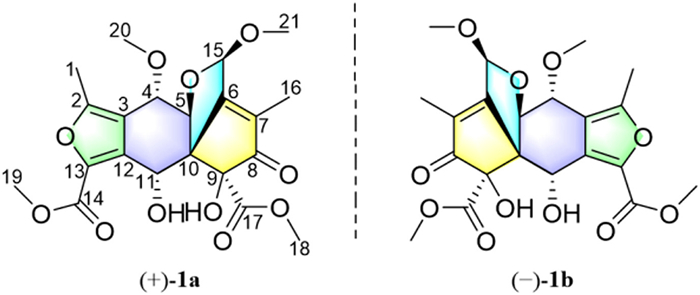
Structure of mycosphatide A (1).
(+)/(−)-Mycosphatide A, a pair of highly oxidized polyketides with lipid-lowering activity from the mangrove endophytic fungus Mycosphaerella sp. SYSU-DZG01
Qi Tan , Run-Zhu Fan , Wencong Yang , Ge Zou , Tao Chen , Jianying Wu , Bo Wang , Sheng Yin , Zhigang She

Obesity represents an important public health problem, which is associated with various complications, particularly cardiovascular diseases, type Ⅱ diabetes mellitus, obstructive sleep apnea hypopnea syndrome and several cancers [1–3]. So the prevention and control of obesity have become an increasing concern. Although behavioral modification is important for the treatment of obesity, it is a challenge for obese people to sustain weight loss in a long-term by means of lifestyle adjustments [4]. Consequently, clinical practice guidelines strongly recommend medical treatment for the overweight people [5]. Currently, six anti-obesity medications (orlistat, phentermine/topiramate, naltrexone/bupropion, liraglutide, semaglutide, and setmelanotide) have been approved by Food and Drug Administration (FDA). However, they have certain side effects, such as gastrointestinal symptoms, insomnia, and hypertension [6]. Therefore, the discovery of anti-obesity new drugs with low toxicity is necessary.
Natural products are a definitely important supplier for innovative drug discovery, and over the nearly four decades, more than a third of FDA-approved medications are related with natural products [7,8]. Mangrove-associated fungi, living in a complex ecosystem, have reported to be prolific source of fascinating natural products possessing diverse pharmacological activities [9], such as antitumor diterpenoids [10], antifouling indole diketopiperazine alkaloid dimers [11], antituberculous and antiplasmodial sesterterpenoids [12,13], antimicrobial polyketides [14]. Hence, the discovery of new drugs from the secondary metabolites of the mangrove-associated fungi seems feasible.
During our ongoing search for novel and bioactive natural products from mangrove endophytic fungi, a pair of highly oxidized novel polyketides (±)-mycosphatide A (1a and 1b), featuring a fantastic 5/5/6/5 tetracyclic ring system, were isolated from a strain of Mycosphaerella sp. SYSU-DZG01 (Fig. 1). In the bioassay, compounds (+)−1a and (−)−1b showed potent lipid-lowering effects in 3T3-L1 adipocytes. Herein, the details of isolation, structure elucidation, putative biogenetic pathway, and lipid-lowering activity of these polyketides are described.
Mycosphatide A (1), which has the form of colorless needles, has a molecular formula of C21H24O11, as deduced by high-resolution electrospray ionization mass spectrometry (HRESIMS) ([M + Na]+ m/z 475.12123, calcd. 475.12108), corresponding to 10 degrees of unsaturation (DOUs). The 1H nuclear magnetic resonance (NMR) data (Table 1) revealed two singlet methyls [δH 2.00 (s, H3–16) and 2.41 (s, H3–1)], four singlet methoxyls [δH 3.53 (s, H3–21), 3.65 (s, H3–20), 3.68 (s, H3–18) and 3.90 (s, H3–19)], four oxygenated methines [δH 4.05 (d, J = 5.2 Hz, H-5), 4.34 (d, J = 5.2 Hz, H-4), 5.06 (d, J = 3.8 Hz, H-11), and 5.68 (s, H-15)], and two hydroxy groups [δH 3.99 (s, 9-OH) and 6.50 (d, J = 3.8 Hz, 11-OH)]. The 13C and heteronuclear single quantum coherence (HSQC) spectra indicated the presence of 21 carbon signals, including one ketone carbonyl (δC 203.3), two ester carbonyls (δC 161.2, and 169.2), six sp2 quaternary carbons (δC 117.4, 134.7, 135.2, 136.3, 152.8, and 163.5), two sp3 quaternary carbons [δC 62.8, and δC 89.1 (oxygenated)], four oxygenated methines (δC 67.0, 78.1, 85.8, and one acetal carbon at δC 100.1), four methoxyls (δC 52.7, 53.6, 55.6, and 58.9), and two methyls (δC 9.9 and 13.3).
The planar structure of 1 was established by 2D NMR analysis (Fig. 2). The 1H−1H correlation spectroscopy (COSY) spectrum revealed connectivity of H-4/H-5. This fragment was further connected with hydrogen-free tertiary carbons and functionalities by detailed heteronuclear multiple-bond correlation (HMBC) analysis. The HMBC associations from an olefinic methyl singlet (H3–16) to two hydrogen-free sp2 carbon (C-6 and C-7) and a ketocarbon (C-8) linked C-6, C-16, and C-8 to C-7. The HMBC associations from 9-OH to an oxygenated quaternary carbon (C-9), a sp3 quaternary carbon (C-10), an ester carbonyl (C-17), and C-8 linked C-10, C-17, and C-8 to C-9. Two methoxy groups were connected to C-17 and an acetal carbon (C-15), respectively, by HMBC associations from H3–18 to C-17, and from H3–21 to C-15. Moreover, The HMBC associations from H-15 to C-6 linked C-15 and C-6, while an oxygenated methine (C-5) was connected to C-6 via C-10 by HMBC associations of H-5/C-10, C-15 and C-6. The above connections generated a 5/5 (A/B) fused ring system. In addition, the oxygenated methine hydrogen (H-11) was observed in the HMBC spectrum to correlated with C-10 and a hydrogen-free sp2 carbon (C-12), suggesting the connectivities of C-10 and C-12 via C-11, while another oxygenated methine (C-4) was connected to C-12 via a hydrogen-free sp2 carbon (C-3) by HMBC associations of H-4/C-3, and C-12. Therefore, a six-membered carbon C-ring was established, sharing C-5 and C-10 with ring B. A methoxyl was located at C-4 by HMBC associations from H3–20 to C-4. On the basis of the degree of unsaturation and the HMBC associations from an olefinic methyl singlet (H3–1) to two hydrogen-free sp2 carbon (C-2 and C-3), from a methoxyl singlet (H3–19) to an ester carbonyl (C-14) and a hydrogen-free sp2 carbon (C-13), and from H-11 to C-13, the furan ring D was then built. To sum up, the planar structure of 1 with an unusual 5/5/6/5 fused ring system was established.
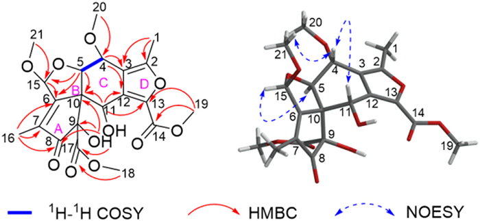
The relative stereochemistry of 1 was partly elucidated based on the interpretation of NOESY correlations (Fig. 2). The nuclear overhauser effect (NOE) correlations of H-4/H-11 and H-4/H3–21 indicated that H-4, H-11 and H3–21 were co-facial and arbitrarily designated as β-orientations, while the NOE cross-peak of H-5/H-15 assigned these protons as α-oriented. However, due to the complex multi-ring system with densely substituted, the assignment of remaining relative configuration of mycosphatide A (1) by further analyses of its NOESY correlations faced great challenges. Ultimately, the single-crystal X-ray diffraction structure (Fig. S8 in Supporting information) established the relative configuration of 1, which unexpectedly showed that compound 1 was a racemic mixture.
Semi-preparative enantioseparation (Fig. S9 in Supporting information) was achieved by chiral-phase high performance liquid chromatography (HPLC) using a chiral ND column (5 µm, 250 mm × 4.6 mm) to yield the enantiomers, (+)−1a ([α]D25 = +19.1, tR = 6.5 min) and (−)−1b ([α]D25 = −18.8, tR = 9.0 min).
Fortunately, the qualified single crystal of (−)−1b was acquired [Flack parameter of 0.01(11)], which defined the absolute configuration of (−)−1b (Fig. 3). Combined with the experimental electronic circular dichroism (ECD) spectra (Fig. 4), the absolute configurations of (+)−1a and (−)−1b were unambiguously identified as 4S, 5S, 9S, 10R, 11R, 15R and 4R, 5R, 9R, 10S, 11S, 15S, respectively.


To the best of our knowledge, mycosphatide A (1) represents the first example of polyketide with an unprecedented 5/5/6/5-fused ring system to date. What is more, according to the single crystal structure of (−)−1b, ring C presents rare boat conformation. A hypothetical biogenetic pathway for compound 1 was proposed (Scheme 1).

First, one acetyl-CoA, six malonyl-CoA and two S-adenosyl-l-methionine (SAM) molecules as starter substrates are condensed under polyketide synthase [15,16], then intermediate I is generated catalyzing by the thioesterase (TE) domain [17], which via esterification and epoxidation between C-5 and C-10 forms intermediate II. The rearrangement reactions in intermediate II generates a spiro[4.5]decane scaffold (key intermediate III). After that, a further reduction and dehydration of III followed by methylation [18] affords an intermediate IV, which undergoes a series of oxidation at C-9, C-16, C-17 and C-13 to yield intermediate V. Subsequently, the enol interconversion in V followed by 1,4-conjugated addition between H2O and the α, β-unsaturated ketone (C-11 to C-13) generates intermediate VI, which ultimately constructed a 5/5/6/5 fused tetracyclic intermediate VII through two simultaneous nucleophilic additions. Finally, the (±)-mycosphatide A is formed through methylation reactions [18].
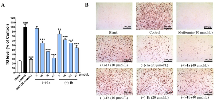
Compounds (+)−1a and (−)−1b were evaluated for the triglyceride (TG)-lowering activity in a 3T3-L1 adipocyte model, and metformin (10 mmol/L) was used as the positive control. As shown in Fig. 5A, the treatment of (+)−1a and (−)−1b significantly decreased the TG levels in a dose-dependent manner from 5 µmol/L to 40 µmol/L, with a TG-reduction ratio of 67.3% and 45.0% at 40 µmol/L, respectively. Besides, no obvious cytotoxicity to 3T3-L1 cells was observed under the effective doses (Fig. S14 in Supporting information). Subsequent study showed that the median effective concentration (EC50) values of (+)−1a and (−)−1b were 7.85 ± 1.56 and 8.87 ± 0.80 µmol/L, respectively. The apparent TG-lowering efficacy of (+)−1a and (−)−1b was also demonstrated by an Oil-Red O (ORO) staining experiment (Fig. 5B), indicating that the 5/5/6/5 tetracyclic chemical scaffold could be a candidate for future lipid-lowering drug development.
In summary, (±)-mycosphatide A (1a and 1b), a pair of highly oxidized and densely substituted novel polyketides featuring an unprecedently constructed 5/5/6/5 tetracyclic ring system, were isolated from the mangrove endophytic fungus Mycosphaerella sp. SYSU-DZG01. The optically pure enantiomers were separated by chiral-phase HPLC, and their absolute configurations were undoubtedly solved by single crystal X-ray diffraction and experimental ECD spectra comparison. The single crystal structure of (−)−1b displayed an uncommon boat conformation of ring C. (+)−1a and (−)−1b with significant lipid-lowering activity may serve as privileged structures for future anti-obesity drug discovery.
The authors declare that they have no known competing financial interests or personal relationships that could have appeared to influence the work reported in this paper.
We thank the National Natural Science Foundation of China (Nos. U20A2001, 81973195, 21877133) and the Guangdong Marine Economy Development Special Project (Nos. GDNRC[2022]35, GDNRC[2023]39) for generous support. We thank Prof. Hang Li in Pharmaceutical Sciences School at Sun Yat-sen University, for his guidance and help in hypothetical biogenetic pathway for (±)- mycosphatide A proposed in the manuscript.
Supplementary material associated with this article can be found, in the online version, at doi:
G.A. Bray, G. Frühbeck, D.H. Ryan, et al., Lancet 387 (2016) 1947–1956. doi: 10.1016/S0140-6736(16)00271-3
S.A. Polyzos, J. Kountouras, C.S. Mantzoros, Metabolism 92 (2019) 82–97. doi: 10.1016/j.metabol.2018.11.014
T.D. Müller, M. Blüher, M.H. Tschöp, et al., Nat. Rev. Drug. Discov. 21 (2022) 201–223. doi: 10.1038/s41573-021-00337-8
K.D. Hall, S. Kahan, Med. Clin. North. Am. 102 (2018) 183–197. doi: 10.1016/j.mcna.2017.08.012
Y.J. Tak, S.Y. Lee, Curr. Obes. Rep. 10 (2021) 14–30. doi: 10.1007/s13679-020-00422-w
C.M. Perdomo, R.V. Cohen, P. Sumithran, et al., Lancet 401 (2023) 1116–1130. doi: 10.1016/S0140-6736(22)02403-5
D.J. Newman, G.M. Gragg, J. Nat. Prod. 83 (2020) 770–803. doi: 10.1021/acs.jnatprod.9b01285
A.G. Atanasov, S.B. Zotchev, V.M. Dirsch, et al., Nat. Rev. Drug. Discov. 20 (2021) 200–216. doi: 10.1038/s41573-020-00114-z
S. Chen, R. Cai, Z. Liu, et al., Nat. Prod. Rep. 39 (2022) 560–595. doi: 10.1039/D1NP00041A
M. Zhang, J.M. Liu, J.L. Zhao, et al., Chin. Chem. Lett. 27 (2016) 957–960. doi: 10.1016/j.cclet.2016.02.008
R. Cai, H. Jiang, Z. Xiao, et al., Org. Lett. 21 (2019) 9633–9636. doi: 10.1021/acs.orglett.9b03796
X. Huang, H. Huang, H. Li, et al., Org. Lett. 15 (2013) 721–723. doi: 10.1021/ol303549c
W. Yang, T. Chen, Y. Chen, et al., J. Org. Chem. 87 (2022) 16807–16819. doi: 10.1021/acs.joc.2c02501
L.H. Meng, X.M. Li, Y. Liu, et al., Chin. Chem. Lett. 26 (2015) 610–612. doi: 10.1016/j.cclet.2015.01.024
H. Zhou, Y. Li, Y. Tang, Nat. Prod. Rep. 27 (2010) 839–868. doi: 10.1039/b911518h
D.T. Wagner, D.C. Stevens, M.R. Mehaffey, et al., Chem. Commun. 52 (2016) 8822–8825. doi: 10.1039/C6CC04418B
H.H. Yeh, S.L. Chang, Y.M. Chiang, et al., Org. Lett. 15 (2013) 756–759. doi: 10.1021/ol303328t
E. Abdelraheem, B. Thair, R.F. Varela, et al., ChemBioChem 23 (2022) e202200212. doi: 10.1002/cbic.202200212

Figure 3 Single-crystal X-ray structure of (−)−1b (the ellipsoid contour probability level is 50%).

Figure 5 TG-lowering efficacy of (+)−1a and (−)−1b in 3T3-L1 cells. (A) TG levels under the treatments of (+)−1a and (−)−1b with different concentrations. (B) Representative ORO staining photomicrographs under the treatment of (+)−1a and (−)−1b with different concentrations. Blank: undifferentiated cells; control: differentiated cells without compound treatment. Data represented the mean ± standard deviation (SD) of at least three independent experiments. |||P < 0.001 vs. the blank group; **P < 0.01, ***P < 0.001 vs. the control group.

 扫一扫看文章

扫一扫关注我们
 DownLoad:


 下载:
下载:


 下载:
下载: