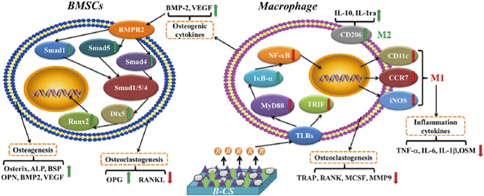
Figure 1.
Immunomodulatory and osteogenesis-promoting effects of the B-incorporated calcium silicate (B-CS) coating. Copied with permission [10]. Copyright 2018, Wiley Publishing Group.
Biomaterial-induced macrophage polarization for bone regeneration
Long Chen , Zhicheng Yao , Siqin Zhang , Kuihan Tang , Qiming Yang , Yuanzheng Wang , Bohan Li , Yingjie Nie , Xiaobin Tian , Li Sun

The innate immune system has received increasing attention, especially the role of macrophages in bone regeneration. When any such material is implanted into the human body for bone repair, it will elicit an innate immune response. In the past, biomaterials were designed under the goal of biological inertia to isolate the material from the host immune system as much as possible. However, in recent years, as our understanding of the heterogeneity and plasticity of immune cells has grown, especially of macrophages, and their role in bone regeneration and repair has become better appreciated, researchers have begun to explore the design of biomaterials that interact with the immune system more actively to induce the body to generate a certain immune response that is conducive to bone regeneration. Macrophages are heterogeneous cell populations, and each subset has different markers and functions. The polarization and plasticity of macrophages allow them to play different roles in the process of tissue regeneration. Broadly speaking, M0 macrophages are nonactivated macrophages. Polarized macrophages can be divided into M1 and M2 phenotypes. The M1 type is considered to significantly promote inflammation and catabolism. In contrast, M2 type can reduce inflammation and promote anabolism.
For bone healing, macrophages have a dual regulatory role, and the phenotype of M1/M2 macrophages in the inflammatory response can undergo adaptive polarization with changes in the microenvironment, play different roles, and participate in the regulation of downstream bone healing responses. M1 macrophages secrete a variety of inflammatory factors, such as tumor necrosis factor-α (TNF-α), interleukin-1 (IL-1) and IL-6, in this process, induce local inflammatory responses, and promote the differentiation of monocytes into multinucleated osteoclasts, resulting in bone tissue inflammatory resorption and loss of bone mass. M2 macrophages release cytokines such as transforming growth factor-β (TGF-β), bone morphogenetic protein-2 (BMP-2), and vascular endothelial growth factor (VEGF) to promote osteogenic differentiation, angiogenic differentiation, and matrix deposition of bone marrow mesenchymal stem cells (BMSCs) [1], while enhancing the expression of the anti-inflammatory factors IL-4 and IL-10 to promote the inflammation resolution and tissue remodeling. At 3–7 days after injury, the acute inflammation gradually subsides, and the hematoma is organized to form granulation tissue. Many BMSCs infiltrate the granulation tissue, where they differentiate into osteoblasts under the action of cytokines such as BMP-2 and TGF-β, secrete extracellular matrix (ECM), and gradually mineralize. M1 macrophages promote the migration of BMSCs, while M2 macrophages significantly increase the activity, proliferation, and osteogenic differentiation of BMSCs, so an appropriate M1/M2 ratio is conducive to bone formation. Excessive M2 polarization will cause fibrosis, and the defect area can even become wrapped by fibrous tissue, resulting in nonunion of bone. Excessive polarization to M1 will lead to a prolonged inflammatory response, resulting in delayed healing or persistent nonhealing of bone tissue. Therefore, the correct understanding the polarization function of macrophages in the bone healing process can not only shed light on the mechanism of tissue scar healing and fibrosis adhesion to surrounding tissues but also provide better guidance on the research and development of new biomaterials for bone regeneration.
Focusing on the polarization of macrophages, researchers have developed many new biomaterials for bone regeneration through different strategies with the goal of altering macrophage ratios by adjusting specific factors such as the chemical composition, structure, hardness, roughness, pore size, and adhesion characteristics of the implanted materials. Interactions between tissue microenvironmental changes, various cellular influences, and material properties can also change the phenotype of macrophages. This paper reviews the current studies on the regulation of macrophage polarization by biomaterials to provide new ideas for the design of biomaterials for bone regeneration.
The early interaction between the material and the tissue interface can drive the host immune response and subsequent tissue remodeling, which has an important impact on the downstream response of bone regeneration. Previous studies have focused on the design of inert coatings to inhibit the activation and regulation of macrophages. The immune response to biomaterials, however, is not conducive to tissue regeneration. The coating of biologically active chemical elements on the surface of biomaterials can make the surface functionalized, which can prevent the adsorption of serum proteins on the surface of the material, thereby preventing the further activation of complement and coagulation reactions and reducing the inflammatory response.
Chen et al. found that, as a commonly used coating material, β-tricalcium phosphate (β-TCP) extract could induce macrophages to polarize toward the M2 type, the mechanism of which was related to the activation of the CaSR pathway. The expression of BMP-2 was also significantly increased, suggesting that macrophages may be involved in the β-TCP-induced bone regeneration response; when the macrophage-conditioned β-TCP extract was applied to BMSCs, the osteogenic differentiation of BMSCs was significantly enhanced, indicating that macrophages play an important role in the osteogenic response induced by biomaterials [2]. Magnesium (Mg) is widely used in orthopedics due to its unique biodegradability, biocompatibility, good mechanical properties, and osteogenic action, but the rapid degradation of Mg in the body can trigger an acute inflammatory response [3]. Therefore, its further application in bone tissue engineering is limited. In a later study, Chen et al. coated a Mg scaffold with β-TCP to construct a Mg-β-TCP scaffold with excellent bone immunomodulatory properties. The β-TCP coating reduced the degradation rate of the Mg scaffold and induced the polarization of macrophages to the M2 type, which may have inhibited the TLR pathway. VEGF and BMP-2 were significantly upregulated in macrophages exposed to the Mg-β-TCP scaffold, indicating that the macrophages in the β-TCP-modified Mg scaffold had osteogenic properties; when BMSCs were stimulated by the conditioned medium of the macrophages cultured on the Mg-β-TCP scaffold, the osteogenic differentiation of BMSCs was significantly upregulated. Thus, the macrophages in the Mg-β-TCP scaffold had osteogenic properties [4]. Li et al. used plasma spraying technology to combine cerium dioxide having antioxidative activity with hydroxyapatite coating and found that after coculture with macrophages, the expression of the M2 macrophage markers CD163 and CD206 was significantly upregulated and anti-inflammatory cytokines (IL-10 and IL-1) were elevated, suggesting that mixed coatings could induce macrophage polarization [5]. The hypoxic microenvironment can regulate local immune responses, thereby promoting tissue repair. Zhou et al. added copper ion (Cu2+) to mesoporous bioactive glass to generate a hypoxia-like microenvironment, activate the HIF-1α pathway, and enhance host angiogenesis. They also found that local M2 macrophage infiltration increased, indicating regulation of macrophages by Cu2+. Others studies showed that the surface of Cu2+-containing nanobioceramics (Cu-Hier-Ti) enhanced osteogenesis and bactericidal activity [6,7]. Titanium and titanium alloys have been widely used as implant materials in the body due to their unique physical and chemical properties, but these take a long time to achieve sufficient bone ingrowth and bone remodeling [8]. Excessive differentiation of M1 macrophages around pure titanium artificial joints can lead to bone resorption and implant loosening around the prosthesis [9]. Lu et al. sprayed a calcium borosilicate (Ca11Si4B2O22) coating on pure titanium material, finding that its degradation products promoted the transition of macrophages from M1 to M2 by inhibiting the TLR signaling pathway, increased anti-inflammatory factor (IL-10 and IL-1) levels, reduced the expression of proinflammatory cytokines (iNOS, IL-6, and TNF-α), stimulated the osteogenic differentiation of BMSCs, and inhibited the differentiation of macrophages into osteoclasts (Fig. 1) [10]. Zhang et al. also found that aminated salinized titanium could reduce the inflammatory response and promote the chemical immunomodulatory effect of M2 polarization in macrophages [11]. Lu et al. found that a phase-transited lysozyme (PTL) coating on the surface of titanium materials improved the migration and adhesion of cells, and the PTL with Sr coating stably released strontium ions to promote cell migration and bone formation. In addition, PTL with Sr regulated the immune response of macrophages, thereby enhancing the recruitment of BMSCs and their osteogenic differentiation [12].
The metal particles produced by prosthesis wearing after hip arthroplasty can act as haptens to activate T cells to trigger delayed hypersensitivity, which can induce the polarization of macrophages to the M1 phenotype, leading to the formation of granulation tissue, periprosthetic osteolysis, and necrosis of surrounding tissues, which can further lead to serious complications such as prosthesis loosening [13-16]. In response to this problem, many researchers have modified the coating of prostheses to regulate the polarization of macrophages to reduce osteolysis and promote bone ingrowth. Hachim et al. developed an IL-4-loaded polypropylene material coating, which significantly increased the number of M2 macrophages around the implant, reduced the fibrotic adhesion around the implant, and improved implant-surrounding tissue integration [17]. The study of Yang et al. confirmed that titanium nanoparticles produced by wear can activate the expression of M1 macrophages, and lithium chloride (LiCl) can drive the polarization of macrophages to the M2 phenotype and reduce the inflammatory response mediated by titanium nanoparticles, eventually promoting the osteogenic differentiation of rat BMSCs [18]. Mahon et al. also showed that pretreatment of macrophages with a Syk inhibitor (R788/piceatannol) or a MAPK inhibitor (SB203580/PD98059) could prevent the M1 phenotype polarization and attenuate the production of proinflammatory mediators [19]. The regulation of macrophage polarization through the coating can greatly improve the immune response around the implant and ultimately achieve the goal of reducing fibrotic adhesion and improving the integration of the implant interface with the surrounding tissue.
Strontium plays an important role in bone remodeling. Incorporation of strontium ions into materials with different structures is a common method to improve the immune response to materials [20]. Liu et al. constructed multifunctional collagen scaffolds by doping strontium ions into self-assembly/mineralization collagen, and strontium released by Sr-doped collagen scaffolds stimulated the polarization of macrophages from M1 to M2, which significantly improved the osteogenic ability of rat BMSCs and enhanced the effect of bone regeneration (Fig. 2) [21]. As a bone tissue repair material, hydroxyapatite can easily induce many macrophages to differentiate into the M1 phenotype. Wang et al. used the function of lanthanum (La3+) to inhibit LPS, then doped it with magnetic SrFe12O19 nanoparticles to recruit endogenous stem cells, finally constructing a magnetic lanthanum-doped hydroxyapatite/chitosan scaffold, which not only reduced the proportion of macrophages that differentiated into M1 but also promoted the polarization of macrophages to the M2 phenotype to inhibit the inflammatory response [22].

Several cytokines are known to affect the polarization of macrophages, including IL-10, IL-13 and IL-4. IL-4 induces M1 macrophages to re-enter the cell cycle and to polarize toward the M2 phenotype, reduce M1 activation, and induce in situ proliferation of M2 macrophages. IL-4 can also promote IL-10, IL-4, PDGF and TGF-β secretion, leading to the amplification of the M2 macrophage response and promoting wound/tissue healing [23]. Jin et al. implanted a biomimetic hierarchical intrafibrillarly mineralized collagen (HIMC) three-dimensional scaffold loaded with IL-4 into an animal model of mandibular defects. The scaffold material significantly enhanced the polarization of macrophages toward the M2 phenotype, thereby promoting the osteogenic differentiation of BMSCs [24]. Zhang et al. confirmed that calcium-enriched gellan gum loaded with IL-4 promoted the polarization of macrophages to the M2 phenotype, increased the expression level of TGF-β1, and activated the TGF-β1/Smad pathway in BMSCs, thereby promoting osteogenic differentiation. This material also reduced cell apoptosis in vivo [25]. Li et al. prepared a polydopamine coating on TiO2 nanotubes, assembled the anti-inflammatory cytokine IL-4 and osteogenic RGD peptide layer by layer, and then coated it with a carboxymethyl chitosan hydrogel layer to control IL-4 production and fix an RGD peptide. The results showed that the scaffold materials not only drove the phenotypic polarization of macrophages to the anti-inflammatory M2 phenotype and induced repair cytokines such as IL-10 but also enhanced the osteogenic differentiation associated with BMP/Smad/RUNX2 signaling pathway [26]. How to regulate the immune response of the host accurately and actively is another important issue. In the process of normal tissue repair, macrophages exhibit the proinflammatory M1 phenotype in the early stage and the healing-promoting M2 phenotype in the later stage. To maintain normal tissue repair, M1 and M2 macrophages need to be synergistically activated; otherwise, the imbalance of M1/M2 will lead to the obstruction of vascular and bone regeneration. Spiller et al. constructed a composite scaffold based on an acellular bone matrix, which successively released IFN-γ and IL-4, and short-term release of IFN-γ promoted the polarization of macrophages to the M1 phenotype to initiate angiogenesis; the more sustained release of IL-4 promoted polarization to the M2 phenotype to promote vascular maturation; this design significantly enhanced the vascularization of local tissues [27]. Gao et al. loaded IL-4 into TiO2 nanotubes through a hydrogel coating and then coated the hydrogel coating with IFN-γ to achieve IL-4 release after IFN-γ release. These results indicated that TiO2 nanotubes loaded with IL-4 and IFN-γ could stimulate the polarization of macrophages from the M1 phenotype to the M2 phenotype. TiO2 nanotubes loaded with IL-4 alone also regulated the transition of macrophages from the M1 phenotype to M2 phenotype, but their effect was weaker than that of IL-4- and IFN-γ-loaded TiO2 nanotubes [28]. Zheng et al. implanted acellular bone matrix materials into the cranial defects in rats, then delivered four different doses of IL-4 (0 ng, 10 ng, 50 ng, and 100 ng) to the defect 3 days after implantation. The immunomodulatory effects of the 10 ng group were significantly better than those of other groups in terms of new bone formation and angiogenesis, suggesting that accurate delivery of IL-4 within a certain time frame can coordinate M1 and M2 macrophages to better cooperate in tissue regeneration and healing [29]. Kumar et al. used the concepts of synthetic peptide chemistry, supramolecular self-assembly, and cytokine delivery to construct a novel hydrogel (multidomain peptide complex with IL-4 and monocyte chemoattractant protein-1 delivery). This hydrogel induces the spatiotemporal activation of monocytes and macrophages through the biphasic release of cytokines. The results showed that the hydrogel activated various macrophages sequentially and inertially according to the stage of tissue healing, thereby better promoting tissue regeneration [23].
The above results show that the regulation of macrophage polarization by cytokines is a complex process. A full understanding of the role of M1 macrophages and M2 macrophages in the process of tissue regeneration will help us rationally use cytokines to regulate macrophage polarization.
Hydrophobic substances can enhance the adhesion of monocytes, and the protein molecules on the hydrophobic surface are easier to spread and diffuse, while a water layer with a hydrophilic or neutral surface is more conducive to the expansion of macrophages, thereby reducing the foreign body giant cell formation and inhibiting the inflammatory response [30,31]. Lv et al. used the atomic layer deposition technique combined with ultraviolet irradiation and self-assembled monolayers (SAMs) to prepare hydrophilic and hydrophobic TiO2 thin films with similar surface morphologies. The team also used the microcontact printing technique to prepare a surface with alternating hydrophilic and hydrophobic bands on the same substrate. Compared to the hydrophobic surface, the hydrophilic surface was more conducive to the expansion of macrophages. They compared the adhesion morphology of macrophages in the hydrophilic region and the hydrophobic region of the surface of the hydrophilic and hydrophobic bands. Compared with the hydrophobic surface, the hydrophilic surface promoted the transition of macrophages to the anti-inflammatory M2 phenotype and promoted osteogenic differentiation and mineralization of preosteoblasts (MC3T3-E1) through the secretion of more osteogenesis-related factors (BMP-2 and TGF-β1) that have a beneficial effect on tissue repair [32]. These results indicate that macrophages cultured on materials with high surface wettability can generate an anti-inflammatory microenvironment, which can be used to improve the body's healing response to biomaterials. Not all hydrophobic and hydrophilic surfaces showed the above results. To evaluate the effects of different surface chemicals on macrophage polarization, Rostam et al. cultured monocytes on untreated hydrophilic O2 plasma-etched polystyrene (O2-PS40) surface for 6 days. The results suggested that monocytes cultured on the hydrophilic O2-PS40 surface were polarized to the M1 phenotype, and the expression levels of proinflammatory transcription factors STAT1 and IRF5 were significantly increased. In contrast, monocytes cultured on the hydrophobic polystyrene surface exhibited an M2-like phenotype, their expression of mannose receptor was high, and they produced anti-inflammatory cytokines IL-10 and CCL18 [33]. This phenomenon, contrary to the above results, may have been due to the adsorption of different biomolecules on these surface chemical substances, resulting in the polarization reaction of macrophages.
Previously, it was believed that rougher surfaces were more likely to induce the secretion of inflammatory factors by macrophages. Refai et al. produced four surface morphologies on the surface of titanium materials: mechanical polishing, coarse sandblasting, acid etching, sandblasting, and acid etching and cultured macrophages on them. They found that the surface morphology caused by sandblasting and acid etching regulated the secretion of proinflammatory cytokines and chemokines by macrophages in a time-dependent manner [34]. Li et al. showed that the cytoskeleton of macrophages changed with the change in the surface roughness of titanium in the roughness range of 100 nm to 400 nm, as did the factors they produced (TNF-α, IL-6 and IL-4). The production of IL-10 can be regulated by the surface roughness of titanium. In addition, with the increase in surface roughness, the macrophages cultured on the titanium surface showed a trend of polarization toward the M1 phenotype [35]. In an in-depth study, Chen et al. found that the surface of large nanogrooves could reduce the adhesion, migration, and cytokine secretion of macrophages relative to the planar surface [36]. Luu et al. confirmed that on a titanium surface with 400–500-nm-wide grooves, the spread of macrophages peaked, and the phenotypes of macrophages were polarized toward anti-inflammatory and promoting healing phenotype (M2) [37]. Wang et al. found that macrophages elongated along the direction of wrinkles made of shape-memory polymers and that they expressed more arginase-1 and IL-10 and less TNF-α, indicating that the polarization was toward the anti-inflammatory M2 type. After the material was implanted into the subcutaneous tissue of mice, it was also found that the surface morphology of the material changed the distribution of collagen deposition in the adjacent tissues. Compared with the wrinkled material, denser collagen tissue was observed near the flat material, and the cells surrounding the wrinkled material exhibited higher expression of arginase-1. These results suggest that the surface of the wrinkled material promotes the polarization of macrophages to the M2 phenotype and may affect the body's response to the implant [38]. Ma et al. used 5 V and 20 V anodic oxidation and ultraviolet irradiation on a titanium surface to produce hydrophilic TiO2 nanotube structure surfaces (denoted NT5 and NT20, respectively) and cocultured them with macrophages. The results suggested that the NT5 surface induced the polarization of macrophages toward the healing related M2 type both in vitro and in vivo. The surface of NT20 promoted the polarization of macrophages toward the proinflammatory-related M1 type [39]. In contrast, Wang et al. generated two different sizes of TiO2 nanotube structure surfaces by anodic oxidation at 10 V (NT10) and 20 V (NT20) on the titanium surface and found that cocultured with macrophages, NT20 induced macrophages to polarize toward the anti-inflammatory M2 macrophages, and their expression of IL-10 and Arg-1 was enhanced, while NT10 induced macrophages to polarize toward the proinflammatory M1 macrophages, and IL-1β, iNOS and TNF-α were upregulated [40]. The two studies obtained different results, but the methods used to form the surface of the TiO2 nanotube structure were different, as were the morphology, hydrophilicity, and roughness of the titanium surfaces. Therefore, the two studies obtained different results, but it can still be concluded that the polarization of macrophages is affected by the characteristics of the titanium surface, including its morphology, roughness, and wettability. It was also confirmed that the reason that different roughness affect the immune response is that the surface morphology of the material changes the distribution of collagen deposition in the adjacent tissues, smooth surfaces being able to significantly reduce the adhesion and activation of platelets and cells compared to the rough surface [41]. The modification of the roughness and wettability of the titanium surface by Hotchkiss et al. induced the body's adaptive immune response to Th2 polarization (promoting wound healing), which made the inflammation subside more quickly [42]. Hamlet et al. observed that the CD163 protein expression and Arg-1 gene expression of M1 macrophages cultured on the surface of microrough and hydrophilic modified titanium significantly increased, confirming that M1 macrophages differentiated toward the M2 phenotype [43]. Hotchkiss et al. also confirmed that a smooth titanium surface could induce the differentiation of macrophages toward the M1 phenotype, and it significantly increased the expression levels of IL-1β, IL-6 and TNF-α. In contrast, the hydrophilic rough titanium surface induced macrophages to differentiate toward the M2 phenotype, and the expression levels of IL-4 and IL-10 were significantly increased, suggesting that the combination of hydrophilicity and roughness on the surface of the material may have a synergistic effect. The study also further confirmed that the increase in surface hydrophilicity had a stronger immunomodulatory effect than increased surface roughness [44].
The charge on the surface of the material also has a certain impact on the immune response. Chang et al. confirmed that the surface of hydrophilic/neutral and anionic materials stimulated the activation of M1 macrophages, induced the expression of IL-8, IL-6, and TNF-α, and lowered the expression of IL-10, thereby promoting the inflammatory response. The surface of the hydrophilic cationic material promoted the activation of M2 macrophages, thereby promoting the anti-inflammatory response [45]. Supported lipid bilayers can indirectly control ligand-mediated cell signal transduction by changing the concentrations of their components or inserting molecules such as proteins and cholesterol. Phosphatidylserine is the most abundant negatively charged phospholipid on cell membranes. It can produce anti-inflammatory effects by binding to macrophages. Quan et al. prepared phosphatidylserine-containing supported lipid bilayers on the surface of titanium materials to induce the polarization of macrophages toward the M2 type, suggesting it can be used as an immunomodulatory coating material to alleviate the response of the host immune system to the prosthesis [46].
Due to its special effect on cell adhesion and proliferation, the morphology of the material has a more extensive regulatory effect on cell function, and a slight change may lead to significantly different responses of macrophages. The nanofiber structure mimics the phenotypic characteristics of the natural ECM and can regulate the transition of macrophages to the anti-inflammatory phenotype in vitro. Wang et al. applied electrospinning technology to spin thermoplastic polyurethane into nano and microfiber membranes. Compared with microfiber membranes, nanofiber membranes caused a weaker macrophage response in vitro and in vivo and only caused minor foreign body reactions. In their further studies, the device made of nanofibers could effectively protect the allograft from host immune attack, suggesting that nano topography can endow the material with biocompatibility and that the nanofiber material is worthy of further development, which can be used to develop "invisible" immune isolation devices for cell transplantation (Fig. 3) [47]. Shayan et al. used bulk metallic glass to nanopattern and explored its interaction with macrophages. The results showed that after nanopatterning, the bulk metallic glass could regulate the polarization of macrophages to the M2 phenotype and transiently induce less fibrosis and more angiogenesis [48]. Castellano et al. used the electrospinning method to prepare poly(3-hydroxybutyrate) (PHB) nanofiber scaffolds. Neither the non-cross-linked collagen membrane nor PHB induced fiber encapsulation, and they gradually degraded. In the myocardial tissues of the rat myocardial infarction model, both the non-cross-linked collagen membrane and PHB induced the inflammatory response of macrophages to differentiate into the M2 phenotype, and PHB induced angiogenesis [49]. Saino et al. cultured macrophages on four types of electrospun poly(l-lactic acid) scaffolds (orderly arranged microfibers, neatly arranged nanofibers, randomly arranged microfibers, and randomly arranged nanofibers). The morphology of the scaffold or membrane affected the polarized immune response of macrophages, especially in the early stage of inflammation. The secretion of proinflammatory molecules by macrophages was mainly determined by the fiber diameter. Nanofiber poly(l-lactic acid) scaffolds minimized the inflammatory response compared to microfiber scaffolds [50].
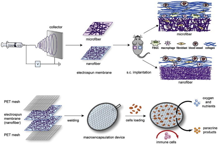
The above results suggest that we can control the fiber diameter and thickness of electrospun nanofiber membranes by simply adjusting the relevant parameters and conveniently manipulating and controlling the composition and structure of the mesh. Their unique nanofiber structure is not only semipermeable but can also be used as an immune barrier to protect allografts from immune attack, improve the biocompatibility of the material, and reduce the foreign body reaction. The surface morphology, diameter, and arrangement of nanofibers will affect the direction of macrophage polarization.
The porosity and pore size of biomaterials are also important factors that regulate macrophage polarization and affect bone and soft tissue repair. Increasing the pore size, porosity, and fiber diameter of the material can promote the polarization of macrophages to the M2 phenotype, the migration of BMSCs, and the recruitment of endogenous cells [51,52]. Wang et al. found that vascular grafts prepared by electrospinning usually have relatively small pores, which limits the infiltration of cells into the graft and hinders the regeneration and remodeling of the graft into new blood vessels. They prepared polycaprolactone scaffolds with coarser fibers (5–6 µm) and larger pores (~30 µm diameter), and in vitro cell culture showed that macrophages cultured on coarser fibers and pore size scaffolds tended to polarize to the M2 phenotype, which promoted tissue remodeling, while the macrophages cultured on the thinner fiber scaffold were polarized to the proinflammatory M1 phenotype. The in vivo results showed that large-pore grafts exhibited more cellular infiltration and ECM deposition, and the ECM was similar to that of natural arteries. These results indicate that thicker and larger-pored fibrous scaffolds can induce many M2 macrophages to infiltrate the grafts, which will further promote cell infiltration and vascularization [53]. Pan et al. found that macrophages were polarized toward the M2 phenotype on the stratified macropore/nanosurface, with downregulated inflammatory cytokines and upregulated anti-inflammatory cytokines, which may be related to the cytoskeletal tension induced by specific cell shapes [54].
Although the porous structure has a certain impact on the polarization of macrophages, the composition of the material itself plays a more important role. Chen et al. studied the effects of porous ceramic calcium phosphates with different phase compositions on macrophage polarization and bone repair and found that porous biphasic calcium phosphate ceramics induced macrophages to polarize to the M2 phenotype and promoted bone repair, while porous β-TCP induced macrophages to polarize toward the M1 phenotype [55]. Porous biomaterials can also be used as carriers to deliver drugs or cytokines to promote repair. Wagner et al. inoculated PLGA microspheres containing different growth factors (VEGF, BMP-2, or FGF-2) on porous polycaprolactone fumarate polymer scaffolds, which yielded a greater vascular in growth rate, vascular penetration, and mineral deposition [56]. Yang et al. inoculated sodium butyrate on three-dimensional porous sulfonated polyetheretherketone, which improved the phagocytic ability of macrophages and the anti-infection property of the material and induced the polarization of macrophages to M2 to promote bone regeneration [57]. The porosity and pore size of the material also affect the overall structural stability of the material. When considering the use of the porous structure of the material to regulate the polarization of macrophages, the required repair strength at the implantation site should also be considered so it will reach the optimal equilibrium point to better complete bone repair [58].
After biomaterials are implanted in the human body for bone regeneration, they will have effects on local tissues and the whole body, mainly in the form of local tissue responses and immune responses in the body. The implantation of biomaterials into the body is a traumatic process, so the local reaction after implantation is similar to the typical bone healing process, which mainly includes an inflammatory phase and the repair phase. As the material remains in the body for a long time and becomes a persistent inflammatory irritant, local inflammatory cell activity is enhanced, the inflammatory period is prolonged, and the repair period is also prolonged. Therefore, the length and extent of the posttraumatic inflammatory repair mainly depend on the biocompatibility and degradability of the implant. Although biomaterials have good biocompatibility and degradability, they will still be seen as foreign bodies after implantation. Therefore, there will be foreign body reactions surrounding the material generated by the host's immunity. The intensity and extent of the foreign body reaction is closely related to the immunomodulatory properties of the material.
Macrophages are important immune cells that have the functions of phagocytosis of pathogens, antigen presentation, and secretion of a variety of cytokines. In different stages of bone repair, various types of macrophages play different roles. M1 macrophages can produce large amounts of proinflammatory cytokines. They have a highly phagocytic function, as they can phagocytose apoptotic neutrophils and remove pathogens or debris from the injury site. M2 macrophages have anti-inflammatory effects. Their differentiation can be induced by IL-4, IL-13, or apoptotic neutrophils in vitro. M2 macrophages can regulate vascular regeneration and collagen production. In recent years, rational design of the configuration of biomaterials to regulate macrophage polarization has been a research hotspot. The immune response after the implantation of a material into a bone can be adjusted by fine-tuning the physical parameters and chemical properties of the biomaterial, and the regulation of the polarization of macrophages is particularly important for bone repair. Surface modification, coating, and synthesis techniques have given biomaterials new properties, such as material hydrophilicity/hydrophobicity, charge expression, roughness, pore size/porosity and nanofiber diameter and arrangement. The immune response induced by implantation of biomaterials into the body is a complex process. The best strategy to ensure the immune compatibility of biomaterials and their ability to promote bone repair and regeneration is to regulate the immune response rather than inhibit it. As an ideal implant material for bone regeneration, it not only needs to meet the specific functional requirements of tissues but also must have immune characteristics to ensure the success and long-term survival of the implant.
The polarization of macrophages after biomaterials are implanted in the body for bone regeneration is related not only to the material itself but also to the physiological and pathological conditions of the host and the local tissue microenvironment. For example, there are significant differences in the response of young and old host to substances. Therefore, the design of the material should fully consider the onset characteristics and pathological state of the disease. The local microenvironment can significantly affect the behavior of macrophages, leading to different results in vitro, in vivo, and at different implantation sites. Therefore, it is difficult to mimic the real situation in the body through in vitro analysis alone. Due to the differences in various objective factors between different studies, such as different structures and different mechanical properties of materials in different parts, different immune properties may be in play, resulting in contradictory and difficult-to-compare results. Therefore, when designing various parameters of immune-regulating biomaterials, the effects of the local microenvironment of the implantation site and other external signals should be fully considered, and a consistent animal model that can simulate the pathological state should be constructed to verify the repair effect.
M1 and M2 macrophages play an indispensable role in the regeneration of bone. The M1 macrophage-mediated inflammatory response is an essential step in the repair process. Most current biomaterial designs focus on simple induction of M2 polarization but ignore the role of M1 macrophages in the bone repair process. The excessive polarization of macrophages to the M2 phenotype can lead to fibrosis. A higher M2 profile could be achieved through an M1 intermediary. According to previous study indicating that a predominantly M2 type response and constructive remodeling in tissue defects could be led by the acellular scaffold, as compared to cellular scaffold, which resulted in a predominantly M1 response and deposition of dense connective tissue and/or scarring [9]. Therefore, further studies should continue to investigate the time, sequence, and intensity of the transition between different phenotypes of macrophages during the bone repair process and combine the above different methods to improve the design of immunomodulatory materials for bone regeneration.
The authors declare that they have no known competing financial interests or personal relationships that could have appeared to influence the work reported in this paper.
The work was supported by the National Natural Science Foundation of China (Nos. 81960404, 81960401 and 82060308), Guizhou Province Science and Technology Project (No. [2019]1429) and Guizhou Provincial high-level innovative talents of Science and Technology Department (No. GCC[2022]037–1).
F. Loi, L.A. Córdova, J. Pajarinen, et al., Bone 86 (2016) 119–130. doi: 10.1016/j.bone.2016.02.020
Z. Chen, C. Wu, W. Gu, et al., Biomaterials 35 (2014) 1507–1518. doi: 10.1016/j.biomaterials.2013.11.014
L. Sun, X. Li, M. Xu, et al., Regen. Biomater. 7 (2020) 391–401. doi: 10.1093/rb/rbaa010
Z. Chen, X. Mao, L. Tan, et al., Biomaterials 35 (2014) 8553–8565. doi: 10.1016/j.biomaterials.2014.06.038
K. Li, Q. Shen, Y. Xie, et al., J. Biomater. Appl. 31 (2017) 1062–1076. doi: 10.1177/0885328216682362
C. Gao, Y. Deng, P. Feng, et al., Int. J. Mol. Sci. 15 (2014) 4714–4732. doi: 10.3390/ijms15034714
Y. Zhou, S. Han, L. Xiao, et al., J. Mater. Chem. B 6 (2018) 3274–3284. doi: 10.1039/C8TB00683K
Y. Oshida, E.B. Tuna, O. Aktören, et al., Int. J. Mol. Sci. 11 (2010) 1580–1678. doi: 10.3390/ijms11041580
A.J. Rao, E. Gibon, T. Ma, et al., Acta Biomater. 8 (2012) 2815–2823. doi: 10.1016/j.actbio.2012.03.042
X. Lu, K. Li, Y. Xie, et al., J. Biomed. Mater. Res. A 107 (2019) 12–24. doi: 10.1002/jbm.a.36456
H. Zhang, X. Wu, G. Wang, et al., Colloid. Surf. B 166 (2018) 269–276. doi: 10.1016/j.colsurfb.2018.03.029
X. Lu, W. Zhang, Z. Liu, et al., Med. Sci. Monit. 25 (2019) 2658–2671. doi: 10.12659/MSM.914269
S.B. Goodman, Biomaterials 28 (2007) 5044–5048. doi: 10.1016/j.biomaterials.2007.06.035
T. Lähdeoja, J. Pajarinen, V.P. Kouri, et al., J. Orthop. Res. 28 (2010) 184–190. doi: 10.1002/jor.20979
A. Jonitz-Heincke, J. Tillmann, A. Klindere, et al., Materials 10 (2017) 566 (Basel).
G. Liu, T. Guo, Y. Zhang, et al., APMIS 125 (2017) 565–578. doi: 10.1111/apm.12679
D. Hachim, S.T. LoPresti, C.C. Yates, B.N. Brown, Biomaterials 112 (2017) 95–107. doi: 10.1016/j.biomaterials.2016.10.019
C. Yang, W. Wang, K. Zhu, et al., Int. J. Nanomed. 14 (2019) 7475–7488. doi: 10.2147/IJN.S210834
O.R. Mahon, S. O'Hanlon, C.C. Cunningham, et al., Acta Biomater. 65 (2018) 426–435. doi: 10.1016/j.actbio.2017.10.041
C.H. Lee, Y.J. Kim, J.H. Jang, J.W. Park, Nanotechnology 27 (2016) 085101. doi: 10.1088/0957-4484/27/8/085101
H. Liu, M. Lin, X. Liu, et al., Bioact. Mater. 5 (2020) 844–858. doi: 10.1016/j.bioactmat.2020.06.005
Q. Wang, Y. Tang, Q. Ke, et al., J. Mater. Chem. B 8 (2020) 5280–5292. doi: 10.1039/D0TB00342E
V.A. Kumar, N.L. Taylor, S. Shi, et al., Biomaterials 52 (2015) 71–78. doi: 10.1016/j.biomaterials.2015.01.079
S.S. Jin, D.Q. He, D. Luo, et al., ACS Nano 13 (2019) 6581–6595. doi: 10.1021/acsnano.9b00489
J. Zhang, H. Shi, N. Zhang, et al., Cell Prolif. 53 (2020) e12907. doi: 10.1111/cpr.12907
M. Li, F. Wei, X. Yin, et al., Mater. Sci. Eng. C: Mater. 109 (2020) 110508. doi: 10.1016/j.msec.2019.110508
K.L. Spiller, S. Nassiri, C.E. Witherel, et al., Biomaterials 37 (2015) 194–207. doi: 10.1016/j.biomaterials.2014.10.017
L. Gao, M. Li, L. Yin, et al., J. Biomed. Mater. Res. A 106 (2018) 1878–1886.
Z.W. Zheng, Y.H. Chen, D.Y. Wu, et al., Theranostics 8 (2018) 5482–5500. doi: 10.7150/thno.28315
J.A. Jones, D.T. Chang, H. Meyerson, et al., J. Biomed. Mater. Res. A 83 (2007) 585–596.
M.A. Alfarsi, S.M. Hamlet, S. Ivanovski, J. Biomed. Mater. Res. A 102 (2014) 60–67. doi: 10.1002/jbm.a.34666
L. Lv, Y. Xie, K. Li, et al., Adv. Healthc. Mater. 7 (2018) e1800675. doi: 10.1002/adhm.201800675
H.M. Rostam, S. Singh, F. Salazar, et al., Immunobiology 221 (2016) 1237–1246. doi: 10.1016/j.imbio.2016.06.010
A.K. Refai, M. Textor, D.M. Brunette, J.D. Waterfield, J. Biomed. Mater. Res. A 70 (2004) 194–205.
X. Li, Q. Huang, T.A. Elkhooly, et al., Biomed. Mater. 13 (2018) 045013. doi: 10.1088/1748-605X/aabe33
S. Chen, J.A. Jones, Y. Xu, et al., Biomaterials 31 (2010) 3479–3491. doi: 10.1016/j.biomaterials.2010.01.074
T.U. Luu, S.C. Gott, B.W. Woo, M.P. Rao, W.F. Liu, ACS Appl. Mater. Inter. 7 (2015) 28665–28672. doi: 10.1021/acsami.5b10589
T. Wang, T.U. Luu, A. Chen, M. Khine, W.F. Liu, Biomater. Sci. 4 (2016) 948–952. doi: 10.1039/C6BM00224B
Q.L. Ma, L.Z. Zhao, R.R. Liu, et al., Biomaterials 35 (2014) 9853–9867. doi: 10.1016/j.biomaterials.2014.08.025
J. Wang, S. Qian, X. Liu, et al., J. Mater. Chem. B 5 (2017) 3364–3376. doi: 10.1039/C6TB03364D
J.A. Anderson, S. Lamichhane, G. Mani, J. Biomed. Mater. Res. A 104 (2016) 2658–2672. doi: 10.1002/jbm.a.35808
K.M. Hotchkiss, N.M. Clark, R. Olivares-Navarrete, Biomaterials 182 (2018) 202–215. doi: 10.1016/j.biomaterials.2018.08.029
S.M. Hamlet, R.S.B. Lee, H.J. Moon, M.A. Alfarsi, S. Ivanovski, Clin. Oral Implan. Res. 30 (2019) 1085–1096. doi: 10.1111/clr.13522
K.M. Hotchkiss, G.B. Reddy, S.L. Hyzy, et al., Acta Biomater. 31 (2016) 425–434. doi: 10.1016/j.actbio.2015.12.003
D.T. Chang, J.A. Jones, H. Meyerson, et al., J. Biomed. Mater. Res. A 87 (2008) 676–687.
H. Quan, Y. Kim, H.C. Park, H.C. Yang, J. Biomed. Mater. Res. A 106 (2018) 2625–2633.
K. Wang, W.D. Hou, X. Wang, et al., Biomaterials 102 (2016) 249–258. doi: 10.1016/j.biomaterials.2016.06.028
M. Shayan, J. Padmanabhan, A.H. Morris, et al., Acta Biomater. 75 (2018) 427–438. doi: 10.1016/j.actbio.2018.05.051
D. Castellano, M. Blanes, B. Marco, et al., Stem Cells Dev. 23 (2014) 1479–1490. doi: 10.1089/scd.2013.0578
E. Saino, M.L. Focarete, C. Gualandi, et al., Biomacromolecules 12 (2011) 1900–1911. doi: 10.1021/bm200248h
V. Karageorgiou, D. Kaplan, Biomaterials 26 (2005) 5474–5491. doi: 10.1016/j.biomaterials.2005.02.002
Y. Dai, X. Li, R. Wu, Y. Jin, C. Gao, Biotechnol. J. 13 (2018) 1700297. doi: 10.1002/biot.201700297
Z. Wang, Y. Cui, J. Wang, et al., Biomaterials 35 (2014) 5700–5710. doi: 10.1016/j.biomaterials.2014.03.078
H. Pan, Y. Xie, Z. Zhang, et al., Biomed. Mater. 12 (2017) 045006. doi: 10.1088/1748-605X/aa6b7c
X. Chen, M. Wang, F. Chen, et al., Acta Biomater. 103 (2020) 318–332. doi: 10.1016/j.actbio.2019.12.019
E.R. Wagner, J. Parry, M. Dadsetan, et al., Connect. Tissue Res. 59 (2018) 542–549. doi: 10.1080/03008207.2018.1424145
C. Yang, L. Ouyang, W. Wang, et al., J. Mater. Chem. B 7 (2019) 5541–5553. doi: 10.1039/C9TB01298B
Q.L. Loh, C. Choong, Tissue Eng. Part B 19 (2013) 485–502. doi: 10.1089/ten.teb.2012.0437

Figure 1 Immunomodulatory and osteogenesis-promoting effects of the B-incorporated calcium silicate (B-CS) coating. Copied with permission [10]. Copyright 2018, Wiley Publishing Group.

Figure 2 Sr doped collagen scaffolds (Sr-CS) promoted in vitro cell proliferation and osteogenic differentiation of bone marrow mesenchymal stromal cells (BMSCs) and synergistically improved osteogenesis of BMSCs by altering the macrophage response. Copied with permission [21]. Copyright 2020, KeAi Communications Co., Ltd.

Figure 3 Electrospun membranes based on thermoplastic polyurethane (TPU) were fabricated to contain microfibers (PU-micro) or nanofibers (PU-nano): PU-nano caused minimal macrophage responses in vitro and in vivo and induced only mild foreign body reactions compared to PU-micro membranes. Copied with permission [47]. Copyright 2016, Elsevier Ltd.

 扫一扫看文章

扫一扫关注我们
 DownLoad:
 下载:
下载:
 下载:
下载: