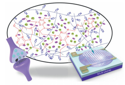
Figure 1.
Schematic of thermally cross-linked network structure which accommodates ions and facilitates ion migration (up), a biological synapse (left) and a correlated synaptic transistor (right).

Neuromorphic electronics have received considerable attention recent years for applications in brain-inspired computation systems and artificial sensorimotor nerves [1-3]. In these systems, artificial synapses are the basic structural and functional units; they emulate basic functions of computation, memory in a brain, and information filtering and delivery functions in the peripheral nervous system (PNS) [4]. Recently, great efforts have been made to design artificial synapses using phase-change materials [5-7], resistive switching materials [8-10], ferroelectric materials [11-13] and ionic materials [14-16]. Synaptic characteristics have also been achieved using floating gate [17-19] and ferroelectric mechanisms [20, 21].
To emulate natural synapses, neuromorphic electronics use synaptic transistors, which have a structure similar to a field-effect transistor, but operate by ion migration and injection in an ion-rich gate-insulator layer. In this structure, ion gels which usually consist of block copolymers and ionic liquids, are an important form that serve as a reservoir of ions [4, 22, 23]. In this structure, block copolymers usually form a physically cross-linked network to serve as a matrix to hold ionic liquids. In this case, the content of ionic liquid can be high, sometimes > 90% by weight. Application of an external electrical field drives migration of ions to form an electrical double layer near the ion gel/semiconductor interface, or injects the ions into the semiconductor, where they can be trapped for a long time to yield long-term plasticity [23-25].
Physical cross-links are usually weak, such as hydrogen bonds, and the formation of these 3D structures usually requires special structures of block copolymers [26]. The synthesis process of block copolymers is usually very strict [27-29], so they are too expensive for industrial production, and the modulation capacity of block copolymers is very limited [28], so they are not easily tuned for different designs and applications. Versatile design of synaptic transistor with easily-tuned properties requires solutions to these problems.
In this paper, we demonstrate a thermally cross-linked matrix for the formation of ion gels instead of traditional physical cross-linked ones. A synaptic transistor was then fabricated using the ion gel to emulate the synaptic cleft, and a thin film of SnO2 nanoparticles as the conductive channel. The device demonstrated important synaptic functions for neuromorphic computation and neural information transmission, such as paired-pulse facilitation, spike-voltage dependent plasticity, spike-rate dependent plasticity, and spike-number dependent plasticity. The device also achieved logic and high-pass filtering functions. This work provides a versatile strategy for fabrication of robust and versatile ion gels, which are applicable to future design and fabrication of neuromorphic electronic devices.
SnO2 precursor solution (100 µL) was spin-coated on the SiO2/Si substrate at 4000 rpm for 30 s, then annealed at 150 ℃ for 30 min. Gold source/drain electrodes were thermally deposited through a multi-branched shadow mask with the channel width and length of 48,400 µm and 100 µm, respectively.
A cross-linkable solution was prepared in a co-solvent (N, N-dimethylformamide: acetonitrile 1:1, v: v) with 5 wt% cyanoethylated pullulan (CEP) (Mw ~489,000, Shin Etsu Chemical Co.) and 1.5 wt% poly(methylated melamine-co-formaldehyde) (PMMF). The solutions were mixed with ionic liquid [EMIM]-[TFSI] (4 times of the weight of CEP), then cast onto the channel region of the synaptic transistor, which was then baked in a vacuum oven at 180 ℃ for 30 min to ensure efficient chemical reactions. The thickness of the ion gel layer is about 100 µm.
FT-IR spectra of solid films were recorded using a VERTEX70. Atomic Force Microscopy (AFM) images were obtained using a Bruker dimension icon microscope in a tapping mode. Electrical characteristics of the electronic devices were measured using a Keithley 4200 semiconductor analyzer in an N2 atmosphere.
The artificial synaptic device (Fig. 1, right) emulates a biological synapse (Fig. 1, left). A basic biological synaptic structure is a connection between two neurons, including several parts of the presynaptic membrane, the postsynaptic membrane, and the synaptic cleft. To prepare the corresponding artificial synapses, we used metal oxide nanoparticle thin film as a conductive channel on an insulative substrate, then evaporated source-drain electrodes through shadow masks. We used a metal probe as an electrode to supply electrical pulses to the ionic gels (Fig. 1, up).
To evaluate the quality of the SnO2 thin film, atomic force microscopy (AFM) images were captured using tapping mode (Fig. 2a). The top-view AFM image shows a dense film with no pinholes. The surface of the film was very smooth with a root-mean-squared roughness of 0.782 nm. Dense and smooth thin film is suitable to provide a high-quality interface with stacked functional layers on it. A high-quality interface is good for the elimination of defects and trap sites to improve conductivity of the channel regime.
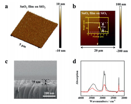
The AFM image near the edge of the thin film (Fig. 2b), revealed that the height difference between the substrate and the thin film was ~75 nm, which is thus the thickness of the film. Scanning electron microscopy (SEM) image of the cross-sectional view of the thin film confirmed the thickness to be about 75 nm (Fig. 2c).
FTIR spectra were of the ion gel were recorded before and after the thermal crosslinking process (Fig. 2d). Different absorption bands were recorded. The band between 3300 and 3600 cm−1 is assigned to polar functional groups, such OH. After curing, the peaks were significantly reduced and shifted to the higher wavenumbers. The reduction of the peaks implies reaction and reduction of OH groups, and the shift to higher wavenumbers is due to partial break of H-bonds. The absorption band between 2700 and 3000 cm−1 is assigned to C—H bonds. After curing, the peaks were reduced obviously; this change implies removal of side products of CH3OH during the cross-linking reaction. Thermal crosslinking is a facile process that has a wide selectivity of polymers and crosslinking agents, requiring only very easy thermal baking process. It complements with UV irradiation curing in real applications [30, 31].
The capacitance of the ion gel film as a function of frequency was tested using an Electrochemical Workstation (Fig. S1 in Supporting information). The capacitance of the film reached as high as 138.57 µF/cm2 at a low frequency (0.01 Hz). This attributed to sufficient polarization time for mobile ions under external electrical field. However, as the frequency increased, the capacitance of the gel film decreased.
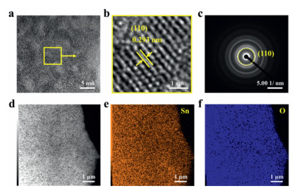
Transmission electron microscope (TEM) images was captured to study the nanostructures of the thin film. Nanocrystallites were observed (Fig. 3a), which showed average diameter of ~5 nm. The lattice structure (Fig. 3b) has a 0.293 nm spacing, which confirms the crystal structure of SnO2. A fast Fourier transform (FFT) image (Fig. 3c) shows clear (110) lattices. Energy Dispersive Spectrometer (EDS) images of the selected area (Fig. 3d) showed uniform Sn (Fig. 3e) and O (Fig. 3f) element distributions. Transfer curve of the electronic device was recorded (Fig. S2 in Supporting information).
In a biological synapse, action potentials are generated from a preneuron, then propagate from an axon to the presynaptic membrane. The action potentials can cause the release of neurotransmitters into the synaptic cleft. The neurotransmitters diffuse through the cleft, and bind to relevant receptors on the postsynaptic membrane, to open the relevant ionic channel to induce ionic flux that cause postsynaptic current (PSC). Correspondingly, we use a metal probe to deliver voltage spikes (5 V, 50 ms), which are applied to ion gel to cause ion migration similar to the release of neurotransmitters (Fig. 4a and Fig. S3 in Supporting information).
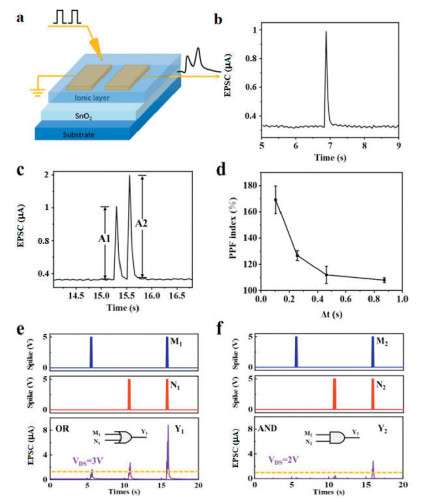
We applied positive spikes to repel cations to the surrounding channel region, and triggered an abrupt change in the current in the channel (Fig. 4b). The current then degraded abruptly, but retained a small current for a short time. This is very similar to that of the postsynaptic current in a biological synapse. This phenomenon underlies basic functions of neural network. After removal of the spike, most of the ions migrate back to the equilium position, whereas the few ions that had adsorbed to the semiconductor/ion gel interface would take longer to spontaneously drift back than the majority of ions due to energy barriers and ion traps at the interface; this delay causes existence of an extended channel current.
Paired-pulse facilitation (PPF) is an important form of short-term plasticity of homosynaptic facilitation in which the second of a pair of action potentials produces a larger EPSC as compared to the first [32]. In response to two successive action potentials (5 V, 50 ms), the second excitatory post-synaptic current (EPSC) peak following is stronger than the first peak. This phenomenon is important for many biological functions. Two successive presynaptic spikes were applied to the ion gel, and triggered two successive EPSC peaks (Fig. 4c); the second one was higher than the first one, as occurs in PPF in a biological synapse. In this process, the first spike induces a certain amount of ions to migrate to the surrounding channel region to cause the first EPSC peak. After the removal of the first spike, most ions diffuse back the equilibrated positions, whereas the few trapped at the interface take longer time than the majority to return. The second spike drives the migration of a similar amount of ions to the channel region, in addition to the remnant ions to induce a stronger effect on the EPSC.
The ratio of the current intensities of the current I2 generated by the second pulse to the current I1 generated by the first pulse is called the paired-pulse facilitation index, PPF index. PPF index decreased as the time interval between pulses increased (Fig. 4d). PPF index can be particularly relevant to temporal processing because the amplitudes of excitatory postsynaptic potentials provide temporal information about recent spike occurrence: These differences could be used to encode temporal information. Voltage spikes drive ions to the interface of ion gels and semiconductors, but after removal of external pulse, ions did not immediately drift back to the equilibrium distribution. The application of a second voltage spike could then induce an increased I2 due to the additional effects of remnant ions.
Using the synaptic device, basic logic functions include "OR" and "AND" were emulated. Herein, EPSC of 1 µA was set as the threshold to define output states. We realized the "OR" logic function under the V of 3 V (Fig. 4e). When the spike signals M1 and N1 were inputted into the postsynaptic membrane respectively, Whether M1 or N1 spike singles were utilized, EPSCs were greater than the threshold value, demonstrating the realization of the "OR" logic function. Different from the "OR" logic function, the "AND" logic function was implemented with the VDS of 2 V (Fig. 4f). Either M2 or N2 was inputted separately, the EPSCs were less than the threshold; only when M2 and N2 were inputted at the same time, the EPSC was greater than the threshold.
Synaptic strength, as measured by current, and can be sensitive to the number of pulses; this response is spike-number dependent plasticity (SNDP) (5 V, 50 ms, n = 1, 2, 3, …, 10) (Fig. 5a). Synaptic strength is also increased by increased voltage-pulse amplitude; this response is spike-voltage dependent plasticity (SVDP) (V = 0.5 V, 1 V, 1.5 V, …5 V, 50 ms) (Fig. 5b). In the central nervous system and peripheral nervous system, information is usually encoded, transmitted and recognized in the form of trains of spikes at different frequencies.

Spike-rate dependent plasticity (SRDP) is an important form of synaptic plasticity, because frequency information is more reliable and robust than voltage amplitudes. Therefore, emulation of frequency-dependent information processing in neural system is very important. We applied 10 identical presynaptic spikes at different frequencies (f = 3.20 Hz, 3.83 Hz, 4.78 Hz, 6.35 Hz, 9.43 Hz) to the synaptic transistor, which caused distinct output behaviors (Fig. 5c). EPSC peaks as triggered at different frequency was plotted (Fig. 5d and Fig. S4 in Supporting information). The dependence of EPSC on peak frequency was well fitted with the sigmoidal function [33, 34]:
|
|
(1) |
where p is the order of the function, fC is the cut-off frequency, a1 and a2 are the initial and final amplitude. The fitting gave rise to the value of ~7.4 Hz for fC. Biologically, synapses can act as a dynamic filter for the processing of the input information, depending on the frequency of the excitation. A synapse with a low probability of vesicle release can act as a high-pass filter, which only respond to high-frequency signals to trigger the neurotransmitter-vesicle release. Therefore, the synapse allows the signals with high frequency exceeding a certain cut-off value to pass through, and immensely weakens low frequency signals. This process is analogous to the high-pass filtering behavior in a biological synapse. The high-pass filtering behavior allows signals at higher frequency than a certain cut-off value to pass through, and blocks low-frequency signals.
To evaluate the short-term and long-term synaptic plasticity of the device, we applied a series of sequential spike to see the EPSC change (Fig. 5e). After only 1 spike, the EPSC was about 1 µA. When additional spikes are applied, the current level increases, and reaches 1400% of the original peak, illustrating good short-term plasticity, which is important for neuromorphic computation. However, after the removal of the spikes, the EPSC immediately drops to the initial current level. These special synaptic behaviors imply fast migration in the cross-linked network, in that the ions could migrate to equilibrated states. The short-term only based synaptic plasticity is nerve signal transmission. Without cross-linking agent, the device was not working properly, which is possibly caused by high electrical leakage of the thin film (Fig. S5 in Supporting information).
To further evaluate energy consumption of the devices, presynaptic spikes with much lower amplitude of 10 mV, 20 mV, 30 mV, 40 mV and 50 mV were applied, and caused obvious change in EPSC (Fig. S6 in Supporting information). After stimulation using different spike numbers, different accumulative EPSC were observed, demonstrating efficient tuning of synaptic plasticity. Recently, great progress has been made to reduce synaptic energy consumption [35]. According to the responsive signals, our device achieved a low-energy consumption of 0.35 fJ per synaptic event, which is below that of biological synapses.
In conclusion, we have fabricated a synaptic transistor using a thin film of SnO2 nanoparticles as the conductive channel and a thermally cross-linked ion gel as an ion-rich layer to emulate the synaptic cleft, where neurotransmitters were released. The device demonstrated essential synaptic functions for neuromorphic computation and neural information transmission, such as paired-pulse facilitation, spike-voltage dependent plasticity, spike-rate dependent plasticity, and spike-number dependent plasticity. Ultralow energy consumption was achieved below femtojoule, which is so far the lowest among the ion-gel gated synaptic transistors. Logic functions are also realized. Moreover, logic operations and high-pass filtering functions have been achieved using the device. This demonstration of ion gels composed of thermally crosslinked matrix represents a new strategy for fabrication of robust and versatile ion gels, and may provide a useful resource for design and modulation of information processing units in neuromorphic electronics.
There are no conflicts to declare.
We gratefully acknowledge the financial support from the National Natural Science Foundation of China (No. 21601076), the Natural Science Foundation of Liaoning Province (No. 2019-ZD-0266).
Supplementary material associated with this article can be found, in the online version, at doi:
Chinese Chemical Letters (CCL) (ISSN 1001-8417) was founded in July 1990. The journal publishes preliminary accounts in the whole field of chemistry, including inorganic chemistry, organic chemistry, analytical chemistry, physical chemistry, polymer chemistry, applied chemistry, etc., satisfying a real and urgent need for the dissemination of research results, especially hot topics. The journal does not accept articles previously published or scheduled to be published. To verify originality, your article may be checked by the originality detection service CrossCheck.
The types of manuscripts include Perspective, Highlight, Communication and Review. The experimental evidence necessary to support your manuscript should be supplied for the referees and eventual publication as Electronic Supplementary Information. The reviews are written by leading scientists within their field and summarized recent work from a personal perspective. They cover many exciting and innovative fields and are of general interest to all chemists.
The contents of papers are the sole responsibility of the authors, and publication shall not imply the concurrence of the Editors or Publisher.
Authors should submit their manuscripts via the online submission page of this journal at
The following items should be submitted via the online submission page:
1. Cover letter: highlighting the novelty, significance, and urgency of the submitted work, which merits rapid publication and providing details of other relevant information, e.g., submitted or in press manuscripts.
2. Graphical abstract for the contents list (submitted as a separate document).
3. Manuscript.
Authors are also suggested to supply the names of two suitable referees for their manuscript upon submission. Authors are also permitted to provide the names of scientists that they would prefer not to review their manuscript. If the names of non-preferred reviewers are provided, authors are requested to provide a brief note stating why these reviewers should not be used.
Upon acceptance of an article, authors will be asked to complete a 'Copyright Transfer Statement'. An e-mail will be sent to the corresponding author confirming receipt of the manuscript together with a 'Copyright Transfer Statement' form. Subscribers may reproduce tables of contents or prepare lists of articles including abstracts for internal circulation within their institutions. Permission of the Publisher is required for resale or distribution outside the institution and for all other derivative works, including compilations and translations. If excerpts from other copyrighted works are included, the author(s) must obtain written permission from the copyright owners and credit the source(s) in the article. Note that all authors should sign the Statement and publication of the accepted manuscript in an online issue is suspended until the Statement is received.
Graphical abstract is mandatory for our journal. It should summarize the contents of the article in a concise, pictorial form designed to capture the attention of a wide readership online. Authors must provide images that clearly represent the work described in the article. Graphical abstract, including title, authors, affiliations, the graph, and short abstract in one or two sentences, should be submitted as a separate file in the online submission system.
Text should be subdivided in the simplest possible way consistent with clarity. Ensure that all tables, figures, and schemes are cited in the text in numerical order. The preferred position for chemical structures should be indicated. Trade names should have an initial capital letter. All measurements and data should be given in SI units where possible, or in other internationally accepted units. Abbreviations should be used consistently throughout the text, and all non-standard abbreviations should be defined on first usage. Authors are requested to draw attention to hazardous materials or procedures by adding the word CAUTION followed by a brief descriptive phrase and literature references if appropriate.
Title. Concise and informative. Titles are often used in informationretrieval systems. Avoid abbreviations and formulae where possible.
Author names and affiliations. The authors' names are listed with the given name first and surname last (capitalizing the first letter, the best is use the spelling they are commonly using in scientific papers). Present the authors' affiliation addresses (where the actual work was done) below the names. Indicate all affiliations with a lower-case superscript letter immediately after the author's name and in front of the appropriate address. Provide the full postal address of each affiliation, including the country name.
Corresponding author. Clearly indicate who will handle correspondence at all stages of refereeing and publication, also post-publication. Ensure that the e-mail address is provided and contact details must be kept up to date by the corresponding author.
Changes to authorship. Authors are expected to consider carefully the list and order of authors before submitting their manuscript and provide the definitive list of authors at the time of the original submission. Any addition, deletion or rearrangement of author names in the authorship list should be made only before the manuscript has been accepted and only if approved by the journal Editor. To request such a change, the Editor must receive the following from the corresponding author: (a) the reason for the change in author list and (b) written confirmation (e-mail, letter) from all authors that they agree with the addition, removal or rearrangement. In the case of addition or removal of authors, this includes confirmation from the author being added or removed.
Abstract. Authors must include a short abstract of approximately that states briefly the purpose of the research, the principal results, and major conclusion(s).
Introduction. State the objectives of the work and provide an adequate background, avoiding a detailed literature survey or a summary of the results.
Experimental. Provide sufficient detail to allow the work to be reproduced. Methods already published should be indicated by a reference, only relevant modifications should be described. Other unimportant information can be deposited in Supporting information which will be available in online version.
Results and discussion. Results should be clear and concise. Discussion should explore the significance of the results of the work, not repeat them. A combined Results and Discussion section is often appropriate. Avoid extensive citations and discussion of published literature.
Conclusions. The main conclusions of the study may be presented in a short Conclusions paragraph, which may stand alone or form a subsection of a Discussion or Results and Discussion section.
Please note: All these sections need to be combined in a single untitled section and only hold essential information, others can be put in Supporting files. In general, communication should not more than four printed pages.
Artwork. Figures, schemes, and equations must be cited in the text and numbered in order of appearance with Arabic numerals. All graphics (including chemical structures) must be provided at the actual size that they are to appear (single-column width is 8.4 cm, double-column width is 17.7 cm). Please ensure that all illustrations within a paper are consistent in type, quality, and size. Legends should be included as part of the graphic. Color figures may be printed in the journal, provided that the editor considers the color necessary to convey scientific information. Ensure that all figures cited in reviews are permitted by the authority.
Tables. All tables should be cited in the text, and numbered in order of appearance with Arabic numerals. All table columns should have a brief explanatory heading and, where appropriate, units of measurement. Vertical lines should not be used. Footnotes to tables should be typed below the table and should be referred to by superscript letters. Each table should have a descriptive heading, which, together with the individual column headings, should make the table, as nearly as possible, self-explanatory. In setting up tabulations, authors are requested to keep in mind the column widths (8.4 cm and 17.7 cm), and to make the table conform to the limitations of these dimensions.
An acknowledgement section may be included. It should be placed after the manuscript text and before the references.
Please ensure that each reference cited in the text is also present in the reference list and indicate references by number(s) in square brackets in line with the text. Formatting for the common references is show below:
Reference to a journal publication
[1] J. van Geer, J.A.J. Hanraads, R.A. Lupton, et al., J. Sci. Commun. 163 (2010) 51-59.
[2] Z. Wu, Z. Zhen, J.H. Jiang, G.L. Shen, R.Q. Yu, J. Am. Chem. Soc. 131 (2009) 12325-12332.
Reference to a book:
[3] W. Strunk Jr., E.B. White, The Elements of Style, fourth ed., Longman, New York, 2000.
Reference to a chapter in an edited book:
[4] G.R. Mettam, L.B. Adams, How to prepare an electronic version of your article, in: B.S. Jones, R.Z. Smith (Eds.), Introduction to the Electronic Age, E-Publishing Inc., New York, 2009, pp. 281-304.
Citing and listing of Web references: As a minimum, the full URL should be given. Any further information, if known (author names, dates, reference to a source publication, etc.), should also be given. Web references should be included in the reference list.
This journal accepts electronic supplementary material to support and enhance your scientific research. Supplementary files offer the author additional possibilities to publish supporting applications, high-resolution images, background datasets, and more. This will depend on the nature of the research being reported. Please note that such items are published online exactly as they are submitted; there is no typesetting involved (supplementary data supplied as appear as such online). Supplementary files supplied will be published online alongside the electronic version of your article in Elsevier web products, including ScienceDirect.
It is the responsibility of the author(s) to provide the reviewers with the necessary information to evaluate the merit of the manuscript in terms of its scientific content. Failure to provide the necessary experimental evidence and data may result in the manuscript being withdrawn by the Editor.
Supplementary data must be saved in files separate from those for the manuscript and figures, and all file names must be supplied. Supplementary files should be referred to within the text of your manuscript in the same way as for figures or tables.
Proofs will be dispatched via e-mail and should be returned with corrections as quickly as possible, normally within 48 hours of receipt. Please check carefully before replying, as inclusion of any subsequent corrections cannot be guaranteed. Proofreading is solely your responsibility. Please use this proof only for checking the typesetting, editing, completeness and correctness of the text, tables and figures. Significant changes will only be considered at this stage with permission from the Editor. We will do everything possible to get your article published quickly and accurately. Note that we may proceed with the publication of your article if no response is received. The final article will then be published online as an Article in Press and any further amendments will not be incorporated.
According to relevant policies and international practice, Chinese Chemical Letters (CCL) charges publication fee 2000/paper for all manuscripts to be published.
B.J. Shastri, A.N. Tait, T.F. de Lima, et al., Nat. Photonics 15 (2021) 102–114. doi: 10.1038/s41566-020-00754-y
K. Roy, A. Jaiswal, P. Panda, Nature 575 (2019) 607–617. doi: 10.1038/s41586-019-1677-2
Y. Ni, J. Feng, J. Liu, et al., Adv. Sci. 9 (2022) 2102036. doi: 10.1002/advs.202102036
Y. Kim, A. Chortos, W. Xu, et al., Science 360 (2018) 998–1004. doi: 10.1126/science.aao0098
T. Tuma, A. Pantazi, M. Le Gallo, A. Sebastian, E. Eleftheriou, Nat. Nanotechnol. 11 (2016) 693–699. doi: 10.1038/nnano.2016.70
H. Bian, Y.Y. Goh, Y. Liu, et al., Adv. Mater. 33 (2021) 2006469. doi: 10.1002/adma.202006469
A. Sebastian, M. Le Gallo, R. Khaddam-Aljameh, E. Eleftheriou, Nat. Nanotechnol. 15 (2020) 529–544. doi: 10.1038/s41565-020-0655-z
Z. Lv, Y. Wang, J. Chen, et al., Chem. Rev. 120 (2020) 3941–4006. doi: 10.1021/acs.chemrev.9b00730
Y. Shi, X. Liang, B. Yuan, et al., Nat. Electron. 1 (2018) 458–465. doi: 10.1038/s41928-018-0118-9
S. Yu, B. Gao, Z. Fang, et al., Adv. Mater. 25 (2013) 1774–1779. doi: 10.1002/adma.201203680
D.M. Evans, T.S. Holstad, A.B. Mosberg, et al., Nat. Mater. 19 (2020) 1195–1200. doi: 10.1038/s41563-020-0765-x
Y. Kim, K. Lee, J. Lee, et al., ACS Nano 15 (2021) 20116–20126. doi: 10.1021/acsnano.1c08005
J. Gao, Y. Zheng, W. Yu, et al., SmartMat 2 (2021) 88–98. doi: 10.1002/smm2.1020
B. Jeong, P. Gkoupidenis, K. Asadi, Adv. Mater. 33 (2021) 2104034. doi: 10.1002/adma.202104034
C.J. Wan, L.Q. Zhu, J.M. Zhou, Y. Shi, Q. Wan, Nanoscale 5 (2013) 10194–10199. doi: 10.1039/c3nr02987e
L. Zhou, S. Yang, G. Ding, et al., Nano Energy 58 (2019) 293–303. doi: 10.1016/j.nanoen.2019.01.045
Y. Ren, J.Q. Yang, L. Zhou, et al., Adv. Funct. Mater. 28 (2018) 1805599. doi: 10.1002/adfm.201805599
W. Li, F. Guo, H. Ling, et al., Adv. Sci. 4 (2017) 1700007. doi: 10.1002/advs.201700007
L. Xiang, J. Ying, J. Han, L. Zhang, W. Wang, Appl. Phys. Lett. 108 (2016) 173301. doi: 10.1063/1.4947576
B. Tian, L. Liu, M. Yan, et al., Adv. Electron. Mater. 5 (2019) 1800600. doi: 10.1002/aelm.201800600
Y. Choi, J.H. Kim, C. Qian, et al., ACS Appl. Mater. Interfaces 12 (2020) 4707–4714. doi: 10.1021/acsami.9b17742
J. Zhu, T. Zhang, Y. Yang, R. Huang, Appl. Phys. Rev. 7 (2020) 011312. doi: 10.1063/1.5118217
C. Wan, P. Cai, X. Guo, et al., Nat. Commun. 11 (2020) 4602. doi: 10.1038/s41467-020-18375-y
D. Feng, Z. Niu, J. Yang, et al., Nano Energy 90 (2021) 106526. doi: 10.1016/j.nanoen.2021.106526
W. Xu, S.Y. Min, H. Hwang, T.W. Lee, Sci. Adv. 2 (2016) e1501326. doi: 10.1126/sciadv.1501326
S.K. Yang, A.V. Ambade, M. Weck, Chem. Soc. Rev. 40 (2011) 129–137. doi: 10.1039/C0CS00073F
F.H. Schacher, P.A. Rupar, I. Manners, Angew. Chem. Int. Ed. 51 (2012) 7898–7921. doi: 10.1002/anie.201200310
N.A. Cortez-Lemus, A. Licea-Claverie, Prog. Polym. Sci. 53 (2016) 1–51. doi: 10.1016/j.progpolymsci.2015.08.001
Y. Xia, G.M. Scheutz, C.P. Easterling, J. Zhao, B.S. Sumerlin, Angew. Chem. Int. Ed. 60 (2021) 18537–18541. doi: 10.1002/anie.202106418
D. Liu, Q. Shi, S. Dai, J. Huang, Small 16 (2020) 1907472. doi: 10.1002/smll.201907472
D.G. Roe, S. Kim, Y.Y. Choi, et al., Adv. Mater. 33 (2021) 2007782. doi: 10.1002/adma.202007782
D.V. Buonomano, M.M. Merzenich, Adv. Psychol. 121 (1997) 129–139.
W. Huang, P. Hang, Y. Wang, et al., Nano Energy 73 (2020) 104790. doi: 10.1016/j.nanoen.2020.104790
Z. Hao, H. Wang, S. Jiang, et al., Adv. Sci. 9 (2022) 2103494. doi: 10.1002/advs.202103494
G. Liu, Q. Li, W. Shi, et al., Adv. Funct. Mater. 32 (2022) 2200959. doi: 10.1002/adfm.202200959

Figure 1 Schematic of thermally cross-linked network structure which accommodates ions and facilitates ion migration (up), a biological synapse (left) and a correlated synaptic transistor (right).

Figure 2 AFM images of (a) SnO2 thin film surface and (b) on the edge of SnO2 thin film and the cross-sectional view of the thin film. (c) SEM image of the cross section of the SnO2 thin film. (d) FT-IR of CEP/Melamine before (black) and after (red) curing.

Figure 3 TEM images of (a) SnO2 thin film, (b) a high-resolution magnified image, and (c) FFT image. EDS images as obtained from (d) a selected area for the elements (e) Sn and (f) O.

Figure 4 (a) Schematic of the synaptic transistor. (b) Excitatory postsynaptic current (EPSC). (c) Paired-pulse facilitation. Excitatory postsynaptic current as triggered by two successive presynaptic spikes. (d) PPF index as a function of time interval between spikes. The logic functions including (e) OR and (f) AND.

Figure 5 (a) Spike-number dependent plasticity. (b) Spike-voltage dependent plasticity. (c) Spike-rate dependent plasticity: Excitatory postsynaptic current in response to different frequency of spikes. (d) EPSC as a function of input spike frequency. When the frequency is lower than the critical frequency (fC), there is no obvious change in EPSC; when the frequency is higher than fC, significant change in EPSC occurs, in consistence with high-pass filtering behavior in biological synapses. (e) EPSC in response to 100 presynaptic spikes.

 扫一扫看文章

扫一扫关注我们
 DownLoad:
 下载:
下载:
 下载:
下载: