Citation:
Jiang-Zhen Qiu, Ye Yu, Zhuo-Fan Chen, Min Zhu, Yongxia Lu, Qinwu Wu, Long-Fei Wang, Jiang Liu. Inclusion of methylviologen dication in a cadmium tetracyanoplatinate host clathrate showing photochromism and photoluminescence modulation[J]. Chinese Chemical Letters,
2023, 34(3): 107346.
doi:
10.1016/j.cclet.2022.03.069
Inclusion of methylviologen dication in a cadmium tetracyanoplatinate host clathrate showing photochromism and photoluminescence modulation
English
Inclusion of methylviologen dication in a cadmium tetracyanoplatinate host clathrate showing photochromism and photoluminescence modulation
Received Date:
09 February 2022 Accepted Date:
15 March 2022 Revised Date:
07 March 2022 Available Online:
15 March 2023
Abstract:
A cadmium tetracyanoplatinate host clathrate, (MV)[Cd2{Pt(CN)4}3]⋅2(H2O) (1), including a methylviologen dication (MV2+) was synthesized, and the crystal structures, photochromic and photoluminescence properties were investigated. In 1, the alternatively parallel stacking between the MV2+ dications as electron acceptors in the channels and the electron donors [Pt1(CN)4]2– units in the host frameworks give a unique donor-acceptor (DA) system. Under UV irradiation, the electron transfer between MV2+ and [Pt(CN)4]2– ions generates MV·+ radicals with a photochromic behavior from pale-yellow to blue. This process occurs through single-crystal-to-single-crystal (SCSC) transformation and obvious structure variation of viologen cations is successfully observed. Moreover, the spectral overlap between the emission bands of 1 and the absorption around 623 nm for the MV·+ radicals leads to a modulation of the photoluminescence.
Cyanide-containing coordination frameworks have been attracting increasing attention in the exploration of advanced functional materials owing to their intriguing architectures [1-4] and topologies as well as a variety of important functional properties. Examples include the high Tc in Prussian blue analogs [5, 6], photoluminescence in lanthanides polycyanidometallates compounds [7-9], colossal positive and negative thermal expansion in Ag3[Co(CN)6] [10], heat-, light-, and pressure-induced spin-crossover in Hofmann-type framework [11, 12], and so on. Owning to a wide selection of polycyanidometallates, e.g., [MI(CN)2]− (M = Cu, Ag, Au), [MⅡ(CN)4]2− (M = Ni, Pd, Pt), [MⅡ/Ⅲ(CN)6]4-/3− (M = Fe, Co, Ru, Os, Pt), [Mo(CN)7]4−, and [MⅣ/Ⅴ(CN)8]4-/3− (M = Mo, W, Nb), as building blocks showing various connection abilities, a various neutral or charged frameworks with different topologies, such as α-Po [13-16], diamondoid [17-19], quartz [20], PtS [21-23], could be obtained depending on the geometry of the nodes (metal centers) and/or the flexibility of the connectors (cyanometallates). Among the numerous polycyanidometallates based coordination frameworks, another interesting feature for some of these materials is the ability to form clathrates to include neutral and/or cationic molecules [24-27] depending on the porosity and charge of the structure, giving the possibility to control the functional properties of the host through guest exchange [28] or introduce additional unique properties [26, 29, 30]. For example, the famous Fe(Ⅱ)-based Hofmann-type frameworks show guest-modulated spin-crossover behavior [12].
Methylviologen (MV2+) dication and its derivates are well-known as electron acceptors that show a wide application in the field of nonlinear optics [31], electrochemistry [32], photochemistry [33], and conductivity [34]. In recent years, ionic MV derivates have been widely incorporated into crystalline open frameworks, such as open inorganic frameworks [35-40] or metal-organic frameworks (MOFs) [41-44] to fabricate novel photochromic materials with unusual properties and understand the charge transfer mechanism. Among them, polycyano–polycadmate complexes, which show remarkable structural variations and inclusion ability for small molecules, was successfully demonstrated by Nishikiori et al. to be rational host to accommodate MV2+ dications [26, 29, 30]. Some of such viologen polycyano-polycadmate materials show photochromic behavior, however, no crystal structure after irradiation was studied due to the decomposition. Moreover, as far as we know, the studies on constructing MV derivates decorated cyanide-containing coordination frameworks were rarely reported [45].
Inspired by the fascinating properties of MV2+ dications and the inclusion ability of polycyanidometallates based coordination frameworks, herein we develop a new MV decorated polycyanidometallate clathrate (MV)[Cd2{Pt(CN)4}3]⋅2(H2O) (1). For 1, it is found the MV2+ dications are loaded in the anionic one-dimensional (1D) channels of the host frameworks [Cd2{Pt(CN)4}3]2−. Moreover, the interactions between the MV2+ guest and the [Pt(CN)4]2− ions from the host lead to a unique donor-acceptor (DA) system. Under UV irradiation, the electron transfer between MV2+ and [Pt(CN)4]2− generates the MV·+ radicals accompanied by a color change from pale-yellow to blue. Moreover, the photochromic behavior of 1 proceeds through single-crystal-to-single-crystal (SCSC) transformation, and the structural variation of MV before and after UV irradiation is discussed in detail. Additionally, a switching of the photoluminescence is observed for 1 during the photochromic process.
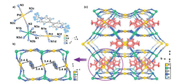
The slow diffusion method of CdCl2 and MV[Pt(CN)4] (MV2+ = N, N'-dimethyl-4,4′-bipyridinium dication) in H2O affords pale yellow block crystals of complex 1 (see experimental section in Supporting information). The purity of the polycrystalline sample of 1 was determined by Powder X-ray diffraction (PXRD) (Fig. S1 in Supporting information) and elemental analysis (EA). The single-crystal X-ray diffraction (SCXRD) of 1 at 293 K and 150 K revealed that complex 1 crystallizes in the monoclinic C2/m space group (Table S1 in Supporting information). Due to the same structure at both temperatures, the structure for 1 at 150 K is only discussed. In the structure of 1, only one crystallographically unique Cd(Ⅱ) atom (labeled as Cd1) is found (Fig. 1a) and located at an inversion center. The Cd1 atom is coordinated by six N atoms which come from the square-planar [Pt(CN)4]2− anions, adopting a pseudo-octahedral [CdN6] coordination sphere. Along the bc plane of the structure, each [Pt1(CN)4]2− anion, which equatorially coordinates to the Cd1 atom, bridges four Cd1 atoms to give an extended wavelike layer. Then, the other [Pt2(CN)4]2− anions as pillars axially connected the Cd1 atoms, resulting in a three-dimensional (3D) negative framework [Cd2{Pt(CN)4}3]2− (Fig. S2a in Supporting information) showing two crossover one-dimensional (1D) channels of 6.5 × 7.3 Å and 6.3 × 10.1 Å along the respective b and c axis of the structure. A TOPOS analysis [46] of the host framework defined a (4,4,6)-linked fsy network (point symbol (44·62)2(42·84)(48·62·85)2) (Fig. S2b in Supporting information) once the Cd(Ⅱ) atoms and [Pt(CN)4]2− moieties are regarded as 6-connected nodes and 4-connected linkers, respectively. It should be noted the host framework for 1 shows the same structural topology with the reported complex [Fe2{Pt(CN)4}3] [47]. However, for the compound [Fe2{Pt(CN)4}3], the selection of positive trivalent Fe(Ⅲ) atoms results in a neutral framework while the positive divalent Cd(Ⅱ) atoms in 1 give the negatively charged host framework. Therefore, for maintaining the charge balance, the MV2+ counterions are encapsulated in the channels of the host framework for 1 (Fig. 1c and Fig. S3 in Supporting information). Due to the confined effect of the channel, no structural disorder is observed for MV2+ cations. Moreover, the two pyridinium rings of MV2+ cation is coplanar and adopt the orientation almost parallel with the [Pt1(CN)4]2− moieties, resulting in the presence of host-guest stacks that contain the sequence MV2+[Pt1(CN)4]2−MV2+. The separation distance between MV2+ and [Pt1(CN)4]2− moieties is around 3.4 Å (Fig. 1b). Additionally, according to the elemental analysis, two H2O molecules should exist in channels of the host framework. Actually, some void cavities, which can confine the solvent guests, are observed in the 1D channels along the c axis of the host framework and are separated by the methyl groups of MV2+ cations (Fig. S4 in Supporting information). However, the disorder of water molecules hinders further precise positioning through structural refinement. Further thermo-gravimetric (TG) measurement shows 1 starts to decompose at 435 ℃ (Fig. S5 in Supporting information), showing high thermal stability.
Figure 1
Figure 1.
(a) A view showing the coordination environment of Cd(Ⅱ) ions and the guest MV2+ ions in 1 at 150 K. Symmetry code: (a) (1/2-x, 1/2+y, -z), (b) (1/2-x, 3/2-y, 1-z), (c) (x, 1-y, z), (d) (x, 1-y, 1+z), (e) (1-x, y, 1-z), (f) (1-x, 2-y, 1-z); (b) A side view of the 3D framework and the loaded MV2+ ions in 1; (c) The encapsuled MV2+ ions in the channel of 1, showing the sequence MV2+[Pt1(CN)4]2−MV2+ packing arrangement, the distance between Pt(Ⅱ) ions and MV2+ ions is calculated as 3.4 Å.
Upon irradiation by a UV led lamp (γ = 365 nm, P = 10 W), the crystalline sample of compound 1 shows an obvious color change from pale-yellow to blue and then undergo gradually decoloration in the dark, indicating a reversible photochromic behavior (Fig. 2a and Fig. S6 in Supporting information). Further UV-vis spectra measurements (Fig. S7 in Supporting information) of 1 reveal that two absorption bands, which center at 401 nm (sharp) and 623 nm (wide), become stronger as the lengthening of UV irradiation time and approach saturation within 10 min (Fig. 2b). Such spectrum changes are in accordance with the existence of MV·+ radicals which are generated from the reduction of MV2+ cations in 1. Additionally, under dark, it is found the absorption band belonging to the MV·+ radicals drops within 14 h, revealing a recovery of MV2+ cations (Fig. 2c). Such reversible photochromic process for 1 could be repeated for more than 5 times, revealing a higher repeatability and photostability than the reported MV2+-G polycyano–polycadmate (PCPC) clathrates, which become insensitive to UV irradiation due to structural decompose after several repetitions of the UV irradiation and discoloration [29]. Moreover, the PXRD pattern for the bleaching sample (1-Bleach) is the same with 1 and irradiated sample 1-UV, indicating the structure maintains intact during the reversible photochromic process (Fig. S1).
Figure 2
Figure 2.
(a) A view showing the color changes between pale-yellow and blue for the crystal samples of 1 through continuous UV irradiation and relaxation under dark. (b) The change of solid state UV-vis spectra in the range of 350–800 nm for 1 under UV irradiation within 20 min. (c) The change of solid state UV-vis spectra in the range of 350–800 nm for 1-UV under dark within 10 h. (d) The ESR spectrum of 1 before (blue solid line) and after UV irradiation (red solid line).
In order to determine the occurrence of MV·+ radicals, EPR measurements were performed at room temperature for the polycrystalline samples of 1 during the photochromic process. For the as-synthesized 1, no EPR signal is detected. After UV irradiation, a noticeable signal at g = 2.01 is observed (Fig. 2d), confirming the reduction of MV2+ ions to MV·+ radicals upon UV irradiation. Moreover, the MV·+ radicals are also observed in the precursor compound (MV)[Pt(CN)4] with an EPR signal at g = 2.01 (Fig. S8 in Supporting information) upon 365 nm UV irradiation. Therefore, considering the similar alternatively stacked MV2+[Pt1(CN)4]2-MV2+ packing arrangement in both 1 and (MV)[Pt(CN)4], the generation of MV·+ could be attributed to the electron transfer between MV2+ and [Pt1(CN)4]2− ions. The single-crystal property of 1 remained intact after UV irradiation, giving us a chance to compare the structural information changes before and after UV-induced photochromism.
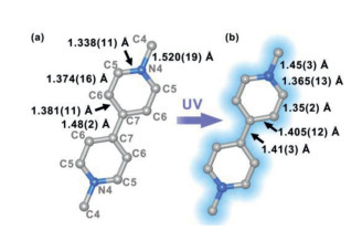
The crystallographic data (Table S1) at 150 K revealed that the irradiated sample 1-UV belongs to the same monoclinic C2/m space group as 1, while the unit cell parameters undergo non-negligible variation, suggesting the occurrence of structural distortion during the generation of MV·+ radicals. Moreover, it is well known the redox states of viologen entities are correlated to the bond length around the heterocyclic moiety. Compared to the central single bond (dCγ-Cγ = 1.48 Å) of MV2+ dication, the central bond in the di-reduced form MV0 can be considered as a polyene-like structure and give a shorter central double bond (dCγ-Cγ = 1.36 Å), therefore the central bond is expected to be intermediate for MV2+ radical cations [48]. Fig. 3 shows the MV structure with labeled C–N and C–C bond lengths in 1 and 1-UV. For 1, the MV ion is clearly an MV2+ dication due to the double bond nature of the central C7–C7′ bond with the distance of 1.48(2) Å. After the 1→1-UV conversion, although the twist angle of the two aromatic rings remains invariant, it is found the central C7–C7′ bond distance is obviously shortened from 1.48(2) Å to 1.41(3) Å, in line with the change of a radical-delocalized bond. Simultaneously, the shortening of the C5-C6 (1.374(11) Å→1.35(2) Å) bond and lengthening of C6-C7 (1.381(11) Å→1.4055(12) Å) and N4-C5 (1.338(11) Å→1.365(13) Å) bonds in the pyridine rings are observed and lead to the structure which is close to some observed viologen radical compounds such as (MV·+)(BF4) [49], confirming the occurrence of MV·+ in 1-UV.
Figure 3
Figure 3.
The bond length changes for the MV ions in 1 before (a) and after (b) UV irradiation.
Additionally, the generation of MV·+ radicals is also accompanied by the host structural adjustment (Table S2 in Supporting information). For the electron donors [Pt(CN)4]2− in the host, it is found the Pt-C bonds length increase while the C≡N bonds undergo a shortening (Table S2). Compared to [Pt2(CN)4]2, such bond length changes are more obvious for [Pt1(CN)4]2 ions. Moreover, considering the reported charge transfer between [C≡N]− ions and MV2+ cations in some polycyanidometallates-based frameworks [26, 45], we further analyzed the distance changes between MV2+ guest and the [C≡N]− before and after UV irradiation. Between [Pt1(CN)4]2 ions and MV2+, the shortest N(MV2+)–N1(CN) distances change from 3.883(6) Å to 3.881(7) Å after irradiation. Between [Pt2(CN)4]2 ions and MV2+, the shortest N(MV2+)–N3(CN) distances are 3.978(7) Å before irradiation and 3.953(8) Å after irradiation. Therefore, considering the shorter N(MV2+)–N1(CN) distance, a faster electron transfer rate between MV2+ and [Pt1(CN)4]2− ions should be expected.
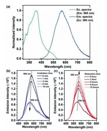
Beside the photochromic behavior, the photoluminescence property of 1 was further studied. According to the previous photoluminescence study on (MV)[Pt(CN)4] by Shiota and Matsushita [50], the columnar structure with the alternatively stacked anionic complex [Pt(CN)4]2– and cationic MV2+ will give intermolecular charge transfer between [Pt(CN)4]2– and MV2+, thus exhibiting a strong intense polarized green emission. For 1, the excitation peak of the polycrystalline samples at 360 nm produces a broad emission with the maximum intensity at 565 nm (Fig. 4a). Compared to the maximum emission intensity at 535 nm for (MV)[Pt(CN)4] (Fig. S9 in Supporting information), a slight redshift of the emission spectra for 1 is observed and may be ascribed to the slight structural difference (e.g., Intermolecular distance) of the parallel packed MV2+[Pt(CN)4]2-MV2+ columnar structure between 1 and (MV)[Pt(CN)4].
Figure 4
Figure 4.
(a) The excitation and emission spectra of 1 in the solid state recorded at room temperature. (b) Photoluminescence spectral variations (Ex: 360 nm) of compound 1 upon UV light irradiation in the air. (c) Photoluminescence spectral variations (Ex: 360 nm) of 1-UV under dark.
Interestingly, when the polycrystalline samples of 1 are irradiated under a UV lamp (λ = 365 nm) for 10 min, emission with a color change from yellow to olivine could be captured with the naked eyes (Fig. S10 in Supporting information). A more detailed photoluminescence spectral study shows that the luminescent intensity of 1 decrease rapidly as the irradiation time increase, and reduce to about 30% of the original intensity with a maximum emission at 550 nm when the irradiation time is 10 min (Fig. 4b). Considering the generation of MV·+ radicals during UV irradiation, such behavior should be attributed to the good spectral overlap between the emission bands of 1 and the broad absorption bands around 623 nm for the colored MV·+ radical units [51]. In fact, under dark, a gradual recovering of the emission spectra for 1 is observed within 12 h, in consistent with the decoloration process of 1-UV revealed by UV-vis spectra (Fig. 4c), demonstrating a switching of photoluminescence during the 1↔1-UV conversion. Moreover, due to the high photostability of the structure, the switching of photoluminescence upon 1↔1-UV conversion could be successfully repeated for at least 3 repetitions (Fig. S11a in Supporting information) and realize a controlling of the emission intensity at 565 nm (Fig. S11b in Supporting information).
In conclusion, we develop a unique cadmium tetracyanoplatinate host clathrate, (MV)[Cd2{Pt(CN)4}3]⋅2(H2O) (1), which is loaded with methylviologen dication in the 1D channel. Under UV irradiation, the electron transfer between MV2+ and the [Pt(CN)4]2− generates MV·+ radicals accompanied by the modulation of the photoluminescence and the reversible photochromism behavior between pale-yellow for 1 and blue for the irradiated product 1-UV. Moreover, in contrast to the previously reported structural decomposition for the viologen polycyano–polycadmate materials during the photochromism process, the single-crystal for 1 keep intact during photochromism, showing a higher photostability. Our result develop new photochromic polycyanidometallates based materials through the introduction of viologen ions, showing the potential application in sensors and switches.
Declaration of competing interest
The authors declare no competing financial interest.
Acknowledgment
We gratefully acknowledge National Natural Science Foundation (No. 21801258) for financial support.
Supplementary materials
Supplementary material associated with this article can be found, in the online version, at doi:10.1016/j.cclet.2022.03.069.
Figure 1
(a) A view showing the coordination environment of Cd(Ⅱ) ions and the guest MV2+ ions in 1 at 150 K. Symmetry code: (a) (1/2-x, 1/2+y, -z), (b) (1/2-x, 3/2-y, 1-z), (c) (x, 1-y, z), (d) (x, 1-y, 1+z), (e) (1-x, y, 1-z), (f) (1-x, 2-y, 1-z); (b) A side view of the 3D framework and the loaded MV2+ ions in 1; (c) The encapsuled MV2+ ions in the channel of 1, showing the sequence MV2+[Pt1(CN)4]2−MV2+ packing arrangement, the distance between Pt(Ⅱ) ions and MV2+ ions is calculated as 3.4 Å.
Figure 2
(a) A view showing the color changes between pale-yellow and blue for the crystal samples of 1 through continuous UV irradiation and relaxation under dark. (b) The change of solid state UV-vis spectra in the range of 350–800 nm for 1 under UV irradiation within 20 min. (c) The change of solid state UV-vis spectra in the range of 350–800 nm for 1-UV under dark within 10 h. (d) The ESR spectrum of 1 before (blue solid line) and after UV irradiation (red solid line).
Figure 4
(a) The excitation and emission spectra of 1 in the solid state recorded at room temperature. (b) Photoluminescence spectral variations (Ex: 360 nm) of compound 1 upon UV light irradiation in the air. (c) Photoluminescence spectral variations (Ex: 360 nm) of 1-UV under dark.

 DownLoad:


 下载:
下载:


 下载:
下载:
