
Figure 1.
Chemical structures of 1, 1a and 2.
Arnequinol A and arnequinone A, two unique meroterpenoids from Arnebia euchroma
Hai-Wei Yan , Rong-Rong Du , Xu Zhang , Ya-Nan Yang , Xiang Yuan , Zi-Ming Feng , Jian-Shuang Jiang , Pei-Cheng Zhang

The Boraginaceae family, composed of about 130 genera, encompasses about 2700 species, which are distributed in tropical, sub-tropical, and warmer regions world-wide [1, 2]. Extensive phytochemical investigations have revealed that some genera of this family, especially Cordia [3-5], Arnebia [6-8] and Auxemma [9, 10], presented an abundant source of structurally diverse and biologically active meroterpenoids, consisting of a quinone or hydroquinone linked to a monoterpene motif. The linkage patterns and positions of these two units, and the intramolecular cyclization and rearrangement reactions of the monoterpene moiety provide great potential to the structural diversity of meroterpenoids from this family [4, 9, 11-13]. To our knowledge, the benzogeijerene skeleton formed by the Cope rearrangement of monoterpene moiety was exclusively found in the Boraginaceae plants [3, 14, 15].
Arnebia euchroma (Royle) Johnst. belonging to the family Boraginaceae, is a commonly prescribed traditional herbal medicine to treat skin eruption and ulcers, smallpox, jaundice, burn wounds, eczema, and constipation [16]. Up to now, there have been about 40 meroterpenoids reported from A. euchroma, of which some compounds displayed potent antiviral, anti-microbial, and cytotoxic activities [13, 17-20]. During our further endeavor to discover novel and bioactive meroterpenoids, arnequinol A (1) and arnequinone A (2) were isolated from the roots of A. euchroma. Compound 1 possesses a new 6/6/3 tricyclic carbon skeleton fused with a heptatomic oxo-bridge. And compound 2 bears a novel highly conjugated aromatic system after the 1, 2-migration of its tertiary methyl and elimination reaction (Fig. 1). This study describes the isolation, structure elucidation, and proposed biosynthetic pathways of 1 and 2. The in vitro cytotoxic activity of 1, 1a (the oxidation product of the aglycone of 1) and 2, as well as neuroprotective activity of 1 and 2 were also evaluated.
Arnequinol A (1) was obtained as a white amorphous powder. Its molecule formula C23H28O11 with ten indices of hydrogen deficiency was deduced from the high resolution electrospray ionization mass spectroscopy (HRESIMS) sodium adduct ion peak at m/z 503.1521 (calcd. for C23H28O11Na, 503.1524). The infrared (IR) absorptions suggested the existence of hydroxyl (3378 cm−1), carbonyl (1698 cm−1), and phenyl (1604, 1505 and 1459 cm−1) functionalities. The 1H nuclear magnetic resonance (NMR) data (Table 1) showed signals for a pentasubstituted benzene ring proton (δH 6.61, s), a monosubstituted vinyl [δH 6.12 (dd, J = 17.5, 11.0 Hz), 5.42 (d, J = 17.5 Hz), and 5.31 (d, J = 11.0 Hz)], an oxymethine proton (δH 5.46, s), an anomeric proton [δH 4.82 (d, J = 7.5 Hz)], a hydroxymethyl [δH 3.94 (d, J = 12.0 Hz) and 3.89 (d, J = 12.0 Hz)], a methoxy (δH 3.86, s), two mutual coupling sp3 methine protons [δH 3.57 (d, J = 7.5 Hz) and 2.44 (d, J = 7.5 Hz)], and a tertiary methyl group (δH 0.88, s). Additionally, four oxymethine resonances (δH 3.31−3.62, m) and an oxymethylene group (δH 3.77, br s) attributable to a glucosyl moiety were observed. The 13C NMR associated with the heteronuclear single quantum coherence (HSQC) spectra assigned 23 carbon signals to an ester carbonyl (δC 176.5), a monosubstituted double bond (δC 144.3 and 118.1), a phenyl group (δC 156.0, 153.7, 139.4, 131.7, 113.2 and 102.2), a glucosyl unit (δC 106.9, 78.8, 78.7, 76.5, 72.0 and 63.2), three sp3 methines involving one oxygenated (δC 78.6, 32.2 and 28.6), a hydroxymethyl (δC 66.7), a methoxy (δC 58.8), a methyl (δC 23.8) and two sp3 nonprotonated carbons (δC 41.0 and 37.8). Since a carbonyl, a phenyl, a double bond, and a glucosyl accounted for seven of the ten indices of hydrogen deficiency, the remaining ones required another three rings for 1.
 DownLoad:
CSV
DownLoad:
CSV

|
Interpretation of the 1H-1H COSY spectrum (Fig. 2) indicated two key spin systems, H-5/H-6 and H-10/H2-11. The heteronuclear multiple bond correlation (HMBC) (Fig. 2) correlations from H-2 to C-1, C-3, C-4 and C-8a evidenced the presence of a pentasubstituted benzene ring. The HMBC correlations from H-5 to C-4, C-4a and C-8a, and from H-8 to C-1, C-4a and C-8a established the linkages of C-5 to C-4a and C-8 to C-8a. Meanwhile, MeO-15 and the glucosyl were positioned at C-3 and C-4, respectively, in the light of the HMBC correlations from MeO-15 and H-1′ to the corresponding carbons. Further HMBCs of H-8 with C-6, H-6 with C-8, H-5 with C-7, CH3-9 with C-6, C-7, C-8 and C-10, and H2-11 with C-7 constructed a six-membered carbon ring fused with the benzene ring at C-4a/C-8a and located the monosubstituted double bond and Me-9 at C-7. Moreover, a cyclopropane ring fused to the six-membered carbon ring at C-5 and C-6 was evidenced from the HMBCs of H2-13/C-5, C-6, and C-12, H-5/C-12 and C-13, and H-6/C-5 and C-13. The last degree of unsaturation was furnished with an ester bridge between C-8 and C-12 based on the HMBC correlations from H-5, H-6, H-8, and H2-13 to C-14. Therefore, the planar structure of 1 was unequivocally established (Fig. 1), featuring a unique 6/7/6/3 tetracyclic system.

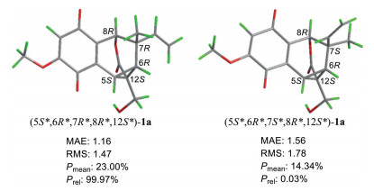
The β-anomeric configuration of the glucosyl moiety was defined by the coupling constant [δH 4.82 (d, J = 7.5 Hz)] of the anomeric proton, and then the d-glucose was ascertained by gas chromatography (GC) analysis [21] (Fig. S2 in Supporting information) after the snailase hydrolysis of 1. Interestingly, during the hydrolysis process, the aglycone of 1 was autoxidized [22] to its quinone form 1a (for NMR data, see Table 1). The relative configuration of 1a was deduced via the rotating frame overhauser spectroscopy (ROESY) experiment, conformation analysis, and 13C NMR calculation. The ROESY correlations (Fig. 2) of H-5/H2-13, H-6/H2-13, and H-5/H-6 revealed that H-5, H-6, and CH2-13 were positioned at the same face of the cyclopropane ring. Given the rigidity of the cyclopropane ring and the ester bridge, H-5, H-6, and H-8 must be all in the equatorial orientation (Fig. 2 and Fig. S1 in Supporting information), resulting that the relative configuration at C-7 cannot be resolved though the ROESY correlations of both H-6 and H-8 with Me-9 exist. Hence there are two possible relative configurations for 1a [(5S*, 6R*, 7R*, 8R*, 12S*)-1a and (5S*, 6R*, 7S*, 8R*, 12S*)-1a, Fig. 3]. The theoretical calculations of 13C NMR chemical shifts of the above two possible isomers were conducted using the GIAO method (Supporting information), following a reported STS protocol [23], which corrected systematic errors according to the types of carbon. As shown in Fig. 3 and Fig. S4 (Supporting information), compared with the experimental data of 1a, (5S*, 6R*, 7R*, 8R*, 12S*)-1a exhibited smaller mean absolute error (MAE) and root mean square (RMS) values with a Prel probability of 99.97%, defining the relative configuration of 1a as 5S*, 6R*, 7R*, 8R*, 12S* with no doubt. Finally, a pair of eligible enantiomers (5S, 6R, 7R, 8R, 12S)-1a and (5R, 6S, 7S, 8S, 12R)-1a were employed for electronic circular dichroism (ECD) calculations using the time-dependent density functional theory (TDDFT) method at the CAM-B3LYP/6-31+g (d, p) level (Supporting information). The calculated ECD spectrum (Fig. 4) of (5S, 6R, 7R, 8R, 12S)-1a matched well with the experimental ECD curve of 1a allowing the absolute configuration of 1a determined.

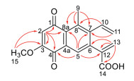
Arnequinone A (2), yellowish amorphous powder, had a molecular formula C17H14O5 as determined by the HRESIMS peak at m/z 299.0914 [M + H]+ (calcd. for C17H15O5, 299.0914), indicative of eleven degrees of unsaturation. The IR spectrum revealed absorption bands for carbonyl (1682, 1645 cm−1) and phenyl (1615, 1581, 1445 cm−1) groups. The 1H NMR data (Table 1) displayed signals for an active carboxy hydrogen (δH 13.07, br s), a monosubstituted vinyl [δH 7.08 (dd, J = 18.0, 11.5 Hz), 5.51 (dd, J = 11.5, 1.5 Hz), and 5.07 (dd, J = 18.0, 1.5 Hz)], a pair of geminal olefinic protons [δH 6.51 (d, J = 1.0 Hz) and 5.90 (d, J = 1.0 Hz)], two single aromatic protons [δH 7.70 (s) and 6.25 (s)], a methoxy (δH 3.83, s), and a methyl (δH 2.22, s). The 13C NMR data indicated three carbonyls, twelve sp2 carbons, a methoxy, and a methyl. All these aromatic carbons represented nine out of eleven degrees of unsaturation, disclosing a dicyclic nucleus for 2. The key HMBC correlations (Fig. 5) from H-2 to C-1, C-3, C-4 and C-8a, from H-5 to C-4, C-7 and C-8a, and from Me-9 to C-7, C-8 and C-8a constructed the dicyclic nucleus as a 1, 4-naphthoquinone with the methyl situated at C-8. Furthermore, HMBCs between H-10 and C-8, between H2-11 and C-7, between H2-13 and C-6/C-12/C-14, between H-5 and C-12, and between MeO-15 and C-3 established the linkages of C-7/C-10, and C-6/C-12, and locations of the carboxy and methoxy substituents at C-12 and C-3, respectively. All the above clarified the structure of 2 as a highly conjugated aromatic meroterpenoid with a methyl-shifting benzogeijerene skeleton (Fig. 1) [3, 14, 15].
Biosynthetically, the key intermediate b (Scheme 1) with a benzogeijerene skeleton was yielded via a Cope rearrangement of a [3, 15, 24]. Subsequent oxidation and esterification cyclization could generate d. Furthermore, intermediate d underwent a nucleophilic reaction to produce e with the 6/7/6/3 ring system, which would be readily transformed to 1 through selective oxidation methylation and glycosylation. Additionally, the dehydration of intermediate c, followed by the subsequent methyl 1, 2-shift and elimination reaction generated intermediate g, which proceeded oxidation methylation at C-3, cooperated with an autoxidation process to yield compound 2. Notably, the autoxidation of methoxylated g and the aglycone of 1 might be a common feature for 2-methoxy-hydroquinones [22].

The cytotoxic activities of 1, 1a and 2 against five human tumor cell lines (HCT116, U87-MG, BGC803, HepG2 and PC9) were evaluated using the MTT assay as described in the literature [25]. 1a exhibited cytotoxicity against all the tested cell lines except HepG2 with the half maximal inhibitory concentration (IC50) values ranging from 19.9 µmol/L to 25.9 µmol/L (Table S3 in Supporting information). Compounds 1 and 2 showed no cytotoxicity (IC50 > 50 µmol/L, Table S3). Then 1 and 2 were tested for their neuroprotective activities against serum-deprivation induced PC12 cell damage using nerve growth factor (NGF) as the positive control [26]. Compound 2 showed neuroprotective activity with a cell survival rate of 66.50% ± 1.53% compared to that of the model group (51.93% ± 1.36%) at a concentration of 10 µmol/L (Supporting information Table S4).
In conclusion, arnequinol A (1) represents an unprecedented 6/6/3 tricyclic carbon skeleton fused with a heptatomic oxo-bridge. Arnequinone A (2) bearing a novel highly conjugated aromatic system, was the first example with a methyl-shifting benzogeijerene skeleton in the meroterpenoids from Boraginaceae plants. Their structural novelty could enrich the chemical diversity of meroterpenoids and the chemical constituents of A. euchroma.
The authors declare that they have no known competing financial interests or personal relationships that could have appeared to influence the work reported in this paper.
This work was supported by the CAMS Innovation Fund for Medical Sciences (No. 2016-I2M-1-010, China).
Supplementary material associated with this article can be found, in the online version, at doi:10.1016/j.cclet.2021.09.064.
N. Kumar, R. Kumar, K. Kishore, Pharmacogn. Rev. 7 (2013) 140-151. doi: 10.4103/0973-7847.120513
R. Gupta, G.D. Gupta, Pharmacogn. Rev. 9 (2015) 127-131. doi: 10.4103/0973-7847.162124
M. Moir, R.H. Thomson, J. Chem. Soc. Perkin Trans. I. 13 (1973) 1352-1357.
M.J. Oza, Y.A. Kulkarni, J. Pharm. Pharmacol. 69 (2017) 755-789. doi: 10.1111/jphp.12715
T.S. Matos, A.K.O. Silva, A.L. Quintela, et al., Fitoterapia 123 (2017) 65-72. doi: 10.1016/j.fitote.2017.09.021
X.S. Yao, E. Yutaka, N. Hiroshi, et al., Tetrahedron. Lett. 25 (1984) 5541-5542. doi: 10.1016/S0040-4039(01)81620-8
A.F. Ahmed, H.A. Saad, E.M.A. El-Karim, Molecules 19 (2014) 5940-5951. doi: 10.3390/molecules19055940
M. Liao, P. Yan, X. Liu, et al., J. Chromatogr. B 1136 (2020) 121924. doi: 10.1016/j.jchromb.2019.121924
O.D.L. Pessoa, T.L.G. Lemos, M.G. Calvalho, et al., Phytochemistry 40 (1995) 1777-1786. doi: 10.1016/0031-9422(95)00397-P
G.M. Costa, T.L.G. Lemos, O.D.L. Pessoa, et al., J. Nat. Prod. 62 (1999) 1044-1045. doi: 10.1021/np980575y
S. Dettrakul, S. Surerum, S. Rajviroongit, et al., J. Nat. Prod. 72 (2009) 861-865. doi: 10.1021/np9000703
P.P. Painter, B.M. Wong, D.J. Tantillo, Org. Lett. 16 (2014) 4818-4821. doi: 10.1021/ol5023036
Y. Wang, Y. Zhu, L. Xiao, et al., Fitoterapia 131 (2018) 236-244. doi: 10.1016/j.fitote.2018.11.005
M. Moir, R.H. Thomson, J. Chem. Soc. Perkin Trans. I 15 (1973) 1556-1561.
K. Mori, M. Kawano, H. Fuchino, et al., J. Nat. Prod. 71 (2008) 18-21. doi: 10.1021/np070211i
J.M. He, S.C. Sun, Z.L. Sun, et al., Int. J. Antimicrob. Ag. 53 (2019) 70-73. doi: 10.1016/j.ijantimicag.2018.08.021
H.M. Li, Y.L. Tang, Z.H. Zhang, et al., Planta Med. 78 (2012) 39-45. doi: 10.1055/s-0031-1280266
L. Wang, F. Li, X. Liu, et al., Planta Med. 81 (2015) 320-326. doi: 10.1055/s-0035-1545693
H. Cao, W. Zhang, D. Liu, et al., Bioorg. Chem. 96 (2020) 103655. doi: 10.1016/j.bioorg.2020.103655
A. Kumar, S. Shashni, P. Kumar, et al., J. Ethnopharmacol. 271 (2021) 113896. doi: 10.1016/j.jep.2021.113896
K. Xu, J.S. Jiang, Z.M. Feng, et al., J. Nat. Prod. 79 (2016) 1567-1575. doi: 10.1021/acs.jnatprod.6b00066
S. Kim, R. Matsubara, M. Hayashi, J. Org. Chem. 84 (2019) 2997-3003. doi: 10.1021/acs.joc.8b02961
J. Li, J.K. Liu, W.X. Wang, J. Org. Chem. 85 (2020) 11350-11358. doi: 10.1021/acs.joc.0c01451
G.D. Manners, L. Jurd, J. Chem. Soc. Perkin Trans. I. 4 (1977) 405-410.
Z.F. Hu, A.D. Gu, L. Liang, et al., Green Chem. 21 (2019) 3286-3299. doi: 10.1039/C8GC04066D
S.Y. Shao, F. Zhang, Y.N. Yang, et al., Bioorg. Chem. 113 (2021) 105025. doi: 10.1016/j.bioorg.2021.105025

Figure 3 The 13C NMR chemical shifts calculation results of a pair of C-7 epimers of (5S*, 6R*, 7R*, 8R*, 12S*)-1a and (5S*, 6R*, 7S*, 8R*, 12S*)-1a.
Table 1. 1H (500 MHz) and 13C (125 MHz) NMR data for compounds 1, 1a, and 2 (δ in ppm, J in Hz).
|
|
 下载: 导出CSV

 扫一扫看文章

扫一扫关注我们


 下载:
下载: