Figure 1.
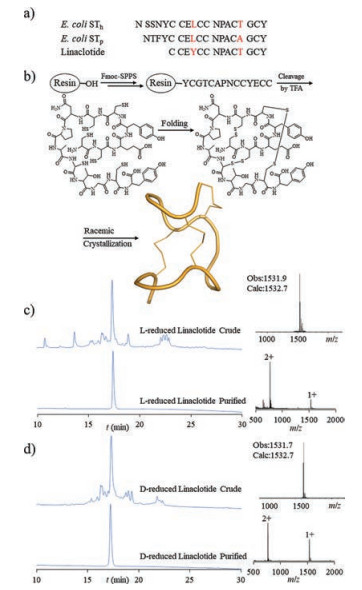
(a) Sequence comparison among STh, STp and linaclotide. (b) Synthetic route. HPLC chromatogram and mass spectra of L-reduced linaclotide (c) and D-reduced linaclotide (d).
Chemical synthesis and structural analysis of guanylate cyclase C agonist linaclotide
Chenchen Chen , Shuai Gao , Qian Qu , Pengcheng Mi , Anjin Tao , Yi-Ming Li

Irritable bowel syndrome (IBS) is a common chronic functional gastrointestinal disorder and studies show that about 7%–15% of North Americans and about 11.5% of Europeans are affected [1-4]. IBS patients have frequent and intermittent abdominal pain and discomfort, which not only adversely affect the patient's health, but also reduce work efficiency and increase the use of related medical resources [5, 6]. IBS with constipation (IBS-C) patients account for about one-third of the total number of IBS patients, and the number of female patients is more than male [7]. Apart from abdominal pain and reduced the frequency of stool, IBS-C patients also suffer from bloating, straining and incomplete sense of evacuation [1, 4]. Because traditional therapies have poor efficacy on the main symptoms of IBS-C patients, more treatment options will be valuable [6].
Linaclotide is a 14-amino acid peptide approved by the FDA for the treatment of IBS-C, which not only accelerates intestinal transit but also improves abdominal pain [8, 9]. This peptide belongs to heat-stable enterotoxin analog of E. coli, which activates GC-C on the surface of intestinal epithelium, resulting in increased level of cyclic guanosine monophosphate (cGMP). Increased cGMP could activate cystic fibrosis transmembrane conductance regulator (CFTR). This activation results in the secretion of bicarbonate and chloride into the lumen, increasing intracavital fluid secretion and accelerating intestinal transit [10, 11]. Although linaclotide is effective for the treatment of IBS-C, there are some side effects such as dose-dependent diarrhoea, abdominal discomfort and flatulence in clinical trials [5, 6]. Therefore, it is still necessary to improve the activity of linaclotide based on drug molecule design [12, 13]. However, the PDB files of linaclotide remains unknown. For this reason, the determination of the three-dimensional structure of linaclotide is of importance.
Racemic protein crystallography has been proven to be a powerful technique for crystallizing proteins [14-18]. The structures of many difficult-to-crystallize peptides/proteins of different sizes were resolved by the racemic protein crystallography combined with the protein chemical synthesis [19-24]. In this work, we report the chemical synthesis of natural linaclotide (Ltype) and its enantiomeric D-peptide (D-type) by using Fmoc solid phase peptide synthesis (Fmoc-SPPS) method. The X-ray crystal structure of linaclotide was determined in 1.59 Å by racemic protein crystallography. The crystal structure showed that linaclotide has a tight, three-beta turns spatial structure immobilized by three pairs of disulfide bonds.
Linaclotide is a heat-stable enterotoxin analog of E. coli consisting of 14 amino acids with three pairs of intramolecular disulfide bonds (C1-C6, C2-C10 and C5-C13) (Fig. 1a) [25]. The use of racemic crystallography for the crystallization of linaclotide needs to obtain L-type and D-type proteins. To this end, we used Wang resin as a solid scaffold and DIC (N, N'-diisopropylcarbodiimide) as condensing reagent to synthesize L-type and D-type linaclotide directly through Fmoc-SPPS [26]. After the completion of the synthesis, the crude linaclotide was cleaved from the resin and the side chain protecting group was removed by using a cleaving reagent (TFA/H2O/phenol/TIPS = 87.5/5/5/2.5, v/v/v/v) (Fig. 1b) [27-42]. The L-type and D-type crude peptides were analyzed by analytical HPLC and verified by ESI-MS. As show in Fig. 1c/d, both crude peptides had good purity. After purification by RP-HPLC and lyophilized, we obtained L-type and D-type reduced linaclotide (isolated yield: 44.9% for L-reduced linaclotide and 51.6% for D-reduced linaclotide).
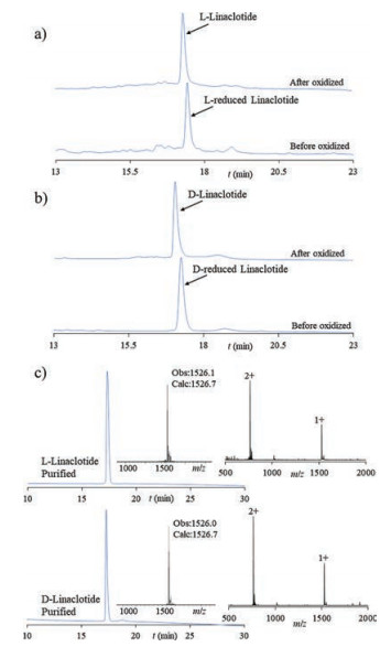
We then folded the L- and D-type reduced linaclotide by using a redox pair consisting of oxidized glutathione (GSSH) and reduced glutathione (GSH). Both L- and D-type reduced linaclotide were first dissolved in 50 mmol/L tris buffer, respectively, then GSSH and GSH were added to each buffer (molar ratio: peptide/GSSH/GSH = 1/10/ 100). The pH of the buffer was adjusted to 8.0 and folding process was performed overnight at 25 ℃ [43]. As shown in Fig. 2a/b, the Land D-type reduced linaclotide were almost completely converted to the corresponding L- and D-type linaclotide. After folding, the pH of the folded solution was adjusted to 2.0, then purified by HPLC immediately to give L- and D-type linaclotide (isolated yield: 51.7% for L-linaclotide and 59.2% for D-linaclotide).
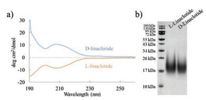
With L- and D-type linaclotide in hands, we first verified the difference in secondary structure between L-type and D-type linaclotide by circular dichroism (CD) spectra. As show in Fig. 3a, the CD spectrum of L-type linaclotides indicates a negative and a positive peak in the range of 205–215 nm and 195–205 nm, respectively, while the curve of D-linaclotide is symmetrically opposite to the L-linaclotide. SDS-PAGE analysis (Fig. 3b) shows that L-type and D-type linaclotide appeared between 17 and 26 kDa with a diffuse appearance. These results imply that the structure of linaclotide was sufficiently rigid, which could not be completely denatured under SDS conditions.
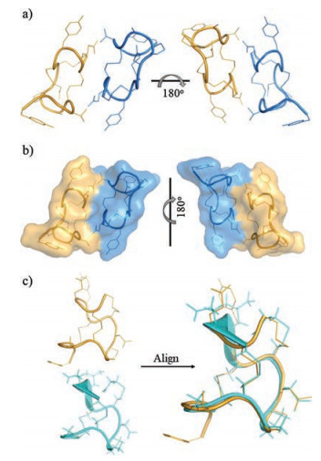
After confirming the protein characterization, we carried out the racemic crystallization experiment to obtain the X-ray structure of linaclotide. Crystals were screened using a Hampton crystallization kit in 18 ℃ crystal chamber after mix the L- and D-type linaclotide to their respective final concentrations of 10 mg/mL. After three days, we obtained the racemic crystals of linaclotide. The crystals were grown in 0.2 mol/L potassium sodium tartrate tetrahydrate, 20% (w/v) polyethylene glycol 3, 350 to form a P1 space group with diffraction resolution of 1.59 Å. Structural information of linaclotide was obtained after molecular replacement and refinement.
As shown in Figs. 4a and b, the crystals of L- and D-type linaclotide pack together and are almost mirror-image linked together. Linaclotide stabilizes its spatial structure by three pairs of intramolecular disulfide bonds, giving it a compact spatial structure containing three-beta turns. Linaclotide has a similar amino acid sequence to heat-stable enterotoxins, which is a class of diarrhea-causing toxins secreted by intestinal pathogens. Through the structural comparison between linaclotide and heat-stable enterotoxin of E. coli (STP mimic), we conclude that linaclotide has similar spatial arrangement (Fig. 4c) [25, 44]. The acquisition of linaclotide crystals potentially provides the basis for the subsequent optimization of the activity.
In summary, we obtained L- and D-type of linaclotide by protein chemical synthesis and acquired the X-ray crystal structure of linaclotide for the first time through racemic crystallization technique. The structure of linaclotide forms a compact spatial structure containing three-β turns through three intramolecular disulfide bonds, which has a similar spatial arrangement to heat-stable enterotoxin of E. coli (STP mimic). We anticipated that the X-ray crystal structure of linaclotide provides the basis for the optimization and improvement of the activity of linaclotide. Our work also highlights the power of chemical protein synthesis in obtaining peptide drugs for biochemical and structural studies.
This work was supported by the National Natural Science Foundation of China (NSFC No. 21572043) and the Fundamental Research Funds for the Central Universities (No. PA2017GDQT0021).
Supplementary data associated with this article can be found, in the online version, at https://doi.org/10.1016/j.cclet.2018.01.005.
G.F. Longstreth, W.G. Thompson, W.D. Chey, et al., Gastroenterology 130(2006) 1480-1491. doi: 10.1053/j.gastro.2005.11.061
A.P. Hungin, L. Chang, G.R. Locke, et al., Aliment. Pharmacol. Ther. 21(2005) 1365-1375. doi: 10.1111/apt.2005.21.issue-11
L.J. Brandt, W.D. Chey, A.E. Foxx-Orenstein, et al., Am. J. Gastroenterol. 104(Suppl. 1) (2009) S1-S35. http://europepmc.org/abstract/MED/19521341
A.P. Hungin, P.J. Whorwell, J. Tack, et al., Aliment. Pharmacol. Ther. 17(2003) 643-650. doi: 10.1046/j.1365-2036.2003.01456.x
W.D. Chey, A.J. Lembo, B.J. Lavins, et al., Am. J. Gastroenterol. 107(2012) 1702-1712. doi: 10.1038/ajg.2012.254
S. Gao, A.J. Lembo, S.J. Shiff, et al., Am. J. Gastroenterol. 107(2012) 1714-1724. doi: 10.1038/ajg.2012.255
E.A. Mayer, N. Engl. J. Med. 358(2008) 1692-1699. doi: 10.1056/NEJMcp0801447
R.W. Busby, A.P. Bryant, W.P. Bartolini, et al., Eur. J. Pharmacol. 649(2010) 328-335. doi: 10.1016/j.ejphar.2010.09.019
J. Gastro, A.M. Harrington, P.A. Hughes, et al., Gastroenterology 145(2013) 1334-1346. doi: 10.1053/j.gastro.2013.08.017
A.P. Bryant, R.W. Busby, W.P. Bartolini, et al., Life Sci. 86(2010) 760-765. doi: 10.1016/j.lfs.2010.03.015
H. Eutamene, S. Bradesi, M. Larauche, et al., Neurogastroenterol. Motil. 22(2010) 312-e84. doi: 10.1111/nmo.2010.22.issue-3
C.D. Fjell, J.A. Hiss, R.E.W. Hancock, G. Schneider, Nat. Rev. Drug Discov. 11(2012) 37-51. doi: 10.1038/nrd3591
B.G. Livett, K.R. Gayler, Z. Khalil, Curr. Med. Chem. 11(2004) 1715-1723. doi: 10.2174/0929867043364928
T.O. Yeates, S.B.H. Kent, Annu. Rev. Biophys. 41(2012) 41-61. doi: 10.1146/annurev-biophys-050511-102333
B.J. Yan, L.Z. Ye, W.L. Xu, L. Liu, Bioorg. Med. Chem. 25(2017) 4953-4965. doi: 10.1016/j.bmc.2017.05.020
H. Yeung, C.J. Squire, Y. Yosaatmadja, et al., Angew. Chem. Int. Ed. 55(2016) 7930-7933. doi: 10.1002/anie.201602719
K. Mandal, B. Dhayalan, M. Avital-Shmilovici, et al., ChemBioChem 17(2016) 421-425. doi: 10.1002/cbic.v17.5
R.D. Bunker, K. Mandal, G. Bashiri, et al., Proc. Natl. Acad. Sci. U. S. A. 112(2015) 4310-4315. doi: 10.1073/pnas.1422387112
C.K. Wang, G.J. King, S.E. Northfield, et al., Angew. Chem. Int. Ed. 53(2014) 11236-11241. doi: 10.1002/anie.201406563
S. Gao, M. Pan, Y. Zheng, et al., J. Am. Chem. Soc. 138(2016) 14497-14502. doi: 10.1021/jacs.6b09545
M. Pan, S. Gao, Y. Zheng, et al., J. Am. Chem. Soc. 138(2016) 7429-7435. doi: 10.1021/jacs.6b04031
C.L. Zhang, S. Liu, X.C. Liu, et al., Chin. Chem. Lett. 28(2017) 1523-1527. doi: 10.1016/j.cclet.2017.03.010
Y. Huang, W.H. Feng, Chin. Chem. Lett. 27(2016) 357-360. doi: 10.1016/j.cclet.2015.11.012
Z.M. Wu, S.Z. Liu, X.Z. Cheng, et al., Chin. Chem. Lett. 27(2016) 1731-1739. doi: 10.1016/j.cclet.2016.04.024
H. Ozaki, T. Sato, H. Kubota, et al., J. Biol. Chem. 266(1991) 5934-5941.
M. Gongora-Benitez, J. Tulla-Puche, M. Paradis-Bas, et al., Pept. Sci. 96(2011) 69-80. doi: 10.1002/bip.v96.1
Y.C. Huang, G.M. Fang, L. Liu, Natl. Sci. Rev. 3(2016) 107-116. doi: 10.1093/nsr/nwv072
G.M. Fang, Y.M. Li, F. Shen, et al., Angew. Chem. Int. Ed. 50(2011) 7645-7649. doi: 10.1002/anie.201100996
X.L. Tan, M. Pan, Y. Zheng, et al., Chem. Sci. 8(2017) 6881-6887. doi: 10.1039/C7SC02937C
Y.T. Li, C. Yi, C.C. Chen, et al., Nat. Commun. 8(2017) 14846. doi: 10.1038/ncomms14846
Z. Wang, W. Xu, L. Liu, T.F. Zhu, Nat. Chem. 8(2016) 698-704. doi: 10.1038/nchem.2517
X.D. Tan, M. Pan, S. Gao, et al., Chem. Commun. 53(2017) 10208-10211. doi: 10.1039/C7CC05504H
Y.C. Huang, C.J. Guan, X.L. Tan, et al., Org. Biomol. Chem. 13(2015) 1500-1506. doi: 10.1039/C4OB02260B
C.C. Chen, Y.C. Huang, L. Xu, et al., Org. Biomol. Chem. 12(2014) 9413-9418. doi: 10.1039/C4OB01885K
Y.C. Huang, C.C. Chen, S. Gao, et al., Chem. Eur. J. 22(2016) 7623-7628. doi: 10.1002/chem.201600101
S. Tang, Y.Y. Si, Z.P. Wang, et al., Angew. Chem. Int. Ed. 54(2015) 5713-5717. doi: 10.1002/anie.201500051
J.X. Wang, G.M. Fang, Y. He, et al., Angew. Chem. Int. Ed. 54(2015) 2194-2198. doi: 10.1002/anie.201408078
G.M. Fang, J.X. Wang, L. Liu, Angew. Chem. Int. Ed. 51(2012) 10347-10350. doi: 10.1002/anie.201203843
S. Bondalapati, E. Eid, S.M. Mali, et al., Chem. Sci. 8(2017) 4027-4034. doi: 10.1039/C7SC00488E
K. Medini, P.W.R. Harris, A. Menorca, et al., Chem. Sci. 7(2016) 2005-2010. doi: 10.1039/C5SC04187B
S. Tang, Z. Wan, Y. Gao, et al., Chem. Sci. 7(2016) 1891-1895. doi: 10.1039/C5SC03404C
T. Wu, Y.H. Li, X. Li, et al., Chem. Sci. 8(2017) 7368-7373. doi: 10.1039/C7SC02420G
M. Pan, Y. He, M. Wen, et al., Chem. Commun. 50(2014) 5837-5839. doi: 10.1039/C4CC00779D
T. Sato, H. Ozaki, Y. Hata, et al., Biochemistry 33(1994) 8641-8650. doi: 10.1021/bi00195a004

Figure 1 (a) Sequence comparison among STh, STp and linaclotide. (b) Synthetic route. HPLC chromatogram and mass spectra of L-reduced linaclotide (c) and D-reduced linaclotide (d).

Figure 2 a) The HPLC traces of folding of L-reduced linaclotide. b) The HPLC traces of folding of D-reduced linaclotide. c) HPLC chromatogram and mass spectrum of purified L-linaclotide and D-linaclotide.

Figure 3 a) CD spectra of L-linaclotide and D-linaclotide. b) SDS-PAGE of L-linaclotide and D-linaclotide.

Figure 4 a) Crystal structure of racemic crystal of L-linaclotide and D-linaclotide. b) Views of L-linaclotide and D-linaclotide crystal structure. c) Comparison of Llinaclotide and STP mimic (PDB: 1ETN). L-linaclotide, D-linaclotide and STP mimic are shown in orange, blue and cyan, respectively.

 扫一扫看文章

扫一扫关注我们
 DownLoad:
 下载:
下载:
 下载:
下载: