Citation:
Yao Chi, Tian Jia, Wang Hui, Zhang Dan-Wei, Liu Yi, Zhang Fan, Li Zhan-Ting. Loading-free supramolecular organic framework drug delivery systems (sof-DDSs) for doxorubicin: normal plasm and multidrug resistant cancer cell-adaptive delivery and release[J]. Chinese Chemical Letters,
2017, 28(4): 893-899.
doi:
10.1016/j.cclet.2017.01.005

Loading-free supramolecular organic framework drug delivery systems (sof-DDSs) for doxorubicin: normal plasm and multidrug resistant cancer cell-adaptive delivery and release
English
Loading-free supramolecular organic framework drug delivery systems (sof-DDSs) for doxorubicin: normal plasm and multidrug resistant cancer cell-adaptive delivery and release
Abstract:
Four water-soluble porous supramolecular organic framework drug delivery systems (sof-DDSs) have been used to adsorb doxorubicin (DOX) in water at physiological pH of 7.4, which is driven exclusively by hydrophobicity.The resulting complexes DOX@SOFs are formed instantaneously upon dissolving the components in water.The drug-adsorbed sof-DDSs can undergo plasm circulation with important maintenance of the drug and overcome the multidrug resistance of human breast MCF-7/Adr cancer cells. DOX is released readily in the cancer cells due to the protonation of its amino group in the acidic medium of cancer cells.In vitro and in vivo experiments reveal that the delivery of SOF-a-d remarkably improve the cytotoxicity of DOX for the MCF-7/Adr cells and tumors, leading to 13-19-fold reduction of the IC50 values as compared with that of DOX.This new sof-DDSs strategy omits the indispensable loading process required by most of reported nano-scaled carriers for neutral hydrophobic chemotherapeutic agents, and thus should be highly valuable for future development of low-cost delivery systems.
-
1. Introduction
Cancer is one of the leading causes of death. In addition to surgical operation, current cancer treatments heavily rely on chemotherapeutic and radiation agents, which, however, may also seriously damage heathy tissues and organs and cause considerable toxicity to patients. Moreover, many drugs suffer from multidrug resistance of cancer cells and/or are poorly soluble in water, which confines their clinical use. To overcome these drawbacks, in the past several decades, great effort has been devoted to the development of drug delivery systems (DDSs), including liposomes and many other kinds of nanoparticle carriers [1-7]. Currently, FDA has approved a number of liposomal delivery systems for cancer therapy in the clinics and many more are under clinical trials or preclinical assessments [8]. To minimize drug leaking in the systemic circulation, delivered drug molecules are typically loaded inside rationally designed nanoparticles. Nevertheless, the loading process and subsequent purification procedures are both complicated and cost-increasing, and any promising drug-carrier system has to pass strict evaluations as new drug before clinical use. As a result, treatment costs related to clinically used liposomal drugs are all remarkably higher than that of the conventional non-liposomal drug treatment [9]. Moreover, efficient drug loading often impedes the release of the drug in cancer cells. In order to reach efficient and controlled release, in the past decade various stimuli-responsive techniques have been developed [10]. Although improved treatment efficacy can be realized, it is expected that the introduction of such stimuli-responsive motifs would further increase the treatment cost of the corresponding DDS-drugs.
Self-assembly has provided robust strategies for the generation of functional supramolecular polymers [11]. In recent years, we and other groups have constructed a variety of homogeneous supramolecular organic frameworks (SOFs) that contain regular nano-scaled pores by using cucurbit [8] uril (CB [8]) encapsulationenhanced aromatic dimerization as driving force in water [12-15]. As supramolecular polycationic electrolytes, three-dimensional (3D) diamondoid SOFs had been found to adsorb organic anions driven by electrostatic attraction, as well as hydrophobicity for some guests, in water [13b, c]. Previously we demonstrated that these SOFs can adsorb pemetrexed (PMX) [16], a dianionic chemotherapeutic agent for the treatment of several cancers. The in situ-prepared PMX@SOFs can maintain important amount of PMX during plasm circulation and deliver the drug into multidrug resistant (MDR) human breast cancer MCF-7/Adr cells. The acidic media of the cancer cells readily pushes the release of the drug in cancer cells owning to the neutralization of its carboxylate groups. Considering the highly hydrophobic nature of their pores, we envisioned that diamondoid SOFs might also be able to accommodate hydrophobic neutral organic drugs. Delivery of neutral hydrophobic drugs are highly desirable because they not only constitute the largest section (>80%) of chemical drugs for cancer treatment [17], but also most frequentlysuffer from poor solubility in aqueous media, MDR and/or high side effects. Actually, both clinically used and preclinical on-assessment DDSs have all involved hydrophobic neutral molecular agents. To test this possibility, we have chosen doxorubicin (DOX) as the model drug to establish a new delivery protocol. DOX is clinically used in the treatment of a wide range of cancers as well as in combination chemotherapy as a component of various chemotherapy regimens, but it also causes many adverse effects such as cardiomyopathy, typhlitis, dyspigmentation, and acral erythema. As a result, DOX has been most widely used as model drug to develop new drug delivery strategies, and several liposomal DOX formulations have been used clinically since 1995 [18]. Herein we report that the binding of the diamondoid SOF-based drug delivery systems (sofDDSs) for DOX exhibits remarkable in vivo adaptivity. They adsorb and maintain DOX in aqueous media at the physiological pH of 7.4, but release the drug readily in acidic MDR MCF-7/Adr cancer cells (pH 4.5-6.8) through the protonation of the amino group of DOX to kill the cancer cells with substantially improved efficacy in both in vitro and in vivo studies. The new sof-DDSs omit the drug-loading process and subsequent purification procedures, which are required for most of reported DDSs for neutral hydrophobic drugs, and avoid the necessity of introducing any stimuli-responsive motif for controlled release, and thus represents a remarkably simplified low-cost delivery strategy for neutral hydrophobic drugs.
2. Results and discussion
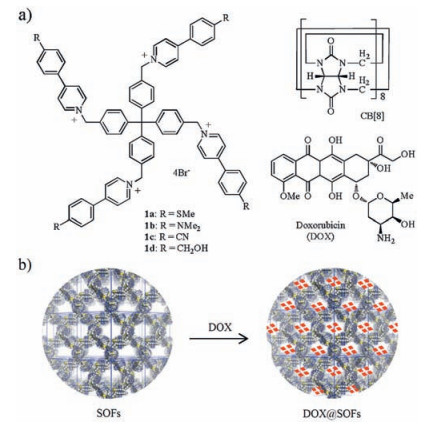
Previous study established that the 1:2 mixtures of compounds 1a-d and CB [8] in water give rise to 3D homogeneous regular diamondoid SOF-a-d (Fig. 1). The pore aperture of these SOFs, defined bysix CB [8] units in one self-assembled macrocycle which adopts a cyclohexane-like chair conformation, has been estimated tobe about2.1nm, andtheir void volume, calculatedon thebasisof the modeled frameworks was approximately 77% [19].The four pyridinium units provide the SOFs with good solubility in water. Nevertheless, the cavity surfaces are expected to be considerably hydrophobic. Thus, the adsorption of these homogeneous frameworks for DOX was first studied (Fig. 1b), using dialysis experiments in phosphate (totally 10mmol/L)-buffered saline (PBS) at the physiological pH of 7.4. As all SOFs are formed instantaneously after the components are dissolved in water, for all experiments, samples were prepared by directly dissolving the molecular components in water, with no SOF samples being prepared in advance. Forall the experiments, 1.5mgof the mixturesof DOX, 1a-d and CB [8] (molar ratio: 20:1:2) were added to a dialysis bag (2mL, cutoff Mn: 1000Da) that was immerged in a PBS buffer (15mL, pH 7.4). The solution, being placed on a shaking orbital shaker, was subjected to dialysis for 3days at 37 ℃ with the buffer being renewed every 6hours. By recording the adsorption of DOX at 480nm in the buffer by UV-vis spectroscopy, we could determine the total amount of DOX that diffused into the buffer. In this way, we established that 1.0g of SOF-a-d could adsorb and maintain 173, 149, 125, and 136mg of DOX, respectively (Fig. 2a). It can be found that all the frameworks exhibited robust adsorption capacity, whereas the largest amount observed for SOF-a may be rationalized by considering the smallest polarity of the methylthio group relative to other three substituents.
图 1
图 2
 图 2 a) The "loading" amount of DOX by SOF-a-d (1.0 g), b) Release of adsorbed DOX in the interior of SOF-a at pH 7.4 (○) and 4.5 (□) at 37 ℃ against timeFigure 2. a) The "loading" amount of DOX by SOF-a-d (1.0 g), b) Release of adsorbed DOX in the interior of SOF-a at pH 7.4 (○) and 4.5 (□) at 37 ℃ against time
Since all the SOFs had been revealed to exhibit low and comparable cytotoxicity [16], we then conducted releasing or leaking experiments for DOX-adsorbed SOF-a, dubbed as DOX@-SOF-a at two pH values. Typically, 1.0mg of the mixture of DOX, 1a and CB [8] (molar ratio: 1:1:2) was added to a dialysis bag (2mL, cutoff Mn: 1000Da) that was immerged in a PBS buffer (15mL, pH 7.4) or AcOH/AcONa buffer (15mL, pH 4.5, [AcOH]+[AcONa]=0.1 mol/L). The solutions were subjected to dialysis at 37 ℃. Again the amount of DOX that leaked into the buffers was determined by recording its absorption using UV-vis spectroscopy (Fig. 2b). It can be found that at pH 7.4, onlyabout 5.1% and 7.8% of DOX leaked into the buffers after 8h and 60h, respectively. In contrast, at pH 4.5, about 53% of DOX already leaked into the buffers after 8h and the value increased to 82% and 89%, respectively, after 24 and 60h. These observations suggest that, when circulating in normal blood plasm, SOF-a would be able to maintain most of the absorbed DOX from leaking within 1-3days and thus avoid important damage to normal tissues during a delivery process. The fact that most of DOX left the SOF at pH 4.5 after 24h implies that, once entering cancer cells, the acidic medium of cancer cells would force the adsorbed DOX torelease fromtheinteriorof theframework. Previousstudies haverevealed that 3D SOFsmaintained their regularityandthe size of their pores did not suffer observable change after adsorbing guests [13b, c]. Considering the relative small size of DOX, it is reasonable to propose that, upon adsorbing the drug, SOF-a-d should also maintain their structural regularity.
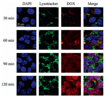
Previous study had confirmed that SOF-a is able to overcome MDR of MCF-7/Adr cancer cells [16], as a result of the enhanced permeability and retention (EPR) effect of the cancer cells [20], and delivered dianionic PMX into the cancer cells. Confocal laser scanning microscopy (CLSM) was used to study the capacity of this sof-DDS in delivering DOX to non-resistant MCF-7 and MDR MCF-7/Adr cancer cells (Fig. 3). DOX@SOF-a sample was prepared in situ by dissolving the three components in water before use. The nuclei of the MCF-7 cells and MDR MCF-7/Adr cells were stained with 4, 6-diamidino-2-phenylindole (DAPI), while their endosome/lysosome were stained with Lysotracker green. The images were all recorded after the cells were incubated with DOX or DOX@SOF-a for 2 h and are shown in Fig. 3. It can be found that, for non-resistant MCF-7 cells, the luminescent signals of DOX, in the absence or presence of SOF-a, and nuclei-stained DAPI were highly overlapping. In contrast, for MDR MCF-7/Adr cells, the luminescence of DOX was only exhibited by the cells which were incubated with DOX@SOF-a. Moreover, this luminescence of DOX was overlapping with that of DAPI. In the absence of SOF-a, only very weak luminescent signal of DOX was displayed, although it also overlapped with that of DAPI. For both kinds of cancer cells, the luminescent signal of 1a of SOF-a and that of endosome/lysosomestained Lysotracker green overlapped very well. These results again supported that SOF-a could enter both non-resistant and MDR cancer cells, while only with the delivery of SOF-a, DOX could enter MDR cells with a considerable amount, even though the drug was readily internalized by non-resistant cancer cells.
图 3
 图 3 Confocal microscopic (CLSM) images of non-resistant MCF-7 cells and MDR MCF-7/Adr cells incubated with free DOX (20 μmol/L) and DOX@SOF-a ([DOX] = 20 mmol/L, [1a] = 0.5 × [CB [8]] = 50 μmol/L) for 2 h. Endosome/lysosome and nuclei were stained with Lysotraker Green DND-26 and DAPI, respectively.Figure 3. Confocal microscopic (CLSM) images of non-resistant MCF-7 cells and MDR MCF-7/Adr cells incubated with free DOX (20 μmol/L) and DOX@SOF-a ([DOX] = 20 mmol/L, [1a] = 0.5 × [CB [8]] = 50 μmol/L) for 2 h. Endosome/lysosome and nuclei were stained with Lysotraker Green DND-26 and DAPI, respectively.
It has been established that SOF-a is captured into MDR MCF-7/ Adr cancer cells through the endocytosis pathway [16]. The framework quickly enters lysosomes and reaches the maximum concentration within 30 min. To get deep insight into the distribution of delivered DOX after being delivered into cancer cells by the frameworks, CLSM images were recorded at different time intervals and are presented in Fig. 4. It can be found that the luminescence of DOX began to appear after incubation for 30 min, and the intensity of luminescence increased and reached the maximum after 2 h. The signals well overlapped with those of nuclei-stained DAPI, supporting that the drug gradually accumulated in the nuclei. As the signal intensity of DOX increased in the nuclei, the luminescence of endosome/lysosome-stained Lysotracker green also became enhanced, which indicated that DOX was gradually released from SOF-a after SOF-a entered the lysosomes. This release process of DOX was slower because DOX had to undergo protonation in the interior of lysosomes where the acidity was strongest. The fact that the luminescence of both Lysotracker green and DOX was very weak at the early stage of incubation might be attributed to that they quenched each other. The hysteretic release of DOX from SOF-a is similar to that observed for PMX [16], supporting that, before release from the sof-DDSs, they must be protonated in the acidic interior of lysosomes.
图 4
 图 4 CLSM images revealing the time-dependent endosomal escape of DOX, reflected by the red fluorescence, from DOX@SOF-a in MDR MCF-7/Adr cancer cells incubated with DOX@SOF-a ([DOX] = 20 μmol/L, [1a] = 0.5 × [CB [8]] = 50 mmol/L). Endosome/lysosome and nuclei were stained with Lysotracker green and DAPI, respectively.Figure 4. CLSM images revealing the time-dependent endosomal escape of DOX, reflected by the red fluorescence, from DOX@SOF-a in MDR MCF-7/Adr cancer cells incubated with DOX@SOF-a ([DOX] = 20 μmol/L, [1a] = 0.5 × [CB [8]] = 50 mmol/L). Endosome/lysosome and nuclei were stained with Lysotracker green and DAPI, respectively.
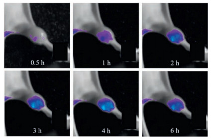
Previous fluorescent imaging study showed that SOF-a could enter and accumulate in the MDR MCF-7/Adr tumor region of nude mice [16]. The accumulation reached maximum after 2 h. The fluorescence signal then became weak gradually, implying that 1a was metabolized in the tumor region, leading the slow decrease of its concentration. The accumulation of DOX in the MDR MCF-7/Adr tumor of nude mice was further studied with DOX@SOF-a by monitoring the fluorescence of DOX. Nude mice bearing MDR MCF-7/Adr tumor were intravenously injected with the DOX@SOF-a solution from the tail, and the fluorescence intensity of the bioimaging signal of DOX at 600 nm in the tumor region was monitored at different time intervals (Fig. 5). After 2 h, the fluorescence intensity reached maximum. Contrast experiment with DOX in the absence of SOF-a showed that no important fluorescence of DOX was observed in the tumor region after 2 h. These observations supported that DOX was delivered into the tumor region by SOF-a. Different from that of SOF-a which gradually weakened after 2 h, the fluorescence of DOX in the tumor region was maintained after 2 h and did not show discernible weakening within recorded time (up to 6 h) (Fig. 5), which indicated that the drug did not suffer from important metabolism or exocytosis in the tumor region, at least during the studied time.
图 5
 图 5 Fluorescence imaging after intravenous injection of DOX@SOF-a (SOF-a dosage: 50 mg/kg, DOX dosage: 8.5 mg/kg, 0.2 mL). In vivo images (bottom view) of a mouse bearing MDR MCF-7/Adr tumor on one hind leg at 0.5, 1, 2, 3, 4, and 6 h after injection.Figure 5. Fluorescence imaging after intravenous injection of DOX@SOF-a (SOF-a dosage: 50 mg/kg, DOX dosage: 8.5 mg/kg, 0.2 mL). In vivo images (bottom view) of a mouse bearing MDR MCF-7/Adr tumor on one hind leg at 0.5, 1, 2, 3, 4, and 6 h after injection.
The cytotoxicity of DOX@SOF-a-d towards MDR MCF-7/Adr cancer cells was then evaluated with CCK-8 assay (Fig. 6). It was revealed that, with the identical DOX dosage, the viability of the cancer cells incubated with DOX@SOF-a-d was all substantially lower that of the cancer cells treated with DOX alone. This result strongly supported that all the four frameworks could deliver DOX to overcome MDR of the cancer cells and enter the cells to kill them more efficiently. The IC50 values of DOX@SOF-a-d and DOX alone were calculated to be 5.35, 6.01, 7.47, 7.13, and 106.8 mmol/L, respectively, which corresponded to 19.0, 16.8, 13.4, and 14.0-fold reduction of the dosage of DOX due to the delivery of SOF-a-d. The above IC50 values were also consistent with the relative adsorption capacity of the four frameworks for DOX (Fig. 2a). This result again supported that the delivery of DOX into the MDR cancer cells by the sof-DDSs played a key role in improving the cytotoxicity of the drug. The fact that the values of the four sof-DDSs-delivered samples were comparable also suggested that the peripheral substituents of tetrahedral 1a-d did not impose important influence on the adsorption and delivery capacity of the framework. This observation is important because it should allow more extensive screening of the tetrahedral component for achieving better water-solubility and lower toxicity for normal tissues.
图 6
 图 6 a) In vitro viabilities of MDR MCF-7/Adr cells treated with DOX@SOF-a-d and free DOX recorded after incubation at 37 ℃ for 24h; b) related IC50 values of DOX@SOF-a-d and free DOX after incubation at 37 ℃ for 24 h. The cytotoxicity was estimated by CCCK-8 proliferation tests ([1a-d]/[DOX] = 2.5).Figure 6. a) In vitro viabilities of MDR MCF-7/Adr cells treated with DOX@SOF-a-d and free DOX recorded after incubation at 37 ℃ for 24h; b) related IC50 values of DOX@SOF-a-d and free DOX after incubation at 37 ℃ for 24 h. The cytotoxicity was estimated by CCCK-8 proliferation tests ([1a-d]/[DOX] = 2.5).
The efficacy of DOX@SOF-a for treatment of subcutaneous MDR MCF-7/ADR tumors, in terms of apoptosis, was further evaluated in vivo. The apoptosis/necrosis of the cells, which were shown by hematoxylin and eosin (H & E) and terminal deoxynucleotidyl transferase dUTP nick-end labeling (TUNEL)-staining images (Fig. 7), were recorded after being treated with PBS as control, SOF-a, DOX@SOF-a. It can be found that, compared to the control group, the SOF-a group did not cause important damage on the tumor cells. However, the cells treated with DOX@SOF-a of the same dosage of DOX were severely destroyed, pointing to the improved treatment efficacy of DOX as a result of the delivery of the sof-DDS.
图 7
 图 7 In vivo images of the H & E and TUNEL-stained tumor tissue sections excised from the MDR MCF-7/Adr tumor of mice 14days after treatment. The mice were injected on one hind leg with normal saline, SOF-a solution or DOX@SOF-a buffer (SOF-a dosage: 50mg/kg; DOX dosage: 10.5mg/kg, 0.2mL). The controls had no injection.Figure 7. In vivo images of the H & E and TUNEL-stained tumor tissue sections excised from the MDR MCF-7/Adr tumor of mice 14days after treatment. The mice were injected on one hind leg with normal saline, SOF-a solution or DOX@SOF-a buffer (SOF-a dosage: 50mg/kg; DOX dosage: 10.5mg/kg, 0.2mL). The controls had no injection.
We further evaluated the in vitro cytotoxicity of SOF-a-d for multidrug resistant (MDR) human breast cancer (MCF-7/Adr) cells using the Cell Counting Kit-8 (CCK-8) assay. It was found that the viability of the MCF-7/Adr cells maintained at about 87%, 89%, 91%, and 90% after incubating for 24h with the SOFs at a relatively high concentration of 200 mg/mL, indicating that all the frameworks were of low cytotoxicity.
3. Conclusion
In summary, we have demonstrated that 3D homogeneous supramolecular organic framework drug delivery systems (sofDDSs) can adsorb doxorubicin in aqueous solution of physiological pH with hydrophobicity as the exclusive driving force. The adsorption is highly efficient and as a result, important amount of the drug can be maintained by the framework during plasm circulation after intravenous injection. The sof-DDSs can further deliver the drug to overcome the multidrug resistance of human breast MCF-7/Adr cancer cells and release the drug in the cells through the protonation of the drug owning to the acidic microenvironment within cancer cells. Compared with that of the drug alone, the IC50 values of all the delivery groups are substantially reduced, which reveals that the hydrophobic interior of the framework plays a major role in encapsulating and delivering the drug. The omission of the loading process, which are indispensable for most of reported drug delivery patterns for hydrophobic chemotherapeutic agents, remarkably simplifies the fabrication of delivery systems. Acidic medium-responsive release of the drug in multidrug resistant tumor cells also makes it unnecessary for the introduction of additional stimuli-responsive motif tothe framework. Thus, the sof-DDSs hold promise for future development of new low-cost techniques for delivery of neutral hydrophobic aza-contained anti-cancer drugs.
4. Experimental
Materials. All reagents were obtained from commercial suppliers and used without further purification unless otherwise noted. The synthesis, fabrication procedures and characterizations of SOF-a-d were according to the reported literature [16].
Methods. Luminescence measurements were performed on a VARIAN CARY Eclipse Fluorescence Spectrophotometer. UV-vis spectra were performed on a Perkin-Elmer 750s instrument. The Spectra/Por 6 Dialysis Tubing (1000Da Molecular Weight Cut Off, 8mm Flat-width) was purchased from Spectrum Laboratories, Inc.
Cell culture. MCF-7 and MCF-7/Adr (human cervical breast cell) lines were obtained from Department of Pathology, School of Basic Medical Sciences, Fudan University, (Shanghai, China) and cultured at 37 ℃ in Dulbecco's modified Eagle medium (DMEM) supplemented with 10% fetal bovine serum (FBS), 2mmol/L L-glutamine, 100 mg/mL penicillin, and 50 mg/mL streptomycin in a 5% CO2 environment.
Cytotoxicity test. The in vitro cytotoxicity was measured using the Cell Counting Kit-8 (CCK-8) assay in human breast carcinoma cell line MCF-7/Adr. Cells growing in log phase were seeded into 96-well cell-culture plate at 2 ×10⁴/well and then incubated for 24h at 37 ℃ under 5% CO2. Phosphate buffer solution (PBS) of the DOX@SOF-a-d samples (10 mL/well) at various concentrations were added to the wells of the treatment group, and PBS (10 mL/ well) to the negative control group, respectively. The cells were incubated for 24h at 37 ℃ under 5% CO2. Subsequently, 10 mL CCK-8 (1×) was added to each well of the 96 well assay plate and incubated for an additional 2h at 37 ℃ under 5% CO2. The absorbance (Avalue) ofeachwellwith background subtraction was measured at 450nm. The following formula was used to calculate the viability of cell growth: Cell viability (%)=(mean of A value of treatment group/mean of A value of control) °100. The statistical evaluation of data was performed using a two-tailed unpaired Student's t-test. A p-value of less than 0.05 was considered statistically significant. Each data point is represented as mean ≈ standard deviation (SD) of eight independent experiments (n=5, n indicates the number of wells in a plate for each experimental condition).
Confocal laser scanning microscopy (CLSM). For CLSM observations, MCF-7 or MCF-7/Adr cells (105 cells per dish) were seeded in coverglass bottom dishes (35mm ×10mm), and then treated with SOF at the final concentration ([1a]=0.05mmol/L, [CB[8]] = 0.1mmol/L). After 10, 15, 20, 25, and 30min of incubation, the cells were softly washed twice to remove excessive SOF. At last, 1mL of PBS solutionwas added and the cells were visualized under a confocal laser scanning microscope (FluoView FV1000, Olympus). The fluorescence images were taken under 60 ×oilimmersion objective.
Animals and tumor models. Female BALB/c nude mice (4 weeks old, ca. 20g body weight) were purchased from Shanghai SLAC Laboratory Animal Co., Ltd., China. Animal procedures were in agreement with the guidelines of the Institutional Animal Care and Use Committee. Tumor cells were harvested when they reached near confluence by incubationwith 0.05% trypsin-EDTA. Cells were pelleted by centrifugation and resuspended in sterile PBS. MCF-7/ Adr cells (2 ×10⁶ cells/site) were implanted subcutaneously into the right fore/hind leg of four-to five-week-old female athymic nude mice, respectively. When the tumors reached 0.4-0.6cm in diameter (14-21days after implant), the tumor-bearing mice were subjected to imaging studies.
Tumor histology. For histology analysis, tumor tissues from control and treated mice were fixed in 10% neutral buffered formalin and frozen sectioned into 5micron thick slices, stained with hematoxylin & eosin (H & E) and terminal deoxynucleotidyl transferase dUTP nick-end labeling (TUNEL), and were examined by a digital microscope (Leica QWin) and a confocal laser scanning microscope (LSM 510 Meta; Zeiss, Germany).
Acknowledgment
We thank the National Natural Science Foundation of China (Nos. 21432004, 21529201, 91527301) and the Ministry of Science and Technology of China (No. 2013CB834501), the Ministry of Education of China Research Fund for the Doctoral Program and of China for financial support. Shanghai Synchrotron Radiation Facility provided BL16B beamline for collecting the synchrotron X-ray scattering and diffraction data, which is also appreciated. YL thanks the support from the Molecular Foundry, Lawrence Berkeley National Laboratory, supported by the Office of Science, Office of Basic Energy Sciences, Scientific User Facilities Division, of the U.S. Department of Energy under Contract No. DE-AC02-05CH11231.
-
-
[1]
(a) D. Ma, G. Hettiarachchi, D. Nguyen, et al. , Acyclic cucurbit[n] uril molecular containers enhance the solubility and bioactivity of poorly soluble pharma-ceuticals, Nature Chem. 4(2012)503-510;
(b) R. Tong, L. Tang, L. Ma, et al. , Smart chemistry in polymeric nanomedicine, Chem. Soc. Rev. 43(2014)6982-7012;
(c) Y. Cao, X. Y. Hu, Y. Li, et al. , Multistimuli-responsive supramolecular vesicles based on water-soluble pillar[6] arene and SAINTcomplexation for controllable drug release, J. Am. Chem. Soc. 136(2014)10762-10769;
(d) Y. Zhou, H. Li, Y. W. Yang, Controlled drug delivery systems based on calixarenes, Chin. Chem. Lett. 26(2015)825-828;
(e) Y. X. Wang, D. S. Guo, Y. C. Duan, Y. J. Wang, Y. Liu, Amphiphilic p-sulfonatocalix[4] arene as“drug chaperoneâ€for escorting anticancer, Drugs. Sci. Rep. 5(2015)9019;
(f) Y. Z. Chen, Y. K. Huang, Y. Chen, et al. , Novel nanoparticles composed of chitosan and β-cyclodextrin derivatives as potential insoluble drug carrier, Chin. Chem. Lett. 26(2015)909-913;
(g) T. Zhang, P. Huang, L. Shi, et al. , Self-assembled nanoparticles of amphiphilic twin drug from floxuridine and bendamustine for cancer therapy, Mol. Pharmaceutics 12(2015)2328-2336;
(h) R. Jia, T. Wang, Q. Jiang, et al. , Self-assembled DNA nanostructures for drug delivery, Chin. J. Chem. 34(2016)265-272;
(i) J. Sun, J. Wang, Z. Yang, Supramolecular assembly models of siRNA delivery systems, Chin. J. Chem. 33(2015)79-89;
(j) Y. Wang, Y. Liu, Supramolecular assemblies based on p-sulfonatocalixarenes and their functions, Acta Chim. Sinica 73(2015)984-991;
(k) C. Yao, P. Wang, X. Li, et al. , Near-infrared-triggered azobenzene-liposome/upconversion nanoparticle hybrid vesicles for remotely controlled drug delivery to overcome cancer multidrug resistance, Adv. Mater. 28(2016)9341-9348;
(l) C. Wang, H. Zhang, D. Zeng, L. San, X. Mi, DNA nanotechnology mediated gold nanoparticle conjugates and their applications in biomedicine, Chin. J. Chem. 34(2016)299-307. -
[2]
(a) R. Tanbour, A. M. Martins, W. G. Pitt, G. A. Husseini, Drug delivery systems based on polymeric micelles and ultrasound: a review, Curr. Pharm. Design 22 (2016)2796-2807;
(b) L. Zhao, J. Ding, C. Xiao, et al. , Poly (L-glutamic acid) microsphere: preparation and application in oral drug controlled release, Acta Chim. Sinica 73(2015)60-65;
(c) T. M. Allen, P. R. Cullis, Liposomal drug delivery systems: from concept to clinical applications, Adv. Drug Deliv. Rev. 65(2013)36-48;
(d) K. Wang, D. S. Guo, X. Wang, Y. Liu, Multistimuli responsive supramolecular vesicles based on the recognition of p-sulfonatocalixarene and its controllable release of doxorubicin, ACS Nano 5(2011)2880-2894. -
[3]
(a) H. Wang, Q. Huang, H. Chang, J. Xiao, Y. Cheng, Stimuli-responsive dendrimers in drug delivery, Biomater. Sci. 4(2016)375-390;
(b) S. Zhang, J. Yang, M. Liu, et al. , Synthesis of peptide dendrimers and their application in the drug delivery system, Acta Chim. Sinica 74(2016)401-409;
(c) Y. Zhou, W. Huang, J. Liu, X. Zhu, D. Yan, Self-assembly of hyperbranched polymers and its biomedical applications, Adv. Mater. 22(2010)4567-4590. -
[4]
(a) H. Q. Wu, C. C. Wang, Biodegradable smart nanogels: a new platform for targeting drug delivery and biomedical diagnostics, Langmuir 32(2016)6211-6225;
(b) S. Liu, Y. Zhou, F. Chen, et al. , Rheological properties, drug release behavior and cytocompatibility of novel hydrogels prepared from carboxymethyl chitosan, Acta Chim. Sinica 73(2015)47-52. -
[5]
(a) M. Prato, K. Kostarelos, A. Bianco, Functionalized carbon nanotubes in drug design and discovery, Acc. Chem. Res. 41(2008)60-68;
(b) F. Du, J. Xu, F. Zeng, S. Wu, Preparation of a multifunctional nano-carrier system based on carbon dots with pH-triggered drug release, Acta Chim. Sinica 74(2016)241-250. -
[6]
(a) I. Brigger, C. Dubernet, P. Couvreur, Nanoparticles in cancer therapy and diagnosis, Adv. Drug Deliv. Rev. 54(2002)631-651;
(b) P. Yang, S. Gai, J. Lin, Functionalized mesoporous silica materials for controlled drug delivery, Chem. Soc. Rev. 41(2012)3679-3698;
(c) T. Sun, Y. S. Zhang, B. Pang, et al. , Engineered nanoparticles for drug delivery in cancer therapy, Angew. Chem. Int. Ed. 53(2014)12320-12364;
(d) P. Yang, L. Wang, H. Wang, Smart supramolecular nanosystems for bioimaging and drug delivery, Chin. J. Chem. 33(2015)59-70;
(e) H. Liang, H. Tian, M. Deng, X. Chen, Gold nanoparticles for cancer theranostics, Chin. J. Chem. 33(2015)1001-1010;
(f) Y. J. Chang, X. Z. Liu, Q. Zhao, et al. , P (VPBA-DMAEA) as a pH-sensitive nanovalve for mesoporous silica nanoparticles based controlled release, Chin. Chem. Lett. 26(2015)1203-1208;
(g) X. Wang, L. Tan, Y. Yang, Controlled drug release systems based on mesoporous silica capped by gold nanoparticles, Acta Chim. Sinica 74(2016) 303-311;
(h) Z. Tang, C. He, H. Tian, et al. , Polymeric nanostructured materials for biomedical applications, Progr. Polym. Sci. 60(2016)86-128. -
[7]
(a) J. Nicolas, Drug-initiated synthesis of polymer prodrugs: combining simplicity and efficacy in drug delivery, Chem. Mater. 28(2016)1591-1606;
(b) Z. Du, Y. Zhang, J. Ye, H. Xu, M. Lang, Synthesis and properties of the poly (ε-caprolactone)-paclitaxel prodrug, Acta Chim. Sinica 73(2015)349-356. -
[8]
(a) D. Landesman-Milo, D. Peer, Transforming nanomedicines from lab scale production to novel clinical modality, Bioconjugate Chem 27(2016)855-862;
(b) T. M. Allen, P. R. Cullis, Adv. Drug Deliv. Rev 65(2013)36-48. -
[9]
Cagnoni P.J., Walsh T.J., Prendergast M.M.. Pharmacoeconomic analysis of liposomal amphotericin B versus conventional amphotericin B in the empirical treatment of persistently febrile neutropenic patients[J]. J.Clin.Oncol., 2000, 18: 2476-2483. doi: 10.1200/JCO.2000.18.12.2476
-
[10]
(a) M. Kanamala, W. R. Wilson, M. Yang, B. D. Palmer, Z. Wu, Mechanisms and biomaterials in pH-responsive tumour targeted drug delivery: A review, Biomaterials 85(2016)152-167;
(b) Y. Zhao, A. C. Tavares, M. A. Gauthier, Nano-engineered electro-responsive drug delivery systems, J. Mater. Chem. B 4(2016)3019-3030;
(c) S. Ganta, H. Devalapally, A. Shahiwala, M. Amiji, A review of stimuli-responsive nanocarriers for drug and gene delivery, J. Controll. Release 126 (2008)187-204;
(d) R. Cheng, F. Meng, C. Deng, H. A. Klok, Z. Zhong, Dual and multi-stimuli responsive polymeric nanoparticles for programmed site-specific drug delivery, Biomaterials 34(2013)3647-3657. -
[11]
(a) L. Brunsveld, B. J. B. Folmer, E. W. Meijer, R. P. Sijbesma, Supramolecular polymers, Chem. Rev. 101(2001)4071-4098;
(b) L. Yang, X. Tan, Z. Wang, X. Zhang, Supramolecular polymers: historical development, preparation, characterization, and functions, Chem. Rev 115 (2015)7196-7239;
(c) T. Aida, E. W. Meijer, S. I. Stupp, Functional supramolecular polymers, Science 335(2012)813-817;
(d) X. Yan, F. Wang, B. Zheng, F. Huang, Stimuli-responsive supramolecular polymeric materials, Chem. Soc. Rev. 41(2012)6042-6065;
(e) D. S. Guo, Y. Liu, Calixarene-based supramolecular polymerization in solution, Chem. Soc. Rev. 41(2012)5907-5921;
(f) S. L. Li, T. Xiao, C. Lin, L. Wang, Advanced supramolecular polymers constructed by orthogonal self-assembly, Chem. Soc. Rev. 41(2012)5950-5968;
(g) X. Ma, H. Tian, Stimuli-responsive supramolecular polymers in aqueous solution, Acc. Chem. Res. 47(2014)1971-1981;
(h) E. A. Appel, J. del Barrio, X. J. Loh, O. A. Scherman, Supramolecular polymeric hydrogels, Chem. Soc. Rev 41(2012)6195-6214. -
[12]
(a) J. Tian, L. Chen, D. W. Zhang, Y. Liu, Z. T. Li, Supramolecular organic frameworks: engineering periodicity in water through host-guest chemistry, Chem. Commun. 52(2016)6351-6362;
(b) H. Wang, D. W. Wang, Z. T. Li, Supramolecular organic frameworks (SOFs): water-phase periodic porous self-assembled architectures, Acta Chim. Sinica 73(2015)471-479;
(c) T. Wan, T. Li, From supramolecular polymers to supramolecular organic frameworks: Engineering the periodicity of solution-phase self-assembled architectures, Imaging Sci. Photochem. 33(2015)3-14;
(d) L. Chen, Y. C. Zhang, W. K. Wang, et al. , Conjugated radical cation dimerization-driven generation of supramolecular architectures, Chin. Chem. Lett. 26(2015)811-816. -
[13]
(a) K. D. Zhang, J. Tian, D. Hanifi, et al. , Toward a single-layer two-dimensional honeycomb supramolecular organic framework in water, J. Am. Chem. Soc. 135 (2013)17913-17918;
(b) J. Tian, T. Y. Zhou, S. C. Zhang, et al. , Three-dimensional periodic supramolecular organic framework ion sponge in water and microcrystals, Nat, Commun. 5(2014)5574;
(c) J. Tian, Z. Y. Xu, D. W. Zhang, et al. , Supramolecular metal-organic frameworks that display high homogeneous and heterogeneous photo-catalytic activity for H2 production, Nat. Commun. 7(2016)11580. -
[14]
Pfeffermann M., Dong R., Graf R.. Free-standing monolayer two-dimensional supramolecular organic framework with good internal order[J]. J. Am.Chem.Soc., 2015, 137: 14525-14532. doi: 10.1021/jacs.5b09638
-
[15]
(a) Y. Zhang, T. G. Zhan, T. Y. Zhou, et al. , Fluorescence enhancement through the formation of a single-layer two-dimensional supramolecular organic frame-work and its application in highly selective recognition of picric acid, Chem. Commun. 52(2016)7588-7591;
(b) S. Q. Xu, X. Zhang, C. B. Nie, et al. , The construction of a two-dimensional supramolecular organic framework with parallelogram pores and stepwise fluorescence enhancement, Chem. Commun. 51(2015)16417-16420. -
[16]
Tian J., Yao C., Yang W.L.. In situ-prepared homogeneous supramolecular organic framework drug delivery systems (sof-DDSs):overcoming cancer multidrug resistance and controlled release[J]. Chin.Chem.Lett., 2017, 28: 798-806. doi: 10.1016/j.cclet.2017.01.010
-
[17]
Data from U. S. Food&Drug Administration: https: //www. cancer. gov/about-cancer/treatment/drugs.
-
[18]
(a) Y. Barenholz, Doxil®-The first FDA-approved nano-drug: Lessons learned, J. Control. Release 160(2012)117-134;
(b) F. Meng, Y. Zhong, R. Cheng, C. Deng, Z. Zhong, pH-sensitive polymeric nanoparticles for tumor-targeting doxorubicin delivery: concept and recent advances, Nanomedicine 9(2014)487-499. C. Yao et al. /Chinese Chemica -
[19]
Accelrys Materials Studio Release Notes, Release 5. 0, Accelrys Software Inc. , San Diego, 2008.
-
[20]
(a) J. Fang, H. Nakamura, H. Maeda, The EPR effect: unique features of tumor blood vessels for drug delivery, factors involved, and limitations and augmentation of the effect, Adv. Drug Deliv. Rev. 63(2011)136-151.
-
[1]
-

Figure 1 a) The structures of compounds 1a-d, CB [8], and doxorubicin, b) and illustration of hydrophobicity-driven formation of DOX@SOF-a-d.
Figure 2 a) The "loading" amount of DOX by SOF-a-d (1.0 g), b) Release of adsorbed DOX in the interior of SOF-a at pH 7.4 (○) and 4.5 (□) at 37 ℃ against time
Figure 3 Confocal microscopic (CLSM) images of non-resistant MCF-7 cells and MDR MCF-7/Adr cells incubated with free DOX (20 μmol/L) and DOX@SOF-a ([DOX] = 20 mmol/L, [1a] = 0.5 × [CB [8]] = 50 μmol/L) for 2 h. Endosome/lysosome and nuclei were stained with Lysotraker Green DND-26 and DAPI, respectively.
Figure 4 CLSM images revealing the time-dependent endosomal escape of DOX, reflected by the red fluorescence, from DOX@SOF-a in MDR MCF-7/Adr cancer cells incubated with DOX@SOF-a ([DOX] = 20 μmol/L, [1a] = 0.5 × [CB [8]] = 50 mmol/L). Endosome/lysosome and nuclei were stained with Lysotracker green and DAPI, respectively.
Figure 5 Fluorescence imaging after intravenous injection of DOX@SOF-a (SOF-a dosage: 50 mg/kg, DOX dosage: 8.5 mg/kg, 0.2 mL). In vivo images (bottom view) of a mouse bearing MDR MCF-7/Adr tumor on one hind leg at 0.5, 1, 2, 3, 4, and 6 h after injection.
Figure 6 a) In vitro viabilities of MDR MCF-7/Adr cells treated with DOX@SOF-a-d and free DOX recorded after incubation at 37 ℃ for 24h; b) related IC50 values of DOX@SOF-a-d and free DOX after incubation at 37 ℃ for 24 h. The cytotoxicity was estimated by CCCK-8 proliferation tests ([1a-d]/[DOX] = 2.5).
Figure 7 In vivo images of the H & E and TUNEL-stained tumor tissue sections excised from the MDR MCF-7/Adr tumor of mice 14days after treatment. The mice were injected on one hind leg with normal saline, SOF-a solution or DOX@SOF-a buffer (SOF-a dosage: 50mg/kg; DOX dosage: 10.5mg/kg, 0.2mL). The controls had no injection.
-

 下载:
下载:


 下载:
下载:


 扫一扫看文章
计量
- PDF下载量: 1
- 文章访问数: 1908
- HTML全文浏览量: 113
 下载:
下载:
