Citation:
Ye Tao, Jin Xin-Yi, Chen Liao, Hu Chong, Ren Jie, Liu Yu-Jing, Wu Gang, Chen Lu-Jian, Chen Hong-Zheng, Li Han-Ying. Shape change of calcite single crystals to accommodate interfacial curvature: Crystallization in presence of Mg2+ ions and agarose gel-networks[J]. Chinese Chemical Letters,
2017, 28(4): 857-862.
doi:
10.1016/j.cclet.2016.12.005
Shape change of calcite single crystals to accommodate interfacial curvature: Crystallization in presence of Mg2+ ions and agarose gel-networks
MOE Key Laboratory of Macromolecular Synthesis and Functionalization, State Key Laboratory of Silicon Materials, Department of Polymer Science and Engineering, Zhejiang University, Hangzhou 310027, China
b.
Department of Electronic Engineering, School of Information Science and Engineering, Xiamen University, Xiamen 361005, China
Received Date:
04 November 2016 Accepted Date:
02 December 2016 Revised Date:
25 January 2016 Available Online:
01 April 2017
Abstract:
Synthetic calcite single crystals, due to their strong crystal habit, tend to grow into characteristic rhombohedra.In the nature, biogenic calcite crystals form composites together with biomacromolecular materials, spurring investigations of how the growing calcite single crystals change their habit to satisfy the curvature of the organic phase.In this work, we examine calcite crystallization on a flat surface of glass slide and a curved surface of polystyrene (PS) sphere.The crystals exhibit tiny contact area onto the glass substrate that is averagely only 15% of their projected area on the substrate.In sharp contrast, the contact area greatly increase to above 75% of the projected area, once magnesium ions or agarose gel networks are introduced into the crystallization media.Furthermore, the calcite crystals form rough and step-like interfaces with a curved surface.However, the interfaces become smooth and curved as the crystals grow in presence of magnesium ions or agarose gel networks.The discrepancy between the interfacial structures implies kinetic effects of the additives on the crystallization around the surfaces. This work may provide implications for understanding the formation mechanisms of single-crystal composite materials.
Interfacing multi-component solids is widely used to construct hybrid materials in both nature and technological world [1-3]. In these hybrid materials, ordered arrangement of the components is highly associated with their properties. For example, the alignment of the fibers in bone contributes greatly the mechanical toughness [4]. Also, highly ordered molecular packing of single crystallinity is desired for electronic applications such as solar cells [5, 6]. Interestingly, ordered arrangement in single crystals leads to anisotropy that, in turn, affects adversely the interfacial structures of the hybrid materials. Considering a single crystal growing toward a surface with an arbitrary shape, the energetic and kinetic anisotropy of the crystal should result in its faceted habit [7] and non-intimate contact with the surface. As such, to construct hybrid materials with single-crystalline components depends on the strategies to tune the shapes of single crystals to accommodate interfacial structures.
Nature provides a variety of examples, dealing with challenges of this kind. Interfacing single-crystals of calcite with organic components is widely seen in biominerals including echinoderms and mollusk shells [8-18]. Intrinsically, calcite has an anisotropic crystallographic structure and synthetic calcite single crystals show a strong habit expressed as {104} rhombohedra. However, this characteristic crystal habit is missing from the biominerals where wavy and even fenestrated shapes of the calcite single crystals dominate [9, 10]. The non-classic shapes of the biominerals indicate the possibility to mold calcite single crystals to accommodate the shape of organic moulds. Accordingly, efforts have been made to investigate the possible mechanisms by which crystals can be molded. It is widely believed that the key process is associated with crystallization through amorphous calcium carbonate (ACC) precursor that is isotropic and moldable [9, 10, 19, 20]. Experimentally, calcite has been molded into large micropatterned single crystals [21], 3D ordered macroporous single crystals [22], and single crystals with triple periodic minimal surface (TPMS) structure [23] using ACC precursor. Interestingly, amorphous precursors are also found to be critical in the biomineralization of calcium phosphate [24-28]. Instead of moldable amorphous precursors, crystallization in confined spaces through classic ion-by-ion precipitation method also results in molded crystals and porous calcite single crystals formed in the confinement of colloidal crystal templates [29]. In addition to the amorphous precursors and confined crystallization, another very relevant research is the morphology modification of calcite single crystals. Shapes different from the typical rhombohedra can be obtained as organic or inorganic additives are introduced during crystallization process. Atomic force microscope (AFM) imaging has clearly shown that varied additives, such as D-aspartic acid, allow specific interactions with the step edges to affect the crystallization kinetics and the macroscopic crystal shape [30, 31]. Although the previous studies have comprehensively show the approaches for morphology modification of calcite single crystals, the knowledge on the interface formation between crystals and foreign solids is still limited. It is desired to know, as a growing calcite single crystal is interfaced with a solid, whether the kinetic effect of the additives will lead to flat or curved surfaces to accommodate the curvature of the solid. In this work, we examined calcite crystallization on foreign solids of flat substrates and colloidal particles, in presence of additives. And we found that these additives enable the formation of extended flat surfaces and surfaces with negative curvatures on calcite crystals to allow intimate contact with the foreign solids.
2.
Results and discussion
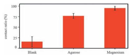
We first examine the calcite crystallization from solution on glass slides. As the crystal is nucleated from one of the {104} faces, it is expressed as the characteristic six {104} rhombohedra. Once the crystal is nucleated from other faces, one more face (Fig. 1a, red) will form to contact the glass slide and it can be observed from the bottom view of the crystal. Contact ratio is defined as the ratio of this contact area to the projected area of the whole crystal (Fig. 1a). The contact ratio reflects how intimate the crystal contacts the glass slide. We randomly selected 50 crystals with seven faces expressed and measured their contact ratios. The obtained average contact ratio is 15% (Fig. 2), indicating of poor contact. As the solution was replaced by hydrogel (1 w/v% agarose), the obtained crystals show a higher contact ratio of 75% and three {104} faces cap the crystals with a slightly rough surface (Fig. 1d). In sharp contrast, calcite crystals grown from solution with magnesium ions present a contact ratio as high as 95% and a new set of facets that lie approximately parallel to the h0 01i axis of pure calcite. Three {104} facets cap the {hk0} cylinder (Fig. 1f) [32]. For comparison, the values of contact ratio are plotted in Fig. 2. The increased contact ratios indicate that adding agarose fibers and magnesium ions effectively change the crystallization kinetics of calcite to accommodate the shape of glass slides.
图 1
图 1
(a) Schematic representation of a calcite crystal nucleating on a substrate with a contact face (red). The contact ratio is defined as the contact area to the projected area (red and blue) of the whole crystal, (b, d and f) Scanning electron microscopy (SEM) images of top view of calcite single crystals grown in 5 mmol/L CaCl2 with no additive, 1 w/v% agarose gel and 5 mmol/L MgCl2 respectively, (c, e and g) Optical microscopy (OM) images of bottom view of the calcite single crystals contacting with glass slides grown in 5 mM CaCl2 with no additive, 1 w/v% agarose gel and 5 mmol/L MgCl2 respectively.
Figure 1.
(a) Schematic representation of a calcite crystal nucleating on a substrate with a contact face (red). The contact ratio is defined as the contact area to the projected area (red and blue) of the whole crystal, (b, d and f) Scanning electron microscopy (SEM) images of top view of calcite single crystals grown in 5 mmol/L CaCl2 with no additive, 1 w/v% agarose gel and 5 mmol/L MgCl2 respectively, (c, e and g) Optical microscopy (OM) images of bottom view of the calcite single crystals contacting with glass slides grown in 5 mM CaCl2 with no additive, 1 w/v% agarose gel and 5 mmol/L MgCl2 respectively.
图 2
The contact ratio of the calcite single crystals grown on glass slides in solution (5 mmol/L CaCl2), agarose gel (1 w/v%; 5 mmol/L CaCl2) and solution (5 mmol/L CaCl2; 5 mmol/L MgCl2). 50 crystals from each experiment were measured.
Figure 2.
The contact ratio of the calcite single crystals grown on glass slides in solution (5 mmol/L CaCl2), agarose gel (1 w/v%; 5 mmol/L CaCl2) and solution (5 mmol/L CaCl2; 5 mmol/L MgCl2). 50 crystals from each experiment were measured.
Next, we proceed to examine interfacing calcite single crystals with curved surfaces. Compared to the flat glass slides, the introduced curvature will require the crystal to express complementary curvature to form intimate contact. Polystyrene (PS) spheres (500 nm or 3 mm in diameters) were dispersed on the glass slides with preformed calcite seed crystals. Subsequent crystallization from the seeds resulted in interfacing between the crystals and the PS spheres and the interfacial structures were imaged after dissolving the PS spheres (Fig. 3a). As grown from solutions, SEM images show concave surfaces on the crystals and the interfaces between calcite single crystals and PS spheres were rough with a step-like morphology (Fig. 3b and c). This result indicates that the PS spheres confine the crystallization space for the crystals that exhibit concave surface morphologies. Also, the anisotropic growth of calcite single crystals leads to the angular step-like morphology. Interestingly, the interfaces between calcite single crystals and PS spheres turn from rough to smooth with the addition of either agarose or magnesium ions in the crystallization media (Fig. 3d-g). In comparison, the effect of magnesium ions is more remarkable and more smooth interfaces are expressed, consistent with the larger contact ratio measured on flat surface (Fig. 2). The smooth interfaces indicate that the crystals express negative curvature to form intimate contact with the PS spheres. Previously, calcite single crystals were grown on PS spheres modified with sulfonic acid groups which can greatly change the growth kinetics around the interface [33]. As a result, smooth curved interfaces were obtained to make microlens arrays, without any additive used in the crystallization media. Similarly, it has been reported calcite crystals nucleating from self-assembled monolayers (SAMs) exhibited different shapes because the modification of crystallization kinetics [27_TD$DIF]via lattice mismatch between the SAMs and the nucleation planes [34]. Instead of modifying the surfaces that crystals grow onto, interfacial morphologies are dramatically changed by the additives in the crystallization media in this work.
图 3
图 3
(a) A schematic representation of the method to image the interface between calcite crystals and PS spheres, (b, d, and f) SEM images of the interface morphologies between calcite single crystals and 500 nm PS spheres with no additive, 1 w/v% agarose, and 5 mmol/L MgCl2, respectively, (c, e, and g) SEM images of the interface morphologies between calcite single crystals and 3 mm PS spheres with no additive, 1 w/v% agarose, and 5 mmol/L MgCl2, respectively.
Figure 3.
(a) A schematic representation of the method to image the interface between calcite crystals and PS spheres, (b, d, and f) SEM images of the interface morphologies between calcite single crystals and 500 nm PS spheres with no additive, 1 w/v% agarose, and 5 mmol/L MgCl2, respectively, (c, e, and g) SEM images of the interface morphologies between calcite single crystals and 3 mm PS spheres with no additive, 1 w/v% agarose, and 5 mmol/L MgCl2, respectively.
Magnesium ions have been widely reported to affect the crystallization of calcium carbonate, by changing the morphology of calcite crystals [35-37], stabling the ACC [38] and incorporating into the crystals [38-40]. Particularly, magnesium ions were proved to interact with the step edge in molecular scale, preferentially inhibiting step motion at the corners of growth hillocks [30, 41]. In this way, the original rectangular step morphology of calcite becomes rounded. Therefore, as magnesium is used as the additive, the growth front of the calcite crystals changes the shape gradually to accommodate the curvatures of the flat glass slide and the curved PS spheres.
Different from the magnesium ions, agarose molecules are insoluble and exist in the crystallization media as fiber networks [42]. During crystallization, the interaction between agarose and the step edge is not strong enough to cause the shape change from the characteristic six rhombohedra of calcite crystals, as reported previously [43, 44]. On the other hand, shape evolution of gel-grown crystals was reported before and the faceted shape even turned into sphere at very high gel concentration [45, 46]. Another mechanism associated with gel-network incorporation inside the crystals was proposed for the shape change. This mechanism might contribute to the formation of curved surface of the calcite crystals because the incorporation of gel networks inside a variety of crystals including calcite has been demonstrated [44, 47-54]. To verifythis mechanism, we compared the interface morphologies of crystals with varied amounts of incorporated gel networks. First, the gel concentration was reduced to 0.1 w/v% that is 10% of the typical value and lower amount of gel networks was incorporated [55]. As a result, the interface became rough and step-like (Fig. 4c), in sharp contrast to the smooth interface for 1 w/v% agarose (Fig. 4b). Furthermore, we grew crystals in another type of agarose (type Ⅸ) that could not be incorporated under the experimental condition due to the weak gel strength[49].Also, rough interface was observed (Fig. 4d).Therefore, the interface become smooth when the concentration of the incorporated gel networks reaches high enough (e.g. 1 w/v%). The incorporated gel networks might reduce the surface energy anisotropy [46]. In addition, the gel networks divide the crystallographic face at the growth front into smaller pieces by the pores of thegels.The crystallization in these pores is partially separatedasion communication between the pores is impeded by the gel network (Fig. 4a), leading to the macroscopic shapes defined by the curvature of the glass slide and PS sphere.
图 4
图 4
(a) Schematic representation of the gel effect, (b) SEM images of the interface morphology between calcite single crystal and PS sphere with 1 w/v% agarose, (c) with 0.1 w/v% agarose, and (d) with 1 w/v% agarose (Ⅸ).
Figure 4.
(a) Schematic representation of the gel effect, (b) SEM images of the interface morphology between calcite single crystal and PS sphere with 1 w/v% agarose, (c) with 0.1 w/v% agarose, and (d) with 1 w/v% agarose (Ⅸ).
In summary, we have investigated the interface formation as growing calcite single crystals approach flat glass slides or PS spheres. The results show that the glass slides and the spheres confine the crystallization space to shape the growth fronts. Further introduction of magnesium ions and agarose gel networks, as additives, into the crystallization media lead to intimate contact between the crystals and the glass slides as well as the spheres. Crystals change shapes to accommodate the curvature they interface and the mechanism of the shape evolution is mainly attributed to the modification of the crystallization kinetics through the additives. This work may provide new insight for the design of composite materials composed of crystalline components.
4.
Experimental
PS sphere: PS spheres were purchased commercially (Sphere Scientific Corporation) without chemical modification. PS spheres were rinsed with ethanol to remove the surfactants physically absorbed on them.
Crystal growth of calcite on glass slides: The glass slide was washed with DI water for three times, then rinsed with ethanol for 30 min before washed with DI water again and finally dried at room temperature. The slides were transferred to Petri dishes (35 mm × 10 mm) and covered with 3 mL filtered solution of 5 mmol/L CaCl2·2H2O (Sigma). The Petri dishes were covered with aluminum foil with one small hole. The Petri dishes with the glass slides were placed in a closed desiccator containing one vial of ammonium carbonate (Sigma-Aldrich). After 24 h in the desiccator, the slides were removed from the Petri dishes and rinsed with DI water.
Second crystal growth on glass slides with PS sphere: Glass slides with calcite seed crystals preformed in the crystallization method mentioned above were washed with DI water, and then a small amount of PS spheres were dispersed on the slides. The slides were rinsed with ethanol for 2 h to remove the surfactants on the PS sphere and rinsed with DI water for 30 min and dried at room temperature before second crystal growth through the crystallization method mentioned above.
Calcite growth in presence of agarose networks or Mg2+ ions: Agarose (Type IB, Sigma-Aldrich) was mainly used in this work to prepare hydrogelswith the gelconcentrationof 1w/v% or 0.1w/v%. For comparison, another type of agarose (type Ⅸ, Sigma-Aldrich) was also used. Agarose powder was dissolved in hot solution of 5mmol/L CaCl2·2H2O. Subsequently, 3mL of the agarose solutions were filtered (syringe filter; 0.7 mm, Glass Fibre, iLab) into a Petri dish at ambient temperature for gelation. Similarly, magnesium chloride (99%, Sigma-Aldrich) was added to the 5mM calcium chloride solution with a fixed concentration of Mg2+/Ca2+=1.
Analysis: Optical microscope (Nikon LV100 POL) equipped with a digital camera was used to observe the contact area between calcite single crystals and glass slides directly by turning the bottom surface of the slides up. The measurement of contact area and projected area was carried out using ImageJ. 50 crystals were randomly selected to calculate theratioof contact areatoprojected area. Removal of PS spheres was achieved by immersing the slides supportingthe crystals in toluene overnight.Samples for SEMwere sputter-coated with Au and imaged in a HITACHI S4800 SEM operating at 3kV.
Acknowledgment
This work was supported by 973 Program (No. 2014CB643503), National Natural Science Foundation of China (Nos. 51625304, 51373150, 51461165301) and Zhejiang Province Natural Science Foundation (No. LZ13E030002).
[1]
Sanchez C., Shea K.J., Kitagawa S. Recent progress in hybrid materials science[J]. Chem.Soc.Rev.,
2011, 40:
471-472.
doi: 10.1039/c1cs90001c
[2]
H. A. Lowenstam, S. Weiner, On Biomineralization, Oxford University Press, Oxford, 1989.
[3]
S. Mann, Biomineralization: Principles and Concepts in Bioinorganic Materials Chemistry, Oxford University Press, Oxford, 2001.
[4]
Fratzl P. A composite matter of alignment[J]. Science,
2012, 335:
177-178.
doi: 10.1126/science.1215841
[5]
Li H.Y., Fan C.C., Fu W.F., Xin H.L., Chen H.Z. Solution-grown organic single-crystalline donor-acceptor heterojunctions for photovoltaics[J]. Angew.Chem.,
2015, 127:
970-974.
doi: 10.1002/ange.201408882
[6]
I. Sunagawa, J. van Suchtelen, Morphology of Crystals, Springer, Netherlands, 1995.
[7]
P. M. Dove, J. J. de Yoreo, S. Weiner, Biomineralization, The Mineralogical Society of America, Washington, 2003.
[8]
E. Weber, B. Pokroy, Intracrystalline inclusions within single crystalline hosts: from biomineralization to bio-inspired crystal growth, CrystEngComm 17 (2015)5873-5883.
[9]
Politi Y., Arad T., Klein E., Weiner S., Addadi L. Sea urchin spine calcite forms via a transient amorphous calcium carbonate phase[J]. Science,
2004, 306:
1161-1164.
doi: 10.1126/science.1102289
[10]
Aizenberg J., Tkachenko A., Weiner S., Addadi L., Hendler G. Calcitic microlenses as part of the photoreceptor system in brittlestars[J]. Nature,
2001, 412:
819-822.
doi: 10.1038/35090573
[11]
Nudelman F., Chen H.H., Goldberg H.A., Weiner S., Addadi L.. Spiers Memorial Lecture.Lessons from biomineralization:comparing the growth strategies of mollusc shell prismatic and nacreous layers in Atrina rigida[J]. Faraday Discuss.,
2007, 136:
9-25.
doi: 10.1039/b704418f
[12]
Aizenberg J., Hanson J., Koetzle T., Weiner S., Addadi L. Control of macromolecule distribution within synthetic and biogenic single calcite crystals[J]. J.Am.Chem.Soc.,
1997, 119:
881-886.
doi: 10.1021/ja9628821
[13]
Berman A., Hanson J., Leiserowitz L.. Crystal-protein interactions: controlled anisotropic changes in crystal microtexture[J]. J.Phys.Chem.,
1993, 97:
5162-5170.
doi: 10.1021/j100121a052
Gries K., R.Kröger , C.Kübel , Fritz M., Rosenauer A. Investigations of voids in the aragonite platelets of nacre[J]. Acta Biomater.,
2009, 5:
3038-3044.
doi: 10.1016/j.actbio.2009.04.017
[16]
Robach J.S., Stock S.R., Veis A. Mapping of magnesium and of different protein fragments in sea urchin teeth via secondary ion mass spectroscopy[J]. J.Struct. Biol.,
2006, 155:
87-95.
doi: 10.1016/j.jsb.2006.03.002
[17]
Dauphin Y. Soluble organic matrices of the calcitic prismatic shell layers of two pteriomorphid bivalves:Pinna nobilis and Pinctada margaritifera[J]. J.Biol. Chem.,
2003, 278:
15168-15177.
doi: 10.1074/jbc.M204375200
[18]
Nudelman F., Sommerdijk N.A. Biomineralization as an inspiration for materials chemistry[J]. Angew.Chem.Int.Ed.,
2012, 51:
6582-6596.
doi: 10.1002/anie.201106715
[19]
M. H. Nielsen, S. Aloni, J. J. De Yoreo, In situ TEM imaging of CaCO3 nucleation reveals coexistence of direct and indirect pathways, Science 345(2014)1158-1162.
[20]
Wang D.B., Wallace A.F., J.J.De Yoreo, Dove P.M. Carboxylated molecules regulate magnesium content of amorphous calcium carbonates during calcification[J]. Proc.Natl.Acad.Sci.U.S.A.,
2009, 106:
21511-21516.
doi: 10.1073/pnas.0906741106
[21]
Aizenberg J., Muller D.A., Grazul J.L., Hamann D.R. Direct fabrication of large micropatterned single crystals[J]. Science,
2003, 299:
1205-1208.
doi: 10.1126/science.1079204
[22]
Li C., Qi L.M. Bioinspired fabrication of 3D ordered macroporous single crystals of calcite from a transient amorphous phase[J]. Angew.Chem.Int.Ed.,
2008, 47:
2388-2393.
doi: 10.1002/(ISSN)1521-3773
Habraken W.J.E.M., Tao J.H., Brylka L.J.. Ion-association complexes unite classical and non-classical theories for the biomimetic nucleation of calcium phosphate[J]. Nat.Commun.,
2013, 4:
1507.
doi: 10.1038/ncomms2490
[25]
Dey, Bomans, F.A.Müller. The role of prenucleation clusters in surface-induced calcium phosphate crystallization[J]. Nat.Mater.,
2010, 9:
1010-1014.
doi: 10.1038/nmat2900
Tao J.H., Pan H.H., Zeng Y.W., Xu X.R., Tang R.K. Roles of amorphous calcium phosphate and biological additives in the assembly of hydroxyapatite nanoparticles[J]. J.Phys.Chem.B,
2007, 111:
13410-13418.
doi: 10.1021/jp0732918
[28]
Lai R.H., Dong P.J., Wang Y.L., Luo J.B. Redispersible and stable amorphous calcium phosphate nanoparticles functionalized by an organic bisphosphate[J]. Chin.Chem.Lett.,
2014, 25:
295-298.
doi: 10.1016/j.cclet.2013.11.012
[29]
Hetherington N.B.J., Kulak A.N., Kim Y.Y.. Porous single crystals of calcite from colloidal crystal templates:ACC is not required for nanoscale templating[J]. Adv.Funct.Mater.,
2011, 21:
948-954.
doi: 10.1002/adfm.201001366
[30]
K. J. Davis, P. M. Dove, J. J. De Yoreo, The role of Mg2+ as an impurity in calcite growth, Science 290(2000)1134-1137.
[31]
Han Y.J., Wysocki L.M., Thanawala M.S., Siegrist T., Aizenberg J. Template-dependent morphogenesis of oriented calcite crystals in the presence of magnesium ions[J]. Angew.Chem.,
2005, 117:
2438-2442.
doi: 10.1002/(ISSN)1521-3757
[32]
C. A. Orme, A. Noy, A. Wierzbicki, et al. , Formation of chiral morphologies through selective binding of amino acids to calcite surface steps, Nature 411 (2001)775-779.
[33]
Ye X.Z., Zhang F., Ma Y.R., Qi L.M. Brittlestar-inspired microlens arrays made of calcite single crystals[J]. Small,
2015, 11:
1677-1682.
doi: 10.1002/smll.v11.14
[34]
Pokroy B., Aizenberg J. Calcite shape modulation through the lattice mismatch between the self-assembled monolayer template and the nucleated crystal face[J]. CrystEngComm,
2007, 9:
1219-1225.
doi: 10.1039/b710294a
[35]
Berner R.A. The role of magnesium in the crystal growth of calcite and aragonite from sea water[J]. Geochim.Cosmochim.Acta,
1975, 39:
489-504.
doi: 10.1016/0016-7037(75)90102-7
[36]
Meldrum F.C., Hyde S.T. Morphological influence of magnesium and organic additives on the precipitation of calcite[J]. J.Cryst.Growth,
2001, 231:
544-558.
doi: 10.1016/S0022-0248(01)01519-6
[37]
Bischoff J.L. Kinetics of calcite nucleation:magnesium ion inhibition and ionic strength catalysis[J]. J.Geophys.Res.,
1968, 73:
3315-3322.
doi: 10.1029/JB073i010p03315
[38]
Loste E., Wilson R.M., Seshadri R., Meldrum F.C. The role of magnesium in stabilising amorphous calcium carbonate and controlling calcite morphologies[J]. J.Cryst.Growth,
2003, 254:
206-218.
doi: 10.1016/S0022-0248(03)01153-9
[39]
Kunitake M.E., Baker S.P., Estroff L.A. The effect of magnesium substitution on the hardness of synthetic and biogenic calcite[J]. MRS Commun.,
2012, 2:
113-116.
doi: 10.1557/mrc.2012.20
[40]
Xu J., Yan C., Zhang F.F.. Testing the cation-hydration effect on the crystallization of Ca-Mg-CO3 systems[J]. Proc.Natl.Acad.Sci.U.S.A.,
2013, 110:
17750-17755.
doi: 10.1073/pnas.1307612110
[41]
J. J. De Yoreo, P. M. Dove, Shaping crystals with biomolecules, Science 306 (2004)1301-1302.
[42]
Arnott S., Fulmer A., Scott W.E.. The agarose double helix and its function in agarose gel structure[J]. J.Mol.Biol.,
1974, 90:
269-284.
doi: 10.1016/0022-2836(74)90372-6
[43]
Yang D., Qi L.M., Ma J.M. Well-defined star-shaped calcite crystals formed in agarose gels[J]. Chem.Commun.,
2003, 118:
1180-1181.
[44]
Li H.Y., Xin H.L., Muller D.A., Estroff L.A. Visualizing the 3D internal structure of calcite single crystals grown in agarose hydrogels[J]. Science,
2009, 326:
1244-1247.
doi: 10.1126/science.1178583
[45]
J.M.García-Ruiz , Gavira J.A., F.Otálora , Guasch A., Coll M. Reinforced protein crystals[J]. Mater.Res.Bull.,
1998, 33:
1593-1598.
doi: 10.1016/S0025-5408(98)00172-X
[46]
Li H.Y., Estroff L.A. Hydrogels coupled with self-assembled monolayers:an in vitro matrix to study calcite biomineralization[J]. J.Am.Chem.Soc.,
2007, 129:
5480-5483.
doi: 10.1021/ja067901d
[47]
Y. J. Liu, W. T. Yuan, Y. Shi, et al. , Functionalizing single crystals: incorporation of nanoparticles inside gel-grown calcite crystals, Angew. Chem. Int. Ed. 53 (2014)4127-4131.
[48]
Li H.Y., Estroff L.A. Porous calcite single crystals grown from a hydrogel medium[J]. CrystEngComm,
2007, 9:
1153-1155.
doi: 10.1039/b709068d
[49]
Liu Y.J., Zhang H.D., Wang L.. Nanoparticles incorporated inside single-crystals:enhanced fluorescent properties[J]. Chem.Mater.,
2016, 28:
7537-7543.
doi: 10.1021/acs.chemmater.6b03589
[50]
Liu W., Liu Y.J., Chen L.. Gel-incorporated PbS and PbI2 single-crystals[J]. Chin.Chem.Lett.,
2015, 26:
504-508.
doi: 10.1016/j.cclet.2015.01.020
[51]
Chen L., Ye T., Liu Y.J.. Gel network incorporation into single-crystals: effects of gel structures and crystal-gel interaction[J]. CrystEngComm,
2014, 16:
6901-6906.
doi: 10.1039/C4CE00243A
[52]
Chen L., Ye T., Jin X.Y.. Gel network incorporation into single crystals grown by decomplexation method[J]. CrystEngComm,
2015, 17:
8113-8118.
doi: 10.1039/C5CE01085C
[53]
Ren J., Huang B.N., Chen L.. Constructing bulk-contact inside single crystals of organic semiconductors through gel incorporation[J]. CrystEngComm,
2016, 18:
800-806.
doi: 10.1039/C5CE02383A
[54]
Liu Y.J., Chen L., Liu W.. Synthetic polymer/single-crystal composite[J]. Polym.Adv.Technol.,
2014, 25:
1189-1194.
doi: 10.1002/pat.v25.11
[55]
Li H.Y., Estroff L.A. Calcite growth in hydrogels:assessing the mechanism of polymer-network incorporation into single crystals[J]. Adv.Mater.,
2009, 21:
470-473.
doi: 10.1002/adma.v21:4
Figure 1
(a) Schematic representation of a calcite crystal nucleating on a substrate with a contact face (red). The contact ratio is defined as the contact area to the projected area (red and blue) of the whole crystal, (b, d and f) Scanning electron microscopy (SEM) images of top view of calcite single crystals grown in 5 mmol/L CaCl2 with no additive, 1 w/v% agarose gel and 5 mmol/L MgCl2 respectively, (c, e and g) Optical microscopy (OM) images of bottom view of the calcite single crystals contacting with glass slides grown in 5 mM CaCl2 with no additive, 1 w/v% agarose gel and 5 mmol/L MgCl2 respectively.
Figure 2
The contact ratio of the calcite single crystals grown on glass slides in solution (5 mmol/L CaCl2), agarose gel (1 w/v%; 5 mmol/L CaCl2) and solution (5 mmol/L CaCl2; 5 mmol/L MgCl2). 50 crystals from each experiment were measured.
Figure 3
(a) A schematic representation of the method to image the interface between calcite crystals and PS spheres, (b, d, and f) SEM images of the interface morphologies between calcite single crystals and 500 nm PS spheres with no additive, 1 w/v% agarose, and 5 mmol/L MgCl2, respectively, (c, e, and g) SEM images of the interface morphologies between calcite single crystals and 3 mm PS spheres with no additive, 1 w/v% agarose, and 5 mmol/L MgCl2, respectively.
Figure 4
(a) Schematic representation of the gel effect, (b) SEM images of the interface morphology between calcite single crystal and PS sphere with 1 w/v% agarose, (c) with 0.1 w/v% agarose, and (d) with 1 w/v% agarose (Ⅸ).


 下载:
下载:


 下载:
下载:


 下载:
下载:
